Basics of Image and data analysis in 3D
|
|
|
- Kristian Bryant
- 8 years ago
- Views:
Transcription
1 Basics of Image and data analysis in 3D outline Why image processing, and how? Image processing in 2D What is an ideal image? Histogram tells stories! Before taking the image: the right imaging conditions! Tools for 2D image analysis/processing 3D volume reconstruction by FIB/SEM Processing steps for 3D reconstruction Registration, Segmentation, Quantification Visualisation

2 Image processing basics the bible John C. Russ (2002) The Image Processing Handbook 5 th edition, CRC Press The (free) tools

3 What is a good image..? Histogram of 8 bit Tif image 8-bit= 256 grey levels 0= Black 255= White Number of pixels with a certain grey level different grey levels

4 A little too dark Adjusted contrast/brightness (after recording) missing grey levels! After resampling the image (inventing pixels)

5 BSE SE
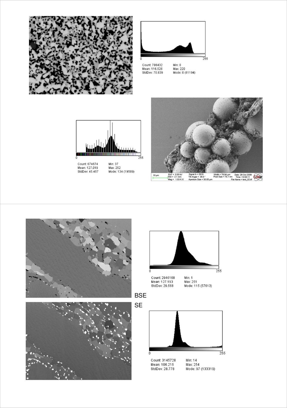
6 dealing with noise noisy original median filter original smooth very noisy

7 Filters. Median Gaussian blur Mean reduce in general the resolution They take into account the neighbourhood of a pixel FFT (fast fourier transform) filter (bandpass) FFT bandpass filter High-pass, Small size details (high space frequency) Low-pass, Large size details (low space frequency)
 FFT bandpass filter High-pass, Small size details")
8 Image processing in 2D Best: start with a good image: equalized histogram, use all 256 grey levels Reduce noise during acquisition Carefully use filters Filtering means mixing in of information from neighbouring pixels: loss of resolution try several to see which one works best do resampling only in the very end 3D imaging with a FIB/SEM FIB Nanotomography 3D Microscopy

9 Image processing in 3D 2D processing (shading correction) Image alignment (registration) Cropping (subvolume) Noise reduction (2D or 3D) Segmentation Visualization Shading correction background

10

11 Registration before after Registration before after

12 cropping Cutting of the border Post acquisition alignment (registration) banana < > cucumber problem

13 4800 images (5nm pixel size) at 1.7nm slice thickness Alignment marks 8kx8k image 20sec. For 2kx2k ROI 5nm pixel Alignment on notches raw StackReg on unprocessed images StackReg on binary enhanced images

14 StackReg Aligned on Notches

15 X-Z plane StackReg Aligned on Notches

16

17
18 Aligned with StackReg

19 Segmentation Identifying the different phases/volumes/objects Creating binary images/stacks of the different objects Pb free solder SnAgCu: one detector is not enough InLens: SE low energy M. Maleki, EPFL-LMAF EsB: Energy selective Backscattered ETD (SE classic)

20 EsB 10µm 10x10x10nm voxel size, 2048x1536x images (2x3Mb) / slice! (DUAL Channel!) 1.6keV, EsB & InLens-SE detector 12Gb data InLens SE

21
22 Phase 1. Dark in EsB image, White in SE InLens 10x10x10nm voxel size, 2048x1536x2000 pixel/slices 2 images (3Mb) / slice 12Gb data Phase 2: White in SE InLens Dark in EsB image 10x10x10nm voxel size, 2048x1536x2000 pixel/slices 2 images (3Mb) / slice 12Gb data
23 3D FIB/SEM: volume reconstruction Nb 3 Sn multifilament superconducting cable 0.5 mm Nb 3 Sn superconductor multifilament cable: Nb 3 Sn filaments (diameter ~5um) in Cu matrix 1.8kV EsB detector: Materials & orientation contrast
24 Es B Threshold SE Solid Oxide Fuel Cell cathode P. Tanasini, LENI
25 The right conditions 1.87kV, EsB detector Image: 2048x nm pixel size 2200 images 36hours
26 Segmentation and analysis Comparison with Transmission X ray Microscopy (TXM) Joy C. Andrews, Yijin Liu, and Piero Pianetta Stanford Synchrotron Radiation Lightsource Stanford Linear Accelerator Center Yong S. Chu National Synchrotron Light Source II Brookhaven National Laboratory beam stop capillary condenser pin hole tomography rotation axis sample objective ZP optically coupled CCD at image plane L C L C S L S ZP L ZP CCD George J. Nelson, William M. Harris, Jeffrey J. Lombardo, John R. Izzo, Jr., and Wilson K. S. Chiu *
27 YSZ LSM FIB data down-sampled to 25nm voxel size TXM Pore FIB The segmentation dilemma Median filter
28 thresholding
29 The gradient problem Erode (shrink 1 pixel) Dilate (grow 1 pixel)
30 Segmentation tools for life science Problem: Structure (shape) determines function of objects Single cell on substrate Image stack: 1024x786 pixel: (10nm image pixel size) 2kV, 60um Aperture, high current, EsB detector (grid 1.5kV) 600 slices, 20nm thickness, milling current 700pA
31 Segmentation based on grey levels Medical steel Ceramic coating: TiO 2 Cell outer membrane and more
32 A little bit more complex. brain tissue, resin embedded
33 Which detector? In chamber SE (Everhard Thornley) in Lens SE in Lens BSE (energy selective) Big volumes Voxel: 7.5x7.5x7.5nm Image 3096x slices (48hours) 23x17x24 um 9700um 3 ~7000 synapses 23Gb data
34 Reconstruction: Christel Genoud 2weeks of work Automated segmentation of neuronal structures Ilastik v0.5 Fred Hamprecht, University of Heidelberg
35 Synapse recognition Anna Kreshuk Automated segmentation of neuronal structures Ilastik v0.5 Fred Hamprecht, University of Heidelberg FIB Nanotomography in life science Specimen preparation (fixation, staining, dehydration, resin infiltration same as for BIO TEM) Image contrast and resolution TEM quality Stable and reliable automated acquisition (less artifacts than ultramicrotomy)
36 Image processing in 3D Start with a good image (low noise, equilibrated histogram, right detector) 2D processing (shading correction) Image alignment (registration) Cropping (subvolume) Noise reduction (2D or 3D) Segmentation (most important) Visualization (strong area for commercial software)
7. advanced SEM. Latest generation of SEM SEM
 7. advanced SEM SEM Low voltage SE imaging Condition of the surface, coatings, plasma cleaning Low voltage BSE imaging Polishing for BSE, EDX and EBSD, effect of ion beam etching/polishing 1 Latest generation
12. FIB. Marco Cantoni 021/693.48.16. Centre Interdisciplinaire de Microscopie Electronique CIME. Focused Ion Beam
 12. FIB Marco Cantoni 021/693.48.16 Centre Interdisciplinaire de Microscopie Electronique CIME 1 Focused Ion Beam a) Principles How does it work..? Ion source, optics, interaction with the sample b) Basic
The Focused Ion Beam Scanning Electron Microscope: A tool for sample preparation, two and three dimensional imaging. Jacob R.
 The Focused Ion Beam Scanning Electron Microscope: A tool for sample preparation, two and three dimensional imaging Jacob R. Bowen Contents Components of a FIB-SEM Ion interactions Deposition & patterns
View of ΣIGMA TM (Ref. 1)
") Overview of the FESEM system 1. Electron optical column 2. Specimen chamber 3. EDS detector [Electron Dispersive Spectroscopy] 4. Monitors 5. BSD (Back scatter detector) 6. Personal Computer 7. ON/STANDBY/OFF
Overview of the FESEM system 1. Electron optical column 2. Specimen chamber 3. EDS detector [Electron Dispersive Spectroscopy] 4. Monitors 5. BSD (Back scatter detector) 6. Personal Computer 7. ON/STANDBY/OFF
Micro-CT for SEM Non-destructive Measurement and Volume Visualization of Specimens Internal Microstructure in SEM Micro-CT Innovation with Integrity
 Micro-CT for SEM Non-destructive Measurement and Volume Visualization of Specimens Internal Microstructure in SEM Innovation with Integrity Micro-CT 3D Microscopy Using Micro-CT for SEM Micro-CT for SEM
Electron Microscopy 3. SEM. Image formation, detection, resolution, signal to noise ratio, interaction volume, contrasts
 Electron Microscopy 3. SEM Image formation, detection, resolution, signal to noise ratio, interaction volume, contrasts 3-1 SEM is easy! Just focus and shoot "Photo"!!! Please comment this picture... Any
MODERN VOXEL BASED DATA AND GEOMETRY ANALYSIS SOFTWARE TOOLS FOR INDUSTRIAL CT
 MODERN VOXEL BASED DATA AND GEOMETRY ANALYSIS SOFTWARE TOOLS FOR INDUSTRIAL CT C. Reinhart, C. Poliwoda, T. Guenther, W. Roemer, S. Maass, C. Gosch all Volume Graphics GmbH, Heidelberg, Germany Abstract:
3D EDX MICROANALYSIS IN A FIB/SEM:
 3D EDX MICROANALYSIS IN A FIB/SEM: WHAT CAN WE EXPECT, WHERE ARE THE LIMITS...? Marco Cantoni, Pierre Burdet Centre Interdisciplinaire de Microscopie Electronique (EPFL-CIME) CIME Since August 2008: Nvision
The Basics of Scanning Electron Microscopy
 The Basics of Scanning Electron Microscopy The small scanning electron microscope is easy to use because almost every variable is pre-set: the acceleration voltage is always 15kV, it has only a single
Name: Due: September 21 st 2012. Physics 7230 Laboratory 3: High Resolution SEM Imaging
 Name: Due: September 21 st 2012 Physics 7230 Laboratory 3: High Resolution SEM Imaging 1. What is meant by the term resolution? How does this differ from other image variables, such as signal to noise
Lectures 6&7: Image Enhancement
 Lectures 6&7: Image Enhancement Leena Ikonen Pattern Recognition (MVPR) Lappeenranta University of Technology (LUT) leena.ikonen@lut.fi http://www.it.lut.fi/ip/research/mvpr/ 1 Content Background Spatial
Physics 441/2: Transmission Electron Microscope
 Physics 441/2: Transmission Electron Microscope Introduction In this experiment we will explore the use of transmission electron microscopy (TEM) to take us into the world of ultrasmall structures. This
CT Traceability - Prof. Wim Dewulf, Group T - KU Leuven
 CT Traceability - Calibration and Accuracy Prof. Wim Dewulf, Group T - KU Leuven Outline Introduction: terminology and procedures Voxel size calibration Edge offset calibration Conclusions Outline Introduction:
Ion Beam Sputtering: Practical Applications to Electron Microscopy
 Ion Beam Sputtering: Practical Applications to Electron Microscopy Applications Laboratory Report Introduction Electron microscope specimens, both scanning (SEM) and transmission (TEM), often require a
Avizo Inspect New software for industrial inspection and materials R&D
 Avizo Inspect New software for industrial inspection and materials R&D Reduce your design cycle, inspection times, and meet higher-level quality standards at a lower cost. Avizo Inspect software streamlines
Medical Image Processing on the GPU. Past, Present and Future. Anders Eklund, PhD Virginia Tech Carilion Research Institute andek@vtc.vt.
 Medical Image Processing on the GPU Past, Present and Future Anders Eklund, PhD Virginia Tech Carilion Research Institute andek@vtc.vt.edu Outline Motivation why do we need GPUs? Past - how was GPU programming
AxioCam MR The All-round Camera for Biology, Medicine and Materials Analysis Digital Documentation in Microscopy
 Microscopy from Carl Zeiss AxioCam MR The All-round Camera for Biology, Medicine and Materials Analysis Digital Documentation in Microscopy New Dimensions in Performance AxioCam MR from Carl Zeiss Both
Basics of Quantitative Image Analysis
 QuickTime_ and a decompressor areneeded to seethispicture. Basics of Quantitative Image Analysis What you need to know about Image Processing but never thought to ask Before you start writing... See these
Basic 3D reconstruction in Imaris 7.6.1
 Basic 3D reconstruction in Imaris 7.6.1 Task The aim of this tutorial is to understand basic Imaris functionality by performing surface reconstruction of glia cells in culture, in order to visualize enclosed
CONFOCAL LASER SCANNING MICROSCOPY TUTORIAL
 CONFOCAL LASER SCANNING MICROSCOPY TUTORIAL Robert Bagnell 2006 This tutorial covers the following CLSM topics: 1) What is the optical principal behind CLSM? 2) What is the spatial resolution in X, Y,
Standard Test Method for Classification of Film Systems for Industrial Radiography 1
 Designation: E 1815 96 (Reapproved 2001) Standard Test Method for Classification of Film Systems for Industrial Radiography 1 This standard is issued under the fixed designation E 1815; the number immediately
Basic Image Processing (using ImageJ)
") Basic Image Processing (using ImageJ) Dr. Arne Seitz Swiss Institute of Technology (EPFL) Faculty of Life Sciences Head of BIOIMAGING AND OPTICS BIOP arne.seitz@epfl.ch Overview File formats (data storage)
Basic Image Processing (using ImageJ) Dr. Arne Seitz Swiss Institute of Technology (EPFL) Faculty of Life Sciences Head of BIOIMAGING AND OPTICS BIOP arne.seitz@epfl.ch Overview File formats (data storage)
Convolution. 1D Formula: 2D Formula: Example on the web: http://www.jhu.edu/~signals/convolve/
 Basic Filters (7) Convolution/correlation/Linear filtering Gaussian filters Smoothing and noise reduction First derivatives of Gaussian Second derivative of Gaussian: Laplacian Oriented Gaussian filters
Electron Microscopy 3. SEM. Image formation, detection, resolution, signal to noise ratio, interaction volume, contrasts
 Electron Microscopy 3. SEM Image formation, detection, resolution, signal to noise ratio, interaction volume, contrasts SEM is easy! Just focus and shoot "Photo"!!! Please comment this picture... Any idea
Lenses and Apertures of A TEM
 Instructor: Dr. C.Wang EMA 6518 Course Presentation Lenses and Apertures of A TEM Group Member: Anup Kr. Keshri Srikanth Korla Sushma Amruthaluri Venkata Pasumarthi Xudong Chen Outline Electron Optics
http://dx.doi.org/10.1117/12.906346
 Stephanie Fullerton ; Keith Bennett ; Eiji Toda and Teruo Takahashi "Camera simulation engine enables efficient system optimization for super-resolution imaging", Proc. SPIE 8228, Single Molecule Spectroscopy
A pretty picture, or a measurement? Retinal Imaging
 Big Data Challenges A pretty picture, or a measurement? Organelles Dynamics Cells Retinal Imaging Physiology Pathology Fundus Camera Optical coherence tomography Fluorescence Histology High Content Screening
Industrial X-ray for Nondestructive Testing Unrestricted Siemens AG 2014. All rights reserved
 Overview of X-ray Technology and Competence offered by Corporate Technology Industrial X-ray for Nondestructive Testing Nondestructive Testing (NDT) with X-rays: Our offer at a glance High-tech X-ray lab
Microscopy. MICROSCOPY Light Electron Tunnelling Atomic Force RESOLVE: => INCREASE CONTRAST BIODIVERSITY I BIOL1051 MAJOR FUNCTIONS OF MICROSCOPES
 BIODIVERSITY I BIOL1051 Microscopy Professor Marc C. Lavoie marc.lavoie@cavehill.uwi.edu MAJOR FUNCTIONS OF MICROSCOPES MAGNIFY RESOLVE: => INCREASE CONTRAST Microscopy 1. Eyepieces 2. Diopter adjustment
Applications of confocal fluorescence microscopy in biological sciences
 Applications of confocal fluorescence microscopy in biological sciences B R Boruah Department of Physics IIT Guwahati Email: brboruah@iitg.ac.in Page 1 Contents Introduction Optical resolution Optical
Planetary Imaging Workshop Larry Owens
 Planetary Imaging Workshop Larry Owens Lowell Observatory, 1971-1973 Backyard Telescope, 2005 How is it possible? How is it done? Lowell Observatory Sequence,1971 Acquisition E-X-P-E-R-I-M-E-N-T-A-T-I-O-N!
Lecture 14. Point Spread Function (PSF)
") Lecture 14 Point Spread Function (PSF), Modulation Transfer Function (MTF), Signal-to-noise Ratio (SNR), Contrast-to-noise Ratio (CNR), and Receiver Operating Curves (ROC) Point Spread Function (PSF) Recollect
Lecture 14 Point Spread Function (PSF), Modulation Transfer Function (MTF), Signal-to-noise Ratio (SNR), Contrast-to-noise Ratio (CNR), and Receiver Operating Curves (ROC) Point Spread Function (PSF) Recollect
Image Segmentation and Registration
 Image Segmentation and Registration Dr. Christine Tanner (tanner@vision.ee.ethz.ch) Computer Vision Laboratory, ETH Zürich Dr. Verena Kaynig, Machine Learning Laboratory, ETH Zürich Outline Segmentation
APPLICATION OF X-RAY COMPUTED TOMOGRAPHY IN SILICON SOLAR CELLS
 APPLICATION OF X-RAY COMPUTED TOMOGRAPHY IN SILICON SOLAR CELLS V.A. Popovich 1, W. Verwaal 2, M. Janssen 1, I. J. Bennett 3, I.M.Richardson 1, 1. Delft University of Technology, Department of Materials
WHITE PAPER. Methods for Measuring Flat Panel Display Defects and Mura as Correlated to Human Visual Perception
 Methods for Measuring Flat Panel Display Defects and Mura as Correlated to Human Visual Perception Methods for Measuring Flat Panel Display Defects and Mura as Correlated to Human Visual Perception Abstract
Advances in scmos Camera Technology Benefit Bio Research
 Advances in scmos Camera Technology Benefit Bio Research scmos camera technology is gaining in popularity - Why? In recent years, cell biology has emphasized live cell dynamics, mechanisms and electrochemical
Study of the Human Eye Working Principle: An impressive high angular resolution system with simple array detectors
 Study of the Human Eye Working Principle: An impressive high angular resolution system with simple array detectors Diego Betancourt and Carlos del Río Antenna Group, Public University of Navarra, Campus
Performance testing for Precision 500D Classical R/F System
 Performance testing for Precision 500D Classical R/F System John M. Boudry, Ph.D. Image Quality Systems Engineer GE Healthcare Technologies Outline System background Image Quality Signature Test (IQST)
Multiphysics Software Applications in Reverse Engineering
 Multiphysics Software Applications in Reverse Engineering *W. Wang 1, K. Genc 2 1 University of Massachusetts Lowell, Lowell, MA, USA 2 Simpleware, Exeter, United Kingdom *Corresponding author: University
Lecture 20: Scanning Confocal Microscopy (SCM) Rationale for SCM. Principles and major components of SCM. Advantages and major applications of SCM.
 Rationale for SCM. Principles and major components of SCM. Advantages and major applications of SCM.") Lecture 20: Scanning Confocal Microscopy (SCM) Rationale for SCM. Principles and major components of SCM. Advantages and major applications of SCM. Some limitations (disadvantages) of NSOM A trade-off
Lecture 20: Scanning Confocal Microscopy (SCM) Rationale for SCM. Principles and major components of SCM. Advantages and major applications of SCM. Some limitations (disadvantages) of NSOM A trade-off
MetaMorph Software Basic Analysis Guide The use of measurements and journals
 MetaMorph Software Basic Analysis Guide The use of measurements and journals Version 1.0.2 1 Section I: How Measure Functions Operate... 3 1. Selected images... 3 2. Thresholding... 3 3. Regions of interest...
Contents. X-ray and Computed Tomography. Characterization of X-rays. Production of X-rays
 J. E. Wilhjelm Ørsted TU Technical University of enmark, Bldg. 348, K-2800 Kongens Lyngby, enmark. X-ray and Computed Tomography Contents History and characterization of X-rays Conventional (projection)
Electron Microscopy SEM and TEM
 Electron Microscopy SEM and TEM Content 1. Introduction: Motivation for electron microscopy 2. Interaction with matter 3. SEM: Scanning Electron Microscopy 3.1 Functional Principle 3.2 Examples 3.3 EDX
Acoustic GHz-Microscopy: Potential, Challenges and Applications
 Acoustic GHz-Microscopy: Potential, Challenges and Applications A Joint Development of PVA TePLa Analytical Systems GmbH and Fraunhofer IWM-Halle Dr. Sebastian Brand (Ph.D.) Fraunhofer CAM Fraunhofer Institute
Neuro imaging: looking with lasers in the brain
 Neuro imaging: looking with lasers in the brain Aim: To image life cells, label free, with cellular resolution in deep tissue Marloes Groot Vrije Universiteit Amsterdam Faculteit Exacte Wetenschappen Natuurkunde
Introduction to CCDs and CCD Data Calibration
 Introduction to CCDs and CCD Data Calibration Dr. William Welsh San Diego State University CCD: charge coupled devices integrated circuit silicon chips that can record optical (and X-ray) light pixel =
MRC High Resolution. MR-compatible digital HD video camera. User manual
 MRC High Resolution MR-compatible digital HD video camera User manual page 1 of 12 Contents 1. Intended use...2 2. System components...3 3. Video camera and lens...4 4. Interface...4 5. Installation...5
Dynamic Neutron Imaging
 Wir schaffen Wissen heute für morgen Pierre Boillat Neutron Imaging & Activation Group (NIAG) and Electrochemistry Laboratory (LEC), Paul Scherrer Institute, Switzerland Dynamic Neutron Imaging PSI, 8.
Application of Data Matrix Verification Standards
 Data Matrix symbol verification at its most basic level eliminates the subjective quality determination that causes discord between marking and reading suppliers, and replaces those subjective opinions
Volume visualization I Elvins
 Volume visualization I Elvins 1 surface fitting algorithms marching cubes dividing cubes direct volume rendering algorithms ray casting, integration methods voxel projection, projected tetrahedra, splatting
Cirrus 0.2T. MRI for Everyone. North America, Asia, Europe. contact: kturek@mri-tech.pl
 Cirrus 0.2T MRI for Everyone North America, Asia, Europe contact: kturek@mri-tech.pl MRI-TECH inc. Cirrus MRI system for all your needs: Low costs Low maintenance High quality Open geometry Imaging of
Encoders for Linear Motors in the Electronics Industry
 Technical Information Encoders for Linear Motors in the Electronics Industry The semiconductor industry and automation technology increasingly require more precise and faster machines in order to satisfy
Usage of AFM, SEM and TEM for the research of carbon nanotubes
 Usage of AFM, SEM and TEM for the research of carbon nanotubes K.Safarova *1, A.Dvorak 2, R. Kubinek 1, M.Vujtek 1, A. Rek 3 1 Department of Experimental Physics, Faculty of Science, Palacky University,
MATRIX TECHNICAL NOTES
 200 WOOD AVENUE, MIDDLESEX, NJ 08846 PHONE (732) 469-9510 FAX (732) 469-0418 MATRIX TECHNICAL NOTES MTN-107 TEST SETUP FOR THE MEASUREMENT OF X-MOD, CTB, AND CSO USING A MEAN SQUARE CIRCUIT AS A DETECTOR
MEDICAL IMAGING 2nd Part Computed Tomography
 MEDICAL IMAGING 2nd Part Computed Tomography Introduction 2 In the last 30 years X-ray Computed Tomography development produced a great change in the role of diagnostic imaging in medicine. In convetional
Mars Atmosphere and Volatile EvolutioN (MAVEN) Mission
 Mission") Mars Atmosphere and Volatile EvolutioN (MAVEN) Mission MAVEN Science Community Workshop December 2, 2012 Particles and Fields Package Solar Energetic Particle Instrument (SEP) Davin Larson and the SEP
Mars Atmosphere and Volatile EvolutioN (MAVEN) Mission MAVEN Science Community Workshop December 2, 2012 Particles and Fields Package Solar Energetic Particle Instrument (SEP) Davin Larson and the SEP
Fundamentals of Cone-Beam CT Imaging
 Fundamentals of Cone-Beam CT Imaging Marc Kachelrieß German Cancer Research Center (DKFZ) Heidelberg, Germany www.dkfz.de Learning Objectives To understand the principles of volumetric image formation
Chapter 13 Confocal Laser Scanning Microscopy C. Robert Bagnell, Jr., Ph.D., 2012
 Chapter 13 Confocal Laser Scanning Microscopy C. Robert Bagnell, Jr., Ph.D., 2012 You are sitting at your microscope working at high magnification trying to sort out the three-dimensional compartmentalization
Automatic and Objective Measurement of Residual Stress and Cord in Glass
 Automatic and Objective Measurement of Residual Stress and Cord in Glass GlassTrend - ICG TC15/21 Seminar SENSORS AND PROCESS CONTROL 13-14 October 2015, Eindhoven Henning Katte, ilis gmbh copyright ilis
Thinking ahead. Focused on life. REALIZED: GROUNDBREAKING RESOLUTION OF 80 µm VOXEL
 Thinking ahead. Focused on life. REALIZED: GROUNDBREAKING RESOLUTION OF 80 µm VOXEL X-ray ZOOM RECONSTRUCTION Flat Panel Detector (FPD) Automatic Positioning Function For ø 40 x H 40 mm, ø 60 x H 60 mm,
Development of High Speed Oblique X-ray CT System for Printed Circuit Board
 計 測 自 動 制 御 学 会 産 業 論 文 集 Vol. 6, No. 9, 72/77 (2007) Development of High Speed Oblique X-ray CT System for Printed Circuit Board Atsushi TERAMOTO*, Takayuki MURAKOSHI*, Masatoshi TSUZAKA**, and Hiroshi
Statistical Considerations in Magnetic Resonance Imaging of Brain Function
 Statistical Considerations in Magnetic Resonance Imaging of Brain Function Brian D. Ripley Professor of Applied Statistics University of Oxford ripley@stats.ox.ac.uk http://www.stats.ox.ac.uk/ ripley Acknowledgements
Fluorescent Array Imaging Reader
 Fluorescent Array Imaging Reader Multiplex Enabled For 2-Color Fluorescent Microarrays Extended Dynamic Range Automated Spot Analysis Compact and Affordable Microarray Analysis With FLAIR FLAIR Sensovation
Scanning Electron Microscopy Primer
 Scanning Electron Microscopy Primer Bob Hafner This primer is intended as background for the Introductory Scanning Electron Microscopy training offered by the University of Minnesota s Characterization
Nanometer-scale imaging and metrology, nano-fabrication with the Orion Helium Ion Microscope
 andras@nist.gov Nanometer-scale imaging and metrology, nano-fabrication with the Orion Helium Ion Microscope Bin Ming, András E. Vladár and Michael T. Postek National Institute of Standards and Technology
Face detection is a process of localizing and extracting the face region from the
 Chapter 4 FACE NORMALIZATION 4.1 INTRODUCTION Face detection is a process of localizing and extracting the face region from the background. The detected face varies in rotation, brightness, size, etc.
Measuring large areas by white light interferometry at the nanopositioning and nanomeasuring machine (NPMM)
") Image Processing, Image Analysis and Computer Vision Measuring large areas by white light interferometry at the nanopositioning and nanomeasuring machine (NPMM) Authors: Daniel Kapusi 1 Torsten Machleidt
Image Processing, Image Analysis and Computer Vision Measuring large areas by white light interferometry at the nanopositioning and nanomeasuring machine (NPMM) Authors: Daniel Kapusi 1 Torsten Machleidt
Scanning Electron Microscopy tools for material characterization
 5th International Workshop on Mechanisms of Vacuum Arcs 02-04/09/2015 Scanning Electron Microscopy tools for material characterization Focus on EBSD for characterisation of dislocation structures Floriane
Image Processing of Atomic Resolution Transmission Electron Microscope Images
 Journal of the Korean Physical Society, Vol. 48, No. 2, February 2006, pp. 250 255 Image Processing of Atomic Resolution Transmission Electron Microscope Images Young-Min Kim, Jong-Man Jeong, Jin-Gyu Kim
Data. microcat +SPECT
 Data microcat +SPECT microcat at a Glance Designed to meet the throughput, resolution and image quality requirements of academic and pharmaceutical research, the Siemens microcat sets the standard for
IMAGING SOFTWARE. Image-Pro Insight Image Analysis Made Easy. Capture, Process, Measure, and Share
 IMAGING SOFTWARE Image-Pro Insight Image Analysis Made Easy Capture, Process, Measure, and Share Image-Pro Insight Image Analysis Made Easy Capture, Process, Measure, and Share Image-Pro Insight, the latest
Scan Time Reduction and X-ray Scatter Rejection in Dual Modality Breast Tomosynthesis. Tushita Patel 4/2/13
 Scan Time Reduction and X-ray Scatter Rejection in Dual Modality Breast Tomosynthesis Tushita Patel 4/2/13 Breast Cancer Statistics Second most common cancer after skin cancer Second leading cause of cancer
WHITE PAPER. Are More Pixels Better? www.basler-ipcam.com. Resolution Does it Really Matter?
 WHITE PAPER www.basler-ipcam.com Are More Pixels Better? The most frequently asked question when buying a new digital security camera is, What resolution does the camera provide? The resolution is indeed
DIGITAL IMAGE PROCESSING AND ANALYSIS
 DIGITAL IMAGE PROCESSING AND ANALYSIS Human and Computer Vision Applications with CVIPtools SECOND EDITION SCOTT E UMBAUGH Uffi\ CRC Press Taylor &. Francis Group Boca Raton London New York CRC Press is
Star Detection and Removal in Night Airglow Images
 Star Detection and Removal in Night Airglow Images Rohit P. Patil *1, S. B. Patil 1, R. N. Ghodpage 2, P. T. Patil 2 1 D.Y. Patil College of, Kolhapur, Maharashtra, India 2 M.F. Radar, Indian Institute
MDCT Technology. Kalpana M. Kanal, Ph.D., DABR Assistant Professor Department of Radiology University of Washington Seattle, Washington
 MDCT Technology Kalpana M. Kanal, Ph.D., DABR Assistant Professor Department of Radiology University of Washington Seattle, Washington ACMP Annual Meeting 2008 - Seattle, WA Educational Objectives Historical
ZEISS Axiocam 506 color Your Microscope Camera for Imaging of Large Sample Areas Fast, in True Color, and High Resolution
 Product Information Version 1.0 ZEISS Axiocam 506 color Your Microscope Camera for Imaging of Large Sample Areas Fast, in True Color, and High Resolution ZEISS Axiocam 506 color Sensor Model Sensor Pixel
Admin stuff. 4 Image Pyramids. Spatial Domain. Projects. Fourier domain 2/26/2008. Fourier as a change of basis
 Admin stuff 4 Image Pyramids Change of office hours on Wed 4 th April Mon 3 st March 9.3.3pm (right after class) Change of time/date t of last class Currently Mon 5 th May What about Thursday 8 th May?
Radiography: 2D and 3D Metrics of Performance Towards Quality Index
 AAPM COMP 011 Radiography: D and 3D Metrics of Performance Towards Quality Index Ehsan Samei, Sam Richard Duke University Learning objectives Outlook 1. To understand methods for D and 3D resolution measurements..
Lecture 12: Cameras and Geometry. CAP 5415 Fall 2010
 Lecture 12: Cameras and Geometry CAP 5415 Fall 2010 The midterm What does the response of a derivative filter tell me about whether there is an edge or not? Things aren't working Did you look at the filters?
26/06/2011. Electron Microscopy: TEM, Immunogold Labeling, SEM, Correlative Microscopy
 26/06/2011 Electron Microscopy: TEM, Immunogold Labeling, SEM, Correlative Microscopy Prof. Dr. Rainer Duden duden@bio.uni-luebeck.de 1 Resolution Comparison Light vs Electron Microscopy Microscope Resolution
Technical Information
 Technical Information TI No. WL 80-63 E April 2001 Rolling Bearing Diagnosis with the FAG Bearing Analyser Rolling Bearings State-Of-The-Art, Condition-Related Monitoring of Plants and Machines Unforeseen
Characterization and Kinetics of the Interfacial Reactions in Solder Joints of Tin-Based Solder Alloys on Copper Substrates
 Characterization and Kinetics of the Interfacial Reactions in Solder Joints of Tin-Based Solder Alloys on Copper Substrates J. C. Madeni*, S. Liu* and T. A. Siewert** *Center for Welding, Joining and Coatings
DETERMINATION OF WOOD MOISTURE PROPERTIES USING CT- SCANNER IN A CONTROLLED ENVIRONMENT
 DETERMINATION OF WOOD MOISTURE PROPERTIES USING CT- SCANNER IN A CONTROLLED ENVIRONMENT Cherepanova, E. 1 & Hansson, L. 2 ABSTRACT The aim of the present work was to examine the existing algorithm for
AMINO ACID ANALYSIS By High Performance Capillary Electrophoresis
 AMINO ACID ANALYSIS By High Performance Capillary Electrophoresis Analysis of Amino Acid Standards Label free analysis using the HPCE-512 ABSTRACT Capillary electrophoresis using indirect UV detection
CT for the production floor: Recent developments in Hard- and Software for industrial CT Systems
 CT for the production floor: Recent developments in Hard- and Software for industrial CT Systems GE s Industrial X-ray and CT Forum, Cologne July 7 th 2015 Oliver Brunke GE Sensing & Inspection Technologies
LED red (-): Measuring value < lower tolerance threshold LED red (+): Measuring value > upper tolerance threshold. Page 1/6
: Measuring value < lower tolerance threshold LED red (+): Measuring value > upper tolerance threshold. Page 1/6") L-LAS Series L-LAS-LT-165-CL - Line laser 1 mw, laser class 2 - Visible laser line (red light 670 nm), typ. 2 mm x 3 mm - Reference distance approx. 165 mm - Measuring range typ. 65... 265 mm - Resolution
L-LAS Series L-LAS-LT-165-CL - Line laser 1 mw, laser class 2 - Visible laser line (red light 670 nm), typ. 2 mm x 3 mm - Reference distance approx. 165 mm - Measuring range typ. 65... 265 mm - Resolution
Software Packages The following data analysis software packages will be showcased:
 Analyze This! Practicalities of fmri and Diffusion Data Analysis Data Download Instructions Weekday Educational Course, ISMRM 23 rd Annual Meeting and Exhibition Tuesday 2 nd June 2015, 10:00-12:00, Room
Principles of Microscopy and Confocal and Fluorescence Microscopy
 Principles of Microscopy and Confocal and Fluorescence Microscopy Content This course in Light Microscopy follows the series of successful courses in Light Microscopy, Confocal and Fluorescence Microscopy
Digital Image Processing
 GONZ_FMv3.qxd 7/26/07 9:05 AM Page i Digital Image Processing Third Edition Rafael C. Gonzalez University of Tennessee Richard E. Woods MedData Interactive Upper Saddle River, NJ 07458 GONZ_FMv3.qxd 7/26/07
Bildverarbeitung und Mustererkennung Image Processing and Pattern Recognition
 Bildverarbeitung und Mustererkennung Image Processing and Pattern Recognition 1. Image Pre-Processing - Pixel Brightness Transformation - Geometric Transformation - Image Denoising 1 1. Image Pre-Processing
Image Analysis for Volumetric Industrial Inspection and Interaction
 Image Analysis for Volumetric Industrial Inspection and Interaction Rasmus Larsen Mads F. Hansen, Hildur Olafsdottir, Thomas H. Mosbech, Lasse F. Laursen DTU Informatics (CT) reconstruction from few projections
Fig.1. The DAWN spacecraft
 Introduction Optical calibration of the DAWN framing cameras G. Abraham,G. Kovacs, B. Nagy Department of Mechatronics, Optics and Engineering Informatics Budapest University of Technology and Economics
Environmental Remote Sensing GEOG 2021
 Environmental Remote Sensing GEOG 2021 Lecture 4 Image classification 2 Purpose categorising data data abstraction / simplification data interpretation mapping for land cover mapping use land cover class
CONCEPT OF DETERMINISTIC ION IMPLANTATION AT THE NANOSCALE
 CONCEPT OF DETERMINISTIC ION IMPLANTATION AT THE NANOSCALE Daniel Spemann Jan Meijer 1, Jürgen W. Gerlach, Paul Räcke 1, Susann Liedtke, Stephan Rauschenbach 2, Bernd Rauschenbach 1 University of Leipzig,
Chapter 12 Filters for FISH Imaging
 Chapter 12 Filters for FISH Imaging Dan Osborn The application of in situ hybridization (ISH) has advanced from short lived, non-specific isotopic methods, to very specific, long lived, multiple color
3-D Printing Artificial Reservoir Rocks to Test Their Petrophysical Properties*
 3-D Printing Artificial Reservoir Rocks to Test Their Petrophysical Properties* Sergey Ishutov 1 and Franciszek Hasiuk 1 Search and Discovery Article #41427 (2014) Posted August 29, 2014 *Adapted from
Zeiss 780 Training Notes
 Zeiss 780 Training Notes 780 Start Up Sequence Do you need the argon laser, 458,488,514nm lines? No Turn on the Systems PC Switch Turn on Main Power Switch Yes Turn on the laser main power switch and turn
Purchasing a cardiac CT scanner: What the radiologist needs to know
 Purchasing a cardiac CT scanner: What the radiologist needs to know Maria Lewis ImPACT St George s Hospital, London maria.lewis@stgeorges.nhs.uk CT scanner development Slice wars 1998 Increased z-coverage