26/06/2011. Electron Microscopy: TEM, Immunogold Labeling, SEM, Correlative Microscopy
|
|
|
- Marcia Lang
- 8 years ago
- Views:
Transcription
1 26/06/2011 Electron Microscopy: TEM, Immunogold Labeling, SEM, Correlative Microscopy Prof. Dr. Rainer Duden 1

2 Resolution Comparison Light vs Electron Microscopy

3 Microscope Resolution ability of a lens to separate or distinguish small objects that are close together wavelength of light used is major factor in resolution shorter wavelength greater resolution 3

4 Electron Microscopy beams of electrons are used to produce images wavelength of electron beam is much shorter than light, resulting in much higher resolution 4

5 Light microscopy Glass lenses Source of illumination is usually light of visible wavelengths Electron microscopy Electromagnetic lenses Source of illumination is electrons Hairpin tungsten filament (thermionic emission) Pointed tungsten crystal (cold cathode field emission)
 Pointed tungsten crystal (cold cathode field")
6 The Transmission Electron Microscope electrons scatter when they pass through thin sections of a specimen transmitted electrons (those that do not scatter) are used to produce image denser regions in specimen, scatter more electrons and appear darker

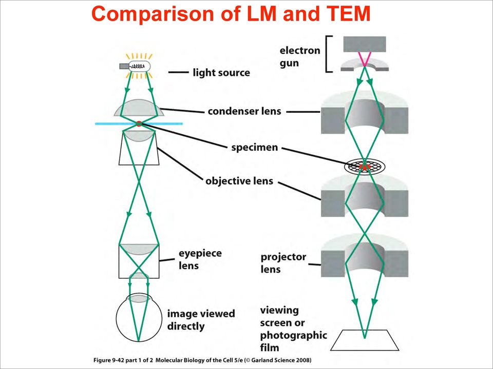
7 Comparison of LM and TEM
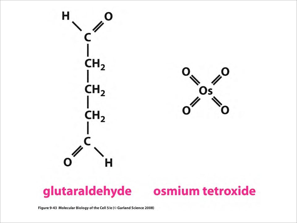
8 Specimen Preparation analogous to procedures used for light microscopy for transmission electron microscopy, specimens must be cut very thin specimens are chemically fixed and stained with electron dense material

9
10 Transmission Electron Microscopy (TEM) Zeiss 10/A conventional TEM Excellent for training Film only

11 Negative Staining Viruses, small particles, proteins, molecules No sectioning Same day results

12 negative staining particles Electron dense negative stain

13 negative staining requires minimal interaction between particle & stain to avoid binding, heavy metal ion should be of same charge +/- as the particle positive staining usually destructive of bio-particles biological material usually -ve charge at neutral ph widely used negative contrast media include: anionic cationic phosphotungstate uranyl actetate/formate molybdate (ammonium) ph ~ 4)
 (@")
14 Negative Stain Ebola

15 Double Immunogold Labeling of Negatively Stained Specimens Bacterial pili serotypes dried onto grid and sequentially labeled with primary antibody, then Protein-A-5nm-gold and Protein-A-15- nm-gold before negative staining

16 metal shadowing - rotary

17 metal shadowing - rotary Contrast usually inverted to give dark shadows > resolution 2-3 nm - single DNA strand detectable - historic use for molecular biology (e.g. heteroduplex mapping) > good preservation of shape, but enlargement of apparent dimensions > in very recent modification (MCD - microcrystallite decoration), resolution ~1.1 nm
 > good preservation of shape, but enlargement of apparent")
18 s e n Clathrin: a major and evolutionarily conserved coat protein a r b s V C em C m d e e d fi i u r Cr Pu clathrin heavy chain ~100kD proteins 66 ~50kD proteins 45 clathrin light chains ~20kD proteins

19 Rotary shadowing EM images of purified Clathrin triskelia

20 Rotary shadowing EM images of purified Clathrin triskelia Kirchhausen and Harrison (1981). Cell 23, : see EM image above Ungewickell and Branton (1981). Nature 289, : - reversible dissassembly of Clathrin triskelions into clathrin - coats in vitro
.")
21 Clathrin triskelions 3 heavy chains 3 light chains
22 s e n Adaptors: a r b s V C em C m d e e d fi i u r Cr Pu essential for cargo sorting clathrin heavy chain ~100kD proteins 66 ~50kD proteins 45 clathrin light chains ~20kD proteins
23 Protein pattern of Adaptor Complexes extracted from purified brain CCVs after SDS-PAGE β1 γ AP-1 AP-2 αa β2 αc µ1 µ2 σ1 σ2
24 Rotary shadowing EM images of purified AP-2 complex
25 Adaptor proteins mediate sorting of specific cargo from different compartments α µ2 σ2 β2 γ µ1 σ1 β1 δ µ3 σ3 β3 ε µ4 σ4 β4 AP-1 AP-2 AP-3 AP-4 Margaret Robinson, Univ. Cambridge
26 Overview of Biological Specimen Preparation Killing & Fixation - Death; Molecular stabilization Dehydration - Chemical removal of H 2 O Infiltration - Replace liquid phase with resin Embedding & Polymerization - Make solid, sectionable block Sectioning - Ultramicrotome, mount, stain
27 Preparing for cutting sections for TEM 27
28
29
30 Estimating Section Thickness Interference reflection angle from Sjöstrand (1967)
31
32 Serial section 3-D reconstruction
33 The Freeze Fracture Technique
34
35
36 Gap Junctions in negative stain, freeze fracture & TEM
37 Tight Junction structure in TEM, freeze fracture, and live fluorescence microscopy
38 Cryotechniques Ultrarapid cryofixation Metal mirror impact Liquid propane plunge Freeze fracture with Balzers 400T Cryosubstitution Cryo-ultramicrotomy Ultrathin frozen sections (primarily for antibody labeling)
39 Clathrin - coated vesicles - the minimal machinery Clathrin triskelions 3 heavy chains 3 light chains Adaptor (AP2) α - four adaptins β2 σ2 µ2 Tom Kirchhausen, Harvard Medical School
40 John Heuser s Quick Freeze Deep Etch Technique
41 Quick freeze - deep etch technique John Heuser Washington University School of Medicine, USA
42 Clathrin - coated Vesicles J. Heuser Inner layer : membrane containing cargo Middle layer : adaptors and accessory proteins Outer layer : mechanical scaffold
43 Now please put on the 3-D glasses
44 John Heuser, Washington Univ. School of Medicine
45
46
47
48
49
50
51
52 Immunolabeling for Transmission Electron Microscopy Normally do Two-Step Method Primary antibody applied followed by colloidal gold-labeled secondary antibody May also be enhanced with silver Can also do for LM
53 Preparation of Biological Specimens for Immunolabeling The goal is to preserve tissue as closely as possible to its natural state while at the same time maintaining the ability of the antigen to react with the antibody Chemical fixation of whole mounts prior to labeling for LM Chemical fixation, dehydration, and embedment in paraffin or resin for sectioning for LM or TEM Chemical fixation for cryosections for LM Cryofixation for LM or TEM
54 Chemical Fixation Antigenic sites are easily denatured or masked during chemical fixation Glutaraldehyde gives good fixation but may mask antigens, plus it is fluorescent Paraformaldehyde often better choice, but results in poor morphology, especially for electron microscopy May use e.g., 4% paraformaldehyde with 0.5% glutaraldehyde as a good compromise
55 Specimen Preparation for TEM Chemical fixation with buffered glutaraldehyde Or 4% paraformaldehyde with >1% glutaraldehyde Postfixation with osmium tetroxide Or not, or with subsequent removal from sections Dehydration and infiltration with liquid epoxy or acrylic resin Polymerization of hard blocks by heat or UV Ultramicrotomy 60-80nm sections Labeling and/or staining View with TEM
56 Approaches to Immunolabeling Direct Method: Primary antibody contains label Indirect Method: Primary antibody followed by labeled secondary antibody Amplified Method: Methods to add more reporter to labeled site Protein A Method: May be used as secondary reagent instead of antibody
57 Colloidal gold of defined sizes, e.g., 5 nm, 10 nm, 20 nm, easily conjugated to antibodies Results in small, round, electron-dense label easily detected with EM Can be enhanced after labeling to enlarge size for LM or EM
58 Immunolocalization LM Fluor/confocal TEM SEM with backscatter detector
59 Preembedding or Postembedding Labeling May use preembedding labeling for surface antigens or for permeabilized cells The advantage is that antigenicity is more likely preserved Postembedding labeling is performed on sectioned tissue, on grids, allowing access to internal antigens Antigenicity probably partially compromised by embedding
60 Steps in Labeling of Sections Chemical fixation Dehydration, infiltration, embedding and sectioning Optional etching of embedment, permeabilization Blocking Incubation with primary antibody Washing Incubation with secondary antibody congugated with reporter (fluorescent probe, colloidal gold) Washing, optional counterstaining Mount and view
61 Controls! Controls! Controls! Omit primary antibody Irrelevant primary antibody Pre-immune serum Perform positive control Check for autofluorescence Check for non-specific labeling Dilution series
62 Desmosomes and IFs in primary mouse keratinocytes Duden & Franke, 988 (J. Cell Biol.)
63 Pre-embedding labelling of desmosomal vesicles in primary mouse keratinocytes Duden and Franke, 1988 (J. Cell Biol.)
64 Visualization of desmosomal vesicles in A431 cells grown on glass coverslips Duden and Franke, 1988 (J. Cell Biol.)
65 Pre-embedding labelling of desmosomal vesicles in A431 cells Duden & Franke, 1988 (J. Cell Biol.)
66
67 Double-labeling Method Use primary antibodies derived from different animals (e.g., one mouse antibody and one rabbit antibody) Then use two secondary antibodies conjugated with reporters that can be distinguished from one another
68 George Palade 1974 Nobel Prize for Physiology or Medicine Analysis of the secretory pathway by a combination of EM and autoradiography ER --> Golgi --> Vesicles --> PM
69 The Scanning Electron Microscope uses electrons reflected from the surface of a specimen to create image produces a 3-dimensional image of specimen s surface features
70 TEM vs SEM
71 Scanning Electron Microscopy
72 SEM
73
74
75 Correlative Light/EM microscopy - SEM: visualization of virus particles on a cell surface
76 Correlative Light/EM microscopy & electron tomography Diaminobenzidine (DAB) photooxidation by GFP (GalT-GFP) Grabenbaur et al., Nat. Meth
77 Correlative Light/EM microscopy & electron tomography Grabenbaur et al., Nat. Meth
78
79 minisog
80 minisog
81
82
Ion Beam Sputtering: Practical Applications to Electron Microscopy
 Ion Beam Sputtering: Practical Applications to Electron Microscopy Applications Laboratory Report Introduction Electron microscope specimens, both scanning (SEM) and transmission (TEM), often require a
Chapter 4. Microscopy, Staining, and Classification. Lecture prepared by Mindy Miller-Kittrell North Carolina State University
 Chapter 4 Microscopy, Staining, and Classification 2012 Pearson Education Inc. Lecture prepared by Mindy Miller-Kittrell North Carolina State University Microscopy and Staining 2012 Pearson Education Inc.
ISOLATION AND PROPERTIES OF SECRETORY GRANULES FROM RAT ISLETS OF LANGERHANS. II. Ultrastructure of the Beta Granule
 ISOLATION AND PROPERTIES OF SECRETORY GRANULES FROM RAT ISLETS OF LANGERHANS II Ultrastructure of the Beta Granule MARIE H GREIDER, S L HOWELL, and P E LACY From the Department of Pathology, Washington
Introduction to flow cytometry
 Introduction to flow cytometry Flow cytometry is a popular laser-based technology. Discover more with our introduction to flow cytometry. Flow cytometry is now a widely used method for analyzing the expression
Serology: Fluorescent antibody tests and other tests employing conjugated antibodies
 Serology: Fluorescent antibody tests and other tests employing conjugated antibodies Authors: Adapted by Prof M van Vuuren. Originally compiled by Dr RW Worthington. (Retired) Licensed under a Creative
Microscopy. MICROSCOPY Light Electron Tunnelling Atomic Force RESOLVE: => INCREASE CONTRAST BIODIVERSITY I BIOL1051 MAJOR FUNCTIONS OF MICROSCOPES
 BIODIVERSITY I BIOL1051 Microscopy Professor Marc C. Lavoie marc.lavoie@cavehill.uwi.edu MAJOR FUNCTIONS OF MICROSCOPES MAGNIFY RESOLVE: => INCREASE CONTRAST Microscopy 1. Eyepieces 2. Diopter adjustment
The microscope is an important tool.
 KEY CONCEPT Microscopes allow us to see inside the cell. BEFORE, you learned Some organisms are unicellular and some are multicellular A microscope is necessary to study most cells The cell theory describes
PROTOCOL. Immunocytochemistry (ICC) MATERIALS AND EQUIPMENT REQUIRED
 MATERIALS AND EQUIPMENT REQUIRED") PROTOCOL Immunocytochemistry (ICC) 1850 Millrace Drive, Suite 3A Eugene, Oregon 97403 11-07 MATERIALS AND EQUIPMENT REQUIRED Materials: MitoSciences primary monoclonal antibody/antibodies Fluorophore-conjugated
PROTOCOL Immunocytochemistry (ICC) 1850 Millrace Drive, Suite 3A Eugene, Oregon 97403 11-07 MATERIALS AND EQUIPMENT REQUIRED Materials: MitoSciences primary monoclonal antibody/antibodies Fluorophore-conjugated
Microscopy: Principles and Advances
 Microscopy: Principles and Advances Chandrashekhar V. Kulkarni University of Central Lancashire, Preston, United kingdom May, 2014 University of Ljubljana Academic Background 2005-2008: PhD-Chemical Biology
Fluorescence Microscopy for an NMR- Biosensor Project
 Fluorescence Microscopy for an NMR- Biosensor Project Ole Hirsch Physikalisch-Technische Bundesanstalt Medical Optics Abbestr. -1, 10587 Berlin, Germany Overview NMR Sensor Project Dimensions in biological
Outline. 1. Experiment. 2. Sample analysis and storage. 3. Image analysis and presenting data. 4. Probemaker
 Tips and tricks Note: this is just an informative document with general recommendations. Please contact support@olink.com should you have any queries. Document last reviewed 2011-11-17 Outline 1. Experiment
View of ΣIGMA TM (Ref. 1)
") Overview of the FESEM system 1. Electron optical column 2. Specimen chamber 3. EDS detector [Electron Dispersive Spectroscopy] 4. Monitors 5. BSD (Back scatter detector) 6. Personal Computer 7. ON/STANDBY/OFF
Overview of the FESEM system 1. Electron optical column 2. Specimen chamber 3. EDS detector [Electron Dispersive Spectroscopy] 4. Monitors 5. BSD (Back scatter detector) 6. Personal Computer 7. ON/STANDBY/OFF
The immune response Antibodies Antigens Epitopes (antigenic determinants) the part of a protein antigen recognized by an antibody Haptens small
 the part of a protein antigen recognized by an antibody Haptens small") The immune response Antibodies Antigens Epitopes (antigenic determinants) the part of a protein antigen recognized by an antibody Haptens small molecules that can elicit an immune response when linked
The immune response Antibodies Antigens Epitopes (antigenic determinants) the part of a protein antigen recognized by an antibody Haptens small molecules that can elicit an immune response when linked
Electron Microscopy SEM and TEM
 Electron Microscopy SEM and TEM Content 1. Introduction: Motivation for electron microscopy 2. Interaction with matter 3. SEM: Scanning Electron Microscopy 3.1 Functional Principle 3.2 Examples 3.3 EDX
Chapter 18: Applications of Immunology
 Chapter 18: Applications of Immunology 1. Vaccinations 2. Monoclonal vs Polyclonal Ab 3. Diagnostic Immunology 1. Vaccinations What is Vaccination? A method of inducing artificial immunity by exposing
Protein immunoblotting
 Protein immunoblotting (Western blotting) Dr. Serageldeen A. A. Sultan Lecturer of virology Dept. of Microbiology SVU, Qena, Egypt seaas@lycos.com Western blotting -It is an analytical technique used to
Chapter 2 Antibodies. Contents. Introduction
 Chapter 2 Antibodies Keywords Immunohistochemistry Antibody labeling Fluorescence microscopy Fluorescent immunocytochemistry Fluorescent immunohistochemistry Indirect immunocytochemistry Immunostaining
How cryo will solve your problems
 How cryo will solve your problems Nadejda B. Matsko FELMI, TU Graz, and ZFE Graz What for do we need cryo microscopy? Cryo fixation Conventional fixation (rapid rapid dehydration) High resolution microscopy
ab183294 TripleStain IHC Kit: M&M&R on rodent tissue (DAB, DAB/Ni & AP/Red)
") ab183294 TripleStain IHC Kit: M&M&R on rodent tissue (DAB, DAB/Ni & AP/Red) Instructions for Use For the detection of Rabbit and Mouse Primary antibodies on Rodent Tissue. This product is for research
ab183294 TripleStain IHC Kit: M&M&R on rodent tissue (DAB, DAB/Ni & AP/Red) Instructions for Use For the detection of Rabbit and Mouse Primary antibodies on Rodent Tissue. This product is for research
protocol handbook 3D cell culture mimsys G hydrogel
 handbook 3D cell culture mimsys G hydrogel supporting real discovery handbook Index 01 Cell encapsulation in hydrogels 02 Cell viability by MTS assay 03 Live/Dead assay to assess cell viability 04 Fluorescent
Lecture 20: Scanning Confocal Microscopy (SCM) Rationale for SCM. Principles and major components of SCM. Advantages and major applications of SCM.
 Rationale for SCM. Principles and major components of SCM. Advantages and major applications of SCM.") Lecture 20: Scanning Confocal Microscopy (SCM) Rationale for SCM. Principles and major components of SCM. Advantages and major applications of SCM. Some limitations (disadvantages) of NSOM A trade-off
Lecture 20: Scanning Confocal Microscopy (SCM) Rationale for SCM. Principles and major components of SCM. Advantages and major applications of SCM. Some limitations (disadvantages) of NSOM A trade-off
EdU Flow Cytometry Kit. User Manual
 User Manual Ordering information: (for detailed kit content see Table 2) EdU Flow Cytometry Kits for 50 assays: Product number EdU Used fluorescent dye BCK-FC488-50 10 mg 6-FAM Azide BCK-FC555-50 10 mg
SILA Sistema Integrato di Laboratori per l Ambiente. CENTRE FOR MICROSCOPY AND MICROANALYSIS Scientific coordinator: Prof.ssa Rosanna De Rosa
 CENTRE FOR MICROSCOPY AND MICROANALYSIS Scientific coordinator: Prof.ssa Rosanna De Rosa 0 The Centre for Microscopy and Microanalysis (CM2) is an interdisciplinary service centre, a comprehensive suite
Immunoblotting (Western blotting)
") Immunoblotting (Western blotting) HIV Transfer to NC membrane membrane gel support buffer Dissociate in SDS Incubate with 1ary antiserum Separate by SDS-PAG Incubate w/ labeled 2nd ab Immunoblotting :
Immunoblotting (Western blotting) HIV Transfer to NC membrane membrane gel support buffer Dissociate in SDS Incubate with 1ary antiserum Separate by SDS-PAG Incubate w/ labeled 2nd ab Immunoblotting :
Nanoelectronics 09. Atsufumi Hirohata Department of Electronics. Quick Review over the Last Lecture
 Nanoelectronics 09 Atsufumi Hirohata Department of Electronics 12:00 Wednesday, 4/February/2015 (P/L 006) Quick Review over the Last Lecture ( Field effect transistor (FET) ): ( Drain ) current increases
Principles of Immunohistochemistry Queen s Laboratory For Molecular Pathology
 Principles of Immunohistochemistry Queen s Laboratory For Molecular Pathology Table of Contents POLYCLONAL VERSUS MONOCLONAL ANTIBODIES...PAGE 3 DIRECT & INDIRECT ASSAYS.PAGE 4 LABELS..PAGE 5 DETECTION...PAGE
Applications of confocal fluorescence microscopy in biological sciences
 Applications of confocal fluorescence microscopy in biological sciences B R Boruah Department of Physics IIT Guwahati Email: brboruah@iitg.ac.in Page 1 Contents Introduction Optical resolution Optical
Forensic Science: The Basics. Microscopy
 Forensic Science: The Basics Microscopy Chapter 6 Jay A. Siegel,Ph.D. Power point presentation by Greg Galardi, Peru State College, Peru Nebraska Presentation by Greg Galardi, Peru State College CRC Press,
Neuro imaging: looking with lasers in the brain
 Neuro imaging: looking with lasers in the brain Aim: To image life cells, label free, with cellular resolution in deep tissue Marloes Groot Vrije Universiteit Amsterdam Faculteit Exacte Wetenschappen Natuurkunde
7.2 Cells: A Look Inside
 CHAPTER 7 CELL STRUCTURE AND FUNCTION 7.2 Cells: A Look Inside Imagine a factory that makes thousands of cookies a day. Ingredients come into the factory, get mixed and baked, then the cookies are packaged.
Protein Analysis. -Detection and quantification. Toby M Holmes
 Protein Analysis -Detection and quantification Toby M Holmes Clinical Research Unit UCD school of Medicine and Medical Sciences Mater Misericordiae University Hospital Dublin Protein structure General
Hypoxyprobe -1 Plus Kit Kit contents:
 Updated 2015 1 PRODUCT INSERT Hypoxyprobe, Inc 121 Middlesex Turnpike Burlington, MA 01803 USA www.hypoxyprobe.com Hypoxyprobe -1 Plus Kit Kit contents: Solid pimonidazole HCl (Hypoxyprobe -1) FITC conjugated
Immunoelectron Microscopy: A Reliable Tool for the Analysis of Cellular Processes
 4 Immunoelectron Microscopy: A Reliable Tool for the Analysis of Cellular Processes Ana L. De Paul, Jorge H. Mukdsi, Juan P. Petiti, Silvina Gutiérrez, Amado A. Quintar, Cristina A. Maldonado and Alicia
INSTRUCTION Probemaker
 INSTRUCTION Probemaker Instructions for Duolink In Situ Probemaker PLUS (Art. no. 92009-0020) and Duolink In Situ Probemaker MINUS (Art. no. 92010-0020) Table of content 1. Introduction 4 2. Applications
Displays. Cathode Ray Tube. Semiconductor Elements. Basic applications. Oscilloscope TV Old monitors. 2009, Associate Professor PhD. T.
 Displays Semiconductor Elements 1 Cathode Ray Tube Basic applications Oscilloscope TV Old monitors 2 1 Idea of Electrostatic Deflection 3 Inside an Electrostatic Deflection Cathode Ray Tube Gun creates
WESTERN BLOTTING TIPS AND TROUBLESHOOTING GUIDE TROUBLESHOOTING GUIDE
 WESTERN BLOTTING TIPS AND TROUBLESHOOTING GUIDE TIPS FOR SUCCESSFUL WESTERB BLOTS TROUBLESHOOTING GUIDE 1. Suboptimal protein transfer. This is the most common complaint with western blotting and could
Basics of Image and data analysis in 3D
 Basics of Image and data analysis in 3D outline Why image processing, and how? Image processing in 2D What is an ideal image? Histogram tells stories! Before taking the image: the right imaging conditions!
Confocal Microscopy and Atomic Force Microscopy (AFM) A very brief primer...
 A very brief primer...") Confocal Microscopy and Atomic Force Microscopy (AFM) of biofilms A very brief primer... Fundamentals of Confocal Microscopy Based on a conventional fluorescence microscope Fluorescent Microscope Confocal
Confocal Microscopy and Atomic Force Microscopy (AFM) of biofilms A very brief primer... Fundamentals of Confocal Microscopy Based on a conventional fluorescence microscope Fluorescent Microscope Confocal
14 The ability of the lenses to distinguish fine detail and structure is called a. Illumination b. Magnification c. Refractive index d.
 1 2 Assume you stain Bacillus by applying malachite green with heat and then counterstain with safranin. Through the microscope, the green structures are a. cell walls. b. capsules. c. endospores. d. flagella.
Experiment #5: Qualitative Absorption Spectroscopy
 Experiment #5: Qualitative Absorption Spectroscopy One of the most important areas in the field of analytical chemistry is that of spectroscopy. In general terms, spectroscopy deals with the interactions
ELECTRON MICROSCOPY PROCEDURES MANUAL
 ELECTRON MICROSCOPY PROCEDURES MANUAL JULY 2010 Electron Microscopy Lab Thomas Building, DE-780 206.667.4289 PROTOCOLS... 1 Specimen Preparation Protocol... 1 1. Fixation:... 1 2. Dehydration:... 1 3.
Covalent Conjugation to Cytodiagnostics Carboxylated Gold Nanoparticles Tech Note #105
 Covalent Conjugation to Cytodiagnostics Carboxylated Gold Nanoparticles Tech Note #105 Background Gold nanoparticle conjugates have been widely used in biological research and biosensing applications.
Chapter 10 Immunofluorescence
 Chapter 10 Immunofluorescence J. Paul Robinson PhD, Jennifer Sturgis BS and George L. Kumar PhD Immunofluorescence (IF) is a common laboratory technique used in almost all aspects of biology. This technique
Electron Microscopy 3. SEM. Image formation, detection, resolution, signal to noise ratio, interaction volume, contrasts
 Electron Microscopy 3. SEM Image formation, detection, resolution, signal to noise ratio, interaction volume, contrasts 3-1 SEM is easy! Just focus and shoot "Photo"!!! Please comment this picture... Any
Preface Light Microscopy X-ray Diffraction Methods
 Preface xi 1 Light Microscopy 1 1.1 Optical Principles 1 1.1.1 Image Formation 1 1.1.2 Resolution 3 1.1.3 Depth of Field 5 1.1.4 Aberrations 6 1.2 Instrumentation 8 1.2.1 Illumination System 9 1.2.2 Objective
JIANGSU CARTMAY INDUSTRIAL CO.,LTD www.labfurniture.asia mail: info@labfurniture.asia
 The basic layout, the main functions and instrumentation concept of micro Inspection Division laboratory, 1, Virology Laboratory 1. Functions: for the city to monitor the prevalence of HIV disease, dealing
Physics 441/2: Transmission Electron Microscope
 Physics 441/2: Transmission Electron Microscope Introduction In this experiment we will explore the use of transmission electron microscopy (TEM) to take us into the world of ultrasmall structures. This
CHAPTER 3 OBSERVING MICROORGANISMS THROUGH A MICROSCOPE. I. UNITS OF MEASUREMENT - See Table 3.1 in text. + Fig. 3.2
 CHAPTER 3 OBSERVING MICROORGANISMS THROUGH A MICROSCOPE I. UNITS OF MEASUREMENT - See Table 3.1 in text. + Fig. 3.2 II. MICROSCOPY: THE INSTRUMENTS A. COMPOUND LIGHT MICROSCOPY Figure 3.3 1. Have ocular
BSC 2010 - Exam I Lectures and Text Pages. The Plasma Membrane Structure and Function. Phospholipids. I. Intro to Biology (2-29) II.
 II.") BSC 2010 - Exam I Lectures and Text Pages I. Intro to Biology (2-29) II. Chemistry of Life Chemistry review (30-46) Water (47-57) Carbon (58-67) Macromolecules (68-91) III. Cells and Membranes Cell structure
BSC 2010 - Exam I Lectures and Text Pages I. Intro to Biology (2-29) II. Chemistry of Life Chemistry review (30-46) Water (47-57) Carbon (58-67) Macromolecules (68-91) III. Cells and Membranes Cell structure
Introduction to Flow Cytometry
 Outline Introduction to Flow Cytometry Basic Concept of Flow Cytometry Introduction to Instrument Subsystems Daisy Kuo Assistant Product Manager E-mail: daisy_kuo@bd.com BDBiosciences Application Examples
Subject Area(s) Biology. Associated Unit Engineering Nature: DNA Visualization and Manipulation. Associated Lesson Imaging the DNA Structure
 Biology. Associated Unit Engineering Nature: DNA Visualization and Manipulation. Associated Lesson Imaging the DNA Structure") Subject Area(s) Biology Associated Unit Engineering Nature: DNA Visualization and Manipulation Associated Lesson Imaging the DNA Structure Activity Title Inside the DNA Header Image 1 ADA Description:
Subject Area(s) Biology Associated Unit Engineering Nature: DNA Visualization and Manipulation Associated Lesson Imaging the DNA Structure Activity Title Inside the DNA Header Image 1 ADA Description:
OBJECTIVES PROCEDURE. Lab 2- Bio 160. Name:
 Lab 2- Bio 160 Name: Prokaryotic and Eukaryotic Cells OBJECTIVES To explore cell structure and morphology in prokaryotes and eukaryotes. To gain more experience using the microscope. To obtain a better
RAD 223. Radiography physiology. Lecture Notes. First lecture: Cell and Tissue
 RAD 223 Radiography physiology Lecture Notes First lecture: Cell and Tissue Physiology: the word physiology derived from a Greek word for study of nature. It is the study of how the body and its part work
Graphical displays are generally of two types: vector displays and raster displays. Vector displays
 Display technology Graphical displays are generally of two types: vector displays and raster displays. Vector displays Vector displays generally display lines, specified by their endpoints. Vector display
Biology 309 Lab Notebook
 Name: Biology 309 Lab Notebook This is a guided lab notebook for you to keep well-organized notes about procedures and record experimental data for experiments as they are performed. It is guided because,
Molecular Cell Biology. Prof. D. Karunagaran. Department of Biotechnology. Indian Institute of Technology Madras
 Molecular Cell Biology Prof. D. Karunagaran Department of Biotechnology Indian Institute of Technology Madras Module 5 Methods in Cell Biology (Methods to Manipulate Protein, DNA and RNA and Methods to
Measuring the Point Spread Function of a Fluorescence Microscope
 Frederick National Laboratory Measuring the Point Spread Function of a Fluorescence Microscope Stephen J Lockett, PhD Principal Scientist, Optical Microscopy and Analysis Laboratory Frederick National
Near-field scanning optical microscopy (SNOM)
") Adviser: dr. Maja Remškar Institut Jožef Stefan January 2010 1 2 3 4 5 6 Fluorescence Raman and surface enhanced Raman 7 Conventional optical microscopy-limited resolution Two broad classes of techniques
Adviser: dr. Maja Remškar Institut Jožef Stefan January 2010 1 2 3 4 5 6 Fluorescence Raman and surface enhanced Raman 7 Conventional optical microscopy-limited resolution Two broad classes of techniques
Annexin V-FITC Apoptosis Detection Kit
 ab14085 Annexin V-FITC Apoptosis Detection Kit Instructions for Use For the rapid, sensitive and accurate measurement of Apoptosis in living cells (adherent and suspension). This product is for research
STANDARD OPERATING PROCEDURE
 STANDARD OPERATING PROCEDURE Title: Antibody Production at Strategic Diagnostics Inc. SOP#: M-119 Version #: 1 Date Approved: August 6, 2009 Author: Strategic Diagnostic Inc. Date Modified: 1. PURPOSE
Dot Blot Analysis. Teacher s Guidebook. (Cat. # BE 502) think proteins! think G-Biosciences www.gbiosciences.com
 think proteins! think G-Biosciences www.gbiosciences.com") PR110 G-Biosciences 1-800-628-7730 1-314-991-6034 technical@gbiosciences.com A Geno Technology, Inc. (USA) brand name Dot Blot Analysis Teacher s Guidebook (Cat. # BE 502) think proteins! think G-Biosciences
PR110 G-Biosciences 1-800-628-7730 1-314-991-6034 technical@gbiosciences.com A Geno Technology, Inc. (USA) brand name Dot Blot Analysis Teacher s Guidebook (Cat. # BE 502) think proteins! think G-Biosciences
Chapter 6: Antigen-Antibody Interactions
 Chapter 6: Antigen-Antibody Interactions I. Strength of Ag-Ab interactions A. Antibody Affinity - strength of total noncovalent interactions between single Ag-binding site on an Ab and a single epitope
RPCI 004 v.002 Staining Procedure For all Directly Conjugated Reagents (Whole Blood Method)
") Immune Tolerance Network RPCI 004 v.002 Staining Procedure For all Directly Conjugated Reagents (Whole Blood Method) Author: Paul Wallace, Director, RPCI Laboratory of Flow Cytometry Approved by: Paul
Immune Tolerance Network RPCI 004 v.002 Staining Procedure For all Directly Conjugated Reagents (Whole Blood Method) Author: Paul Wallace, Director, RPCI Laboratory of Flow Cytometry Approved by: Paul
Atomic Force Microscopy. Long Phan Nanotechnology Summer Series May 15, 2013
 Atomic Force Microscopy Long Phan Nanotechnology Summer Series May 15, 2013 1 World s Smallest Movie 2 Outline What is AFM? How does AFM Work? 3 Modes: Contact mode Non contact mode Tapping mode Imaging
Basic principles and mechanisms of NSOM; Different scanning modes and systems of NSOM; General applications and advantages of NSOM.
 Lecture 16: Near-field Scanning Optical Microscopy (NSOM) Background of NSOM; Basic principles and mechanisms of NSOM; Basic components of a NSOM; Different scanning modes and systems of NSOM; General
PHOTOELECTRIC EFFECT AND DUAL NATURE OF MATTER AND RADIATIONS
 PHOTOELECTRIC EFFECT AND DUAL NATURE OF MATTER AND RADIATIONS 1. Photons 2. Photoelectric Effect 3. Experimental Set-up to study Photoelectric Effect 4. Effect of Intensity, Frequency, Potential on P.E.
Prokaryotic and Eukaryotic Cells
 Lab 2- Bio 201 Prokaryotic and Eukaryotic Cells Name: OBJECTIVES To explore cell structure and morphology in prokaryotes and eukaryotes. To gain more experience using the microscope, and in particular,
Annexin V-EGFP Apoptosis Detection Kit
 ab14153 Annexin V-EGFP Apoptosis Detection Kit Instructions for Use For the rapid, sensitive and accurate measurement of apoptosis in various samples This product is for research use only and is not intended
Methods for Protein Analysis
 Methods for Protein Analysis 1. Protein Separation Methods The following is a quick review of some common methods used for protein separation: SDS-PAGE (SDS-polyacrylamide gel electrophoresis) separates
ArC Amine Reactive Compensation Bead Kit
 ArC Amine Reactive Compensation Bead Kit Catalog no. A1346 Table 1. Contents and storage information. Material Amount Composition Storage Stability ArC reactive beads (Component A) ArC negative beads (Component
WESTERN BLOT DETECTION KIT Buffers and detection reagents for up to ten 10 x 10 cm 2 blots. Fluorescent detection via: Goat anti-mouse SureLight P3
 WESTERN BLOT DETECTION KIT Buffers and detection reagents for up to ten 10 x 10 cm 2 blots Fluorescent detection via: Goat anti-mouse SureLight P3 Cat. #: WK-P112 6440 Dobbin Road, Suite D Phone (443)
Micro-CT for SEM Non-destructive Measurement and Volume Visualization of Specimens Internal Microstructure in SEM Micro-CT Innovation with Integrity
 Micro-CT for SEM Non-destructive Measurement and Volume Visualization of Specimens Internal Microstructure in SEM Innovation with Integrity Micro-CT 3D Microscopy Using Micro-CT for SEM Micro-CT for SEM
Annexin V-FITC Apoptosis Detection Kit
 ab14085 Annexin V-FITC Apoptosis Detection Kit Instructions for Use For the rapid, sensitive and accurate measurement of Apoptosis in living cells (adherent and suspension). This product is for research
Practical Cell Analysis
 Practical Cell Analysis Dimitri Pappas Dept of Chemistry & Biochemistry, Texas Tech University, USA WILEY A John Wiley and Sons, Ltd, Publication Contents Preface Acknowledgments xiii xix 1 Getting Started
Microscopes and the Metric System
 Microscopes and the Metric System BIO162 Fall 2007 Sizes of Microorganisms: -Viruses: 0.01 0.3 um -Bacteria: 1 3 um -Fungi: 3 30 um -Protozoa: 5 1000 um 1 Measuring Microorganisms Ocular Micrometer The
Approaches that can be used to study expression of specific proteins
 Approaches that can be used to study expression of specific proteins Receptors and transporters Homogenate binding studies Receptor autoradiography Radiochemical Western blotting Immunohistochemistry/cytochemistry
Human serum albumin (HSA) nanoparticles stabilized with. intermolecular disulfide bonds. Supporting Information
 nanoparticles stabilized with. intermolecular disulfide bonds. Supporting Information") Human serum albumin (HSA) nanoparticles stabilized with intermolecular disulfide bonds Wentan Wang, Yanbin Huang*, Shufang Zhao, Ting Shao and Yi Cheng* Department of Chemical Engineering, Tsinghua University,
Human serum albumin (HSA) nanoparticles stabilized with intermolecular disulfide bonds Wentan Wang, Yanbin Huang*, Shufang Zhao, Ting Shao and Yi Cheng* Department of Chemical Engineering, Tsinghua University,
ECL Western Blotting Substrate INSTRUCTIONS FOR USE OF PRODUCTS W1001 AND W1015.
 Technical Manual ECL Western Blotting Substrate INSTRUCTIONS FOR USE OF PRODUCTS W1001 AND W1015. PRINTED IN USA. 6/09 ECL Western Blotting Substrate All technical literature is available on the Internet
Laboratory #3 Guide: Optical and Electrical Properties of Transparent Conductors -- September 23, 2014
 Laboratory #3 Guide: Optical and Electrical Properties of Transparent Conductors -- September 23, 2014 Introduction Following our previous lab exercises, you now have the skills and understanding to control
Western Blot Analysis with Cell Samples Grown in Channel-µ-Slides
 Western Blot Analysis with Cell Samples Grown in Channel-µ-Slides Polyacrylamide gel electrophoresis (PAGE) and subsequent analyses are common tools in biochemistry and molecular biology. This Application
BNG 331 Cell-Tissue Material Interactions. Biomaterial Surfaces
 BNG 331 Cell-Tissue Material Interactions Biomaterial Surfaces Course update Updated syllabus Homework 4 due today LBL 5 Friday Schedule for today: Chapter 8 Biomaterial surface characterization Surface
Electron microscopy as an analytical tool for detection and characterization of inorganic nanoparticles in food. EU Framework Programme 7-Nanolyse
 Electron microscopy as an analytical tool for detection and characterization of inorganic nanoparticles in food EU Framework Programme 7-Nanolyse Nanostructures in food 1. Natural e.g. milk proteins 2.
Chapter 12 Filters for FISH Imaging
 Chapter 12 Filters for FISH Imaging Dan Osborn The application of in situ hybridization (ISH) has advanced from short lived, non-specific isotopic methods, to very specific, long lived, multiple color
Problem Set 6 UV-Vis Absorption Spectroscopy. 13-1. Express the following absorbances in terms of percent transmittance:
 Problem Set 6 UV-Vis Absorption Spectroscopy 13-1. Express the following absorbances in terms of percent transmittance: a 0.051 b 0.918 c 0.379 d 0.261 e 0.485 f 0.072 A = log P o /P = log1/t = - log T
Introduction to Flow Cytometry
 Introduction to Flow Cytometry presented by: Flow Cytometry y Core Facility Biomedical Instrumentation Center Uniformed Services University Topics Covered in this Lecture What is flow cytometry? Flow cytometer
Scanning Near Field Optical Microscopy: Principle, Instrumentation and Applications
 Scanning Near Field Optical Microscopy: Principle, Instrumentation and Applications Saulius Marcinkevičius Optics, ICT, KTH 1 Outline Optical near field. Principle of scanning near field optical microscope
Science In Action 8 Unit C - Light and Optical Systems. 1.1 The Challenge of light
 1.1 The Challenge of light 1. Pythagoras' thoughts about light were proven wrong because it was impossible to see A. the light beams B. dark objects C. in the dark D. shiny objects 2. Sir Isaac Newton
Antibody Purification and Labeling
 5 Antibody Purification and Labeling 5.1 Antibody Purification 65 Magne Protein A Beads and Magne Protein G Beads 67 5.2 Antibody Labeling 69 phab Amine and Thiol Reactive Dyes 71 63 Discover Reliable
IMMUNOFLUORESCENCE MODULE 62.1 INTRODUCTION. Notes
 Immunofluorescence MODULE 62 IMMUNOFLUORESCENCE 62.1 INTRODUCTION Immunofluorescence (IF) is one of the very common laboratory techniques used in almost all disciplines of Biology including Medicine for
Lenses and Apertures of A TEM
 Instructor: Dr. C.Wang EMA 6518 Course Presentation Lenses and Apertures of A TEM Group Member: Anup Kr. Keshri Srikanth Korla Sushma Amruthaluri Venkata Pasumarthi Xudong Chen Outline Electron Optics
ZETA POTENTIAL ANALYSIS OF NANOPARTICLES
 ZETA POTENTIAL ANALYSIS OF NANOPARTICLES SEPTEMBER 2012, V 1.1 4878 RONSON CT STE K SAN DIEGO, CA 92111 858-565 - 4227 NANOCOMPOSIX.COM Note to the Reader: We at nanocomposix have published this document
Review of the Cell and Its Organelles
 Biology Learning Centre Review of the Cell and Its Organelles Tips for most effective learning of this material: Memorize the names and structures over several days. This will help you retain what you
MLX BCG Buccal Cell Genomic DNA Extraction Kit. Performance Characteristics
 MLX BCG Buccal Cell Genomic DNA Extraction Kit Performance Characteristics Monolythix, Inc. 4720 Calle Carga Camarillo, CA 93012 Tel: (805) 484-8478 monolythix.com Page 2 of 9 MLX BCG Buccal Cell Genomic
Student name ID # 2. (4 pts) What is the terminal electron acceptor in respiration? In photosynthesis? O2, NADP+
 What is the terminal electron acceptor in respiration? In photosynthesis? O2, NADP+") 1. Membrane transport. A. (4 pts) What ion couples primary and secondary active transport in animal cells? What ion serves the same function in plant cells? Na+, H+ 2. (4 pts) What is the terminal electron
1. Membrane transport. A. (4 pts) What ion couples primary and secondary active transport in animal cells? What ion serves the same function in plant cells? Na+, H+ 2. (4 pts) What is the terminal electron
www.gbo.com/bioscience Tissue Culture 1 Cell/ Microplates 2 HTS- 3 Immunology/ HLA 4 Microbiology/ Bacteriology Purpose Beakers 5 Tubes/Multi-
 11 Cryo 5 Tubes/Multi 2 HTS 3 Immunology / Immunology Technical Information 3 I 2 96 Well ELISA 3 I 4 96 Well ELISA Strip Plates 3 I 6 8 Well Strip Plates 3 I 7 12 Well Strip Plates 3 I 8 16 Well Strip
Microscope Lab Introduction to the Microscope Lab Activity
 Microscope Lab Introduction to the Microscope Lab Activity Wendy Kim 3B 24 Sep 2010 http://www.mainsgate.com/spacebio/modules/gs_resource/ CellDivisionMetaphase.jpeg 1 Introduction Microscope is a tool
Product name Company Cat # PowerPac Basic Power supply Bio Rad 165-6019 Mini Protean electrophoresis system Mini trans blot cell Bio Rad 170-3930
 SDS-PAGE and western blot for low molecular weight proteins (2-20kDa) Merav Marom Shamur, Smart Assays Aim: Analysis of low molecular weight proteins by SDS-PAGE and western blot under reducing conditions.
CSCI 4974 / 6974 Hardware Reverse Engineering. Lecture 8: Microscopy and Imaging
 CSCI 4974 / 6974 Hardware Reverse Engineering Lecture 8: Microscopy and Imaging Data Acquisition for RE Microscopy Imaging Registration and stitching Microscopy Optical Electron Scanning Transmission Scanning
Virological Methods. Flint et al. Principles of Virology (ASM), Chapter 2
, Chapter 2") Virological Methods Flint et al. Principles of Virology (ASM), Chapter 2 Overview The most commonly used laboratory methods for the detection of viruses and virus components in biological samples can be
Virological Methods Flint et al. Principles of Virology (ASM), Chapter 2 Overview The most commonly used laboratory methods for the detection of viruses and virus components in biological samples can be
Introduction. Stefano Ferrari. Università degli Studi di Milano stefano.ferrari@unimi.it. Elaborazione delle immagini (Image processing I)
") Introduction Stefano Ferrari Università degli Studi di Milano stefano.ferrari@unimi.it Elaborazione delle immagini (Image processing I) academic year 2011 2012 Image processing Computer science concerns
Introduction Stefano Ferrari Università degli Studi di Milano stefano.ferrari@unimi.it Elaborazione delle immagini (Image processing I) academic year 2011 2012 Image processing Computer science concerns