LAB 2: INTRODUCTION TO MICROSCOPY, CELL STRUCTURE AND TISSUES
|
|
|
- Bruno Goodman
- 7 years ago
- Views:
Transcription
1 2-1 LAB 2: INTRODUCTION TO MICROSCOPY, CELL STRUCTURE AND TISSUES The purpose of this laboratory is to learn the correct care and use of the microscope. This lab also provides an introduction to epithelial tissues and reviews the proper procedure for making a temporary wet mount of a specimen. Prelab Review microscope use (p 7) Complete introduction to cell structure self review (p 8) Complete table of organelle functions (p 9) Label animal cell diagram (p 10) Complete p 12 Fill in the representative locations for the tissues (p 13-15) Complete p I. INTRODUCTION TO MICROSCOPY Introduction The microscope provides a means of viewing specimens that are not visible with the naked eye and allows small details of visible specimens to be examined. This instrument is expensive and proper care is critical to ensure good results and to prevent damage. In order to understand the instructions for use, you must first become familiar with the components of the microscope and their functions. Once you are familiar with the components, you may proceed to set up critical (Kohler) illumination if time allows. Following the steps for critical Illumination (p 6) will ensure that the proper amount and intensity of light is focused on the specimen to allow you to see minute details. Failure to follow the instructions can cause distortion of the images and interfere with specimen identification. Objectives 1. To identify the component parts of the microscope 2. To learn the functions of the component parts of the microscope 3. To describe and demonstrate the proper techniques for care of the microscope 4. To demonstrate the proper technique of illuminating and focusing the microscope using Kohler illumination 5. To define terms relevant to microscopy
 Complete p 16-17 I.")
2 2-2 PART A. IDENTIFICATION OF COMPONENT PARTS OF THE MICROSCOPE AND THEIR FUNCTIONS With the assistance of your instructor, locate the microscope parts identified below on your microscope, and label them on the diagram provided. Oculars, Head, Head clamp screw, Diopter rings, Revolving nosepiece, Objectives, Stage, Condenser, Iris diaphragm ring (or lever), Mechanical stage with slide holder, Stage travel knobs, Condenser adjustment knob (not shown in diagram), Coarse focus knob, Fine focus knob, Power switch, Rheostat NOTE: there are several different models of microscope in the lab; some of the labeled parts may not be in exactly the same location.
, Mechanical stage with slide")
3 Become familiar with the functions of the component parts 1. Oculars (eyepieces) Function: To magnify the specimen. Magnification is usually 10X and is engraved on the top of the objectives. The oculars are actually simple magnifying glasses and are useful if you wish to examine the lenses for dirt or oil. 2. Diopter rings Function: To correct for differences between the viewer's eyes. Your microscope may have one or two diopter rings, depending on the model. 3. Revolving nosepiece Function: To house the objectives and allow the various objectives to be moved into or away from the viewing position 4. Objectives Function: To increase magnification and resolution. The objectives are engraved with several numbers. These numbers give information about the quality, requirements and capabilities of the objectives. Magnification of the four objectives on the Kyowa Medilux-12 are 4X (scanning), 10X (low), 40X (high dry) and 100X (oil immersion). The 100X objective is only used when immersion oil is placed between the slide and objective. This objective will not be used this term unless instructor demonstrates and/or adds the oil. 5. Stage Function: Where slide is placed (moved up and down by focus knobs) 6. Mechanical stage with slide holder Function: To hold the specimens in place for viewing and allow movement of the slides in two directions to enable the viewer to examine the entire slide. The mechanical stage has readings which permit relocation of specific sites. 7. Condenser clamp screw Function: To hold condenser in place. 8. Condenser Function: To focus the light from the illuminator on the object being viewed. N.B. Incorrect adjustment of the condenser is the most common error in microscopy. The condenser must be adjusted as described in the following section. The condenser is always at or near the stage. Generally the condenser is not moved to reduce light. Moving the condenser out of its correct position causes distortion of the image. 9. Iris diaphragm ring (or lever) Function: Controls the amount of light from the light source.

4 Head clamp screw Function: To hold head in place and to allow rotation of the head. Care should be taken to ensure that the head clamp screw is secure. 11. Stage travel knobs Function: To move the slide over the stage surface 12. Head Function: To house the two oculars 13. Condenser adjustment knob Function: To move the condenser up and down for critical illumination. 14. Coarse focus knob Function: To focus specimen with scanning objective. 15. Fine focus knob Function: To fine focus on the specimen. 16. Power switch for light Function: To provide light for viewing specimens. 17. Interpupillary distance scale Function: To give setting for distance between the viewer's eyes.

5 2-5 PART B. CARE AND USE OF THE MICROSCOPE 1. Students are responsible for the care and maintenance of the microscopes for the semester. 2. The microscopes should always be stored in the microscope cabinet with the 4X objective in place and the cord neatly and securely wrapped around the microscope. 3. Take the microscope assigned to your station from the cupboard. Carry with one hand on the arm or limb and the other underneath the base. Ensure that the microscope is held in its normal position with the base horizontal to the floor. If microscope is tilted, there is a danger of the oculars or condenser falling out. 4. Check your microscope at the beginning of each laboratory period and report any problem or malfunction to your instructor so that you will not be held responsible. 5. Check that all objectives are screwed in firmly and that the head clamp screw is tight. 6. Lower stage as far as possible, using the coarse adjustment to separate the objective and stage as widely as possible. 7. Wipe all lenses of the oculars, objectives, condenser, and the field diaphragm with kimwipes before use. 8. Obtain a stained, prepared slide, turn the light on, center the specimen over the light and using the 4X (scanning) objective, raise the stage as high as it goes. Now lower the stage to focus with the course focus knob. N.B. The coarse focus knob is only used with the scanning (lowest power lens) 9. Using the handling strips, adjust the interpupillary distance so that it is comfortable for you and you see only a single circle of light (viewing field). Note the reading on the scale. My reading on the interpupillary distance scale is 10. Rotate the diopter ring on the right eyepiece tube to the same reading as noted on the interpupillary scale. Switch to the 10X objective. Keep your left eye closed, look through the right ocular and adjust with the fine adjustment until the best image is obtained. Then close the right eye, look through the left ocular and adjust if necessary. Use ONLY the left diopter. Do NOT use either focusing knob during this adjustment. 11. To focus with the 10X objective, rotate the low power objective into place, and focus with the fine focus only. These microscopes are parfocal and remain basically in focus when you change from one objective lens to another. 12. Repeat the last step with the 40X lens to focus at high power. NOTE: the working distance is very small (the lens almost touches the slide); NEVER turn the coarse focus knob with any lens except the scanning objective...you can crush slides and damage lenses. 13. When you are finished using the microscope, rotate the scanning lens back into place, then lower the stage fully to remove your slide. Return the microscope to the cupboard.

6 2-6 CRITICAL ILLUMINATION (optional) 1. With the specimen in focus, move the condenser up toward the top of its range. 2. Open the substage condenser diaphragm. 3. Close the iris diaphragm. 4. Move the condenser up or down until the iris diaphragm is in sharp focus, i.e., the edges are clear and are surrounded by a bluish light. N.B. If the cone of light is not centered, ask the instructor for assistance. 5. Open the iris diaphragm until it clears the periphery of the field. Turn down the light if it is too bright. N.B. Once you have set the condenser, do NOT move it out of this position. The condenser should not be lowered to improve illumination, instead adjust the condenser diaphragm 6. Adjust the condenser diaphragm until the image is optimal. 7. Switch to high dry (40X) power. 8. Adjust the condenser diaphragm until the light is optimal for you. 9. Turn off the light when you have completed the observations. It is not necessary to turn the light off between slides but if the microscope is not being used for part of the lab, the light should be turned off. 10. Clean all lenses, rotate the 4X objective into place, wrap cord firmly and neatly around the microscope and return to the correct location in the microscope cupboard.

7 2-7 RELATED TERMINOLOGY: 1. Magnification - the enlargement of a specimen 2. Primary magnification - magnification of one of the components of the microscope, e.g., the primary magnification of oculars is 10X, objectives are 4X, 10X, 40X or 100X 3. Total magnification - the combined magnification of the oculars and the objective being used, e.g., the total magnification when using the 10X objective is 100X 4. Resolution - the ability to separate fine lines or structures so that they can be seen clearly as separate entities; the ability to see fine differences in detail. With poor resolution, two lines or points that are very close together could or would appear as one line or point. 5. Depth of focus (field) - the vertical range of the specimen that appears to be in sharp focus. As the magnification increases, the depth of field decreases. 6. Working distance - the space between the lowest part of the objective and the specimen when the specimen is in focus. 7. Viewing field the actual area that you see when looking through the microscope. The field of view decreases as the objective magnification increases. 9. Parfocal - the characteristic of a microscope that allows you to maintain focus as you change from one objective to another once the specimen has been focused with one of the objectives. Only small adjustments with the fine focus should be required. Complete the table below for your microscope: Objective lens Objective magnification Ocular magnification Total magnification

8 2-8 II. INTRODUCTION TO CELL STRUCTURE (self review) A. THE EUKARYOTIC - CELL 1. Briefly - distinguish between Eukaryotic Cell Prokaryotic Cell 2. Complete the following flow chart showing the levels of organization in living organisms: (eg. ) (eg. ) cells (eg. ) (eg. ) (eg. ) (eg. )

9 Using your text as a reference, complete the table below by summarizing the function of each of the organelles. Locate the following structures and organelles on the animal cell model and in the electron photomicrographs, then label the diagram of the cell which follows. Table 1. Organelles and their functions ORGANELLE plasma membrane FUNCTION nucleus with its chromatin nucleolus nuclear envelope nuclear pores cytosol ribosomes smooth endoplasmic reticulum rough endoplasmic reticulum golgi complex lysosomes mitochondria centrioles vesicles or vacuoles cilia or flagella cytoskeleton

10 2-10 THE ANIMAL CELL
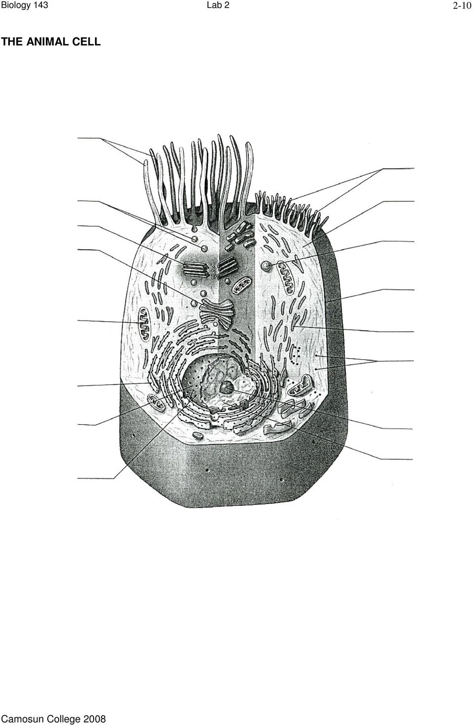
11 Examine a living eukaryotic cell: Using a toothpick, gently scrape the inside of your cheek and spread the material on a slide. Add a drop of methylene blue stain to the smear, and cover the preparation with a coverslip. Examine the preparation with your compound microscope and locate the flat, squamous epithelial cells that line your oral cavity. Bring the cells into focus with the high power objective (400X) and sketch a few cells below (to scale, as you see them in the viewing field) What organelles are visible? Why are most of the "organelles" you just studied not visible? Fig 1. Sketch of squamous epithelial cells from lining of mouth Magnification power =
 and sketch a few cells below (to scale, as you see them in the viewing field) What organelles are visible?")
12 2-12 PART III: INTRODUCTION TO TISSUES In this lab you will be examining various epithelial tissues. You should be able to identify the cells and tissues studied, know the functions of the tissues, and know where in the body each tissue type is found. As the tissues are reviewed in the lab, you should identify each tissue image (at the back of the lab), paste the image in the appropriate space and label any structures / cells indicated. TISSUES 1. Define a "tissue" 2. Name the 4 major tissue types a) b) c) d) EPITHELIAL TISSUES AND GLANDS Epithelial tissues cover body surfaces and line body cavities, organs and vessel lumens. are characterized by a basement membrane, on which the cell layer(s) sit. form both exocrine and endocrine glands You will examine slides of various epithelial tissue types. You should learn to locate and recognize the various types and become familiar with their functions and locations in the body. Describe 4 functions of epithelial tissue:
 b) c) d) EPITHELIAL TISSUES AND GLANDS Epithelial tissues cover body surfaces and line body cavities, organs and vessel lumens.")
13 2-13 Note the flow diagram below indicating the major types of epithelial tissues in terms of cell shape, (squamous, cuboidal, columnar); surface specialization, (plain, ciliated, microvillous): and the number of layers, (simple, pseudostratified, stratified, transitional). Differentiation Simple squamous Apical specializations Layering Nonkeratinized stratified squamous Keratinized stratified squamous Newly mitosed cell Simple cuboidal Microvilli Transitional Cilia Stratified cuboidal Microvilli Simple columnar Stratified columnar Pseudostratified ciliated columnar Cilia SIMPLE EPITHELIAL TISSUES A. Simple squamous FUNCTION: Rapid diffusion of materials through the tissue DESIRED FEATURES: Shortest possible diffusion distance, i.e. thinnest possible tissue REPRESENTATIVE LOCATIONS IN THE BODY: SLIDES: Mammalian lung, section (label squamous cell, alveolar sac)

14 2-14 B. Simple cuboidal FUNCTION: Form walls of small-diameter tubules, some secretion and absorption; general metabolism DESIRED FEATURES: Thicker cells than squamous, but not too thick for small tubules REPRESENTATIVE LOCATIONS IN THE BODY: SLIDES: Mammalian kidney, section. (Note the presence of microvilli in some, but not all of the tubules near the periphery. Those that have them are responsible for about 80% of the kidney's impressive reabsorption capacity) Label cuboidal cell, basement membrane. C. Simple columnar FUNCTION: Absorption of nutrients DESIRED FEATURES: Relatively large cells to house the necessary metabolic machinery and many microvilli REPRESENTATIVE LOCATIONS IN THE BODY: SLIDE: Duodenum, section (label columnar cell nucleus, basement membrane, connective tissue)
 Label cuboidal cell, basement membrane. C.")
15 2-15 STRATIFIED AND PSEUDOSTRATIFIED EPITHELIAL TISSUES A. Stratified squamous FUNCTION: Protection against abrasion and tearing; can be stretched DESIRED FEATURES: Layers of cells on the surface that are expendable and that can be replaced continually from below REPRESENTATIVE LOCATIONS IN THE BODY: SLIDES: Esophagus, section (label squamous cells, basement membrane, connective tissue) B. Pseudostratified ciliated columnar FUNCTION: Trapping of airborne particles that could otherwise become lodged in the lungs DESIRED FEATURES: Production of mucus in which to trap particles and presence of cilia to move the mucus and trapped particles to where they can be disposed of REPRESENTATIVE LOCATIONS IN THE BODY: SLIDE: Trachea, section (label basement membrane, connective tissue, cilia, goblet cell). NOTE: locate a goblet cell; goblet cells are unicellular exocrine glands which secrete mucus NOTE: Glands are typically formed from epithelial tissue.

16 2-16 GLANDULAR EPITHELIUM Epithelial tissue has also become specialized for secretion. Modified epithelial cells form the functional basis of all the glands of the body including both exocrine and endocrine glands. Examine the text and answer the following: A. DISTINGUISH BETWEEN: a) Endocrine gland b) Exocrine gland B. STRUCTURALLY: exocrine glands may be... a) Unicellular: Locate the unicellular exocrine gland, the goblet cell, in the pseudostratified, ciliated, columnar epithelium. Goblet cells secrete a polysaccharide called mucin. When mixed with water, what does this secretion become? What are the functions of this substance? Where else in the body would you expect to find goblet cells? b) Multicellular: If multicellular, duct structure may be i) Simple ii) Compound Both simple and compound multicellular glands may have secretory units described as i) Tubular ii) Alveolar (acinar)
 Unicellular: Locate the unicellular exocrine gland, the goblet cell, in the pseudostratified, ciliated, columnar epithelium. Goblet cells secrete a polysaccharide called mucin.")
17 2-17 C. FUNCTIONAL CLASSIFICATION OF EXOCRINE GLANDS on the basis of how they work. Define each of the following terms - with examples. a) Merocrine b) Holocrine c) Apocrine EXOCRINE GLANDS Unicellular Multicellular Simple Compound Tubular Acinar Tubular Tubuloacinar Acinar D. ENDOCRINE GLANDS (Structure and Function will be studied in association with the endocrine system).

18

19 EPITHELIAL TISSUES

20 1
Name Class Date Laboratory Investigation 4B Chapter 4: Cell Structure
 Name Class Date Laboratory Investigation 4B Chapter 4: Cell Structure The Microscope: A Tool of the Scientist You may refer to pages 66-67, 72-73 in your textbook for a general discussion of microscopes.
Care and Use of the Compound Microscope
 Revised Fall 2011 Care and Use of the Compound Microscope Objectives After completing this lab students should be able to 1. properly clean and carry a compound and dissecting microscope. 2. focus a specimen
MITOSIS IN ONION ROOT TIP CELLS: AN INTRODUCTION TO LIGHT MICROSCOPY
 MITOSIS IN ONION ROOT TIP CELLS: AN INTRODUCTION TO LIGHT MICROSCOPY Adapted from Foundations of Biology I; Lab 6 Introduction to Microscopy Dr. John Robertson, Westminster College Biology Department,
BIO 10 Lab 1 Introduction Pre Lab Test
 BIO 10 Lab 1 Introduction Pre Lab Test 1. Why is the microscope in our lab called a compound microscope? 2. How do you calculate total magnification? 3. What is the lowest and the maximal magnification
Use of the Microscope and Cytology
 Use of the Microscope and Cytology Introduction: A true study of anatomy not only considers the large, visible structures of an organism, but also the small structures that provide the organism its form
EXPERIMENT #1: MICROSCOPY
 EXPERIMENT #1: MICROSCOPY Brightfield Compound Light Microscope The light microscope is an important tool in the study of microorganisms. The compound light microscope uses visible light to directly illuminate
Histology. Epithelial Tissue
 Histology Epithelial Tissue Epithelial Tissue Lines internal and external body surfaces Forms glands Epithelial Tissue Little extracellular matrix Attached on one side Avascular Basement membrane Apical
Lab Exercise 4. Epithelial Tissues. Connective Tissue Proper. What you need to be able to do on the exam after completing this lab exercise:
 Lab Exercise 4 Epithelial Tissues Connective Tissue Proper Textbook Reference: See Chapter 4 What you need to be able to do on the exam after completing this lab exercise: Be able to identify each type
Microscopy and Cellular Morphology
 Microscopy and Cellular Morphology As we discussed in class, many organisms on the planet exist as single cells and are referred to as microorganisms bacteria, protozoans, among others. When a single microorganism
MICROSCOPY. To demonstrate skill in the proper utilization of a light microscope.
 MICROSCOPY I. OBJECTIVES To demonstrate skill in the proper utilization of a light microscope. To demonstrate skill in the use of ocular and stage micrometers for measurements of cell size. To recognize
OBJECTIVES PROCEDURE. Lab 2- Bio 160. Name:
 Lab 2- Bio 160 Name: Prokaryotic and Eukaryotic Cells OBJECTIVES To explore cell structure and morphology in prokaryotes and eukaryotes. To gain more experience using the microscope. To obtain a better
Prokaryotic and Eukaryotic Cells
 Lab 2- Bio 201 Prokaryotic and Eukaryotic Cells Name: OBJECTIVES To explore cell structure and morphology in prokaryotes and eukaryotes. To gain more experience using the microscope, and in particular,
Compound microscope (Hund)
") 1 2 3 4 5 6 7 8 9 10 11 12 13 14 Compound microscope (Hund) 15 16 17 18 19 20 1) Eyepieces (magnifies 10x), one with diopter adjustment, 2) Interp[upillary adjustment, 3) Head, 4) Revolving nosepiece,
1 2 3 4 5 6 7 8 9 10 11 12 13 14 Compound microscope (Hund) 15 16 17 18 19 20 1) Eyepieces (magnifies 10x), one with diopter adjustment, 2) Interp[upillary adjustment, 3) Head, 4) Revolving nosepiece,
Exercise 2. The Compound Light Microscope
 6 Exercise 2 The Compound Light Microscope INTRODUCTION: Student Learning Objectives: After completing this exercise students will: a. Demonstrate proficient use of the microscope using low, high dry,
Animal & Plant Cell Slides
 Animal & Plant Cell Slides Category: Biology Type: Class Experiment, 60 min class Materials: 2 Glass Slides 2 Cover Slips 1 Bottle of methylene blue (optional) 1 Plastic tray 1 Bottle of iodine 1 Plastic
MT-30 & MT-90 Series. Advanced Academic Microscopes/ Advanced Academic Polarizing Microscope INSTRUCTION MANUAL
 Introduction With your purchase of an MT-30/MT-90 series type microscope you have chosen for a quality product. The MT-30/MT-90 series type microscopes are developed for use at schools and laboratories.
THE COMPOUND MICROSCOPE
 THE COMPOUND MICROSCOPE In microbiology, the microscope plays an important role in allowing us to see tiny objects that are normally invisible to the naked eye. It is essential for students to learn how
Biology 13A Lab #3: Cells and Tissues
 Biology 13A Lab #3: Cells and Tissues Lab #3 Table of Contents: Expected Learning Outcomes.... 28 Introduction...... 28 Activity 1: Eukaryotic Cell Structure... 29 Activity 2: Perspectives on Tissue Preparations.
MICROSCOPY OF LIVING MICROBES
 EXPERIMENT 1 MICROSCOPY OF LIVING MICROBES Many students taking microbiology for the first time feel that they are going to have a hard time with the microscope. This lab as an experiment is intended to
Cell Biology Prokaryotic and eukaryotic cells
 Cell Biology Prokaryotic and eukaryotic cells Observation of cells and organelles In this lab you will be looking at an example of a Prokaryotic cell (Bacillus cereus) and a some examples of Eukaryotic
Chapter 4: A Tour of the Cell. 1. Cell Basics. Limits to Cell Size. 1. Cell Basics. 2. Prokaryotic Cells. 3. Eukaryotic Cells
 Chapter 4: A Tour of the Cell 1. Cell Basics 2. Prokaryotic Cells 3. Eukaryotic Cells 1. Cell Basics Limits to Cell Size There are 2 main reasons why cells are so small: If cells get too large: 1) there
Animal Tissues. I. Epithelial Tissue
 Animal Tissues There are four types of tissues found in animals: epithelial tissue, connective tissue, muscle tissue, and nervous tissue. In this lab you will learn the major characteristics of each tissue
Students will identify these animal cell structures: Students should properly answer the pre-activity cell membrane, nucleus. questions.
 WHAT DO PLANT & ANIMAL CELLS LOOK LIKE? Grade Levels: 10-12 Time Frame: 2 periods Big Idea: Students will compare various plant epithelial cells (onion and elodea) with human epithelial cells (cheek lining
The Cell Interior and Function
 The Cell Interior and Function 5 5.0 CHAPTER PREVIEW Investigate and understand the organization and function of the cell interior. Define the differences between eukaryotic and prokaryotic cell structure.
Comparing Plant and Animal Cells
 1.2 Comparing Plant and Animal Cells Here is a summary of what you will learn in this section: Plant and animal cell structures are called organelles. Plant and animal cells perform some similar functions,
Pre-Lab Questions. 1. What is cell theory? 2. What do all cells contain? 3. What is a prokaryote? 4. What is a eukaryote? 5. What is an organelle?
 Name: TOC# Background Ever since the first microscope was used, biologists have been interested in studying the cellular organization of all living things. After hundred s of years of observations by many
LAB 3 Use of the Microscope
 LAB 3 Use of the Microscope Introduction In this laboratory you will be learning how to use one of the most important tools in biology the compound light microscope to view a variety of specimens. You
Section B: Epithelial Tissue 1. Where are epithelial tissues found within the body? 2. What are the functions of the epithelial tissues?
 Tissue worksheet Name Section A: Intro to Histology Cells are the smallest units of life. In complex organisms, cells group together with one another based on similar structure and function to form tissues.
Biology Chapter 7 Practice Test
 Biology Chapter 7 Practice Test Multiple Choice Write the letter that best answers the question or completes the statement on the line provided. 1. The work of Schleiden and Schwann can be summarized by
7.2 Cell Structure. Lesson Objectives. Lesson Summary. Cell Organization Eukaryotic cells contain a nucleus and many specialized structures.
 7.2 Cell Structure Lesson Objectives Describe the structure and function of the cell nucleus. Describe the role of vacuoles, lysosomes, and the cytoskeleton. Identify the role of ribosomes, endoplasmic
Biology 101 Chapter 4 Cells as the Basic Unit of Life. The Cell Theory Major Contributors: Galileo = first observations made with a microscope
 Biology 101 Chapter 4 Cells as the Basic Unit of Life The Cell Theory Major Contributors: Galileo = first observations made with a microscope Robert Hooke = first to observe small compartments in dead
Review of the Cell and Its Organelles
 Biology Learning Centre Review of the Cell and Its Organelles Tips for most effective learning of this material: Memorize the names and structures over several days. This will help you retain what you
Lesson Aim To explain the human body at a microscopic level, including the structure and function of cells, tissues and membranes.
 LESSON 1. CELLS & TISSUES Lesson Aim To explain the human body at a microscopic level, including the structure and function of cells, tissues and membranes. THE CELL All living matter is composed of functional
Microscope Lab Introduction to the Microscope Lab Activity
 Microscope Lab Introduction to the Microscope Lab Activity Wendy Kim 3B 24 Sep 2010 http://www.mainsgate.com/spacebio/modules/gs_resource/ CellDivisionMetaphase.jpeg 1 Introduction Microscope is a tool
Cells. Introduction WSBCTC 1
 Cells Cells are the fundamental unit of life. All living things are composed of cells. While there are several characteristics that are common to all cells, such as the presence of a cell membrane, cytoplasm,
The Cell: Organelle Diagrams
 The Cell: Organelle Diagrams Fig 7-4. A prokaryotic cell. Lacking a true nucleus and the other membrane-enclosed organelles of the eukaryotic cell, the prokaryotic cell is much simpler in structure. Only
Chapter 3. Cellular Structure and Function Worksheets. 39 www.ck12.org
 Chapter 3 Cellular Structure and Function Worksheets (Opening image copyright by Sebastian Kaulitzki, 2010. Used under license from Shutterstock.com.) Lesson 3.1: Introduction to Cells Lesson 3.2: Cell
The microscope is an important tool.
 KEY CONCEPT Microscopes allow us to see inside the cell. BEFORE, you learned Some organisms are unicellular and some are multicellular A microscope is necessary to study most cells The cell theory describes
3.1 AS Unit: Cells, Exchange and Transport
 3.1 AS Unit: Cells, Exchange and Transport Module 1: Cells 1.1.1 Cell Structure Candidates should be able to: (a) state the resolution and magnification that can be achieved by a light microscope, a transmission
Cell Structure and Function. Eukaryotic Cell: Neuron
 Cell Structure and Function Eukaryotic Cell: Neuron Cell Structure and Function Eukaryotic Cells: Blood Cells Cell Structure and Function Prokaryotic Cells: Bacteria Cell Structure and Function All living
Chapter 1 Parts C. Robert Bagnell, Jr., Ph.D., 2012
 Chapter 1 Parts C. Robert Bagnell, Jr., Ph.D., 2012 Figure 1.1 illustrates the parts of an upright compound microscope and indicates the terminology that I use in these notes. Figure 1.1. Parts of a Compound
COMPARING PLANT AND ANIMAL CELLS
 COMPARING PLANT AND ANIMAL CELLS OBJECTIVES: Distinguish between plant and animals cells by their structures Demonstrate the benefit of stains Acquire ability to prepare wet mounts SAFETY: Methylene blue
Microscopy. MICROSCOPY Light Electron Tunnelling Atomic Force RESOLVE: => INCREASE CONTRAST BIODIVERSITY I BIOL1051 MAJOR FUNCTIONS OF MICROSCOPES
 BIODIVERSITY I BIOL1051 Microscopy Professor Marc C. Lavoie marc.lavoie@cavehill.uwi.edu MAJOR FUNCTIONS OF MICROSCOPES MAGNIFY RESOLVE: => INCREASE CONTRAST Microscopy 1. Eyepieces 2. Diopter adjustment
HISTOLOGY LABORATORY. Microscope Orientation and Blood Smear Lab
 HISTOLOGY LABORATORY Microscope Orientation and Blood Smear Lab For practicing how to use the microscope DO NOT use the blood smear slide (it is too boring for the lower mags). Use a slide from the white
Tissues (Histology) Ch. 3 Human Anatomy lecture
 Ch. 3 Human Anatomy lecture") I. Histology the study of tissues A. 4 basic tissue types epithelial connective muscle nervous Tissues (Histology) Ch. 3 Human Anatomy lecture B. Usually found in combinations to form organs. C. As you
I. Histology the study of tissues A. 4 basic tissue types epithelial connective muscle nervous Tissues (Histology) Ch. 3 Human Anatomy lecture B. Usually found in combinations to form organs. C. As you
Human Anatomy & Physiology I with Dr. Hubley. Practice Exam 1
 Human Anatomy & Physiology I with Dr. Hubley Practice Exam 1 1. Which definition is the best definition of the term gross anatomy? a. The study of cells. b. The study of tissues. c. The study of structures
National Optical & Scientific Instruments Inc. 11113 Landmark 35 Drive San Antonio, Texas 78233 Phone (210) 590-9010 Fax (210) 590-1104
 590-9010 Fax (210) 590-1104") National Optical & Scientific Instruments Inc. 11113 Landmark 35 Drive San Antonio, Texas 78233 Phone (210) 590-9010 Fax (210) 590-1104 INSTRUCTIONS FOR MODELS 156, 156-S, 157 COMPOUND BIOLOGICAL MICROSCOPES
National Optical & Scientific Instruments Inc. 11113 Landmark 35 Drive San Antonio, Texas 78233 Phone (210) 590-9010 Fax (210) 590-1104 INSTRUCTIONS FOR MODELS 156, 156-S, 157 COMPOUND BIOLOGICAL MICROSCOPES
Cells & Cell Organelles
 Cells & Cell Organelles The Building Blocks of Life H Biology Types of cells bacteria cells Prokaryote - no organelles Eukaryotes - organelles animal cells plant cells Cell size comparison Animal cell
Compartmentalization of the Cell. Objectives. Recommended Reading. Professor Alfred Cuschieri. Department of Anatomy University of Malta
 Compartmentalization of the Cell Professor Alfred Cuschieri Department of Anatomy University of Malta Objectives By the end of this session the student should be able to: 1. Identify the different organelles
Chapter 2: Cell Structure and Function pg. 70-107
 UNIT 1: Biochemistry Chapter 2: Cell Structure and Function pg. 70-107 Organelles are internal structures that carry out specialized functions, interacting and complementing each other. Animal and plant
Microscopes. Eukaryotes Eukaryotic cells are characterized by having: DNA in a nucleus that is bounded by a membranous nuclear envelope
 CH 6 The Cell Microscopy Scientists use microscopes to visualize cells too small to see with the naked eye. In a light microscope (LM), visible light is passed through a specimen and then through glass
Bacterial (Prokaryotic) Cell. Common features of all cells. Tour of the Cell. Eukaryotic Cell. Plasma Membrane defines inside from outside
 Cell. Common features of all cells. Tour of the Cell. Eukaryotic Cell. Plasma Membrane defines inside from outside") www.denniskunkel.com Tour of the Cell www.denniskunkel.com Today s Topics Properties of all cells Prokaryotes and Eukaryotes Functions of Major Cellular Organelles Information, Synthesis&Transport,, Vesicles
www.denniskunkel.com Tour of the Cell www.denniskunkel.com Today s Topics Properties of all cells Prokaryotes and Eukaryotes Functions of Major Cellular Organelles Information, Synthesis&Transport,, Vesicles
Lecture 4 Cell Membranes & Organelles
 Lecture 4 Cell Membranes & Organelles Structure of Animal Cells The Phospholipid Structure Phospholipid structure Encases all living cells Its basic structure is represented by the fluidmosaic model Phospholipid
CELLS: PLANT CELLS 20 FEBRUARY 2013
 CELLS: PLANT CELLS 20 FEBRUARY 2013 Lesson Description In this lesson we will discuss the following: The Cell Theory Terminology Parts of Plant Cells: Organelles Difference between plant and animal cells
Eukaryotic Cell Structure: Organelles in Animal & Plant Cells Why are organelles important and how are plants and animals different?
 Why? Eukaryotic Cell Structure: Organelles in Animal & Plant Cells Why are organelles important and how are plants and animals different? The cell is the basic unit and building block of all living things.
the plant & animal cell
 6.1 Basic unit of life Biology Biology Structure & functions of 06 the plant & animal cell In 1665, Robert Hooke observed a section of a cork using a microscope prepared by him. He discovered a structure
Chapter 5 Organelles. Lesson Objectives List the organelles of the cell and their functions. Distinguish between plant and animal cells.
 Chapter 5 Organelles Lesson Objectives List the organelles of the cell and their functions. Distinguish between plant and animal cells. Check Your Understanding What is a cell? How do we visualize cells?
The Tissue Level of Organization
 The Tissue Level of Organization Tissues A groups of similar cells, usually having similar embryonic origin and specialized function Histology: the study of tissues Four general types Epithelial Muscle
Plant and Animal Cells
 Plant and Animal Cells a. Explain that cells take in nutrients in order to grow, divide and to make needed materials. S7L2a b. Relate cell structures (cell membrane, nucleus, cytoplasm, chloroplasts, and
Chapter 4. Microscopy, Staining, and Classification. Lecture prepared by Mindy Miller-Kittrell North Carolina State University
 Chapter 4 Microscopy, Staining, and Classification 2012 Pearson Education Inc. Lecture prepared by Mindy Miller-Kittrell North Carolina State University Microscopy and Staining 2012 Pearson Education Inc.
THE HISTORY OF CELL BIOLOGY
 SECTION 4-1 REVIEW THE HISTORY OF CELL BIOLOGY Define the following terms. 1. cell 2. cell theory Write the correct letter in the blank. 1. One early piece of evidence supporting the cell theory was the
Using a Microscope to See Different Types of Cells
 Using a Microscope to See Different Types of Cells copyright 2003 by Dr. Vivianne Nachmias, University of Pennsylvania All organisms are made up of cells - a cell is the simplest collection of matter that
tissues are made of cells that work together, organs are )
") Study Guide Cells Unit Test Matching. Write the letter of the correct response on the line. You may use the responses more than once. A. proteins B. simple carbohydrates C. complex carbohydrates D. lipids
Study Guide Cells Unit Test Matching. Write the letter of the correct response on the line. You may use the responses more than once. A. proteins B. simple carbohydrates C. complex carbohydrates D. lipids
cells - relatively simple cells - lack nuclear membrane and many organelles - bacteria and their relatives are all prokaryotic
 Cell Biology A cell is chemical system that is able to maintain its structure and reproduce. Cells are the fundamental unit of life. All living things are cells or composed of cells. 1 The interior contents
3.1 Cells and cell function
 BTEC s own resources 3.1 Cells and cell function In this section: P1 How you are made Key terms Tissue a group of similar cells acting together to perform a particular function. Epithelial cells one of
How To Use An Asbestos Microscope
 Asbestos Microscopes and Accessories Pyser-SGI has been supplying microscopes and accessories into Asbestos Laboratories for over 40 years PS12 Stage Micrometer with UKAS Certificate of Calibration - For
The illustrations below reflect other scientists results in identifying and counting the stages of the onion root tip and the whitefish blastula.
 Abstract: The purpose of this laboratory experiment was to identify in what stage of mitosis viewed cells were in. The stages of mitosis include prophase, metaphase, anaphase and telophase. Although the
Eukaryotes. www.njctl.org PSI Biology Eukaryotes & Gene Expression
 Eukaryotes The Eukaryotic Cell Classwork 1. Identify two characteristics that are shared by all cells. 2. Suppose you are investigating a cell that contains a nucleus. Would you categorize this cell as
Cytology. Living organisms are made up of cells. Either PROKARYOTIC or EUKARYOTIC cells.
 CYTOLOGY Cytology Living organisms are made up of cells. Either PROKARYOTIC or EUKARYOTIC cells. A. two major cell types B. distinguished by structural organization See table on handout for differences.
Laboratory 3 Histology
 Laboratory 3 Histology Goals: For epithelial tissues: o discuss the major features; o classify based on simple/stratified and squamous/cubodial/columnar; o identify each type by microscopy; o identify
National Optical & Scientific Instruments Inc. 11113 Landmark 35 Drive San Antonio, Texas 78233 Phone (210) 590-9010 Fax (210) 590-1104
 590-9010 Fax (210) 590-1104") National Optical & Scientific Instruments Inc. 11113 Landmark 35 Drive San Antonio, Texas 78233 Phone (210) 590-9010 Fax (210) 590-1104 INSTRUCTIONS FOR MODELS 106, 106-L 107, 107-L 108, 108-L 109-L ELEMENTARY
National Optical & Scientific Instruments Inc. 11113 Landmark 35 Drive San Antonio, Texas 78233 Phone (210) 590-9010 Fax (210) 590-1104 INSTRUCTIONS FOR MODELS 106, 106-L 107, 107-L 108, 108-L 109-L ELEMENTARY
EXTRACTION OF DNA FROM CALF THYMUS CELLS Revised 2/1/96 Introduction
 Revised 2/1/96 Introduction Cells may be classified into two primary types depending on whether they have a discrete nucleus (eukaryotic) or do not (prokaryotic). Prokaryotes include bacteria, such as
DETECTION OF BACTERIAL MOTILITY. To demonstrate bacterial motility by microscopic and macroscopic techniques.
 DETECTION OF BACTERIAL MOTILITY I. OBJECTIVES To demonstrate bacterial motility by microscopic and macroscopic techniques. To observe flagella in prepared slides stained by specific flagellar stains. II.
7.2 Cells: A Look Inside
 CHAPTER 7 CELL STRUCTURE AND FUNCTION 7.2 Cells: A Look Inside Imagine a factory that makes thousands of cookies a day. Ingredients come into the factory, get mixed and baked, then the cookies are packaged.
Introduction to the Cell: Plant and Animal Cells
 Introduction to the Cell: Plant and Animal Cells Tissues, Organs, and Systems of Living Things Cells, Cell Division, and Animal Systems and Plant Systems Cell Specialization Human Systems All organisms
A Fishy Tale. Observing the Circulatory System of a Goldfish with a Compound Light Microscope
 A Fishy Tale Observing the Circulatory System of a Goldfish with a Compound Light Microscope A Fishy Tale About this Lesson In this lesson, students will explore a computer animation of the human body
Plasma Membrane hydrophilic polar heads
 The Parts of the Cell 3 main parts in ALL cells: plasma membrane, cytoplasm, genetic material this is about the parts of a generic eukaryotic cell Plasma Membrane -is a fluid mosaic model membrane is fluid
Quick Hit Activity Using UIL Science Contests For Formative and Summative Assessments of Pre-AP and AP Biology Students
 Quick Hit Activity Using UIL Science Contests For Formative and Summative Assessments of Pre-AP and AP Biology Students Activity Title: Quick Hit Goal of Activity: To perform formative and summative assessments
Prokaryotic and Eukaryotic Cells
 Why? Prokaryotic and Eukaryotic Cells Do all cells have the same structure? An efficiency apartment is a one-room apartment. This one room is where you sleep, eat, shower, and entertain your guests. It
Objective: On a team of no more than (2). Build to illustrate a 3D model of a PLANT or ANIMAL cell. 10 pts.
. Build to illustrate a 3D model of a PLANT or ANIMAL cell. 10 pts.") THE CELL model: Activity 4.1 Science / Biology Objective: On a team of no more than (2). Build to illustrate a 3D model of a PLANT or ANIMAL cell. - Your models should clearly demonstrate the following
THE CELL model: Activity 4.1 Science / Biology Objective: On a team of no more than (2). Build to illustrate a 3D model of a PLANT or ANIMAL cell. - Your models should clearly demonstrate the following
Gymnázium, Brno, Slovanské nám. 7, WORKBOOK - Biology WORKBOOK. www.gymnaslo.agb.cz
 WORKBOOK www.gymnaslo.agb.cz Subjekt: Biology Teacher: Iva Kubištová Student:.. School year:../. This material was prepared with using http://biologygmh.com/ Topics: 1. 2. 3. Cell Structure and Function
Biology I. Chapter 7
 Biology I Chapter 7 Interest Grabber NOTEBOOK #1 Are All Cells Alike? All living things are made up of cells. Some organisms are composed of only one cell. Other organisms are made up of many cells. 1.
CHAPTER 2 : CELL AS THE BASIC UNIT OF LIFE
 CHAPTER 2 : CELL AS THE BASIC UNIT OF LIFE Parts of microscope : An instrument that magnifies minute objects so they can be seen easily. It is one of the most important tools of science. Physicians and
Do Not Write on this Quiz Paper (südamlik aitäh)
") 1. This makes ribosomes. Cell Organelle Quiz Do Not Write on this Quiz Paper (südamlik aitäh) a. Rough ER c. Golgi apparatus (body) b. Nucleolus d. Mitochondria 2. This is an energy producing organelle.
1. This makes ribosomes. Cell Organelle Quiz Do Not Write on this Quiz Paper (südamlik aitäh) a. Rough ER c. Golgi apparatus (body) b. Nucleolus d. Mitochondria 2. This is an energy producing organelle.
Microscopes and the Metric System
 Microscopes and the Metric System BIO162 Fall 2007 Sizes of Microorganisms: -Viruses: 0.01 0.3 um -Bacteria: 1 3 um -Fungi: 3 30 um -Protozoa: 5 1000 um 1 Measuring Microorganisms Ocular Micrometer The
How Well Do You Know Your Cells?
 How Well Do You Know Your Cells? Complete each sentence below with words from the box. One word will not be used. cells cell membrane cell walls chloroplasts cytoplasm Hooke Leeuwenhoek mitochondria nucleus
Biology 105 Human Biology PRACTICE MIDTERM EXAM 1. Essentials of Anatomy and Physiology, 5e (Martini/Nath) Chapter 4 The Tissue Level of Organization
 Chapter 4 The Tissue Level of Organization") Essentials of Anatomy and Physiology, 5e (Martini/Nath) Chapter 4 The Tissue Level of Organization Multiple-Choice Questions 1) The four main types of tissues include A) epithelium. B) muscle. C) nerve.
Essentials of Anatomy and Physiology, 5e (Martini/Nath) Chapter 4 The Tissue Level of Organization Multiple-Choice Questions 1) The four main types of tissues include A) epithelium. B) muscle. C) nerve.
Date: Student Name: Teacher Name: Jared George. Score: 1) A cell with 1% solute concentration is placed in a beaker with a 5% solute concentration.
 A cell with 1% solute concentration is placed in a beaker with a 5% solute concentration.") Biology Keystone (PA Core) Quiz Homeostasis and Transport - (BIO.A.4.1.1 ) Plasma Membrane, (BIO.A.4.1.2 ) Transport Mechanisms, (BIO.A.4.1.3 ) Transport Facilitation Student Name: Teacher Name: Jared
Biology Keystone (PA Core) Quiz Homeostasis and Transport - (BIO.A.4.1.1 ) Plasma Membrane, (BIO.A.4.1.2 ) Transport Mechanisms, (BIO.A.4.1.3 ) Transport Facilitation Student Name: Teacher Name: Jared
The Living Cell from the Biology: The Science of Life Series. Pre-Test
 1 Pre-Test Directions: Answer each question TRUE OR FALSE. 1. The instructions for making proteins are stored in molecules of DNA. 2. Proteins are made in the nucleus. 3. All cells are surrounded by a
Lab 4 Cell Structure, Osmosis, and Diffusion
 Lab 4 Cell Structure, Osmosis, and Diffusion Introduction: Connecting Your Learning The basic building block of life is the cell. Each cell contains several structures, some of which are common to both
The Cell Teaching Notes and Answer Keys
 The Cell Teaching Notes and Answer Keys Subject area: Science / Biology Topic focus: The Cell: components, types of cells, organelles, levels of organization Learning Aims: describe similarities and differences
CHAPTER 5: TISSUES. 2. Name the four primary adult tissue types, and give a brief description of each.
 OBJECTIVES: 1. Define the term tissue. 2. Name the four primary adult tissue types, and give a brief description of each. 3. Describe the functions and types of extracellular fluid (ECF). 4. Compare and
The Cell Grade Ten. Estimated Duration: Three hours
 Ohio Standards Connection: Life Sciences Benchmark A Explain that cells are the basic unit of structure and function of living organisms, that once life originated all cells come from pre-existing cells,
Biological cell membranes
 Unit 14: Cell biology. 14 2 Biological cell membranes The cell surface membrane surrounds the cell and acts as a barrier between the cell s contents and the environment. The cell membrane has multiple
A new advance in routine inspections INVERTED MICROSCOPE CKX41/CKX31
 A new advance in routine inspections INVERTED MICROSCOPE CKX41/CKX31 Phase contrast Relief contrast Incorporation of advanced UIS2 optics ensures the highest level of clarity for cell checking applications.
City Part Function Cell Part Controls what goes in and
 Answer key: CELL CITY INTRODUCTION! Floating around in the cytoplasm are small structures called organelles. Like the organs in your own body, each one carries out a specific function necessary for the
Plant and Animal Cells
 Plant and Animal Cells Cell Scientists Hans and Zacharias Janssen Dutch lens grinders, father and son produced first compound microscope (2 lenses) Robert Hooke (1665) English Scientist looked at a thin
Cells. Structure, Function and Homeostasis
 Cells Structure, Function and Homeostasis Characteristics of Cells Basic unit of life anything alive is made of cells Plasma membrane (skin) that separates them from the environment. Skeletonsfor protection
Cell Structure & Function!
 Cell Structure & Function! Chapter 3! The most exciting phrase to hear in science, the one that heralds new discoveries, is not 'Eureka!' but 'That's funny.! -- Isaac Asimov Animal Cell Plant Cell Cell
Cellular Structure and Function
 Chapter Test A CHAPTER 7 Cellular Structure and Function Part A: Multiple Choice In the space at the left, write the letter of the term or phrase that best answers each question. 1. Which defines a cell?