Study on the Efferent Innervation of the Body Wall Musculature of Lumbricus Terrestris (L)
|
|
|
- Fay Anderson
- 6 years ago
- Views:
Transcription
\" (1975). Master's Theses. Paper 2749.")
1 Loyola University Chicago Loyola ecommons Master's Theses Theses and Dissertations 1975 Study on the Efferent Innervation of the Body Wall Musculature of Lumbricus Terrestris (L) Carol A. Aslam Loyola University Chicago Recommended Citation Aslam, Carol A., "Study on the Efferent Innervation of the Body Wall Musculature of Lumbricus Terrestris (L)" (1975). Master's Theses. Paper This Thesis is brought to you for free and open access by the Theses and Dissertations at Loyola ecommons. It has been accepted for inclusion in Master's Theses by an authorized administrator of Loyola ecommons. For more information, please contact This work is licensed under a Creative Commons Attribution-Noncommercial-No Derivative Works 3.0 License. Copyright 1975 Carol A. Aslam
2 STUDY ON THE EFFERENT INNERVATION OF THE BODY WALL ~USCULATURE OF LUMBRICUS TERRESTRIS (L.) by Carol Aslam A Thesis Submitted to the Faculty of the Graduate School of Loyola University of Chicago in Partial Fulfillment of the Requirements for the Degree of Master of Science November '. s. '.
3 ACKNOWLEDGMENTS The author will always be indebted to her advisor, Dr. Robert Hadek, for unfailing support and scientific criticism throughout the preparation of this manuscript. Special thanks are also due to members of the Department of Anatomy who generously gave of their time, counsel and technical assistance. The encouragement of my husband and enduring patience of my children have made possible the completion of this program. ii
4 BIOGkAPllY Carol A. Aslam was born on November 1, 1943, in Ioledo. Ohio. She was graduated from St. Ursula Academy, in June, 1961 in Toledo, Ohio and continued her studies at the University of Toledo, Toledo, Ohio, where she received her Registry in Radiologic Technology in June, She completed her undergraduate studies at Illinois Benedictine College, Lisle, Illinois, and received the degree of Bachelor of Science in Biology in August, In July, 1973, she began her graduate studies in the Department of Anatomy, Loyola University, Stritch School of Medicine, Maywood, Illinois. In 1974 she elected Dr. Robert Uadel as her advisor and has been working with him since. iii
5 ACKNOWLEDGMENTS.. LIFE... TABLE OF CONTENTS. INTRODUCTION..... A. B. A General Description. The Problem. REVIEW OF THE LITERATURE Page ii 111 A. The Ventral Nerve Cord 5 B. The Peripheral Nervous System.. 7 (1) Segmental Innervation. 8 (2) The Peripheral Plexus. 9 C. Muscle-Nerve Relationships 11 (1) Preterminal Nerve Fiber Characteristics. 11 (2) Terminal Nerve Fiber Characteristics 12 (3) Physiological Implications 13 (4) Neuromuscular Junctions D. An Account of Secretory Vesicles Within the Earthworm Nervous System 14 (1) Central Nervous System 14 (2) Peripheral Nervous System. 14 iv 1 l l s MATERIAL AND METHODS A. A Description of the Animal B. Care c. Light Microscopy D. Electron Microscopy RESULTS AND OBSERVATIONS A. General Organization of the Peripheral Nervous System, Light Microscopy. 23 B. Fine Structure of the Peripheral Nervous System, Electron Microscopy DISCUSSION A. B. c. Light Microscopic Methods..... Electron Microscopic Methods Histological and Ultrastructural Findings iv
6 SUMMARY AND CONCLUSIONS..... BIBLIOGRAPHY PLATES AND FIGURES Page v
7 INTRODUCTION A. A General Description The earthworm Lumbricus terrestris (L.) on which this study was done is an oligochaete of the phylum Annelida. It is characterized by externally visible segments and has a length of approximately 150 millimeters. The shape of this worm is cylindrical and flattened ventrally. The dorsal surface is a shade of deep brown bisected by a median longitudinal dark line running the entire length of the animal that is the mid-dorsal blood vessel. The anterior end exhibits a small knob which is called the prostomium, or lip. The body wall is composed of several layers of tissue including a thin transparent cuticle which covers the whole external surface. Beneath the epidermis are the two muscle layers which are the main constituents of the body wall. The outer circular layer is SOOu thick with fibers oriented circumferentially. while the inner longitudinal muscle layer is thicker (looou) and composed of pinnately arranged bundles. The muscle cells in both layers are morphologically identical (Mill and Knappt 1970). Both muscle layers are interrupted at the border of each somite by a connective tissue septa! wall l
8 .l that partially divides the body celom into individual compartments. The celomic cavities are lined by peritoneum and are filled with fluid which acts as a hydrostatic skeleton, rendering the body semi-rigid. The organization of the nervous system on which this study has been done is intermediate between the nerve net of the radially symmetrical coelenterate and the well organized central nervous system of the arthropods (Dorsett, 1964). The central nervous system consists of: (1) a bilo bed sup rap haryngeal ganglion (the "bra in") situa ted dorsally at the level where the oral cavity joins the pharynx (somite three); (2) a pair of circumpharyngeal connectives that extend from this dorsally situated ''brain" to the ventral region where they fuse to form the paired subpharyngeal ganglion (somite four) and (3) a set of nerve cords originating from this paired structure, fused at somite four that continued along the entire length of the body on the ventral floor of the celom, as the ventral nerve cord. One segmental ganglion in each somite can be seen with a dissecting microscope (Adey, 1951). In each somite three pairs of nerves course in a ventro-lateral direction toward the body wall musculature (Dawson, 1920; Drewes and Pax, 1973). Collectively, the lateral nerves are called the segmental nerves and typically they are present in each segment. These lateral nerves contribute to the innerva-
9 3 tion of the body wall musculature and the mucous gland cells. This is accomplished by means of: (a) a nerve ring located bet ween tile circular and longitudinal muscle 1 ayers, (b) through a subepidermal plexus located betw~en the epidermis and the circular muscle layer and (c) an internal uerve net located in the body wall musculature. The internal nerve net is a plexus of nerve fibers which surround muscles in the circular and longitudinal layers. The subevidermal plexus and internal nerve net are in communicatioo with each other throughout the length of the earthworm (Hess, 1925). B. The Problem While earlier research on the neuromuscular system of the earthworm was concerned with the general features of neuromuscular ju11ctions, (Rctzius, 1892; Smallwood, 1926) contemporary studies have concentrated on its fine structure (Mill and Knapp, 1970, Rosenbluth, 1972). The most recent investigation has revealed the presence of neuromuscular junctions in the earthworm consistent with features demonstrated in a variety of other species (Rosenbluth, 1972). Rosenbluth (1972) described the characteristics of neuromuscular junctions by: {i) morphology of axonal vesicles, {ii) gap width and (iii) postjunctional membrane specialization. Nevertheless, these morphological features proved inadequate to characterize specific
10 4 functions (e.g., fast versus slow muscular contraction; excitatory versus inhibitory responses, etc.). PhyRiological studies indicated that responses observed in the body wall muscle vere a result of dual innervation (Dorsett, 1964; Hidaka, Ito, Kuriyama and Tashiro, 1969: Drewes and Pax, 1974). The purpose of this study is to substantiate earlier descriptions of neuromuscular junctions, using light and electron microscopy. Earlier studies have failed to relate neuromuscular junctions with either a plexi system (a vestige of the nerve net) or the centrally organized segmental nervous system which may dually innervate the body wall musculature of Lumbricus terrestris (L.). This study will attempt to show that earthworm body wall muscle is dually innervated by plexi (whose possible origin is being described for the first time) and the segmental system. Further morphological assess~ent can then be made on the bases of end plate morphology, gap width and postjunctional membrane specialization. These two systems may well represent tbe morphological basis for the differing physiological properties which have not been previously identified morphologically, nor adequately explained.
11 REVIEW OF THE LITERATURE Since the first histological studies on the earthworm by Retzius (1892) theru have been numerous histological, biochemical and physiological studies on the nervous system of annelids (Hess, 1925, 1925a; Smallwood, 1926, 1928; Stough, 1~26; Bullock, 1945; Adey, 1951; Wilson, 1960; Hidaka, Ito, Kuriyama. Tashiro, 1969; Mill and Knapp, 197~, Ehinger and Myhrberg, 1971; ~yhrberg, 1972~ Rosenbluth, 1972; Drewes and Pax, 1974). This review will also include those studies on the neuromuscular junction in Lumbr icl!-_!_!~~-est~is_..j.1.!.). A. The Ventral Nerve Cord The nervous system of the earthworm has been the subject of numerous studies and reviews (Retzius, 1892; Langdon, 1895; Hess, 1925; Smallwood, 1926; Stough, 1926; Bullock. 1945; Adey, 1951~ Horridge, 1959; Wilson, 1960). The consensus of these investigators was that the ventral portion of the central nervous system was composed of a giant fiber system and a ventral nerve cord that arose at the level of the fourth segment and continued to the caudal end of the body. The giant fiber system and the ventral nerve cord have been functionally coupled to the startle response as well as to 5
12 6 reflexes concerned with slower ambulatory and hurrowing movements respectively (Bullock and Horridge. 1965). There were three giant fibers in the ventral nerve cord; one median and two lateral ones. The size of the central fiber as a rule was consistent throughout the cord while the lateral fibers are subject to fluctuations (Stough, 1926). Tlu~ two lateral fibers were freely interconnected throughout the cord (Stough, 1926; Bullock, 1945). Bullock (1945) has shown that in Lumbricus, a tactile stimulation of the anterior segments of the body e&cited the median fiber, while stimulation of the body caudal to segment 40 excited the lateral fibers. The lateral giant f ibcr system was probably intended primarily to subscrve anterior conduction of stimuli from the posterior segments while the median fiber conducted impulses from the anterior region to the posterior part (Bullock, 1945). On transverse section, the ventral nerve cord was found to contain three types of cell bodies: unipolar, bipolar and multipolar (Adey, 1951; Bullock and Horridge, 1965). In transverse section, centrally located nerve cells were within an encircling neuropile masa that in turn was enclosed within a peripheral circle of nerve cell bodies comprising the rind. These cells. the central as well as the peripheral ones were either motor or inter-
13 7 neuron cell bodies. On the strength of their location they could not be segregated into functional groups. (1) Interneurons Sensory information is relayed from the periphery (epidermis) to the ventral nerve cord in the following way; upon receiving a stimulus, receptor cells in the epidermis propogate an impulse through their axons which synapse on interneurons in the segmental ganglia. (These axons are the afferent portion of the lateral nerve in every segment). The interneurons relay information to the giant fiber system. The giant fiber system is the sensory and motor integrator of the central nervous system (Bullock and Horridge, 1965). (ii) Motor Neurons Motor neurons are characterized by a cell body with a diameter of 20u-60u, which possesses an axon that passes toward the adjacent segmental nerve. The axon cleaves into two branches; one courses in the region of the giant fiber system while the other enters the segmental nerve. Adey, (1951) assumed that these cells were at least part of the efferent pathway to the musculature of the body wall. B. The Peripheral Nervous System The peripheral nervous system is composed of: (1) the segmental innervation and (2) the peripheral plexus.
14 8 (1) Segmental Innervation Each segment of the earthworm is supplied by three pairs of lateral nerves whose axons originate in the cell bodies of the ventral nerve cord. Hes~ (1925) found that these lateral nerves send brauches to one segment only. Bullock and Horridge's studies (1965) indicate that all three pairs of lateral nerves contain afferent and efferent fibers. Cell bodies of the afferent fibers are located in the periphery, in or beneath the epidermis, in the muscles, or other tissues along the course of the nerves, but never in the central nervous system (Bullock and Horridge, 1965). The afferents are most heavily pronounced in the second lateral nerve while the efferents are most heavily pronounced in the third. In each segment the first lateral nerve arises more rostrally than the second and the third. The first lateral nerve is responsible for innervation of the segment it arises from and the caudalmost portions of the adjacent cranial segment. The second lateral nerve innervates only the segment from which it arises, while the third lateral nerve innervates its own segment along with the anterior portions of the next most caudal segment (Drewes and Pax, 1973). Upon entering the body wall musculature, branches are given off to the longitudinal muscle layer at its celomic border (Smallwood, 1926}. The bulk of the segmental branches course between
15 9 the circular and longitudinal muscle layers of the body wall, forming a nerve ring. The nerve ring has been shown to send branches into the muscle layers as well as branches to the subepidermal plexus and to the sensory receptors in the epidermis (Hess, 1925; Smallwood, 1928). the subepidermal plexus is an accumulation of anastomosing nerve fibers that show no signs of being segmental in arrangement (tless, 1925). These fibers communicate with an internal nerve net (located within the circular and longitudinal muscle layers) and with the ventral nerve cord where they form synaptic connections with other neurons. It appears that part of the subepidermal network can be classified as efferent in character as it sends fibers into the epidermis and innervates mucous gland cells (Dawson, 1920; Smallwood, 1926). Therefore, the segmentally arranged portion of the peripheral nervous system not only innervates (i) the body wall musculature in a segmental manner but also contributes to the formation of (ii) the nerve ring, (iii) subepidermal plexus and (iv) internal nerve net, the latter two of which are not segmental in distribution. (2) The Peripheral Plexus The peripheral plexus includes: (a) axons originating from sensory receptor cell bodies of the epidermis, (b) the internal nerve net of the body wall musculature, (c) the
16 10 afferent portion of the subepidermal plexus and (d) interneurons. Sensory receptor cells (a) or "sense cells" of the epidermis send impulses directly to the internal nerve plexus within the body wall muscle layers as well as to the central nervous system. The impulses may pass directly from the epidermal cells to muscles without synaptic relations in the central nervous system (Hess, 1925). Therefore, Hess (1925) concluded that the so-called "sense cells" of the epidermis appear to be the cell bodies of sensory neurons. Experiments which involve removal of the suprapharyngeal ganglion, subpharyngeal ganglion and ventral nerve cord showed that the (b) internal nerve net, together with the (c) subepidermal nerve plexus was capable of transmitting tactile impulses over as many as six somites. With heat as a stimulus. impulses were transmitted over as many as 15 somites (Hess, 1925). Interneurons (d) have been reported as multipolar aud bipolar (Bullock and liorridge, 1965). They are found in or between the two peripheral muscle (circular and longitudinal) layers or along peripheral nerves and give origin to relatively long processes that originate from each pole. Morphological characteristics and distribution of these interneurons indicate that more than one physiological function may be attributed to this system (Bullock and
17 11 Eorridge, 1965). o~ the strengtl of a variety of staining techniques, Dawson (1S2C) observed a vast array of int~rmuscular n~rve cells (interneurons) in the earthworms Belodrilus caliginosu~ and Allolobaphora (Eiscnia) foetida. Re (Dawson, 1920) hypothesized that these nerve cells represent vestiges of a primitive nerve net characteristic of lower invertebrates and outlying cells which phylogenetically have not been incorporated in the ventral nerve cord. On the strength of previous investigations and his own work, Ress (1925) concluded that the earthworm represents a transitional stage bet~een those animals that have a nerve plexus and those with a well-defined central nervou system, i.e., from the plexus system to the synaptic system. C. Muscle-Nerve Relationships (l) Preterminal Nerve Fiber Characteristics Retzius, (1892) using the Golgi method demonstrated the first type of finely branched nerves with varicosities that end in minute knobs. His camera lucida drawings indicate that he may have observed both multiterminal and polyneuronal innervation. Smallwood (1926) described a second type of nerve termination characterized by a cluster of branches. These nerves also end with a knob-like terminal and were assumed to be true motor endings. Using levadite
18 and gold chloride stains, Smallwood (1926) found these very minute branches as passing from the nerve ring to the 12 longitudinal muscle layer. This type of ending has since been described as an en grappe'' ending (Gray, 1957; Dorsett, 1964). (2) Terminal Nerve Fiber Characteristics Both types of endings, cluster ending and fine branching, (Smallwood, 1926; Retzius, 1892) were demonstrated to be multiterminal with a variety of staining methods. Thus, morphological evidence indicates that in oligochaetes a relatively small number of motor neurons innervate a large number of muscle fibers. {!here could be as few as 75 nerve cells in a single segmental ganglion sending fibers to the 1500 effector cells in each somite (Smallwood, 1926D. This situation is similar in the polychaeta Nereis and liarmothol, where only a small number of motor axons leave the segmental ganglia (Horridge, 1959). Using the electron microscope, Mill and Knapp, (1970) have described "muscle tails" in Lumbricus terrestr1s ~) which are devoid of contractile elements and structurally differ from those fibers which connect the muscle to connective tissue. These 11 muscle tails" were characterized by the absence of f ibrillar bundles and specialized sarcoplasm. Many of these "muscle tails" have been seen associated with a single axonal ending.
19 Considering the light and electron microscopic evidence, it is possible for a nerve to contact more than one muscle and for one muscle to form junctions with more than a single nerve (Retzius, 1892; Smallwood, 1926; Mill and Knapp, 1970). (3) Physiological Implications Polyneuronal innervation in the earthworm is indicated by the observations of minute excitatory and inhibitory potential changes in the body wall muscles, which on rare occasions could be recorded from the same cell (Hidaka, Ito, Kuriyama, Tashiro, 1969). A further indication of dual innervation is the capacity of certain muscles for fast and slow contraction (Horridge, 1959; Wilson, 1960). This is caused presumably by having a dual innervation similar to the one reported in the crustacea (Uchizono, 1967). Dorsett (1963), and subsequently Mill and Knapp, (1970) have suggested that if the nerve endings found in Lumbricus terrestris (L.) by Retzius (1892) and Smallwood, (1926) do represent two different types of motor endings, these may provide an anatomical basis for the slow and fast systems of muscular contractions. (4) Neuromuscular Junctions While studying the ultrastructure of the earthworm body wall musculature Mill and Knapp (1970) found only a single type of myoneural junction. Rosenbluth (1972) described a second type of ending. Rosenbluth (1972) proposed that
20 14 Type I junctions are the sites of cholinergic transmission, characterized by clear neurosecretory vesicles (measuring 50nm in diameter), and a gap measuring loonm in width whereas the Type II junctions were characterized by (among others) dense core vesicles (measuring loonm in diameter) and a gap width of 15nm or thereabouts. These characteristics indicated that Type II junctions were probably catecholaminergic. D. An Account of Secretory Vesicles Within the Earthworm Nervous System (1) Central Nervous System Perikarya and their associated neuropile mass were of two varieties. The first contained large dense vesicles measuring nm in diameter and was neurosecretory in function (Myhrberg and Ehinger, ). The second contained dense cored vesicles measuring nm in diameter and contained the transmitters norepinephrine and dopamine, as well as smaller granular vesicles measuring 60-90nm in diameter and containing the transmitters norepinephrine, dopamine and 5-hydroxytryptamine (Myhrberg and Ehinger, ). The vesicles containing catecholamine transmitter substances have been associated with sensory reflexes (Clark, 1967}. (2) Peripheral Nervous System Ehinger and Myhrberg (1971) also reported the ultrastruc-
21 15 tural localization of dopamine within the receptor cells of tl:e epidermis and the subepidermal nerve plexus of the peripheral nervous system. Axon terminals associated with the innervation of the body wall musculature contained an abundance of clear spherical vesicles (measuring 50nm in diameter) were believed to contain the transmitter acetycholine (Rosenbluth, 1972). Acetycholine has been shown to be the excitatory transmitter in the body wall musculature of Lumbricus terrestris (L.) (Hidaka, Ito, Kuriyama and Tashiro, 1969).
22 MATERIAL AND METHODS A. Description of the Animal Live, sexually mature specimens of Lumbricus terrestris ~) were obtained from local bait shops. It has been noted that peripheral axons associated with neuromuscular junctions give off many branches to the muscles, and their eudings are dendritic in fully-grown worms but bud-like in young worms (Mill and Knapp, 1970). The presence of well developed clitellum in ~egwents is the external sign of sexual maturity (Elliott, 1968). Therefore, only animals with a well-developed clitellum have been used. B. Care Animals were kept in large plastic containers (8xllx6 inches) filled with three inches of moist potting soil, and fed oak leaves. The leaves were collected dry in the fall and first soaked in tap water prior to placing in a two-inch layer over the potting soil. Old leaves and apparent sick animals were routinely discarded. Containers were covered with tight fitting aluminum foil which had regularly spaced air holes. Io this manner specimens were 0 0 satisfactorily maintained in a refrigerator at C for six mouths. 16
23 17 c. Light Microscopy Survey studies on transversely sectioned earthworms were undertaken for general orientation. Staining techniques for these sections included: Masson's trichrome~ blue and Kluver Barrera's method (Luna, 1968). toluidine Earthworms selected for light microscopy were anesthetized by placing the animals in an aqueous 5% urethane solution for 20 minutes prior to dissection. Tissues were fixed in 10% Formalin 24 hours/cubic centimeter, washed in tap water, dehydrated in baths of ethyl alcohol of increasing strength, embedded in paraffin wax and serially sectioned at 8u with a rotary microtome. To demonstrate the nervous elements in pilot studies the silver staining method of Bodian {1936) was used. Subsequently, however, because of the capriciousness of the method, it was used only for survey work. Gregory (1974) attributed inconsistencies in staining to impurities and aging of the protargol (silver protein). Fitzgerald (1964) reported that following Formalin fixation the silver impregnation of the cyton is poor and the staining of the nervous elements in the central nervous system is rather inconsistent. This inconsistency was eliminated by the method devised by Fitzgerald (1964). Fitzgerald's (1964} double impregnation technique was successful in demonstrating differentiation in the central nervous systea and apparent complete staining of somatic
24 13 motor and sensory fibers. Since this is a rarely used technique, a description of the method follows: Fixative Solution Picric acid, saturated in 90% alcohol 70~ Formaldehyde 25% Glacial acetic acid 5% Following fixation for one to three days, tissue blocks were transferred directly to absolute ethanol for 24 hours (four changes), cleared in benzene and embedded in ss F paraffin wax. Sections were cut at 15u, mounted on albuminized slides, and stored overnight at 37 C. Slides were allowed to stand at room temperature for 20 minutes, then treated in the following manner: 1. Hydration in solutions of decreasing concentrations of ethanol was accomplished within a thirty minute interval after initial clearing in xylene; 2. Slides were rinsed in distilled water and placed in a 10% solution of silver nitrate for hours at 56 C; 3. Subsequently, slides were allowed to stand at room temperature for 20 minutes followed by decantation of the silver nitrate solution and washed in three changes of distilled water for 1.5 minutes; 4. 0 Slides were then stored at 37 C for eighteen hours in a protargol solution which was made up within
25 19 thirty minutp.s of use by dusting 200 mg. of "protarr,ol-s Winthrop'' onto 100 ml distilled water; 5. Prior to development, slides were allowed to stand at room temperature for 20 minutes, then rinsed in distilled water for five to ten seconds; 6. The first step of development was accomplished by placing the slides in a mixture of 10?. sodiun irnlphite (anhydrous) containing 1% hydroquinone for a period of three to five minutes; 7. Following the initial development, slides were washed in running tap water for minutes and then rinsed briefly in distilled water; 8. Toning of the sections was accomplished during a 10-minute interval in a 0.5% aqueous gold chloride solution containing one drop of glacial acetic acid per 100 ml; 9. Following a two to three minute wash in distilled water, the slides were further developed in either 50% alcohol containing three drops of aniline oil per 100 ml for seconds or in a 2% aqueous oxalic acid solution for four to five minutes; 10. Slides were washed again in distilled water for one to two minutes, then fixed in 5% sodium thiosulphate for five to ten minutes; 11. Slides were then dehydrated, cleared, and mounted in the usual manner.
26 20 Serial sections of 15u were obtained through the first five to seven segments in either transverse, longitudinal or parasagittal planes. Micrographs were obtained on a Zeiss photomicroscope with initial magnifications ranging from 10 to 400 times, with subsequent photographic enlargements to 1200 times. D. Electron Microscopy Appropriate tissue samples were obtained from animals anesthetized in 5% urethane. The procedure was as follows: l. The tissue was fixed in 2.5% gluteraldehyde in Sorensen's buffer (1909) at pli 7.4 for one hour and post fixed in 1% osmium tetroxide in the same buffer for an additional one to two hours; 2. The tissue was dehydrated in increasing grades of acetone or ethanol at 4 C until absolute concentration was reached, then was kept at room temperature; 3. The tissues were cleared in propylene oxide; 4. The tissue was infiltrated and embedded in Epon 812 (Luft, 1961). Care was taken to orient the tissues carefully so that they could be cut in planes that one could relate to the axis of the animal; 0 5. Specimen blocks were incubated at 60 C for three days. Variations in this procedure included addition of gm. of sucrose per one ml of fixative (Caulfield, 1957).
27 21 Additional preparation of the tissue was achieved in the following ways: 1. Anesthetized earthworms wer~ flooded with the above described gluteraldchyde fixative at room temperature during dissection and appropriate samples no larger than one cubic millimeter were obtained from the body wall musculature. 2. Uhole earthworms were perfused by means of a tuberculin syringe with 2.5% gluteraldehyde buffered with Sorensen's buffer at pil 7.4 that contained 4.5% sucrose, and subsequently chilled to 4 C. After 45 minutes, earthworms were removed from the fixative, portions of the ventral nerve cord with segmental nerves and body wall musculature attached were dissected out. These tissues were returned to the gluteraldehyde solution for an additional one-half hour and post fixed in osmium tetroxide with the same buff er and sucrose concentrations. Sections were obtaiued on either a Sorvall Porter Blum (MT!) or on a Reichert OmU2) ultramicrotome. One micron sections stained with toluidine blue were used for survey purposes. Thin sections ranging from 80nm to loonm were mounted on 200 or 300 mesh copper grids. Contrast was enhanced with uranyl acetate (Swift and Rash, 1948), or a combination of uranyl acetate and lead citrate (Reynolds, 1963); and examined with an RCA EMU 3F-2 electron microscope
28 22 at an accelerating voltage of 50KV. Electron micrographs were obtained at initial magnifications ranging from 2000 X to 22,000 X with subsequent photographic enlar~ements to 88,000 X original size.
29 RESULTS AND OBSERVATIONS A. General Organization of the Peripheral Nervous System, Light Microscopy Silver impregnation studies of 20 animals {segments one through seven) were obtained in either transverse or longitudinal sections. In transverse sections segmental nerves were observed extending from the ventral nerve cord to the periphery {Fig. 1). In longitudinal sections it was possible to detect the single first and "double" second and third nerves. The second and third nerves invariably separated before entering the body wall musculature (Fig. 2). Tracing these nerves from the cord to the nerve ring, the two types of fibers, thick and thin, were observed in all cases (Fig. 3). Fibers, heavily impregnated with silver dye, were observed coursing from the nerve ring into the septum of the pinnately arranged bundles of the longitudinal muscle layer {Fig. 4). From the septum the nerve fiber traveled a short distance between the muscle fibers and abruptly terminated by a spray which ended in knobs on the sarcolemma (Fig. 4). The internal nerve net was observed in transverse as well as in longitudinal sections of the body wall musculature (Figs. 5, 6). This network was characterized 23
30 24 by apparently auastomosing fibers and varicosities. It was also noted that almost all nerve fibers were wavy in appearance and occasionally spirally arranged (Figs. 3, 4, 5 and 6). Higher magnification frequently resolved an apparent knobbed process to be a "kink" in the fiber. (Fig. 6). Cell bodies were seen along the length of the segmental nerve {Fig. 3) and apparently lying on the surface of the muscles in the longitudinal muscle layer (Fig. 7). These, presumably intermuscular nerve cells, were irregularly shaped and on occasion appeared to issue processes which seemed to contribute to the formation of the internal nerve net of the body wall musculature {Fig. 7). B. Fine Structure of the Peripheral Nervous System, Electron Microscopy The most striking and also the most consistent feature throughout the length of one lateral nerve and portions of six other lateral nerves, when studied in transverse sections, was the existence of two populations of axons. The first type was an irregularly shaped axon of almost uniform size whose appearance could be compared to a pavement stone (Fig. 8). The average width of the axons at their narrowest and widest margins measured 350nm and 800nm respectively. Approximately 50 axons were measured across the narrowest and widest margins. The narrowest margin
31 ranged from 69nm to 460nm with an average width of 350nm. The wider margin ranged from 460nm to 1170nm with an averasa 25 width of 800nm. Although the lateral nerve was sectioned transversely, individual axons (observed in light microscopic studies) did not course in a straight plane, but were assumed to be wavy in form. Due to this light microscopic observation, it was assumed that the axons, in many cases, were cut obliquely and possibly sectioned more than once. The axoplasm showed little density and contained several identifiable inclusions: neurotubules, mitochondria, agranular endoplasmic reticulum and smooth walled vesicles. In any one plane of section, this pavement stone shaped axon was always observed to contain neurotubules but not always mitochondria or vesicles. The population of mitochondria in any axon ranged from zero to five with the average being one to two mitochondria/axon, measuring approximately looum in diameter. The second variety of axon was smaller, tubular shaped and demarcated by a smooth border (Fig. 9). Its average width at its narrowest and widest margins measured llonm and 370nm respectively. The measurements were obtained in the same manner as the larger axon. The narrowest margin ranged from 46nm to 276nm with the average width being llonm. The wider margin ranged from 138nm to 805nm with the average width being 370nm. The axoplasm appeared more dense, with a greater abundance of neurotubules than
32 26 the larger pavement stone axon mentioned above. Mitochon- dria and agranular endoplasmic reticulum were not as frequen t as in the larger axou. ~,rore tha1l on~ mi toe hondr ion was rarely observed in any one plane of Rect1on in these smaller axons. In both varieties of axons the axolemmae were 20nm apart without any intervening cellular process (Figs. 8.9). The two types of axons were generally seen arranged in bundles of their respective types (Figs. 8, 9). At time mixed bundles of axons could be observed originating from the main lateral nerve {Fig. 10). The gliosome supporting cells form a connective tissue lamella (Fig. 10). Frequently~ contact zones characterized by a thickening of opposing cell membranes ~ere observed in this supportive lamella (Fig. 10). Approximately twenty animals were observed in the electron microscopic study of the body wall muscle. In the longitudinal muscle bundles the septal nerve contained: (a) processes which were almost devoid of secretory vesicles. but contained mitochondria. microtubules and profiles of agranular endoplasmic reticulum (Fig. 11); (b) axons with a smooth border which contained either clear spherical secretory vesicles measuring 50nm in diameter or dense vesicles measuring loonm in diameter (Fig. 12);
33 27 (c) axons with an irregular border which contained predominantly clear spherical vesicles measuring 50nm in diameter (Fig. 13). The septal nerve was observed to course in the septum of the pinnatcly arranged muscle fibers of the longitudinal muscle layer along with various blood vessels. The internal nerve net was observed in transverse and longitudinal sections (Figs. 14, 15) to contain: (a) cell processes characterized by profiles of agranular endoplasmic reticulum, neurof ilaments and occasionally clear spherical vesicles (measuring 50nm) as well as dense cored vesicles {measuring loonm) (Fig. 14); (b) small tubular shaped axons contained within a smooth border (Fig. 14). A gliosome supportive cell layer was observed to form a thin connective tissue lamella. The entire bundle was enveloped by a sheath of collagen fibers. Neuromuscular junctions resembling motor end plates in skeletal muscle were observed along the length of septal nerve bundles. In any one pinnate muscle bundle, the septal nerve could be observed to form neuromuscular junctions with as many as twenty muscle fibers and tails. In some instances the septal nerve was observed without formation of neuromuscular junctions. The septa! nerve wae composed exclusively of non-myelinated axons (Fig. 13).
34 28 The similarity between Type I junction and motor end plates in vertebrate s}eletal muscle io due to the overwhelming presence of clear spherical vesicles (measuring 50 nm in diameter). Vithin the nerve endings, however, unlike vertebrate motor end plates, microtubules and neurofilaments were absent from the junctional axoplasm. The scalloped postjunctional membrane displayed surface project ions measur in& 2 Onrn. in length (Fig. 16) The space (juuctional gap) between the two cell membranes contained an amorphous ground substance which nppeared to "layer" parallel with the postjunctional membrane. The junctional gap proper measured approximately nm in width. (Fig. 13). This type of junction (Type I), occurred along the length of the septal nerve, was observed to contact muscle tails as well as individual muscle fibers (Fig. 13). Occasionally the axons contained dense cored vesicles. On the micrograph which is included in this thesis, a small nerve bundle incompletely ensheathed by supportive cells was observed to form a neuromuscular junction between the exposed axons and the adjacent muscle cell (Fig. 17). The interesting feature of this junction was the presence of five axons apparently all of them in contact with the muscle. One of the axons displays predominantly dense cored vesicles (approximately loonm in diameter), a second was mixed, while the other three contain
35 predominantly clear spherical vesicles (measuring 50nm in 29 diameter). dicrotubules and neurofilamcnts were absent from the juuctioual axoplasm. The scalloped postjunctional membrane displayed specializations which include cytoplasmic filaments (Fig. 16) and the previously described peripheral projections measuring ZOnm in length (Fig. 17). The distal ends of the projections were frequently interconnected by a thin linear density (Fig. 16). The presynaptic element averages approximately one micron across (per axon) at the point of contact with the muscle. Neuromuscular junctions characterized by Rosenbluth (1972) to contain dark secretory vesicles and to exhibit a narrow junctional gap were not observed in the earthworms studied. However, a different nerve-muscle relationship was observed in the series of earthworms studied. This nerve-muscle relationship was characterized by single unsheathed axons in close apposition to a muscle cell. The axon was filled with clear spherical vesicles of 50nm in diameter. The nerve and muscle cell membranes did not appear to exhibit any specialization (Fig. 19).
36 DISCUSSION TLe ~ual of this study was to identify neuromuscular junctions wil~ ligi1t an<l electron microhcopy and to unravel the relationship between the dual innervation (plexi and seg~cntal system) of the earthworm body musculature. A second aim was to observe the structure of the axonal endplate aild attempt to assess the functional differences, if any, that may exist between the neuromuscular junctions most frequently observed, on the strength of their morphology. The two morphological techniques used in the study were light and electron microscopy. A. Light Microscopic Methods Since the stant of this work, the most efficient method (technique) to illustrate the structure of nerve terminals was sought. The silver impregnation method of Fitzgerald (1964) was selected as the most effective because with it, metallic silver is absorbed not only by axons, but also by the end terminals themselves. This was borne out in this study. Following silver staining, absorption differences between the fibers in the central nervous system and the fibers in the peripheral nervous system were noted. even with low-power observation. The peripheral fibers appeared wider (thicker) than fibers in the ventral nerve cord. 30
37 31 This was due to the fact tbac peripheral axons absorb silver nore ra;i<lly Chau does the central nervous system. A similar phenono~eu was observed by Adey (1951). He reported that wit1i J!tethyldll'-" blue staining pr.2parations, the peripheral nerves in Lumbricus racgacolex stained more rapidly chau the fibers associatoj with the central nervous system. It was also apparent that when using the standard Bodian's technique the septal nerve and accompanying blood vessel were equally impregnated, making differentiation between the two difficult. The advantage of using the Fitzgerald modification of the Bodian stain was that peripheral nerve fibers appeared co be more consistent in size. Its disadvantage (in the peripheral nervous system) was the difficulty in determining 1 an apparent fiber represented a single axon or several fibers travelling together, owing to heavy impregnation. Therefore, employment of this stain had the obvious advantage of resolving terminals, but did not resolve the question of whether the elements in the nerve net were single or multiple fibers. Possibly because of fixation artifact, a single fiber could often be observe breaking away from the main trunk and returning immediately. indicating that the "single'' nerves observed were actually several fibers. For this reason, measurements of nerve bundles or endings could not be trusted and are not included in this study. The Fitzgerald method stained the elements of the body wall in the followin manner: Blood
38 33 absorbed silver at such a rate that the enclosed individual axons were not able to be observed. B. Electron Microscopic Methods Although the fixative fluid was buffered to the physiological level of the earthworm (ph 7.4), fixation artifacts (e.g., membrane separation, leaching, etc.) were repeatedly observed. The tonicity of the fixative solution appeared to have a direct effect on the appearance of the fixed tissue. In this regard, the use of 4.5% sucrose improved the membrane separation and leaching of connective tissue (Caulfield, 1957). c. Histological and Ultrastructural Findings Lateral nerves containing thick and thin fibers have been observed with both techniques. Electron microscopic studies have shown that in the lateral nerve (a component of the segmentals), both large and small axons contain light and dark vesicles. A predominance of dark vesicles (loonm in diameter) ~ere frequently associated with the tubular shaped smaller axons, although clear vesicles were also present but in a much lower number. Available evidence indicates that dark microvesicles, (containing catecholaminergic transmitter substances) are associated with sensory reflexes (Clark, 1967). Rosenbluth, (1972) has proposed that axons containing dense vesicles may form inhibitory catecholaminergtc neuromuscular junctions.
39 34 It is assumed that dark vesicle filled axon terminals represent a "direct line" be.tween sensory receptor cells and motor terminals, involving at most one relay. This could represent a peripheral inhibitory reflex arc. The larger pavement-stone type of axons described for the first time in this study most frequently contained a predominance of clear spherical vesicles (measuring 50nm in diameter). The available evidence indicates that these vesicles contained acetycholine transmitter which is the excitatory transmitter of the body wall musculature (Hidaka, Ito, Kuriyama, Tashiro, 1969; Rosenbluth, 1972). Cell processes were observed in the periphery of lateral nerves. These structures could be the axons or dendritic processes of interneurons, although the perikaryon of the interneurons was never identified as such in electron microscopic studies of the lateral nerve. Therefore, confirmation of the presence of interneurons along the lateral nerves cannot be made in this study. The nerve ring that is present between the circular and longitudinal muscle layers was observed to be composed of the same elements as the lateral nerve, i.e., bundles of small tubular shaped axons, bundles of larger pavementstone shaped axons and cell processes, all of them invested in a supportive lamella. In addition, cell bodies of varying sizes and shapes were observed in the nerve ring.
40 35 These, presumed interneurons, usually displayed at least t~o poles. On occasion they could be observed sending one of their processes to the periphery of the pinnately arranged muscle bundles of the longitudinal muscle layer. These processes (dendrites or axons) were observed to be incorporated into the internal nerve net, results heretofore not mentioned. The septal nerve in light microscopic studies appeared to be a single thick fiber which continued along the length of the septum within the longitudinal muscle layer. Electron microscopic studies have shown the septal nerve to be composed of many fibers, almost exclusively axons, and was the origin of Type I neuromuscul~r junctions along its entire length. These Type I terminations could end either on muscle fibers or on muscle tails. In some instances small tubular shaped axons which were not associated with neuromuscular junctions were also observed. Large cell processes, usually devoid of secretory vesicles were occasionally present in the septal nerve. Under the light microscope the internal nerve net was observed to be composed of multiple anaetomosing fibers, each showing some varicosities. This nerve net appeared to "lay" on the surface of the body muscle. Electron microscopic studies, primarily in the longitudinal muscle layer, have shown the internal nerve net (surrounding each pinnate muscle bundle) is composed of many axons
41 36 and processes (dendrites or axons). Some of the elements within the internal nerve net were cytoplasmic processes; the others bundles of tubular shaped axons; all ensheathed by supportive cells. No cell bodies that could be described as interneurons were observed to be associated with the cytoplasmic processes. In some areas supportive cells resembling large fibroblasts were observed. These could be the.,interneurons" light microscopists observed. These cells could also represent the varicosities observed in light microscopic studies. No pavement-stone shaped axons could be identified in the electron microscopic studies of the internal nerve net. The term "internal nerve net,'' used by previous authors, morphologically denoted anastomoeing nerve cells and axons and functionally denoted a diffusely conducting system. This study has shown the internal nerve net was actually composed of many axons which do not share the same axoplasm. Hess (1925) has observed that the plexi system was not diffusely conducting (from one end to the other end of the animal) but rather had a local effect restricted to not more than 15 somites. Intermuscular nerve cells, a component of a nerve net, could not be identified. "Internal nerve net 11 is therefore considered a misnomer and is more properly named internal muscle plexus. Occasionally a branch of the internal nerve net
42 37 could be observed extending into the septum of the pinnately arranged longitudinal muscle bundles. This branch, composed of many axons, coursed between individual muscle fibers until it reached the septum and once vithin it. it would accompany the septal nerve. The neuromuscular junctions associated with the internal nerve net occurred in the periphery of the pinnate muscle bundles. They were exclusively Type I junctions characterized by axon terminals filled with clear spherical vesicles, a wide junctional gap, and a postjunctional membrane specialization. The internal nerve net observed in this study confirms the nerve terminals described by Retzius (1892) and Smallwood (1926). These finely branching nerve terminals described by the previous authors are the structures which end in knobbed processes were also observed in this light microscopic study. This substantiates the findings of not only Small~ood (1926) in Lumbricus, but also Retzius (1895) in polychaetes, Dorsett (1963, 1964) in Nereis Vierens and Nereis Diversicolor, Gray (1957) in the tonic muscle of Rana temporaria and Cole (1957) in Natrix oreythrogaster and Felis domesticus. With regard to nerve termination, electron microscope study revealed one predominating type of neuromuscula junction (Type I). These junctions were observed in the septal region as well as in the periphery of the pinnately
43 38 arranged muscle bundles of the longitudinal muscle layer. The axons located in the septum were observed to contact muscle fibers and muscle tails along the entire length of the septum. The limiting membrane of the axons forming Type I junctions was always the irregular variety paralleling observations of the pavement-stone type axon localized in the lateral nerves. The junctions were characterized by a wide junctional gap, specialized postjunctional membrane and axon terminals, predominantly containing clear spherical vesicles. This latter observation is consistent with neuromuscular junctions reported by Mill and Knapp (1970) and Rosenbluth (1972) in Lumbricus; Farnesi and Vagnetti (1975) in Branchiobdella pentodonta; and Rosenbluth (1973) in leech. Infrequently a Type I junction observed in the septal region was composed of more than one axon terminal. This contained two types of axons which contained either clear or dense vesicles (Fig. 18). According to previous authors, these axons may originate from (at least) two different perikarya, indicating polyneuronal origin and innervation (Clark, 1967; Myhrberg, 1972). Parallel with this information it has been reported that more than one transmitter substance may be liberated at a synapse (Pellegrini de Iraldi and De Robertis, 1962). On the strength of this study it is suggested that this nerve
44 39 ending may have a dual function. It is assumed that this junction not only represents polyneuronal innervation but may also be an example of a mixed excitatory and inhibitory termination which, excepting this study, has only been substantiated physiologically (lli<laka, Ito, Kuriyama and Tashiro, 1969). Type II neuromuscular junction (Rosenbluth, 1972) could not be confirmed in this study although occasionally sin3le non-myelinated axons containing predominantly clear spherical vesicles or dense vesicles could be observed coursing near the border of muscle fibers. appeared to be evenly spaced along the axon. The vesicles The axon formed an napparent" neuromuscular junction with the muscle fiber. The junctional cleft was narrow. No postjunctional membrane specialization could be confirmed in this study. This "junction" may represent axons which merely course near the border of muscles. Due to the rarity and inconsistency of these "junctions" this finding was not interpreted as a second type of neuromuscular junction, although the possibility cannot be excludej. Interneurons have been reported to occur along the length of the lateral nerve, nerve ring, subepidermal plexus and in, or between body wall muscles (Dawson, 1920; Bullock and Horridge, 1965). Certain cells were observed in this light microscopic study which appear to confirm this.
45 40 However, examination with the electron microscope did not confirm it. There was an abundance of large, irregularly shaped supportive cells in these areas, but there were no nerve cells and consequently could not be interncurons. The pavement-stone shaped axons of the lateral nerve that were observed in the nerve ring and septal nerve contained predominantly clear spherical vesicles. It is assumed that their axon teroinals containing the clear spherical vesicles form the neuromuscular junctions responsible for excitatory efferent innervation of the earthworm body wall musculature. The tubular shaped smaller axon in the lateral nerve was not observed forming neuromuscular junctions. Axons of similar morphological characteristics were observed in the nerve ring and formed almost entirely the subepidermal plexus. These axons invariably contained dense vesicles. It is the assumption of this study that these smaller tubular shaped axons are the afferent pathway from the epidermal sense cells to either a central connect ion (via the lateral nerves) or to a peripheral interneuron (located in the nerve ring) which in turn may have local effects on the body wall musculature. This pathway could well represent the integration of sensory and motor functions in the peripheral plexus system.
46 I l SUMMARY AND CONCLUSIONS The two nervous systems (segmental and plexi) contributins to the innervation of the body wall musculature of Lumbricus terrestris (L.) werc! studied by light and electron microscopy. The results of this investigation substantiate earlier histological features of neuromuscular junctions. Confirmation of an "en grappeq ending is consistent with findings reported by Smallwood (1926) in Luubricus terrestris (L.) and Gray (1957) in Rana temporaria. The observation of a finely branching network system, the internal nerve net, confirms the results by Retzius (1892) in Lumbricus. Light and electron nicroscopic studies on the peripher~l nervous system revealed two populations of axons comprising the lateral nerves in each segment: the larger "pavement stone" shaped axons and the smaller ntubular" shaped axons and cytoplasmic processes. The nerve ring was also co~posed of the two varieties of axons and the cytoplasmic (dendritic or axon) processes. The latter processes were frequently incorporated into the internal nerve net (plexi system). The nerve ring has been observed to give rise to I I i the septal branches which penetrate the connective tissue 41
47 42 septum of longitudinal mascl~ bundles. Examination of septal nerve bunjles, as in the nerve ring and lateral nerves again showed the existence of large and small axons. The :aore abundant larg,~r axons were observe<l to form neuromuscular junctions (Type I) along their leneth. These junc tions resembled vertebrate skeletal muscle innervation due to the presence of axon terninals filled with cl~ar spherical vesicles and a wide junctional 3np and a specialized postjunctional membrane. The clear spherical vesicles are believed to contain acetylcholine, the excitatory transmitter of earthworm body wall musculature (''loscnbluth, 1972; Hidaka, Ito, Kuriyama and Tashiro, 1969). This observation substantiated earlier studies on earthworm nouromuscular junctions (Mill and Knapp, 1970; Rosenbluth, 1972) Smaller tubular shaped axons in the septum of longi tudiaal muscle bundles were not observed to form neuromuscular junctions. Examination of the internal nerve net revealed a supportive network containing many non-~yelinated fibers (tubular shaped axons and cell processes). nerve The term ''internal nerve net" pre supposes fibers and nerve cell bodies that may be anatomically anastomosing and functionally, diffusely conducting. Hess's work (1925) has shown the internal nerve net to have local effects only. and this study has shown that the axons and cytoplasmic processes do I
48 43 not anastomose. Intermuscular nerve cell~ were not identified. Therefor, the internal nerve net is more properly naned the internal muscle plexus. Occasionally brenches of this internal muscle plexus were observed enterinr the connective tissue between the pinnate muscle bundlee. In the periphery as well as in the septum, neuromuscular junctions resembling vertebrate skeletal muscle innervation were observed. Light and electron microscoric studies confirm that the neuromuscular junctions of the body wall muscle are multiterminal (Smallwood, 1926; Dorsett, 1 f.3;! '.ill and Knapp, 1970; and Rosenbluth, 1972). Type II neuromuscular junctions reported by Rosenbluth (1972) could not be confirmed by this study. Infrequently, a neuro~uscular junction t1ith the previously described characteristics contained, in addition, axon terminals filled with dense vesicles. As the variety of vesicle population is constant from soma to axon terminal (Myhrberg, 1971), this junction represents polyneuronal innervation and possibly an example of a mixed excitatory and inhibitory termination. This observation contributes to the physiological evidence already on hand that earthworm muscle cells are capable of inhibitory and excitatory junction potentials (Hidaka, Ito, Yuriyama and Tashiro, 1969). Large "pavement stone" shaped axons localized in the lateral nerves, nerve ring, and septa! bundle represent I I
49 I l 44 the segmeutally arranged portiou of the peripheral nervous system. The neuromuscular junctions formed by this variety of axons f ille<l with clear spherical vesicles {containing acetycholine) are responsible for the excitatory motor respouses in earthworm body wall ouscle. The centrally orgauizcd, ~egmentally arranged portion of tbe nervous system is believed to be the dominant system in the innervation of the body wall muscle. rhe smaller "tubular" shaped axons were most abundantly observed in the subepidermal plexus. On the strengt~ of this study, this variety of axons most likely represents the afferent pathway from epidermal sensory receptor cell bodies. These axons follow two courses; some are directed to interneurous in the nerve riug, while others are directed centrally through the lateral nerves. Interneurons and tubular shaped axons (comprising a portion of the peripberal plexi system) are assumed to represent the nervous mechanism responsible for the local control of the earthworm body wall muscle. I
50 BIBLIOGRAPHY Adey, W. R "The Nervous System of the Earthworm Hegascolex.'' J. Comp. Neuro., 94: bodian, D "A New Method for Staining Nerve Fibers and Nerve Endings in Mounted Paraffin Sections." Anat. Rec. 65 : Bullock, I. li "Functional Organization of the Giant Fiber System of Lumbricus terrestris." J. ~euro~hys.! ; Bullock, I. li.~ Horridge, G. A Structure and Function in the Nervous System of Invertebrates Vol. 1, W. ll. Freeman and Co., San Francisco. Caulfield, J. B ''Effects of Varying the Vehicle for in Tissue Fixation." Biophys. Biochem. Cytol. No. ~' 1 ; 827-G29. Clark, R. B "Ihe Integrative Action of a Worm's Brain. Nervous and Hormonal Mechanisms of Integration.'' Symposia of the Society for Exp. Bio. No. XX : Academic Press Inc., New York, N.Y. Cole, W. V ''Some Observations on the Comparative Histology of the Motor End Plate.'' Trans. Amer. Mico. Soc. 70 : 23~-244. Dawson, A. B "The Intermuscular Nerve Cells of the Earthworm." J. Comp. Neuro. E_ : Dorsett, D. A "The Sensory and Motor Innervation of Nereis." Proc. Royal Soc. London Series E. 159 : Drewes, C. D.; Pax, R. A "Neuromuscular Physiology of the Longitudinal Muscle of the Earthworm, Lumbricus Terrestris, I. Effects of Different Physiological Salines.'' J. Exp. Bio.~: Drewes, C. D.; Pax, R.. A ':Neuromuscular Physiology of the Longitudinal Muscle of the Earthworm, Lumbricus Terrestris, II. Patterns of Innervation. J. Exp. Bio I I I 45
51 I 46 Drewes, C. D., Pax, R. A "Neuromuscular Physiology of the Longitudinal Muscle of the Earthworm Lumbricus Terrestrts, III. liappiug of 2-fotor Fields." J. Exp. Bio. 60 : Ehinger, B.; Myhrberg, H. E "Neuronal Localization of Dopamine Noradrenalin and 5-hydroxytryptamine in the Central and Peripheral Nervous System of Lumbricus terrestris (L.).".istochemie Elliott, A. M Zoology 4th Ed. Appleton-Century Crofts, New York, N. Y. Farnesi, R.H.; Vagn0tti, D "The Fine Structure of the Myoneural Junctions in the Body Wall Muscles in Branchiobdella pentodonta whit. (Annelida, Oligochaeta).'' Anat. Rec. 182 : Fitzgerald, J. J. T "The Double-Impregnation Silver Technique for :ierve Fibers in Paraffin Sections. 11 Quart. J. Micro. Sci. 105 : Gray, E. G "The Spindle and Extrafusal Innervation of a Frog Huse le." Proc. Roy. Soc. Londo<;.1 B. 146 : Gregory, G. E "Effect of Sodium Sulphite Purity on Color of Bodian Silver Staining of Insect Central Nervous Systems. 0 Stain Tech. 49 : Hess, w. N. 1925a "The Nervous System of the Earthworm Lumhricus terrestris.'' J. ~orph. Physio. 40 : Hess, W. N The Nerve Plexus of the Earthworm Lumbricus terrestris." Anat. Rec. 31 : Hidaka, T.; Ito, Y.; Kuriyama, H.; Tashiro, N "Neuromuscular Transmission in the Longitudinal Layer of Somatic Muscle in the Earthworm." J. Esp. Bio. 50 : Horridge, G. A "Analysis of the Rap id Responses of Nereis and Hormothoe (Annelida)." Proc. Roy. Soc. (London) 150 : Langdon, F "Sense Organs of Lumbricus Agricola.It J. Morph.!.Q. : I
52 Luft. J "I;aprovements in Epoxy Resins.Embedding Methods." J. Biophy. Biochem. Cytol. 2. : Luna, L. G Manual of Histologic Staining Method of the Armed?orces Institute of Pathology. Third Edition, McGraw Hill, New York, N. Y. MacConaill, M Staining of the Central Nervous Syster::i wit:i LcaJ Heuatoxyliu." J. Anat. (Lon<lon) 81 : Mill, P. J.; Knapp, M. F "The Fine Structure of Obliquely Striated Dody Wall tfuscle in the Earthworm Lumbricus terrestris (Linn.)." J. Cell Sci. 7 : ::-1yilrbt:rg, H. E "Localization of ~1onoamines in the Central Nervous System of Lumbricus terrestris (L.) with ReI1a rks on Neuro secretory V es icl es. ' z. Zellforsch 126 : Pellegrinie de Iraldi, A.; De Robertis, E. D. P "Electron : acroscopy Study of a Special r~eurosecretory Neuron in the Nerve Cord of the Earthworm. 11 Fifth Int. Conir Electron Mic. 2 Academic Press, Inc., New York, N. Y. Retzius, G "zur Kentniss der Gehirnganglien und das Sens ibil en ~e rvensy.;t em der Po lye hae ten.,, Biol. Untersuch. (N. F.), l : Retzius, G "Das Nervensystem der Lumbricinen." Eiol. U'nters. (Bd. 3) N. F. III, Reynolds, 1:. S "The lise of Lead Citrate at High ph As an Electron Opaque Stain in Electron Microscopy." J. Cell llio. 17 : Rosenbluth, J "Hyoneural Junctions of Two Ult:rastructurally Distinct Types in Earthworm Body Wall Huscle." J. Cell Bio. 54 : Rosenbluth, J "Postjunctiorial Hembrane Specialization at Cholinergi.c Myoneural June tions in the Leech." J. Comp. Neuro. 151 : Smallwood, W. H "The Peripheral!~ervous System of the Common Earthworm Lumbricus terrestris. '' J. Comp. Neuro. 42 :
53 nallwood, H. M "New light on the Structural Pattern of the Nervous System of Annelida." Anat. Rec Sorensen, S. P. L "Fnzyr.iE1tuciien II. Uber die Hessung und die Bedeuntung der Wasserstoff ionkonzentration bei Enzymatischen Prozessen." Eiochem. Z. 21 : 131, 210. Stough, H. B "The Giant Nerve Fibers of the Earthworl'l." J. Comp. Neuro. 40 : 409-li63. 4B Swift, H.; Rash, Y.: Cytoc hem is try. 1 ' "Studies on Electron Microscope Sci. Instr. News 3 : 1-8. Uchizono, K "Inhibitory Synapses on the Stretch Receptor neuron of the Crayfish.'' Na tu re 21 l Wilson, D. M "Nervous Control of Movements in Annelids. t< J. Exp. Bio. 37 :
54 PLATES AND FIGrRES 49
55 50 PLATE I ~~PLAUATION OF FIGURE Figure 1. Schematic representation of peripheral pathways in the body wall of Lumbricus terrestris (L.), after liess (1925) GF - Giant Fiber System VNC - Ventral Nerve Cord IN - Interneuron LN - Lateral Nerve MN - Motor Neuron EG - En Grappe Ending INN - Internal Nerve Net LM - Longitudinal Muscle Layer SB - Septa! Branch NR - Nerve Ring CM - Circular Muscle Layer SEP - Subepidermal Plexus SC - Sense Cell (Receptor) MC - Mucous Gland Cell
56 s E p EPIDERMIS 1
57 52 PLATE II EXPLA~ATION OF FIG~RES Figure 2. Pliotonicrograph of the ventral nerve cord and lateral nerves (lougitudinal section). The single first nerve (arrow) aud the "double" second and third nerves (double arrow) comprising the segmental nerve in each somite are visible. X32 Figure 3. Longitudinal section of a second and third lateral nerves coursing together; demonstrating coarse and fine fibers. At thi8 sta3e the origin of fibers cannot be determined any longer. rnidentified cells are visible along the pathway of these fibers. Xl200

58
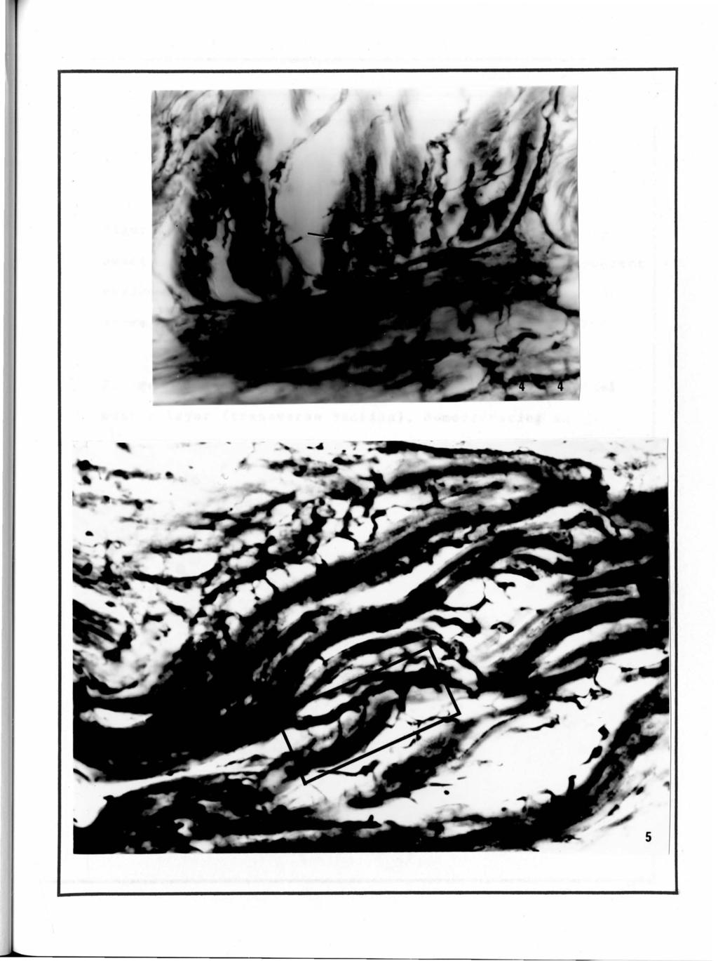
59 54 PLATE III E~PLANhTIG~ OF FIGURES 1''igure 4. l'hotom.icrograpl1 of the earthworm body wall (transverse section). Au "en grappe'' ending with motor enci plates (arro~) is visible n~ar the base of the longitudinal muscle layer. XSOO figure 5. Longitudinal section demonstrating the internal nt!rve net associated with ttte body wall musculature. Xl200
60
61 56 PLATE IV EXPLANATION OF FIGURES Figure 6. Photomicrograph of the earthworm longitudinal muscle layer (longitudinal section), demonstrating apparent varicosities associated with the internal nerve net. Observe also ukink'' (arrow) ref erred to in the text. XSOO Figure 7. Photomicrograph of the earthworm longitudinal muscle layer (transverse section), demonstrating an apparent 11 interneuron 11 (C) X800

62
63 ~. l 58 PLATE V EXPLANATION OF FIGURE Figure 8. Electron micrograph demonstrating a bundle of 11 pavement stone" shaped axons within the lateral nerve (transverse section). The axons show an irregular border and contain microtubules, neurofilaments and flattened membraneous sacs. X44,000

64
65 60 PLATE VI EXPLANATION OF FIGURE Figure 9. Electron micrograph demonstrating a bundle of "tubular" shaped axons within the lateral nerve (transverse section). The axons exhibit a smooth border and contain numerous neurof ilaments. The clefts between the axons are free of formed elements. X44,000

66
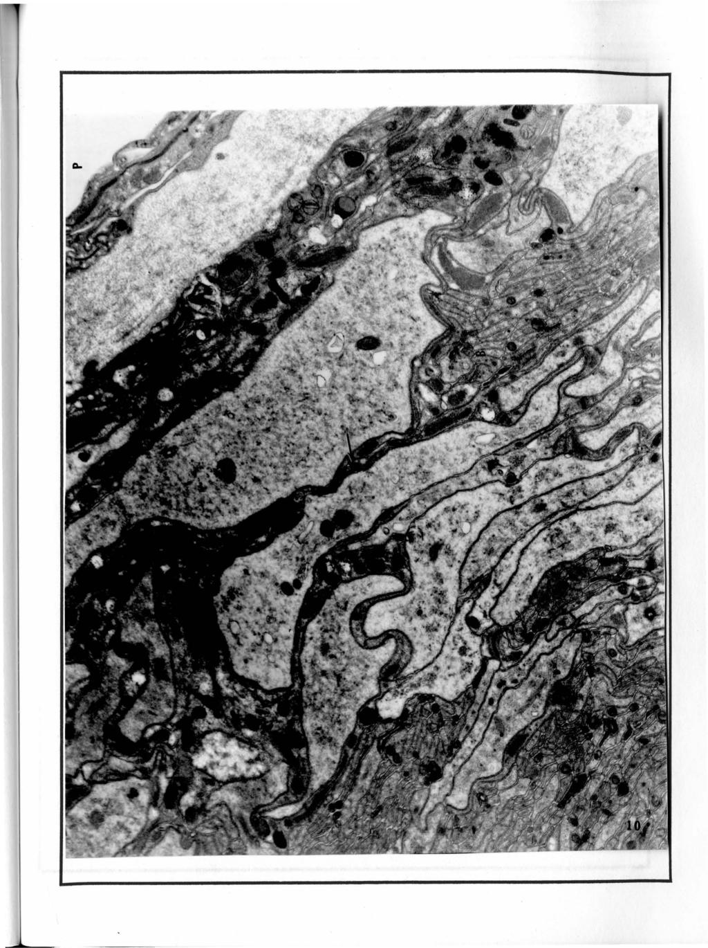
67 62 PLATE VII EXPLANATION OF FIGURE Figure 10. Lateral nerve (transverse section). Axon bundles are ensheathed in a supportive lamella which contains prominent gliosomes (G). The entire bundle is enveloped by a sheath of collagen fibers. X8400 P - B - Arrow - boundary of nerve bundle small axon bundle dense bodies I
68
69 64 PLATE VIII EXPLANATION OF FIGURE Figure 11. Connective tissue septum of the longitudinal muscle bundle which is penetrated by a septal nerve branch. Two kinds of nerve fibers are visible, one containing predominantly dense cored vesicles (D), and a second containing microtubules, and profiles of agranular endoplasmic reticulum. X32,000

70
71 i~.j. 66 PLATE IX EXPLANATION OF FIGURES Figure 12. Connective tissue septum of the longitudinal muacle which contains a bundle of smooth 11 tubular" shaped axons. Prominent gliosomes are visible, partially ensheathing the nerve bundle. X22,400 Figure 13. Neuromuscular junction associated with a radial nerve bundle forming contact with muscle fibers and tails along its pathway. Arrow indicates instances in which a linear density is seen in the junctional cleft parallel to the postjunctional membrane. X32,000

72
73 68 PLATE X EXPLANATION OF FIGURE Figure 14. Electron micrograph of the internal nerve net (transverse sect ion). Small "tubular" shaped axons (arrow) and cell processes (double arrow) which are characterized by profiles of agranular endoplasmic reticulum (ag), and clear spherical vesicles (v) which occasionally are visible. The entire bundle is enveloped by a sheath of collagen fibers. X22,400

74
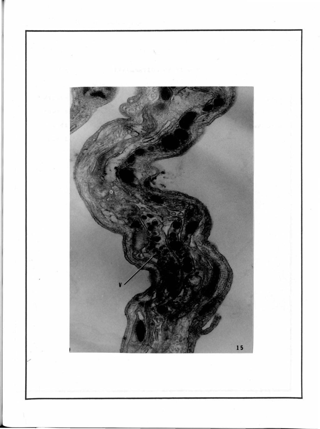
75 70 PLATE XI EXPLANATION OF FIGURE Figure 15. Internal nerve net (longitudinal section) demonstrating a gliosomal {G) supportive lamella and axon bundles containing dense vesicles (V). X32,000
76
77 72 PLATE XII EXPLANATION OF FIGURE Figure 16. The scalloped postjunctional membrane exhibits rows of projections approxinately 20nm in length (arrow). The distal ends of the projections are interconnected by a thin linear density. X88,000

78
79 74 PLATE XIII EXPLAUATION OF FIGURE Figure 17. Neuromuscular junction occurring between a muscle fiber and exposed axons of a small nerve bundle incompletely ensheathed by supportive cells. Axons are packed with vesicles at the point of contact, with either cl~ar spherical vesicles {measuring 50nm in diameter) or dense vesicles {measuring approximately loonm in diameter). The junctional membrane is scalloped and exhibits a cytoplasmic coating. X22,400

80
Nerves and Nerve Impulse
 Nerves and Nerve Impulse Terms Absolute refractory period: Period following stimulation during which no additional action potential can be evoked. Acetylcholine: Chemical transmitter substance released
(From the Department of Anatomy, Harvard Medical School, Boston)
") THE FINE STRUCTURE OF THE ELECTRIC ORGAN OF THE ELECTRIC EEL AND TORPEDO RAY* PRELIMINARY COMMUNICATION BY JOHN H. LUFT, M.D. (From the Department of Anatomy, Harvard Medical School, Boston) PLATE 76 Electric
THE FINE STRUCTURE OF THE ELECTRIC ORGAN OF THE ELECTRIC EEL AND TORPEDO RAY* PRELIMINARY COMMUNICATION BY JOHN H. LUFT, M.D. (From the Department of Anatomy, Harvard Medical School, Boston) PLATE 76 Electric
Nervous Tissue Dr. Archana Rani Associate Professor Department of Anatomy KGMU UP, Lucknow
 13.01.2015 Nervous Tissue Dr. Archana Rani Associate Professor Department of Anatomy KGMU UP, Lucknow Introduction Property of irritability and conductivity Respond to various types of stimuli Distributed
Chapter 9 Nervous System
 Chapter 9 Nervous System Nervous System function: The nervous system is composed of neurons and neuroglia. at the ends of peripheral nerves gather information and convert it into nerve impulses. When sensory
CELLS IN THE NERVOUS SYSTEM
 NEURONS AND GLIA CELLS IN THE NERVOUS SYSTEM Glia Insulates, supports, and nourishes neurons Neurons Process information Sense environmental changes Communicate changes to other neurons Command body response
Biology Slide 1 of 38
 Biology 1 of 38 2 of 38 35-2 The Nervous System What are the functions of the nervous system? 3 of 38 35-2 The Nervous System 1. Nervous system: a. controls and coordinates functions throughout the body
Chapter 7: The Nervous System
 Chapter 7: The Nervous System Objectives Discuss the general organization of the nervous system Describe the structure & function of a nerve Draw and label the pathways involved in a withdraw reflex Define
NEURONS NEUROGLIAL CELLS.
 1 THE NERVOUS TISSUE Definition: The nervous tissue is an assemblage of cells and supportive elements (materials) in which there is a predominance of cells which are highly specialized in the property
12. Nervous System: Nervous Tissue
 12. Nervous System: Nervous Tissue I. Introduction to the Nervous System General functions of the nervous system The nervous system has three basic functions: 1. Gather sensory input from the environment
ISOLATION AND PROPERTIES OF SECRETORY GRANULES FROM RAT ISLETS OF LANGERHANS. II. Ultrastructure of the Beta Granule
 ISOLATION AND PROPERTIES OF SECRETORY GRANULES FROM RAT ISLETS OF LANGERHANS II Ultrastructure of the Beta Granule MARIE H GREIDER, S L HOWELL, and P E LACY From the Department of Pathology, Washington
Name Date Hour. Nerve Histology Microscope Lab
 Name Date Hour Nerve Histology Microscope Lab PRE-LAB: Answer the following questions using your reading and class notes before starting the microscope lab. 1. What is the difference between the functions
Chapter 15. Autonomic Nervous System (ANS) and Visceral Reflexes. general properties Anatomy. Autonomic effects on target organs
 and Visceral Reflexes. general properties Anatomy. Autonomic effects on target organs") Chapter 15 Autonomic Nervous System (ANS) and Visceral Reflexes general properties Anatomy Autonomic effects on target organs Central control of autonomic function 15-1 Copyright (c) The McGraw-Hill Companies,
Chapter 15 Autonomic Nervous System (ANS) and Visceral Reflexes general properties Anatomy Autonomic effects on target organs Central control of autonomic function 15-1 Copyright (c) The McGraw-Hill Companies,
CHAPTER XV PDL 101 HUMAN ANATOMY & PHYSIOLOGY. Ms. K. GOWRI. M.Pharm., Lecturer.
 CHAPTER XV PDL 101 HUMAN ANATOMY & PHYSIOLOGY Ms. K. GOWRI. M.Pharm., Lecturer. Types of Muscle Tissue Classified by location, appearance, and by the type of nervous system control or innervation. Skeletal
What role does the nucleolus have in cell functioning? Glial cells
 Nervous System Lab The nervous system of vertebrates can be divided into the central nervous system, which consists of the brain and spinal cord, and the peripheral nervous system, which contains nerves,
Animal Tissues. I. Epithelial Tissue
 Animal Tissues There are four types of tissues found in animals: epithelial tissue, connective tissue, muscle tissue, and nervous tissue. In this lab you will learn the major characteristics of each tissue
North Bergen School District Benchmarks
 Grade: 10,11, and 12 Subject: Anatomy and Physiology First Marking Period Define anatomy and physiology, and describe various subspecialties of each discipline. Describe the five basic functions of living
Parts of the Nerve Cell and Their Functions
 Parts of the Nerve Cell and Their Functions Silvia Helena Cardoso, PhD [ 1. Cell body] [2. Neuronal membrane] [3. Dendrites] [4. Axon] [5. Nerve ending] 1. Cell body The cell body (soma) is the factory
BIO 201 ANATOMY AND PHYSIOLOGY I with LAB
 BIO 201 ANATOMY AND PHYSIOLOGY I with LAB (Title change ONLY Oct. 2013) Presented and Approved: January 12, 2012 Effective: 2012-13 FA Prefix & Number BIO 201 Course Title: Anatomy and Physiology I Purpose
Anatomy PHL 212. By Dr Tajdar Husain Khan
 Anatomy PHL 212 By Dr Tajdar Husain Khan Overview of Anatomy Anatomy(from the Greek word anatome,"dissection") is a branch of natural science dealing with the structural organization of living things The
AP Biology I. Nervous System Notes
 AP Biology I. Nervous System Notes 1. General information: passage of information occurs in two ways: Nerves - process and send information fast (eg. stepping on a tack) Hormones - process and send information
Chapter 13. The Nature of Somatic Reflexes
 Chapter 13 The Nature of Somatic Reflexes Nature of Reflexes (1 of 3) A reflex is an involuntary responses initiated by a sensory input resulting in a change in a gland or muscle tissue occur without our
BIO 2401 MUSCLE TISSUE page 1 MUSCLES AND MUSCLE TISSUE. Striations Present or Absent?
 BIO 2401 MUSCLE TISSUE page 1 Types of Muscle MUSCLES AND MUSCLE TISSUE Type of Muscle Skeletal Location of Muscle attaches to and covers bony skeleton Striations Present or Absent? present Control of
Chapter 15. The Autonomic Nervous. The Autonomic Nervous System. Autonomic Motor Pathways. ANS vs. SNS
 The Autonomic Nervous System Chapter 15 The subconscious involuntary nervous system Regulates activity of smooth muscle, cardiac muscle & certain glands The Autonomic Nervous System 1 2 ANS vs. SNS Somatic
Please read chapter 15, The Autonomic Nervous System, complete this study guide, and study this material BEFORE coming to the first class.
 Please read chapter 15,, complete this study guide, and study this material BEFORE coming to the first class. I. Introduction to the autonomic nervous system: Briefly describe the autonomic nervous system.
Anatomy Review Graphics are used with permission of: adam.com (http://www.adam.com/) Benjamin Cummings Publishing Co (http://www.awl.com/bc).
 Benjamin Cummings Publishing Co (http://www.awl.com/bc).") Page 1. Introduction The structure of neurons reflects their function. One part of the cell receives incoming signals. Another part generates outgoing signals. Anatomy Review Graphics are used with permission
Page 1. Introduction The structure of neurons reflects their function. One part of the cell receives incoming signals. Another part generates outgoing signals. Anatomy Review Graphics are used with permission
Chapter 15. Sympathetic Nervous System
 Chapter 15 Sympathetic Nervous System Somatic versus Autonomic Pathways Somatic efferent innervation ACh Myelinated fiber Somatic effectors (skeletal muscles) Autonomic efferent innervation ACh ACh or
Nervous System: PNS and CNS
 Nervous System: PNS and CNS Biology 105 Lecture 10 Chapter 8 Outline I. Central Nervous System vs Peripheral Nervous System II. Peripheral Nervous System A. Somatic Nervous System B. Autonomic Nervous
Anatomy & Physiology Bio 2401 Lecture. Instructor: Daryl Beatty Nervous System Introduction Part 1
 Anatomy & Physiology Bio 2401 Lecture Instructor: Daryl Beatty Nervous System Introduction Part 1 Nervous System Introduction Chapter 11 Section A Sequence 4.1 DB Nervous system 1 Intro Presentations 4.2,
Anatomy Review. Graphics are used with permission of: Pearson Education Inc., publishing as Benjamin Cummings (http://www.aw-bc.
 Anatomy Review Graphics are used with permission of: Pearson Education Inc., publishing as Benjamin Cummings (http://www.aw-bc.com) Page 1. Introduction The structure of neurons reflects their function.
Muscles How muscles contract - The Sliding Filament Theory
 Muscles How muscles contract - The Sliding Filament Theory A muscle contains many muscle fibers A muscle fiber is a series of fused cells Each fiber contains a bundle of 4-20 myofibrils Myofibrils are
Origin of Electrical Membrane Potential
 Origin of Electrical Membrane Potential parti This book is about the physiological characteristics of nerve and muscle cells. As we shall see, the ability of these cells to generate and conduct electricity
Chapter 7: The Nervous System
 Chapter 7: The Nervous System I. Organization of the Nervous System Objectives: List the general functions of the nervous system Explain the structural and functional classifications of the nervous system
The Nervous System, Part I.Unlecture
 The Nervous System, Part I.Unlecture Review basic nervous system anatomy before you begin this lecture. The lecture touches on a few of the major characteristics, but you are expected to have already been
CHAPTER 9 BODY ORGANIZATION
 CHAPTER 9 BODY ORGANIZATION Objectives Identify the meaning of 10 or more terms relating to the organization of the body Describe the properties of life Describe the function for the structures of the
Chapter 15 Anatomy and Physiology Lecture
 1 THE AUTONOMIC NERVOUS SYSTEM Chapter 15 Anatomy and Physiology Lecture 2 THE AUTONOMIC NERVOUS SYSTEM Autonomic Nervous System (ANS) regulates the activity of smooth muscles, cardiac muscles, and certain
AUTONOMIC NERVOUS SYSTEM
 AUTONOMIC NERVOUS SYSTEM Somatic efferent and ANS Somatic Efferent Control is over skeletal muscles. External environment This division of the PNS responds to some change in the external environment. single
Nerves and Conduction of Nerve Impulses
 A. Introduction 1. Innovation in Cnidaria - Nerve net a. We need to talk more about nerves b. Cnidaria have simple nerve net - 2 way conduction c. Basis for more complex system in Vertebrates B. Vertebrate
Section B: Epithelial Tissue 1. Where are epithelial tissues found within the body? 2. What are the functions of the epithelial tissues?
 Tissue worksheet Name Section A: Intro to Histology Cells are the smallest units of life. In complex organisms, cells group together with one another based on similar structure and function to form tissues.
Skeletal, Muscular, and Integumentary Systems
 Chapter 36 Skeletal, Muscular, and Integumentary Systems Section 36 1 The Skeletal System (pages 921 925) This section describes the skeletal system and its functions. Introduction (page 921) 1. What forms
CSE511 Brain & Memory Modeling. Lect04: Brain & Spine Neuroanatomy
 CSE511 Brain & Memory Modeling CSE511 Brain & Memory Modeling Lect02: BOSS Discrete Event Simulator Lect04: Brain & Spine Neuroanatomy Appendix of Purves et al., 4e Larry Wittie Computer Science, StonyBrook
Biology 13A Lab #3: Cells and Tissues
 Biology 13A Lab #3: Cells and Tissues Lab #3 Table of Contents: Expected Learning Outcomes.... 28 Introduction...... 28 Activity 1: Eukaryotic Cell Structure... 29 Activity 2: Perspectives on Tissue Preparations.
Tissues (Histology) Ch. 3 Human Anatomy lecture
 Ch. 3 Human Anatomy lecture") I. Histology the study of tissues A. 4 basic tissue types epithelial connective muscle nervous Tissues (Histology) Ch. 3 Human Anatomy lecture B. Usually found in combinations to form organs. C. As you
I. Histology the study of tissues A. 4 basic tissue types epithelial connective muscle nervous Tissues (Histology) Ch. 3 Human Anatomy lecture B. Usually found in combinations to form organs. C. As you
Integration and Coordination of the Human Body. Nervous System
 I. General Info Integration and Coordination of the Human Body A. Both the and system are responsible for maintaining 1. Homeostasis is the process by which organisms keep internal conditions despite changes
Exercise 5 Nervous Tissue
 Exercise 5 Nervous Tissue Objectives After completing the laboratory exercise for nervous tissue you should be able to: Introduction Identify a "typical" multipolar neuron in the light microscope. Identify
the plant & animal cell
 6.1 Basic unit of life Biology Biology Structure & functions of 06 the plant & animal cell In 1665, Robert Hooke observed a section of a cork using a microscope prepared by him. He discovered a structure
Chapter 13: The Spinal Cord, Spinal Nerves, and Spinal Reflexes
 Chapter 13: The Spinal Cord, Spinal Nerves, and Spinal Reflexes I. General Organization of the Nervous System, p. 422 Objectives 1. Describe the basic structural and organizational characteristics of the
Nervous System Organization. PNS and CNS. Nerves. Peripheral Nervous System. Peripheral Nervous System. Motor Component.
 Nervous System Organization PNS and CNS Chapters 8 and 9 Peripheral Nervous System (PNS) connects CNS to sensory receptors, muscles and glands Central Nervous System (CNS) control/integrating center brain
Compartmentalization of the Cell. Objectives. Recommended Reading. Professor Alfred Cuschieri. Department of Anatomy University of Malta
 Compartmentalization of the Cell Professor Alfred Cuschieri Department of Anatomy University of Malta Objectives By the end of this session the student should be able to: 1. Identify the different organelles
Activity 5: The Action Potential: Measuring Its Absolute and Relative Refractory Periods. 250 20 Yes. 125 20 Yes. 60 20 No. 60 25 No.
 3: Neurophysiology of Nerve Impulses (Part 2) Activity 5: The Action Potential: Measuring Its Absolute and Relative Refractory Periods Interval between stimuli Stimulus voltage (mv) Second action potential?
RAD 223. Radiography physiology. Lecture Notes. First lecture: Cell and Tissue
 RAD 223 Radiography physiology Lecture Notes First lecture: Cell and Tissue Physiology: the word physiology derived from a Greek word for study of nature. It is the study of how the body and its part work
Questions on The Nervous System and Gas Exchange
 Name: Questions on The Nervous System and Gas Exchange Directions: The following questions are taken from previous IB Final Papers on Topics 6.4 (Gas Exchange) and 6.5 (Nerves, hormones and homeostasis).
NEURON AND NEURAL TRAMSMISSION: ANATOMY OF A NEURON. created by Dr. Joanne Hsu
 NEURON AND NEURAL TRAMSMISSION: ANATOMY OF A NEURON NEURON AND NEURAL TRAMSMISSION: MICROSCOPIC VIEW OF NEURONS A photograph taken through a light microscope (500x) of neurons in the spinal cord. NEURON
Bi 360: Midterm Review
 Bi 360: Midterm Review Basic Neurobiology 1) Many axons are surrounded by a fatty insulating sheath called myelin, which is interrupted at regular intervals at the Nodes of Ranvier, where the action potential
Chapter 11: Functional Organization of Nervous Tissue
 Chapter 11: Functional Organization of Nervous Tissue Multiple Choice 1. The nervous system A) monitors internal and external stimuli. B) transmits information in the form of action potentials. C) interprets
Lesson Aim To explain the human body at a microscopic level, including the structure and function of cells, tissues and membranes.
 LESSON 1. CELLS & TISSUES Lesson Aim To explain the human body at a microscopic level, including the structure and function of cells, tissues and membranes. THE CELL All living matter is composed of functional
BIOL 1108 Vertebrate Anatomy Lab
 BIOL 1108 Vertebrate Anatomy Lab This lab explores major organs associated with the circulatory, excretory, and nervous systems of mammals. Circulatory System Vertebrates are among the organisms that have
Muscle Tissue. Muscle Physiology. Skeletal Muscle. Types of Muscle. Skeletal Muscle Organization. Myofibril Structure
 Muscle Tissue Muscle Physiology Chapter 12 Specially designed to contract Generates mechanical force Functions locomotion and external movements internal movement (circulation, digestion) heat generation
Smooth Muscle. Learning Objectives.
 Smooth Muscle. Learning Objectives. At the end of this course, you should be able to : 1. describe the structure of smooth muscle 2. describe where smooth muscle occurs within the body 3. discuss the structural
Reflex Physiology. Dr. Ali Ebneshahidi. 2009 Ebneshahidi
 Reflex Physiology Dr. Ali Ebneshahidi Reflex Physiology Reflexes are automatic, subconscious response to changes within or outside the body. a. Reflexes maintain homeostasis (autonomic reflexes) heart
Chapter 10 Muscle Tissue Lecture Outline
 Chapter 10 Muscle Tissue Lecture Outline Muscle tissue types 1. Skeletal muscle = voluntary striated 2. Cardiac muscle = involuntary striated 3. Smooth muscle = involuntary nonstriated Characteristics
Resting membrane potential ~ -70mV - Membrane is polarized
 Resting membrane potential ~ -70mV - Membrane is polarized (ie) Electrical charge on the outside of the membrane is positive while the electrical charge on the inside of the membrane is negative Changes
Introduction to Psychology, 7th Edition, Rod Plotnik Module 3: Brain s Building Blocks. Module 3. Brain s Building Blocks
 Module 3 Brain s Building Blocks Structure of the Brain Genes chains of chemicals that are arranged like rungs on a twisting ladder there are about 100,000 genes that contain chemical instructions that
Biological cell membranes
 Unit 14: Cell biology. 14 2 Biological cell membranes The cell surface membrane surrounds the cell and acts as a barrier between the cell s contents and the environment. The cell membrane has multiple
Student Academic Learning Services Page 1 of 8 Nervous System Quiz
 Student Academic Learning Services Page 1 of 8 Nervous System Quiz 1. The term central nervous system refers to the: A) autonomic and peripheral nervous systems B) brain, spinal cord, and cranial nerves
FUNCTIONS OF THE NERVOUS SYSTEM 1. Sensory input. Sensory receptors detects external and internal stimuli.
 FUNCTIONS OF THE NERVOUS SYSTEM 1. Sensory input. Sensory receptors detects external and internal stimuli. 2. Integration. The brain and spinal cord process sensory input and produce responses. 3. Homeostasis.
Membrane Structure and Function
 Membrane Structure and Function Part A Multiple Choice 1. The fluid mosaic model describes membranes as having A. a set of protein channels separated by phospholipids. B. a bilayer of phospholipids in
An Introduction to Anatomy and Physiology
 An Introduction to Anatomy and Physiology Objectives Define anatomy and physiology Identify the levels of organization in organisms from simplest to most complex Identify the organ systems of the human
Paramedic Program Anatomy and Physiology Study Guide
 Paramedic Program Anatomy and Physiology Study Guide Define the terms anatomy and physiology. List and discuss in order of increasing complexity, the body from the cell to the whole organism. Define the
Explore the Neuroscience for Kids Web Site (ANSWERS) Start at: http://faculty.washington.edu/chudler/neurok.html
 Start at: http://faculty.washington.edu/chudler/neurok.html") NAME Explore the Neuroscience for Kids Web Site (ANSWERS) Start at: http://faculty.washington.edu/chudler/neurok.html On the left side, click on Explore, then click on The Neuron, then click on Millions
NAME Explore the Neuroscience for Kids Web Site (ANSWERS) Start at: http://faculty.washington.edu/chudler/neurok.html On the left side, click on Explore, then click on The Neuron, then click on Millions
Biology 141 Anatomy and Physiology I
 Fall 2016 Biology 141 Anatomy and Physiology I COURSE OUTLINE Faculty Name: Enter Faculty Name Here Program Head: Enter Program Head Here Dean s Review: Dean s Signature: Date Reviewed: / / Revised: Fall
The Cell: Organelle Diagrams
 The Cell: Organelle Diagrams Fig 7-4. A prokaryotic cell. Lacking a true nucleus and the other membrane-enclosed organelles of the eukaryotic cell, the prokaryotic cell is much simpler in structure. Only
Autonomic Nervous System Dr. Ali Ebneshahidi
 Autonomic Nervous System Dr. Ali Ebneshahidi Nervous System Divisions of the nervous system The human nervous system consists of the central nervous System (CNS) and the Peripheral Nervous System (PNS).
7. Skeletal System: Bone Structure and Function
 7. Skeletal System: Bone Structure and Function For the next two chapters (7 and 9) we will study the skeletal system. Although the major feature of this system is the bones, the skeletal system also consists
18. What is limbic system? A. The inner parts of cerebral hemispheres associated with deep structures and from a complex structure. 19.
 CHAPTER 21 NEURAL CONTROL AND COORDINATION One mark Questions: 1. Name the structural and functional unit of nervous system? A. Neuron. 2. What does central Nervous System consists of? A. Brain and spinal
Welcome to Anatomy & Physiology
 Welcome to Anatomy & Physiology Chapter 1 -Human Organization What do you need to do to pass this class? MEMORIZE! The Scope of Human Anatomy Human anatomy is the study of the structure of the human body.
Transverse Sections of the Spinal Cord
 Transverse Sections of the Spinal Cord The spinal cord is perhaps the most simply arranged part of the CNS. Its basic structure, indicated in a schematic drawing of the eighth cervical segment (Figure
Brain & Mind. Bicester Community College Science Department
 B6 Brain & Mind B6 Key Questions How do animals respond to changes in their environment? How is information passed through the nervous system? What can we learn through conditioning? How do humans develop
Vascular System The heart can be thought of 2 separate pumps from the right ventricle, blood is pumped at a low pressure to the lungs and then back
 Vascular System The heart can be thought of 2 separate pumps from the right ventricle, blood is pumped at a low pressure to the lungs and then back to the left atria from the left ventricle, blood is pumped
Module F SKELETAL SYSTEM & ARTICULATIONS
 Module F SKELETAL SYSTEM & ARTICULATIONS Topic from General functions of bone & the skeletal system Structural components microscopic anatomy Structural components gross anatomy Physiology of embryonic
Standards Alignment Minnesota Science Standards Alignment Matrix www.brainu.org/resources/mnstds
 Lesson Summary: Neurons transfer information by releasing neurotransmitters across the synapse or space between neurons. Students model the chemical communication between pre-synaptic and post-synaptic
Structure and Function of Neurons
 CHPTER 1 Structure and Function of Neurons Varieties of neurons General structure Structure of unique neurons Internal operations and the functioning of a neuron Subcellular organelles Protein synthesis
Nerve Cell Communication
 Nerve Cell Communication Core Concept: Nerve cells communicate using electrical and chemical signals. Class time required: Approximately 2 forty minute class periods Teacher Provides: For each student
Neuroscience Methods
 Neuroscience Methods VII. Histology I: An Introduction5 Materials Fixed rat brain Cryostat, microtome, or other means of sectioning Subbed microscope slides Materials for cresyl violet staining protocol
Muscle Fibres. Anatomy and Physiology Advanced Diploma Course Sample Pages Page 1
 Muscle Fibres Muscles are composed of thousands of individual muscle fibres, which are held together by connective tissue. However, muscle fibres may differ in physiological makeup. Anatomy and Physiology
CHAPTER 5 SIGNALLING IN NEURONS
 5.1. SYNAPTIC TRANSMISSION CHAPTER 5 SIGNALLING IN NEURONS One of the main functions of neurons is to communicate with other neurons. An individual neuron may receive information from many different sources.
General A&P Nervous Tissues, Nerves, Spinal Cord and Reflexes Lab Exercises
 1 General A&P Nervous Tissues, Nerves, Spinal Cord and Reflexes Lab Exercises Have someone in your group read the following out loud, while the others read along: In this "Lab Guide", we will be looking
Mammalian Physiology. Autonomic Nervous System UNLV. PHYSIOLOGY, Chapter 11 Berne, Levy, Koeppen, Stanton UNIVERSITY OF NEVADA LAS VEGAS
 Mammalian Physiology Autonomic Nervous System UNLV 1 UNIVERSITY OF NEVADA LAS VEGAS PHYSIOLOGY, Chapter 11 Berne, Levy, Koeppen, Stanton Objectives Describe the organization of the autonomic nervous system
The Huntington Library, Art Collections, and Botanical Gardens
 The Huntington Library, Art Collections, and Botanical Gardens Rooting for Mitosis Overview Students will fix, stain, and make slides of onion root tips. These slides will be examined for the presence
31.1 The Neuron. BUILD Vocabulary. Lesson Objectives
 Name Class Date 31.1 The Neuron Lesson Objectives Identify the functions of the nervous system. Describe the function of neurons. Describe how a nerve impulse is transmitted. BUILD Vocabulary A. The chart
U N IT 10 NE RVOUS SYS TE M REVIEW 1. Which of the following is controlled by the somatic nervous system? A. rate of heartbeat B.
 U N IT 10 NE RVOUS SYS TE M REVIEW 1. Which of the following is controlled by the somatic nervous system? A. rate of heartbeat B. contraction of skeletal muscles C. increased blood flow to muscle tissue
Physical & Chemical Properties. Properties
 Physical & Chemical Properties Properties Carbon black can be broadly defined as very fine particulate aggregates of carbon possessing an amorphous quasi-graphitic molecular structure. The most significant
Chetek-Weyerhaeuser High School
 Chetek-Weyerhaeuser High School Anatomy and Physiology Units and Anatomy and Physiology A Unit 1 Introduction to Human Anatomy and Physiology (6 days) Essential Question: How do the systems of the human
REVIEW SHEET EXERCISE 3 Neurophysiology of Nerve Impulses Name Lab Time/Date. The Resting Membrane Potential
 REVIEW SHEET EXERCISE 3 Neurophysiology of Nerve Impulses Name Lab Time/Date ACTIVITY 1 The Resting Membrane Potential 1. Explain why increasing extracellular K + reduces the net diffusion of K + out of
http://abcnews.go.com/politics/video/obama-says-brain-initiative-will-be-transformative-18861944
 http://abcnews.go.com/politics/video/obama-says-brain-initiative-will-be-transformative-18861944 What are the nervous system s functions? The nervous system organizes and controls an individual s appropriate
Name: Teacher: Olsen Hour:
 Name: Teacher: Olsen Hour: The Nervous System: Part 1 Textbook p216-225 41 In all exercises, quizzes and tests in this class, always answer in your own words. That is the only way that you can show that
組 織 學 實 驗 : 神 經 系 統 Histology Lab : Nervous system
 組 織 學 實 驗 : 神 經 系 統 Histology Lab : Nervous system 實 驗 講 義 : 謝 侑 霖 老 師 Yu-Lin Hsieh, PhD. 劉 俊 馳 Chun-Chih Liu 李 怡 琛 Yi-Chen Lee 張 昭 元 Chao-Yuah Chang 張 瀛 双 Ying-Shuang Chang :07-3121101 ext 2144-18 :littlebu@kmu.edu.tw
Exploring Creation with Advanced Biology ~ Schedule for 2015-2016 Apologia ~ Exploring Creation with Biology ~ Module 1 (Week 1) Date:
 Date:") Apologia ~ Exploring Creation with Biology ~ Module 1 (Week 1) Mon., Aug. 31 Tue., Sep. 1 Wed., Sept. 2 Thurs., Sept. 3 Fri., Sept. 4 Reading Pgs. 1-4; Introduction, Heart of the matter, Pgs. 5-8; Organization
Apologia ~ Exploring Creation with Biology ~ Module 1 (Week 1) Mon., Aug. 31 Tue., Sep. 1 Wed., Sept. 2 Thurs., Sept. 3 Fri., Sept. 4 Reading Pgs. 1-4; Introduction, Heart of the matter, Pgs. 5-8; Organization
An Overview of Cells and Cell Research
 An Overview of Cells and Cell Research 1 An Overview of Cells and Cell Research Chapter Outline Model Species and Cell types Cell components Tools of Cell Biology Model Species E. Coli: simplest organism
Vertebrate Body Organization
 Vertebrate Body Organization Digestive tube suspended in coelom from mouth to anus Body supported by internal skeleton of jointed bones Vertebrae and Cranium protects nervous system Diaphragm divides coelom
MUSCULAR SYSTEM. A. K. Sengupta 9/9/2010 1/12
 MUSCULAR SYSTEM Introduction Functions and basic types of muscle cells Skeletal muscle cells and connective tissues The nervous system Mechanism of muscle contraction Motor unit Action potential basis