Non-invasive Multislice CT Coronary Angiography
|
|
|
- Griselda Warren
- 9 years ago
- Views:
Transcription

1 Non-invasive Multislice CT Coronary Angiography Erasmus Medical Center, Rotterdam, The Netherlands Departments of Radiology en Cardiology Nico R Mollet, MD Filippo Cademartiri, MD Pim J de Feyter, MD
2 Multislice CT Basics
3 Data Acquisition

4 Data Acquisition

5 Data Acquisition
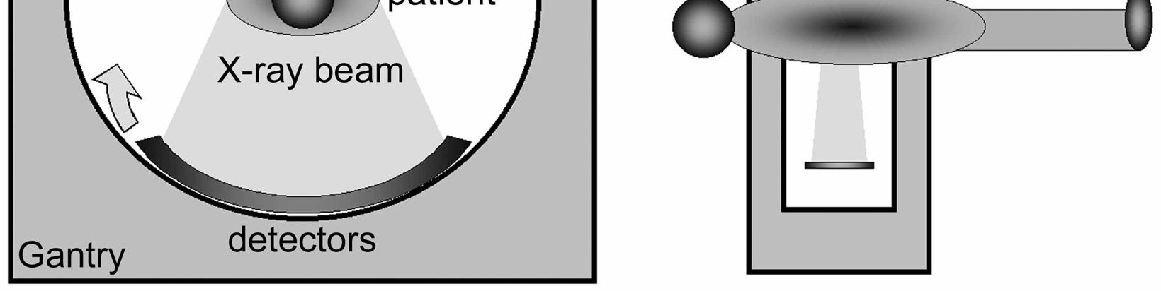
6 Data Acquisition X ray Attenuation Profile Image Reconstruction Detectors

7 Data Acquisition Sequential CT Step-and-Shoot Spiral CT Axial slices obtained Axial slices reconstructed
8 Image Reconstruction -180 GANTRY POSITION +180 PLANE POSITION Z-AXIS
9 Evolution in CT Conventional (Sequential) CT: Axial slices Spiral CT: Volume scanning: Reconstruction of axial slices Multislice or Multidetector Spiral CT: Larger volume in less scan-time
10 Analog-Digital Conversion Acquisition Analog-digital Conversion Digital Reconstruction Storage Archiving Post-processing Quantification Digital-analog Conversion Display

11 Digitization
12 CT Density Values: Hounsfield Units G H Air bubbles Fat Pericardium, thrombus, muscles, plaques Vessel Calc/Clip/Stent Lung Fat Soft tissue CM Bone/Calc T R Air Oil Water Calcium/Metal Density - HU
 Table feed per rotation Total width of")
13 Pitch Pitch 2 Pitch 1 Pitch 0.5 Pitch: (in Multislice CT) Table feed per rotation Total width of collimated beam (n detectors x collimation)
 Pitch =1: Contiguous data Pitch <1:")
14 Pitch Pitch 2 Pitch 1 Pitch 0.5 Pitch >1: Data gaps (interpolation needed) Pitch =1: Contiguous data Pitch <1: Oversampling of a single object (e.g pitch 0.2: 5x oversampling)
15 Collimation X-ray beam X-ray tube Collimator (fan-shaper) X-ray tube Collimated fan beam Table Feed Detectors Collimator (select detectors) Detectors Longitudinal view Short axis view
16 Collimation DAS DAS DAS DAS
17 Collimation DAS DAS DAS DAS
18 Matrix & Field of View CT matrix: 512 x 512 pixels Field of View Pixel size Matrix size CT slice: 512 x 512 voxels

19 Voxel Size Voxel = 3-dimensional Pixel Isotropic spatial resolution: voxel size equal in x,y,z-axis

20 Spatial Resolution & Partial Voluming Averaging of >1 tissue types within 1 voxel resulting in a weighted density value

21 Synchronization With Cardiac Cycle

22 Synchronization With Cardiac Cycle

23 Synchronization With Cardiac Cycle
24 Synchronization With Cardiac Cycle
 ECG signal triggers the acquisition Cardiac phase is")
25 Synchronization With Cardiac Cycle Prospective ECG Triggering (Sequential) ECG signal triggers the acquisition Cardiac phase is pre-defined

26 Synchronization With Cardiac Cycle
27 Synchronization With Cardiac Cycle Retrospective ECG Gating ECG signal is recorded while the whole volume is acquired Cardiac phase is arbitrary
28 Synchronization With Cardiac Cycle Prospective Triggering Retrospective Gating Sequential Scanning Data acquired at a single, predefined phase of the cardiac cycle Trigger time is based on estimation of the R-R intervals from previous heart beats Sensitive to irregular heart rhythm Min. Temporal Resolution - 250ms Spiral Scanning Data acquired for the entire cardiac cycle - parallel ECG recording True match of the phase reconstruction to the ECG retrospectively Less sensitive to irregular heart rhythm Min. Temporal Resolution - 125ms

29 Synchronization With Cardiac Cycle Same patient: Irregular Heart Rate EBT Sequential Trigger 4-MSCT Spiral Gating Courtesy of University Clinic of Grosshadern, Munich, Germany
30 Multislice CT Coronary Angiography Anatomy
")
31 Tomographic (Axial) Images
")
32 Tomographic (Axial) Images

33 Left Coronary Artery

34 Right Coronary Artery

35 Venous Anatomy

36 Myocardium & Valves

37 Anatomical Myocardial Bridging
38 Multislice CT Coronary Angiography Technique

39 The Aim
40 MSCT Coronary Angiography Prerequisites 1. HIGH SPATIAL RESOLUTION Depiction of vessel wall and small coronary branches 2. HIGH TEMPORAL RESOLUTION Reduce motion artifacts 3. FAST COVERAGE Short breath-hold time 4. SYNCHRONIZATION WITH CARDIAC CYCLE ECG-gating for end-diastolic and end-systolic imaging
41 Spatial Resolution RCA 3.9± ±0.5 LM 4.5±0.5 LAD (prox) 3.7±0.4 CX 3.4± ±0.6 LAD (dist) 1.9±0.4 Female : - 9% LV hypertrophy : + 17% Dilated CMP : + 12% Dodge JT Jr., Circulation, 1992

42 Spatial Resolution Courtesy of University Clinic of Grosshadern, Munich, Germany

43 Spatial Resolution Flat Panel Technology Flohr, Herz 03
44 Fast Coverage High resolution MSCT-CA CA 4-MSCTA 64-MSCTA Coll=1.0 mm Rot.=500 ms Coll=0.6 mm Rot.=330 ms s 12 s

45 Fast Coverage 16-slices or more!!!

46 Fast Coverage Nieman, Heart 02

47 Coronary Motion LAD 22.4 mm/s ± 4.0 RCA 69.5 mm/s ± 22.5 CX 48.4 mm/s ± 15 Achenbach, Radiology 02

48 Synchronization with cardiac cycle

49 Synchronization with cardiac cycle 30% 165 ms 65% 165 ms
50 Synchronization with cardiac cycle R Absolute Forward Absolute Reverse ms ms P T S 165 ms
51 Temporal Resolution = ½ Rotation Time ms ms 1

52 Temporal Resolution HR 49 HR 67 HR 82 Nieman, Heart 02 Flohr, Herz 03
53 Temporal Resolution Single segment vs. Multi-segment Image Reconstruction
54 Single segment Data from 1 cardiac cycle ms Temporal resolution = 250 ms
55 Multi segment Data from 2 cardiac cycles Temporal resolution = 125 ms

56 Multi segment Data from 2 cardiac cycles 1 2 >125 Multi-segment is adaptive

57 Multi segment Data from 2 cardiac cycles Flohr, Acad Rad 03

58 Multi segment Temporal Resolution Flohr, Acad Rad 03

59 Temporal Resolution Single-segment vs Bi-segment (high HR) Single Segment Reconstruction 250ms Bi-segment Reconstruction 125msec

60 Temporal Resolution Single segment vs Bi-segment (low HR) Single Segment Reconstruction 250ms Bisegment Reconstruction 125ms
61 Single vs. Multi- Segment Reconstruction Limitations Multi-segment requires a lower pitch Longer scan and higher dose Multi-segment only feasible in consecutive heart beats with equal R-R interval Does not work if small change in R-R
62 Single vs. Multi- Segment Reconstruction Limitations z z - - Position Continuous spiral scan & feed Continuous spiral scan & feed Recon Recon Image data Image data Delay Recon Volume Gaps Pitch must be reduced when using >2-segment reconstruction
63 MSCT Coronary Angiography Prerequisites What do we have?

64 64-slice CT coronary angiography
65 MSCT Coronary Angiography Prerequisites 1. HIGH SPATIAL RESOLUTION (isotropic 0.4 mm 3 ) Smaller coronary branches evaluable More reliable visualization of in-stent lumen 2. HIGH TEMPORAL RESOLUTION Evaluation of entire coronary tree possible in HR<70 bpm 3. FAST COVERAGE Manageable breath-hold hold for nearly all pts Injection of less contrast, higher injection rate ( ml/s) 4. SYNCHRONIZATION WITH CARDIAC CYCLE ECG-editing possible in minor heart rhythm irregularities
66 Patient Preparation Scan Procedure

67 Patient Preparation Phone contact Check for indication Check for contraindication Check for current medication Check for current heart rate

68 Patient Preparation HR>65bpm Contra-indications Asthma AV block Overt Heart Failure 45-60min. 100mg b-blockerb blocker HR<65bpm HR check IV access

69 Patient Preparation Instructions ECG-leads Breath hold / HR check

70 Patient Preparation IV/Injector connect Prepare injector
71 Scan Procedure 1 hour 4 sec Beta-blocker (>70 min -1 ) IV access & ECG leads Overview scan range Contrast injection (CareBolus ) 100 ml Iomeron 5 ml/s API: Breath hold 12s +100 HU
72 Scan Procedure: Bolus Tracking HU Contrast injection 100 ml Iomeron 5 ml/s Transition sec
73 Scan Protocol - slices/rotation: 2 x 32 (S16: 16) - individual detector width: 0.6 mm (S16: 0.75) - rotation time: 330 (S16: 375 ms) - table feed: 3.8 mm/rotation (S16: 3.0) - X-tube voltage: 120 kv - X-tube current: max. 900 mas (S16: max. 700)
74 Image reconstruction Image processing
75 Image Reconstruction - retrospective ECG-gating (editing when necessary) - temporal resolution: 165 ms (single-phase reconstruction) - effective slice thickness: 0.75 mm - increment: 0.4 mm - kernel: medium smooth (b30f) native coronaries sharp (b46f) in-stent lumen or calcified vessels
76 Filtering Axial images and VRT s Measurements, Ca++, Stents MPR, MIP and flexible use B10f-B30f B40f-B60f B30f-B40f

77 Filtering B10f B30f B60f B80f

78 Image Processing MPR MIP VRT Curved MPR MPR: multiplanar reconstructions MIP: maximum intensity projections VRT: volume rendering techniques
79 Limitations & Artefacts
80 Limitations of CT coronary angiography SELECTED PATIENT POPULATION Stable heart rhythm, able to breath hold for 20s, <70 bpm (spontaneous/β-blocker induced) X-RAY DOSE Better images and higher spatial resolution requires higher dose HEAVY CALCIFICATIONS Blooming and partial volume artifacts mask the lumen HIGH AND IRREGULAR HEART RATES Motion artefacts and mis-registration OPERATOR DEPENDENCY Time-consuming and variable diagnostic accuracy
81 Operator dependency Available: Sensitivity: Specificity: PPV: NPV: STD % 96 % 78 % 91 % 3D % 97 % 87 % 99 % Cademartiri, AJC 04
82 Severe calcification
83 Severe calcification

84 High heart rates

85 High heart rates Nieman, Heart 02

86 Respiratory Motion

87 Arrhythmia Premature ventricular contraction after each 4-5 normal cycles

88 Low HR ECG-editing

89 Low HR ECG-editing
90 ECG-editing Extra-systole

91 ECG-editing Missing data

92 ECG-editing
93 ECG-editing: Results Only patients with mild arrhythmia included Available: Sensitivity: Specificity: PPV: NPV: STD % 96% 87% 91% Edit % 96% 87% 97% Cademartiri, AJR (in press)
94 Radiation Exposure

95 Dose Reduction Prospective ECG-tube modulation
96 Dose Reduction Tube Modulation (+) Reduction up to 50% of dose in low heart rates (-) Less reliable in case of mild arrhythmia (-) Unable to reconstruct datasets during early diastolic phase (low-dose area), but often good time interval for evaluation of the RCA
97 Radiation Exposure Estimations / Calculations Flohr, Acad Rad 03
98 Radiation Exposure Estimations / Calculations Exposure Cornerstones: Bi-Plane Chest X-Ray: 0.1 msv EBCT Calcium Scoring: 0.9 msv Diagnostic Coronary Angiography: 2-6 msv Natural Background Radiation: 2-5 msv p.a. 8.6mSv Cardiac MSCT Exposure 0.7mSv 2.6mSv 1.3mSv * 7mSv 3.5mSv * 0.6mSv 2.2mSv 1.1mSv * 4.3mSv * 4-Slice Trigger 2.5mm Score 4-Slice Gating 3mm Score 4-Slice Gating 1.25mm CTA 16-Slice Trigger 3mm Score 16-Slice Gating 3mm Score 16-Slice Gating 0.75mm CTA * ECG-Pulsing Jakobs, Eur. Radiology 02
99 Radiation Exposure Estimations / Calculations male/female (ECG pulsing) Ca Seq. Ca Spiral CT Angio Jakobs (Eur Rad 02): est. 2.0 / MSCT 100 mas 4x2.5mm (1.0 / 1.4) Hunold (Radiol 03): phantom 1-EBCT63 mas 1x3 mm 1.0 / / MSCT 2-400mAs 2 4x1 mm 1.5 / / / Morin (Circ 03): estimations 1-EBCT63 mas 1x3 mm MSCT 150 mas 4x1/1.3 mm Trabold (RoFo 03): phantom 2.9 / / MSCT 300 mas 12x.8mm (1.6) (4.3) Hunold (RSNA 03): phantom 4MSCT 100/default mas 4x1mm 3.0 / / 13.0 Siemens 4MSCT 100/default mas 4x1mm 4.2 / / 15.7 GE 4MSCT 100/default mas 4x1mm 2.4 / / 8.2 Philips 16MSCT 100/default mas 16x.6mm 5.8 / / 18.8 GE
State-of-the-Art Technology in Cardiac CT
 1 2 Next Step Evolution or Revolution? State-of-the-Art Technology in Cardiac CT Stefan Ulzheimer, PhD Global Director of Collaborations CT Siemens Medical Solutions Major Innovations in CT Head The 80
Purchasing a cardiac CT scanner: What the radiologist needs to know
 Purchasing a cardiac CT scanner: What the radiologist needs to know Maria Lewis ImPACT St George s Hospital, London [email protected] CT scanner development Slice wars 1998 Increased z-coverage
MDCT Technology. Kalpana M. Kanal, Ph.D., DABR Assistant Professor Department of Radiology University of Washington Seattle, Washington
 MDCT Technology Kalpana M. Kanal, Ph.D., DABR Assistant Professor Department of Radiology University of Washington Seattle, Washington ACMP Annual Meeting 2008 - Seattle, WA Educational Objectives Historical
Master s Program in Medical Physics. Physics of Imaging Systems Basic Principles of Computer Tomography (CT) III. Prof. Dr. Lothar Schad.
 III. Prof. Dr. Lothar Schad.") 1 12/9/2008 Page 1 Master s Program in Medical Physics Physics of Imaging Systems Basic Principles of Computer Tomography (CT) III Chair in Faculty of Medicine Mannheim University of Heidelberg Theodor-Kutzer-Ufer
1 12/9/2008 Page 1 Master s Program in Medical Physics Physics of Imaging Systems Basic Principles of Computer Tomography (CT) III Chair in Faculty of Medicine Mannheim University of Heidelberg Theodor-Kutzer-Ufer
MYOCARDIAL PERFUSION COMPUTED TOMOGRAPHY PhD course in Medical Imaging. Anne Günther Department of Radiology OUS Rikshospitalet
 MYOCARDIAL PERFUSION COMPUTED TOMOGRAPHY PhD course in Medical Imaging Anne Günther Department of Radiology OUS Rikshospitalet CORONARY CT ANGIOGRAPHY (CTA) Accurate method in the assessment of possible
Low-dose CT for Pulmonary Embolism
 Low-dose CT for Pulmonary Embolism Gautham Gautham P. P. Reddy, Reddy, MD, MD, MPH MPH University University of of Washington Washington Introduction Introduction CT CT accounts accounts for for > 50%
Clinical Training for Visage 7 Cardiac. Visage 7
 Clinical Training for Visage 7 Cardiac Visage 7 Overview Example Usage 3 Cardiac Workflow Examples 4 Remove Chest Wall 5 Edit Chest Wall Removal 6 Object Display Popup 7 Selecting Optimal Phase 8 Thick
How To Improve Your Ct Image Quality
 Translating Protocols Between Scanner Manufacturer and Model Cynthia H. McCollough, PhD, FACR, FAAPM Professor of Radiologic Physics Director, CT Clinical Innovation Center Department of Radiology Mayo
Rb 82 Cardiac PET Scanning Protocols and Dosimetry. Deborah Tout Nuclear Medicine Department Central Manchester University Hospitals
 Rb 82 Cardiac PET Scanning Protocols and Dosimetry Deborah Tout Nuclear Medicine Department Central Manchester University Hospitals Overview Rb 82 myocardial perfusion imaging protocols Acquisition Reconstruction
CT Protocol Optimization over the Range of CT Scanner Types: Recommendations & Misconceptions
 CT Protocol Optimization over the Range of CT Scanner Types: Recommendations & Misconceptions Frank N. Ranallo, Ph.D. Associate Professor of Medical Physics & Radiology University of Wisconsin School of
Cone Beam Reconstruction Jiang Hsieh, Ph.D.
 Cone Beam Reconstruction Jiang Hsieh, Ph.D. Applied Science Laboratory, GE Healthcare Technologies 1 Image Generation Reconstruction of images from projections. textbook reconstruction advanced acquisition
Imaging of Thoracic Endovascular Stent-Grafts
 Imaging of Thoracic Endovascular Stent-Grafts Tariq Hameed, M.D. Department of Radiology and Imaging Sciences, Indiana University School of Medicine, Indianapolis, Indiana Disclosures: No relevant financial
CTA OF THE EXTRACORONARY HEART
 CTA OF THE EXTRACORONARY HEART Charles White MD Director of Thoracic Imaging Department of Radiology University of Maryland NO DISCLOSURES [email protected] CARDIAC CASE DISTRIBUTION Coronary CTA 30% ED chest
Spiral CT: Single and Multiple Detector Systems. AAPM Refresher Course Nashville, TN July 28,1999
 Spiral CT: Single and Multiple Detector Systems AAPM Refresher Course Nashville, TN July 28,1999 Mike McNitt-Gray, PhD, DABR Assistant Professor UCLA Radiological Sciences [email protected] X-Ray
FFR CT : Clinical studies
 FFR CT : Clinical studies Bjarne Nørgaard Department Cardiology B Aarhus University Hospital Skejby, Denmark Disclosures: Research grants: Edwards and Siemens Coronary CTA: High diagnostic sensitivity
Development of SCENARIA New Version Software MEDIX VOL. 62 P.32 P.35
 Development of SCENAIA New Version Software Takayuki Kadomura Toshio Sakamoto Kana Tanaka Takatsugu Ito Yuuko Nishimura Naomi Maekawa MEDIX VOL. 62 P.32 P.35 Development of SCENAIA New Version Software
CT RADIATION DOSE REPORT FROM DICOM. Frank Dong, PhD, DABR Diagnostic Physicist Imaging Institute Cleveland Clinic Foundation Cleveland, OH
 CT RADIATION DOSE REPORT FROM DICOM Frank Dong, PhD, DABR Diagnostic Physicist Imaging Institute Cleveland Clinic Foundation Cleveland, OH CT Patient comes out... Patient goes in... Big Black Box Radiology
Thinking ahead. Focused on life. REALIZED: GROUNDBREAKING RESOLUTION OF 80 µm VOXEL
 Thinking ahead. Focused on life. REALIZED: GROUNDBREAKING RESOLUTION OF 80 µm VOXEL X-ray ZOOM RECONSTRUCTION Flat Panel Detector (FPD) Automatic Positioning Function For ø 40 x H 40 mm, ø 60 x H 60 mm,
Multislice CT: Current Technology and Future Developments
 Multislice CT: Current Technology and Future Developments 1 Stefan Ulzheimer and Thomas Flohr Contents 1.1 Introduction 3 1.2 System Design 5 1.2.1 Gantry 6 1.2.2 X-Ray Tube and Generator 6 1.2.3 MDCT
For the NXT Investigators
 Diagnostic performance of non-invasive fractional flow reserve derived from coronary CT angiography in suspected coronary artery disease: The NXT trial Bjarne L. Nørgaard, Jonathon Leipsic, Sara Gaur,
R/F. Efforts to Reduce Exposure Dose in Chest Tomosynthesis Targeting Lung Cancer Screening. 3. Utility of Chest Tomosynthesis. 1.
 R/F Efforts to Reduce Exposure Dose in Chest Tomosynthesis Targeting Lung Cancer Screening Department of Radiology, National Cancer Center Hospital East Kaoru Shimizu Ms. Kaoru Shimizu 1. Introduction
Brilliance CT 64-channel confi guration
 Brilliance CT 64-channel confi guration with Essence technology The Brilliance CT 64-channel confi guration is designed to help you conduct the most advanced multislice CT studies possible. These systems
Fundamentals of Cone-Beam CT Imaging
 Fundamentals of Cone-Beam CT Imaging Marc Kachelrieß German Cancer Research Center (DKFZ) Heidelberg, Germany www.dkfz.de Learning Objectives To understand the principles of volumetric image formation
CORONARY ARTERY BYPASS GRAFTS, STENTS, AND EXTRACORONARY CARDIAC DZ. Charles White MD
 CORONARY ARTERY BYPASS GRAFTS, STENTS, AND EXTRACORONARY CARDIAC DZ Charles White MD Director of Thoracic Imaging Department of Radiology University of Maryland CORONARY ARTERY BYPASS GRAFTS First performed
GE Healthcare. Revolution EVO. More than just high tech. Higher purpose.
 GE Healthcare Revolution EVO More than just high tech. Higher purpose. Revolution EVO. Designed with purpose. Today s healthcare environment is about creating new solutions to pressing needs. It s about
Chest 1: Pulmonary Nodule Follow-up: Low-Dose Helical CT (Unenhanced) (Non-metastatic) Gantry Rotation Time. mas (Reg-Lg) 40-80
 (Non-metastatic) Gantry Rotation Time. mas (Reg-Lg) 40-80") Revisions Effective January 2012 Chest 1: Pulmonary Nodule Follow-up: Low-Dose Helical CT (Unenhanced) (Non-metastatic) Technologist Instructions Patient must cough several times prior to scan to clear
Revisions Effective January 2012 Chest 1: Pulmonary Nodule Follow-up: Low-Dose Helical CT (Unenhanced) (Non-metastatic) Technologist Instructions Patient must cough several times prior to scan to clear
The disclaimer on page 1 is an integral part of this document. Copyright February 23, 2016 by AAPM. All rights reserved.
 DISCLAIMER: TO THE EXTENT ALLOWED BY LOCAL LAW, THIS INFORMATION IS PROVIDED TO YOU BY THE AMERICAN ASSOCIATION OF PHYSICISTS IN MEDICINE, A NON-PROFIT ORGANIZATION ORGANIZED TO PROMOTE THE APPLICATION
Computer-Assisted Coronary CT Angiography Analysis
 Linköping University Medical Dissertations, No. 1237 Computer-Assisted Coronary CT Angiography Analysis From Software Development to Clinical Application Chunliang Wang Division of Radiological Sciences
scan: : a new tool to analyse
 Gated CT-scan scan: : a new tool to analyse the shape of the aortic annulus. SEROUSSI SAFAR K, JONDEAU G, LANSAC E., SERFATY JM Bichat Hospital, U698, Paris, France Anatomy: 3D + spatial resolution Retrospective
GE Medical Systems Training in Partnership. Module 8: IQ: Acquisition Time
 Module 8: IQ: Acquisition Time IQ : Acquisition Time Objectives...Describe types of data acquisition modes....compute acquisition times for 2D and 3D scans. 2D Acquisitions The 2D mode acquires and reconstructs
Sistemas de reducción de dosis en CT para pediatría. Nuevos avances Ángela de Pinto Siemens CT Business Manager angela.pinto@siemens.
 Sistemas de reducción de dosis en CT para pediatría. Nuevos avances Ángela de Pinto Siemens CT Business Manager [email protected] Copyright Siemens AG 2009. All rights reserved. An Innovation Leader
Multislice Computed Tomography: Basic Principles and Clinical Applications
 New Applications with the Assistance of Innovative Technologies Multislice Computed Tomography: Basic Principles and Clinical Applications A. F. Kopp, K. Klingenbeck-Regn 2, M. Heuschmid, A. Küttner, B.
Cardiac CT for Calcium Scoring
 Scan for mobile link. Cardiac CT for Calcium Scoring Cardiac computed tomography (CT) for Calcium Scoring uses special x-ray equipment to produce pictures of the coronary arteries to determine if they
Patient-centered CT imaging: New methods for patient-specific optimization 1 of image quality and radiation dose
 Patient-centered CT imaging: New methods for patient-specific optimization 1 of image quality and radiation dose ipatient is an advanced platform that delivers focused innovations to facilitate patient-centered
Pediatric Hospitals Bring Low-dose CT to the Middle East
 Pediatric Hospitals ring Low-dose CT to the Middle East For years, radiologists have been cognizant of the importance of limiting pediatric patients exposure to radiation dose. uilding on the LR principle,
CT: Size Specific Dose Estimate (SSDE): Why We Need Another CT Dose Index. Acknowledgements
: Why We Need Another CT Dose Index. Acknowledgements") CT: Size Specific Dose Estimate (SSDE): Why We Need Another CT Dose Index Keith J. Strauss, MSc, FAAPM, FACR Clinical Imaging Physicist Cincinnati Children s Hospital University of Cincinnati College of
CT: Size Specific Dose Estimate (SSDE): Why We Need Another CT Dose Index Keith J. Strauss, MSc, FAAPM, FACR Clinical Imaging Physicist Cincinnati Children s Hospital University of Cincinnati College of
Principles of Medical Ultrasound. Pai-Chi Li Department of Electrical Engineering National Taiwan University
 Principles of Medical Ultrasound Pai-Chi Li Department of Electrical Engineering National Taiwan University What is Medical Ultrasound? Prevention: actions taken to avoid diseases. Diagnosis: the process
Scan Time Reduction and X-ray Scatter Rejection in Dual Modality Breast Tomosynthesis. Tushita Patel 4/2/13
 Scan Time Reduction and X-ray Scatter Rejection in Dual Modality Breast Tomosynthesis Tushita Patel 4/2/13 Breast Cancer Statistics Second most common cancer after skin cancer Second leading cause of cancer
Chest CT protocols. Mannudeep K. Kalra, MD, DNB. Dianna D. Cody, PhD. Massachusetts General Hospital Harvard Medical School
 Chest CT protocols Mannudeep K. Kalra, MD, DNB Dianna D. Cody, PhD Massachusetts General Hospital Harvard Medical School M.D. Anderson Cancer Center Specific principles Routine chest CT Lung nodule follow
Press. Siemens solutions support diagnosis and treatment of cardiovascular diseases
 Press Healthcare Erlangen, August 29, 2015 ESC 2015: ExCel London Exhibition and Convention Center, Booth #G700 Siemens solutions support diagnosis and treatment of cardiovascular diseases New cardiovascular
A d v a n c e d 1 2 8
 Advanced 128 SCENARIA Advanced 128 Hitachi Innovation at Work for You. Continuing its commitment to the SCENARIA scalable CT platform, Hitachi introduces cutting-edge 128-slice technology with the SCENARIA
Concepts for High-Resolution Low-Dose CT of the Breast
 RSNA 2012 Refresher Course 721B, Chicago, Nov. 30, 2012 Concepts for High-Resolution Low-Dose CT of the Breast Disclosures WAK is founder, shareholder and CEO of CT Imaging GmbH, Erlangen, Germany. Willi
American College of Radiology CT Accreditation Program. Testing Instructions
 American College of Radiology CT Accreditation Program Testing Instructions (Revised July 24, 2015) This guide provides all of the instructions necessary for clinical tests, phantom tests and general submission
CT for the production floor: Recent developments in Hard- and Software for industrial CT Systems
 CT for the production floor: Recent developments in Hard- and Software for industrial CT Systems GE s Industrial X-ray and CT Forum, Cologne July 7 th 2015 Oliver Brunke GE Sensing & Inspection Technologies
DENTAL Cone beam 3D X-RAY SYSTEM with
 VERSATILE INTUITIVE efficient DENTAL Cone beam 3D X-RAY SYSTEM with dedicated panoramic imaging With thirty years of experience in designing and manufacturing state-of-the-art dental panoramic and tomographic
CPT Radiology Codes Requiring Review by AIM Effective 01/01/2016
 CPT Radiology Codes Requiring Review by AIM Effective 01/01/2016 When a service is authorized only one test per group is payable. *Secondary codes or add-on codes do not require preauthorization or separate
kv-& MV-CBCT Imaging for Daily Localization: Commissioning, QA, Clinical Use, & Limitations
 kv-& MV-CBCT Imaging for Daily Localization: Commissioning, QA, Clinical Use, & Limitations Moyed Miften, PhD Dept of Radiation Oncology University of Colorado Denver Questions Disease Stage (local, regional,
Tracking Radiation Exposure From Medical Diagnostic Procedures: Siemens Perspectives
 Tracking Radiation Exposure From Medical Diagnostic Procedures: Siemens Perspectives Gilbert W. Beebe Symposium The National Academies Katharine Grant, PhD Staff Scientist 8 December 2011 For internal
Three Dimensional Ultrasound Imaging
 Three Dimensional Ultrasound Imaging Hans Torp/ Sevald Berg/Kjell Kristoffersen m/flere Department of circulation and medical imaging NTNU Hans Torp NTNU, Norway Acquisition Reconstruction Filtering Collecting
CT scanning. By Mikael Jensen & Jens E. Wilhjelm Risø National laboratory Ørsted DTU. (Ver. 1.2 4/9/07) 2002-2007 by M. Jensen and J. E.
 2002-2007 by M. Jensen and J. E.") 1 Overview CT scanning By Mikael Jensen & Jens E. Wilhjelm Risø National laboratory Ørsted DTU (Ver. 1.2 4/9/07) 2002-2007 by M. Jensen and J. E. Wilhjelm) As it can be imagined, planar X-ray imaging has
1 Overview CT scanning By Mikael Jensen & Jens E. Wilhjelm Risø National laboratory Ørsted DTU (Ver. 1.2 4/9/07) 2002-2007 by M. Jensen and J. E. Wilhjelm) As it can be imagined, planar X-ray imaging has
Multi Detector Row CT Systems and Image- Reconstruction Techniques 1
 Special Review Radiology Thomas G. Flohr, PhD Stefan Schaller, PhD Karl Stierstorfer, PhD Herbert Bruder, PhD Bernd M. Ohnesorge, PhD U. Joseph Schoepf, MD Published online before print 10.1148/radiol.2353040037
123 Main St NY, New York 12345 ph: (202) 555 5555 fax: (202) 555 5555
 555 5555 fax: (202) 555 5555") Patient Name: DOE, JOHN D. Gender: M Date of Study: 4/2/2013 Date of birth: 6/28/1962 Age: 50 Medical Record #: 45869725 Ordering Physician: JANE INTERNIST, MD History: Atypical Angina, Abn ECG, High Cholesterol,
Patient Name: DOE, JOHN D. Gender: M Date of Study: 4/2/2013 Date of birth: 6/28/1962 Age: 50 Medical Record #: 45869725 Ordering Physician: JANE INTERNIST, MD History: Atypical Angina, Abn ECG, High Cholesterol,
Mx8000 IDT 16 provides dose control methodologies like DoseRight ACS (Automatic Current Selection) and DoseRight DOM (dynamic dose modulation).
 and DoseRight DOM (dynamic dose modulation).") Philips Mx8000 IDT 16 CT Scanner Philips Medical systems Mx8000 IDT 16 delivers 16 simultaneous slices at sub-millimeter collimator, high resolution and isotropic imaging. New large area detectors allow
Philips Mx8000 IDT 16 CT Scanner Philips Medical systems Mx8000 IDT 16 delivers 16 simultaneous slices at sub-millimeter collimator, high resolution and isotropic imaging. New large area detectors allow
SITE IMAGING MANUAL ACRIN 6698
 SITE IMAGING MANUAL ACRIN 6698 Diffusion Weighted MR Imaging Biomarkers for Assessment of Breast Cancer Response to Neoadjuvant Treatment: A sub-study of the I-SPY 2 TRIAL Version: 1.0 Date: May 28, 2012
Cynthia H. McCollough b) and Michael R. Bruesewitz Department of Radiology, Mayo Clinic College of Medicine, Rochester, Minnesota 55905
 and Michael R. Bruesewitz Department of Radiology, Mayo Clinic College of Medicine, Rochester, Minnesota 55905") The phantom portion of the American College of Radiology ACR Computed Tomography CT accreditation program: Practical tips, artifact examples, and pitfalls to avoid a Cynthia H. McCollough b) and Michael
The phantom portion of the American College of Radiology ACR Computed Tomography CT accreditation program: Practical tips, artifact examples, and pitfalls to avoid a Cynthia H. McCollough b) and Michael
The IAC Standards and Guidelines for CT Accreditation
 The IAC Standards and Guidelines for CT Accreditation Table of Contents All entries in Table of Contents are linked to the corresponding sections. Introduction... 4 Part A: Organization... 5 Section 1A:
Multi-slice Helical CT Scanning of the Chest
 Multi-slice Helical CT Scanning of the Chest Comparison of different low-dose acquisitions Lung cancer is the main cause of deaths due to cancer in human males and the incidence is constantly increasing.
Table of Contents. Scan acquisition and user interface basics. Dose modulation and reduction tools. Multi-Slice Detector Geometry
 Table of Contents Scan acquisition and user interface basics Dose modulation and reduction tools Multi-Slice Detector Geometry Image Reconstruction and Display Contrast Media Tools Multi-planar formats
Doppler. Doppler. Doppler shift. Doppler Frequency. Doppler shift. Doppler shift. Chapter 19
 Doppler Doppler Chapter 19 A moving train with a trumpet player holding the same tone for a very long time travels from your left to your right. The tone changes relative the motion of you (receiver) and
Cardiac CT Emerging Role and Current Indications
 Cardiac CT Emerging Role and Current Indications Dr. Felix Keng MBBS, FRCP (Lond), FAMS, Dip CBNC, Dip CBCCT, MMed (Int Med), FAPSC Director, Nuclear Cardiology National Heart Centre, Singapore Adjunct
The new generation in ECG interpretation
 The new generation in ECG interpretation Philips DXL ECG Algorithm, Release PH100B The Philips DXL ECG Algorithm, developed by the Advanced Algorithm Research Center, uses sophisticated analytical methods
The disclaimer on page 1 is an integral part of this document. Copyright March 1, 2016 by AAPM. All rights reserved.
 DISCLAIMER: TO THE EXTENT ALLOWED BY LOCAL LAW, THIS INFORMATION IS PROVIDED TO YOU BY THE AMERICAN ASSOCIATION OF PHYSICISTS IN MEDICINE, A NON-PROFIT ORGANIZATION ORGANIZED TO PROMOTE THE APPLICATION
GE Healthcare. Optima* CT660
 GE Healthcare Optima* CT660 Your vision of quality care made The Optima CT660 system helps you deliver competent, personalized care that helps fulfill your mission and please your patients. One look at
Copyright March 1, 2016 by AAPM. All rights reserved.
 DISCLAIMER: TO THE EXTENT ALLOWED BY LOCAL LAW, THIS INFORMATION IS PROVIDED TO YOU BY THE AMERICAN ASSOCIATION OF PHYSICISTS IN MEDICINE, A NON-PROFIT ORGANIZATION ORGANIZED TO PROMOTE THE APPLICATION
GE Healthcare. Great treasures in small places. BrightSpeed Elite
 GE Healthcare Great treasures in small places BrightSpeed Elite The BrightSpeed Elite brings you multi-detector CT capabilities with maximum convenience in an ultra-compact design. Built with LightSpeed
4D Scanning. Image Guided Radiation Therapy. Outline. A Simplified View of the RT Process. Outline. Steve B. Jiang, Ph.D.
 4D Scanning Steve B. Jiang, Ph.D. Department of Radiation Oncology [email protected] http://gray.mgh.harvard.edu/ Outline Problems with free breathing 3D scanning What is 4D CT? How does it work?
Computed Tomography, Head Or Brain; Without Contrast Material, Followed By Contrast Material(S) And Further Sections
 And Further Sections") 1199SEIU BENEFIT AND PENSION FUNDS High Tech Diagnostic Radiology and s # 1 70336 Magnetic Resonance (Eg, Proton) Imaging, Temporomandibular Joint(S) 2 70450 Computed Tomography, Head Or Brain; Without
1199SEIU BENEFIT AND PENSION FUNDS High Tech Diagnostic Radiology and s # 1 70336 Magnetic Resonance (Eg, Proton) Imaging, Temporomandibular Joint(S) 2 70450 Computed Tomography, Head Or Brain; Without
Management of Pacing Wires After Cardiac Surgery
 Management of Pacing Wires After Cardiac Surgery David E. Lizotte, Jr. PA C, MPAS, FAPACVS President, Association of Physician Assistants in Cardiovascular Surgery Conflicts: None Indications 2008 Journal
Low-gradient severe aortic stenosis with normal LVEF: A disturbing clinical entity
 Low-gradient severe aortic stenosis with normal LVEF: A disturbing clinical entity Jean-Luc MONIN, MD, PhD Henri Mondor University Hospital Créteil, FRANCE Disclosures : None 77-year-old woman, mild dyspnea
Scott Hubbell, MHSc, RRT-NPS, C-NPT, CCT Clinical Education Coordinator/Flight RRT EagleMed
 Scott Hubbell, MHSc, RRT-NPS, C-NPT, CCT Clinical Education Coordinator/Flight RRT EagleMed Identify the 12-Lead Views Explain the vessels of occlusion Describe the three I s Basic Interpretation of 12-Lead
Procedure Codes. RadConsult provides real-time decision support for physicians who order high-cost imaging procedures RADIATION THERAPY
 Procedure Codes 2011 RadConsult provides real-time decision support for physicians who order high-cost imaging procedures RADIATION THERAPY 2D3D Therapeutic radiology treatment planning; simple 77261 Therapeutic
PET/CT-MRI First clinical experience
 20 th April 2013, Barcelona, Sp PET/CT-MRI First clinical experience Philippe Appenzeller, MD Staff Radiologist and Nuclear Medicine Physician Department Medical Imaging, University Hospital Zurich PET/CT-MR
Implementation of Cone-beam CT imaging for Radiotherapy treatment localisation.
 Implementation of Cone-beam CT imaging for Radiotherapy treatment localisation. Andrew Bridges Clinical Scientist Diagnostic Radiology & Radiation Protection Physics Overview What is CBCT? Use of CBCT
Siemens Computed Tomography
 Siemens Computed Tomography Innovation Leader. The Cosmopolitan Information Topic Waltraud Winter Siemens Copyright Siemens AG 2011. All rights reserved. Page 1 October 2012 Waltraud Winter An Innovation
Tumor. An Brief Introduction to 4D CT Scanning. Outline. Three Types of Motion Artifacts. CT Artifacts in Free Breathing 3D Scan
 An Brief Introduction to 4D CT Scanning Steve B. Jiang, Ph.D. Dept of Radiation Oncology Univ of California San Diego Outline Problems with free breathing 3D scanning What is 4D CT? How does it work? Acknowledgements
M D Anderson Cancer Center Orlando TomoTherapy s Implementation of Image-guided Adaptive Radiation Therapy
 M D Anderson Cancer Center Orlando TomoTherapy s Implementation of Image-guided Adaptive Radiation Therapy Katja Langen, PhD Research supported by TomoTherapy Inc. Today s Lecture Introduction to helical
QUANTITATIVE IMAGING IN MULTICENTER CLINICAL TRIALS: PET
 Centers for Quantitative Imaging Excellence (CQIE) LEARNING MODULE QUANTITATIVE IMAGING IN MULTICENTER CLINICAL TRIALS: PET American College of Radiology Clinical Research Center v.1 Centers for Quantitative
Diagnostic and Therapeutic Procedures
 Diagnostic and Therapeutic Procedures Diagnostic and therapeutic cardiovascular s are central to the evaluation and management of patients with cardiovascular disease. Consistent with the other sections,
Musculoskeletal MRI Technical Considerations
 Musculoskeletal MRI Technical Considerations Garry E. Gold, M.D. Professor of Radiology, Bioengineering and Orthopaedic Surgery Stanford University Outline Joint Structure Image Contrast Protocols: 3.0T
Ny teknologi: Fagdagene ved St. Olavs Hospital Lasse Løvstakken Dept. Circulation and Medical Imaging 11.06.2010
 1 Ny teknologi: Ultralyd måler m blodstrøm Fagdagene ved St. Olavs Hospital Lasse Løvstakken Dept. Circulation and Medical Imaging 11.06.2010 2 Conventional imaging methods of blood flow using ultrasound
The New Enhanced Multiframe CT and MR DICOM Objects
 The New Enhanced Multiframe CT and MR DICOM Objects David Clunie, MB, BS CTO, RadPharm Bangor, PA 18013 Bradley J Erickson, MD PhD Dept Radiology, Mayo Clinic Rochester, MN 55902 (507) 284-8548 Outline
GE Healthcare. Discovery * CT750 HD Great care by design
 GE Healthcare Discovery * CT750 HD Great care by design At GE Healthcare, imag lie at the heart of every than 30 years, we ve d that enhance the ways and treat disease. Bett us to render the inner greater
Global Business Unit Address. Siemens Medical Solutions USA, Inc. 2501 N. Barrington Road Hoffman Estates, IL 60192-5203. Telephone: +1847 304 7700
 Trademarks and service marks used in this material are property of Siemens Medical Solutions USA or Siemens AG. All other company, brand, product and service names may be trademarks or registered trademarks
ParaVision 6. Innovation with Integrity. The Next Generation of MR Acquisition and Processing for Preclinical and Material Research.
 ParaVision 6 The Next Generation of MR Acquisition and Processing for Preclinical and Material Research Innovation with Integrity Preclinical MRI A new standard in Preclinical Imaging ParaVision sets a
5 Factors Affecting the Signal-to-Noise Ratio
 5 Factors Affecting the Signal-to-Noise Ratio 29 5 Factors Affecting the Signal-to-Noise Ratio In the preceding chapters we have learned how an MR signal is generated and how the collected signal is processed
Data. microcat +SPECT
 Data microcat +SPECT microcat at a Glance Designed to meet the throughput, resolution and image quality requirements of academic and pharmaceutical research, the Siemens microcat sets the standard for
Efficient Evaluation of Chest Pain
 Efficient Evaluation of Chest Pain Vikranth Gongidi, DO FACC FACOI Indian River Medical Center Vero Beach, FL No Disclosures Outline Background Chest pain pathway Indications for stress test Stress test
Cardiac Computed Tomographic Angiography (CCTA)
") Cardiac Computed Tomographic Angiography (CCTA) DESCRIPTION Contrast-enhanced computed tomography angiography (CTA) is a noninvasive imaging test that requires the use of intravenously administered contrast
Cardiac Computed Tomographic Angiography (CCTA) DESCRIPTION Contrast-enhanced computed tomography angiography (CTA) is a noninvasive imaging test that requires the use of intravenously administered contrast
IGRT. IGRT can increase the accuracy by locating the target volume before and during the treatment.
 DERYA ÇÖNE RADIOTHERAPY THERAPIST ACIBADEM KOZYATAGI HOSPITAL RADIATION ONCOLOGY DEPARTMENT IGRT IGRT (image-guided radiation therapy) is a technique that reduces geometric uncertainties by considering
Tender Notice No: NEIGR/S&P/03/04/2014-2015 Dated: 13/04/2015 TENDER CORRIGENDUM /ADDENDUM
 North Eastern Indira Gandhi Regional Institute of Health and Medical Sciences (An Autonomous Institute, Ministry of Health and Family Welfare, Government of India) Director s Block, Mawdiangdiang, Shillong
Moving Forward What does this mean for the Medical Physicist and the Imaging Community?
 Moving Forward What does this mean for the Medical Physicist and the Imaging Community? John M. Boone, Ph.D., FAAPM, FACR Professor and Vice Chairman of Radiology University of California Davis Medical
Voxar 3D TM. A suite of advanced visualization and analysis software tools
 Voxar 3D TM A suite of advanced visualization and analysis software tools The power to deliver advanced visualization throughout the enterprise To effectively manage the rapid growth of large volumetric