Data. microcat +SPECT
|
|
|
- Whitney Hopkins
- 8 years ago
- Views:
Transcription
1 Data microcat +SPECT
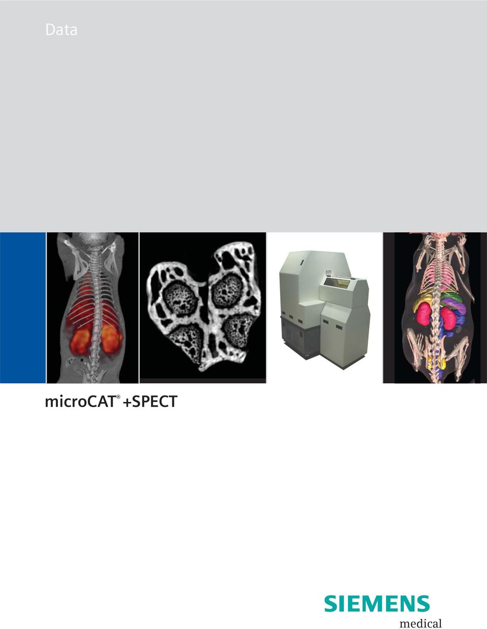
2 microcat at a Glance Designed to meet the throughput, resolution and image quality requirements of academic and pharmaceutical research, the Siemens microcat sets the standard for in vivo preclinical micro computed tomography. With a range of x-ray source and detector configurations available, the ability to adjust the scanner magnification between scans, and a suite of image reconstruction and data analysis tools, the microcat is the most versatile instrument in its class. Key Features Industry leading combination of resolution and Field of View (FOV) Unique capability to adjust x-ray source and detector positions for user-selected resolution and Field of View Superior image quality due to low-noise CCD detectors Multiple options for high speed image reconstruction Dedicated Real Time Reconstruction Linux Cluster Hardware/Software Solution Linux Cluster Software-only Solution (for customers with existing clusters) Compatibility with micropet scanners Optional SPECT detector for integrated SPECT/CT Dedicated preclinical service and applications support team Gantry The microcat gantry is a fully shielded enclosure with redundant safety interlocks in compliance with US FDA standard 21 CFR The gantry is divided into three major components. The upper imaging compartment houses the animal bed, x-ray source and detector and all moving components. The lower compartment houses all support electronics. The detachable front bore shield provides easy access to the animal under test while providing complete radiation shielding to the operator. The gantry rests on retractable vibration isolation pads, which may be raised to allow the gantry to roll between laboratories on its heavy-duty castors. The gantry is sized to fit through standard laboratory doors when the front shield is detached. X-ray Source Standard Source: The standard source is a 40W, tungsten anode, kvp source with <50-micron focal spot. This source provides a high x-ray flux for high speed scanning and a large cone angle for high magnification studies. The maximum achievable resolution with this x-ray source and the standard detector is 27 microns. Typical scan times are less than 5 minutes. Optional Variable-focus Source: For ultra-high resolution studies, an optional x-ray source is available which can operate in microfocus mode (<6 micron focal spot) providing a maximum resolution of 15 microns (10% MTF) with both detector options. The same source can operate with a larger focal spot at up to 65W for high speed studies. X-ray Detector Standard Detector: The standard x-ray detector has 2048 x 3096 pixels and is designed for high-speed, low-noise, whole-mouse imaging. The detector may be configured for a Field of View as large as 8 cm x 5.4 cm. The raw data is 12 bits deep and the detector dynamic range is 69 db with 1 x 1 binning and 72 db with 2 x 2 and 4 x 4 binning. The maximum achievable resolution with this detector and the standard x-ray source is 27 microns. With the optional micro-focus x-ray source, the maximum resolution is 15 microns. Large Area Detector: For larger animals, an optional detector is available with 4096 x 4096 pixels, providing a maximum Field of View of 11 cm x 11 cm. With this detector, the Field of View is software selectable through the graphical user interface, permitting the operator to crop each data set to match the geometry of the subject. This detector also has a superior signal to noise ratio with 14-bit readout and dynamic ranges of 67 db with 1 x 1 binning, 79 db with 2 x 2 binning and 84 db with 4 x 4 binning. The maximum achievable resolution with this detector and the standard x-ray source is 27 microns. With the optional micro-focus x-ray source the maximum resolution is 15 microns. 2

3 Specifications microcat Standard Detector Number of detector pixels 3072 x 2048 Typical Operation FOV Mouse Configuration 8 cm (axial) x 5.4 cm (transaxial) Rat Configuration 5.4 cm (axial) x 8 cm (transaxial) Large Area Detector Number of detector pixels 4096 x 4096 Typical Operation FOV 11 cm (axial) x 11 cm (transaxial) Standard X-ray source Maximum power Focal spot size Voltage range Maximum anode current Microfocus X-ray source Maximum power Focal spot size Voltage range Maximum anode current Spatial resolution Maximum FOV High resolution mode with microfocus x-ray source 40 W < 50 microns kvp (additional shielding required for operation above 80 kv) 500 microamps 65 W < 6 8W; < 60 65W kvp 500 microamps 37 microns (10% MTF) 15 microns (10% MTF) Minimum scan time 360 step scan <6 minutes 180 step scan <3 minutes Continuous rotation <1 minute Reconstruction Algorithm Speed with real time reconstruction Software Safety Modified Feldkamp algorithm volume reconstructed during scan time. Integrated graphical user interface for data acquisition, image reconstruction and data visualization. Amira TM 3D visualization and volume modeling package. 16-bit RAW, BMP and DICOM 3 output formats. Fully shielded cabinet with redundant safety interlocks. FDA registered. Complies with standards set by US FDA, Center for Devices and Radiological Health (21 CFR ). 3
 500 microamps 65 W < 6 microns @ 8W; < 60 microns @ 65W 20 130 kvp 500 microamps 37 microns (10% MTF) 15")
4 microcat Features Animal Bed Three styles of detachable animal beds are available: Mouse bed for up to 50 mm diameter animals Rat bed for up to 100 mm diameter animals Miniature bed for high resolution CT studies of small samples and for high resolution SPECT studies Each removable animal bed is interchangeable with Siemens micropet beds for easy transport of animals between instruments in dual modality studies. Positron emitting fiducial markers visible in the x-ray CT data set may be incorporated into the beds for easy image registration. Respiratory Gating The microcat is configured to accept respiratory gating signals from Biopac and BioTRIG physiological monitoring systems. Precision Motion Systems High-resolution images require high-precision moving components. Only the highest quality motion systems are employed in Siemens preclinical scanners. microspect All microcat systems can be configured with or upgraded to include a state-of-the-art SPECT imaging system for anatomic and functional data acquisition on a single platform. Applications Segmentation tools provide accurate quantification of anatomic data. Newly available preclinical microcat contrast agents provide previously unattainable soft tissue and vascular image contrast. Image courtesy of Oak Ridge National Laboratory. All microcat systems can be configured with optional high resolution SPECT detector heads for multi-modality studies. Image courtesy of the University of Tennessee Graduate School of Medicine, Knoxville, TN. With the optional variable focus x-ray source, images down to 15 micron resolution can be acquired in vivo. 4

5 Specifications Unit weight Unit height Unit width Unit depth (without front shield) Unit depth (with front shield) microcat Approximately 1800 lbs (820 kg) 70 in (1780 mm) 49 in (1245 mm) 33 in (840 mm) 55 in (1395 mm) Temperature and Humidity Operating room temperature F (7 24 C) Operating humidity: 30 70% non-condensing Note: The maximum power consumption of the microcat system is 2 kw (6800 BTU/hr). Electrical Requirements microcat and computer Note: Appropriate power connection will be provided for destination country. 110V/20A isolated outlet (20A outlet required) 240V/10A (Europe) Magnetic Field Magnetic field strength must be less than 10 Gauss. Radiation Safety The microcat is a cabinet x-ray system. The scanner is FDA registered and compliant with title 21 of the Code of Federal Regulations, Part (21 CFR ). 5
 240V/10A (Europe) Magnetic Field Magnetic field strength must be less than 10 Gauss. Radiation Safety The microcat is a cabinet x-ray system.")
6 microcat +SPECT at a Glance Siemens microcat +SPECT generates high resolution, fully registered SPECT/CT data sets. With the largest commercially available pixilated detector heads, an extensive set of collimators, multiple energy windows, and independently adjustable detector head positions, the microcat +SPECT sets the standard for preclinical multimodality imaging. Key Features Largest commercially available pixilated detector heads 150 mm x 150 mm Large detector heads permit greater pinhole magnification, improving sensitivity while maintaining large Field of View Small pixels provide high resolution, even at low magnification and with parallel-hole collimators +SPECT data acquisition system is fully integrated into the microcat hardware and software microcat beds are compatible with micropet scanners for easy dual and triple modality studies microcat +SPECT data sets are fully compatible with the micropet ASIPro software package Detector Heads 150 mm x 150 mm active area Array of position sensitive photomultiplier tubes and pixilated NaI(Tl) 2.0 mm x 2.0 mm x 10 mm crystals 2.2 mm crystal spacing 4,600 pixels per detector head Collimators Parallel hole (1.2 mm and 2 mm aperture) Pinhole (0.5 mm, 1 mm, 2 mm, and 3 mm inserts) Multi-pinhole collimation will be introduced in early

7 Image courtesy of VA Hospital, Columbia, MO. 7

8 Amira is a registered trademark of Mercury Computer Systems. Biopac is a registered trademark of Biopac Systems, Inc. BioTRIG is a registered trademark of Spin Systems (QLD) Pty Ltd. Linux is a registered trademark of Sun Microsystems. microcat and micropet are registered trademark of Siemens reserves the right to modify the design and specifications contained herein without prior notice. Product performance depends on the choice of system configuration. Please contact your local Siemens sales representative for the most current information or contact one of the addresses listed below All rights reserved. All photographs 2005 Siemens Medical Solutions, USA. All rights reserved. Note: Original images always lose a certain amount of detail when reproduced. Molecular Imaging 2501 N. Barrington Road Hoffman Estates, IL USA Contact Addresses Molecular Imaging 2501 N. Barrington Road Hoffman Estates, IL USA Telephone: Headquarters 51 Valley Stream Parkway Malvern, PA USA Telephone: Molecular Imaging 810 Innovation Drive Knoxville, TN USA Telephone: , Siemens AG Order No. A91MI T-7600 Printed in USA 10/05 PA 1005/1.5

Global Business Unit Address. Siemens Medical Solutions USA, Inc. 2501 N. Barrington Road Hoffman Estates, IL 60192-5203. Telephone: +1847 304 7700
 Trademarks and service marks used in this material are property of Siemens Medical Solutions USA or Siemens AG. All other company, brand, product and service names may be trademarks or registered trademarks
Next Generation Small Animal PET Technology
 G4 PET/X-ray P R O D U C T N O T E Pre-clinical in vivo Imaging Key Benefits: High performance, ultra-sensitive PET technology designed for pre-clinical imaging 3D PET and X-ray whole body images Purpose
Thinking ahead. Focused on life. REALIZED: GROUNDBREAKING RESOLUTION OF 80 µm VOXEL
 Thinking ahead. Focused on life. REALIZED: GROUNDBREAKING RESOLUTION OF 80 µm VOXEL X-ray ZOOM RECONSTRUCTION Flat Panel Detector (FPD) Automatic Positioning Function For ø 40 x H 40 mm, ø 60 x H 60 mm,
Automated Pose Determination for Unrestrained, Non-anesthetized Small Animal Micro-SPECT and Micro-CT Imaging
 Automated Pose Determination for Unrestrained, Non-anesthetized Small Animal Micro-SPECT and Micro-CT Imaging Shaun S. Gleason, Ph.D. 1, James Goddard, Ph.D. 1, Michael Paulus, Ph.D. 1, Ryan Kerekes, B.S.
DENTAL Cone beam 3D X-RAY SYSTEM with
 VERSATILE INTUITIVE efficient DENTAL Cone beam 3D X-RAY SYSTEM with dedicated panoramic imaging With thirty years of experience in designing and manufacturing state-of-the-art dental panoramic and tomographic
Remote RadEye Product Family
 Remote RadEye Product Family Overview The Remote RadEye x-ray camera provides the ultimate flexibility in design and product options for your complex imaging applications. Our unique camera design separates
PERFORM-X DIGITAL X-RAY SYSTEM PERFORM-X. with ceiling mounted tube support PERFORM-X
 DIGITAL X-RAY SYSTEM with ceiling mounted tube support Universal Digital Radiographic System Conventional system, full DR components Best-in-class image quality Flexible connectivity to PACS systems General
HIGH PERFORMANCE MOBILE SURGICAL C-ARM KMC-950
 HIGH PERFORMANCE MOBILE SURGICAL C-ARM 1K x 1k CCD Digital Camera System H.F. GENERATOR & ROTATING ANODE TUBE WITH DIGITAL WORKSTATION DESCRIPTION: Mobile Surgical C-arm systems are integrated with a triple
Toshiba Excelart Vantage 1.5T MRI Tech Specs (Technical Specifications)
") Toshiba Excelart Vantage 1.5T MRI Tech Specs (Technical Specifications) Excelart Vantage Magnet Configuration: Ultra-short-bore Strength (or W x H): 1.5 T Homogeneity, ppm V-RMS: Dimensions of maximum
Toshiba Excelart Vantage 1.5T MRI Tech Specs (Technical Specifications) Excelart Vantage Magnet Configuration: Ultra-short-bore Strength (or W x H): 1.5 T Homogeneity, ppm V-RMS: Dimensions of maximum
Agilent Cary 4000/5000/6000i Series UV-Vis-NIR
 Agilent Cary 4000/5000/6000i Series UV-Vis-NIR Guaranteed specifications Design overview Double beam, ratio recording, double out-of-plane Littrow monochromator UV-Vis-NIR spectrophotometer (Agilent Cary
Scan Time Reduction and X-ray Scatter Rejection in Dual Modality Breast Tomosynthesis. Tushita Patel 4/2/13
 Scan Time Reduction and X-ray Scatter Rejection in Dual Modality Breast Tomosynthesis Tushita Patel 4/2/13 Breast Cancer Statistics Second most common cancer after skin cancer Second leading cause of cancer
CT RADIATION DOSE REPORT FROM DICOM. Frank Dong, PhD, DABR Diagnostic Physicist Imaging Institute Cleveland Clinic Foundation Cleveland, OH
 CT RADIATION DOSE REPORT FROM DICOM Frank Dong, PhD, DABR Diagnostic Physicist Imaging Institute Cleveland Clinic Foundation Cleveland, OH CT Patient comes out... Patient goes in... Big Black Box Radiology
Performance testing for Precision 500D Classical R/F System
 Performance testing for Precision 500D Classical R/F System John M. Boudry, Ph.D. Image Quality Systems Engineer GE Healthcare Technologies Outline System background Image Quality Signature Test (IQST)
Rb 82 Cardiac PET Scanning Protocols and Dosimetry. Deborah Tout Nuclear Medicine Department Central Manchester University Hospitals
 Rb 82 Cardiac PET Scanning Protocols and Dosimetry Deborah Tout Nuclear Medicine Department Central Manchester University Hospitals Overview Rb 82 myocardial perfusion imaging protocols Acquisition Reconstruction
A d v a n c e d 1 2 8
 Advanced 128 SCENARIA Advanced 128 Hitachi Innovation at Work for You. Continuing its commitment to the SCENARIA scalable CT platform, Hitachi introduces cutting-edge 128-slice technology with the SCENARIA
Products - Microarray Scanners - SpotLight Two-Color Microarray Fluorescence Scanners New! SpotLight 2 now available!
 Products - Microarray Scanners - SpotLight Two-Color Microarray Fluorescence Scanners New! SpotLight 2 now available! Arrayit SpotLight Two-Color Microarray Scanners provide the market s most affordable
Master s Program in Medical Physics. Physics of Imaging Systems Basic Principles of Computer Tomography (CT) III. Prof. Dr. Lothar Schad.
 III. Prof. Dr. Lothar Schad.") 1 12/9/2008 Page 1 Master s Program in Medical Physics Physics of Imaging Systems Basic Principles of Computer Tomography (CT) III Chair in Faculty of Medicine Mannheim University of Heidelberg Theodor-Kutzer-Ufer
1 12/9/2008 Page 1 Master s Program in Medical Physics Physics of Imaging Systems Basic Principles of Computer Tomography (CT) III Chair in Faculty of Medicine Mannheim University of Heidelberg Theodor-Kutzer-Ufer
Family IMAGINGFORSCIENCE. the next generation four modality preclinical imaging platform SPECT/MRI, PET/MRI, SPECT/CT, PET/CT, SPECT/CT/PET
 Family the next generation four modality preclinical imaging platform SPECT/MRI, PET/MRI, SPECT/CT, PET/CT, SPECT/CT/PET IMAGINGFORSCIENCE nanoscan products are No.1 in the following: - achieve the breakthrough
QUANTITATIVE IMAGING IN MULTICENTER CLINICAL TRIALS: PET
 Centers for Quantitative Imaging Excellence (CQIE) LEARNING MODULE QUANTITATIVE IMAGING IN MULTICENTER CLINICAL TRIALS: PET American College of Radiology Clinical Research Center v.1 Centers for Quantitative
COST EFFECTIVE FLAT PANEL DIGITAL RADIOGRAPHY UPGRADE SOLUTIONS
 COST EFFECTIVE FLAT PANEL DIGITAL RADIOGRAPHY UPGRADE SOLUTIONS DRive is a digital imaging DR hardware & Software solution designed for General Radiography of anatomy. It is intended to replace film/screen
RIEGL VQ-480. Airborne Laser Scanning. Airborne Laser Scanner with Online Waveform Processing. visit our website www.riegl.com
 Airborne Laser Scanner with Online Waveform Processing RIEGL VQ-48 high-accuracy ranging based on echo digitization and online waveform processing high laser repetition rate - fast data acquisition multiple
CTVision System. Accurate Imaging for Precise Treatment. www.siemens.com/medical
 CTVision System Accurate Imaging for Precise Treatment www.siemens.com/medical Adapt in Real Time A radiation therapy treatment plan is a snapshot in time. But as time moves on, changes in patient anatomy
0 EC2 92011 V-,) 133 Lj9a
 133 Lj9a") 0 EC2 92011 V-,) 133 Lj9a Section 5: 5 1 0(k) Summar 5 10(K) SUMMARY FOR SOMATOM DEFINITION Flash (with Stellar Detector) Submitted by: Siemens Medical Solutions USA, Inc. 51 Valley Stream Parkway Malvern,
0 EC2 92011 V-,) 133 Lj9a Section 5: 5 1 0(k) Summar 5 10(K) SUMMARY FOR SOMATOM DEFINITION Flash (with Stellar Detector) Submitted by: Siemens Medical Solutions USA, Inc. 51 Valley Stream Parkway Malvern,
Embedded Systems in Healthcare. Pierre America Healthcare Systems Architecture Philips Research, Eindhoven, the Netherlands November 12, 2008
 Embedded Systems in Healthcare Pierre America Healthcare Systems Architecture Philips Research, Eindhoven, the Netherlands November 12, 2008 About the Speaker Working for Philips Research since 1982 Projects
Purchasing a cardiac CT scanner: What the radiologist needs to know
 Purchasing a cardiac CT scanner: What the radiologist needs to know Maria Lewis ImPACT St George s Hospital, London maria.lewis@stgeorges.nhs.uk CT scanner development Slice wars 1998 Increased z-coverage
CR 30-Xm. CR 30-Xm Digitizer
 D I G I T I Z E R CR 30-Xm Complete solution for digital mammography and all general radiography applications The CR 30-Xm is a versatile computed radiography (CR) digitizer that can handle digital mammography,
Digital panoramic X-ray for practical diagnostics
 ORTHOPHOS XG 3 instruments treatment centers Imaging systems cad/cam systems ORTHOPHOS XG 3 Digital panoramic X-ray for practical diagnostics System concept Standard panoramic X-ray with proven technology.
CT scanning. By Mikael Jensen & Jens E. Wilhjelm Risø National laboratory Ørsted DTU. (Ver. 1.2 4/9/07) 2002-2007 by M. Jensen and J. E.
 2002-2007 by M. Jensen and J. E.") 1 Overview CT scanning By Mikael Jensen & Jens E. Wilhjelm Risø National laboratory Ørsted DTU (Ver. 1.2 4/9/07) 2002-2007 by M. Jensen and J. E. Wilhjelm) As it can be imagined, planar X-ray imaging has
1 Overview CT scanning By Mikael Jensen & Jens E. Wilhjelm Risø National laboratory Ørsted DTU (Ver. 1.2 4/9/07) 2002-2007 by M. Jensen and J. E. Wilhjelm) As it can be imagined, planar X-ray imaging has
Molecular Imaging: from cell to man
 Molecular Imaging: from cell to man Torino, 24 November 2011 Advances in PET/SPECT technology for pre-clinical molecular imaging applications Alberto Del Guerra Dipartimento di Fisica "E.Fermi' Università
ZEISS Axiocam 506 color Your Microscope Camera for Imaging of Large Sample Areas Fast, in True Color, and High Resolution
 Product Information Version 1.0 ZEISS Axiocam 506 color Your Microscope Camera for Imaging of Large Sample Areas Fast, in True Color, and High Resolution ZEISS Axiocam 506 color Sensor Model Sensor Pixel
Power Injectors for Diagnostic Imaging
 Power Injectors for Diagnostic Imaging Contrast under Control A resource for all your contrast delivery needs Contrast under Control 2 1 3 Mallinckrodt provides contrast delivery systems for Computed
ParaVision 6. Innovation with Integrity. The Next Generation of MR Acquisition and Processing for Preclinical and Material Research.
 ParaVision 6 The Next Generation of MR Acquisition and Processing for Preclinical and Material Research Innovation with Integrity Preclinical MRI A new standard in Preclinical Imaging ParaVision sets a
Fundamentals of Cone-Beam CT Imaging
 Fundamentals of Cone-Beam CT Imaging Marc Kachelrieß German Cancer Research Center (DKFZ) Heidelberg, Germany www.dkfz.de Learning Objectives To understand the principles of volumetric image formation
thyroid uptake systems www.capintec.com
 64 thyroid uptake systems Captus 3000 Thyroid Uptake System Comprehensive Software Quick-start feature for rapid set-up User defined, HIPAA compliant counting protocols Thyroid uptake Wipe test Bioassay
Cardius. XPO Series. Cardius 3 XPO
 Cardius XPO Series Cardius 3 XPO Three imaging systems, all of which provide outstanding image quality, superior efficiency, and increased patient comfort. Cardius 2 XPO Includes the standard features
Customer: Cardiff Hospital NHS (United Kingdom) Machine:
 Machine:") Customer: Cardiff Hospital NHS (United Kingdom) Machine: Bench for leak test Model: Easy TS Page 1 of 7 Date: 6 th March 2012 1. Introduction. Easy TS is a compact and reliable table top bench designed
Customer: Cardiff Hospital NHS (United Kingdom) Machine: Bench for leak test Model: Easy TS Page 1 of 7 Date: 6 th March 2012 1. Introduction. Easy TS is a compact and reliable table top bench designed
Applications: X-ray Microtomography, Streak Tube and CRT Readout, Industrial & Medical Imaging X-RAY GROUP
 Now Powered by LightField FEATURES BENEFITS Back Illuminated CCD (248B) For highest sensitivity Front illuminated CCD (248F) Affordable technology for moderate light level applications Ultra low noise
IDM-200 -V. Interchangeable HPGe Detector Module (IDM) for Systems Integrators
 for Systems Integrators") IDM-200 -V Interchangeable HPGe Detector Module (IDM) for Systems Integrators Free from liquid nitrogen, free from maintenance, free from integration complexities. Convenient-to-mount, all-in-one integrated
IDM-200 -V Interchangeable HPGe Detector Module (IDM) for Systems Integrators Free from liquid nitrogen, free from maintenance, free from integration complexities. Convenient-to-mount, all-in-one integrated
X-RAY IMAGING Emerging Digital Technology - CMOS Detectors
 Application Note Case Study Technology Primer White Paper X-RAY IMAGING Emerging Digital Technology - CMOS Detectors Image Sensors X-Ray DETECTORS Scanners Image Processing Custom Solutions In all domains
REGIUS History. Touch REGIUS110. REGIUS170 ImagePilot REGIUS150 REGIUS330 CS-1
 Konica Minolta has been a CR technology leader since 1996, consistently designing, developing, and delivering innovative solutions. The REGIUS Σ represents the sum of this experience. REGIUS Σ delivers
Application of Digital Radiography for the Detection and Classification of Pneumoconiosis
 Application of Digital Radiography for the Detection and Classification of Pneumoconiosis DEPARTMENT OF HEALTH AND HUMAN SERVICES Centers for Disease Control and Prevention National Institute for Occupational
Micro-CT for SEM Non-destructive Measurement and Volume Visualization of Specimens Internal Microstructure in SEM Micro-CT Innovation with Integrity
 Micro-CT for SEM Non-destructive Measurement and Volume Visualization of Specimens Internal Microstructure in SEM Innovation with Integrity Micro-CT 3D Microscopy Using Micro-CT for SEM Micro-CT for SEM
X-Mind. Instinct for perfection
 X-Mind Instinct for perfection X-Mind tubes are located at the back of the head which gives the patient better protection because the distance between the focal spot and the skin is 50% greater than in
Robot Perception Continued
 Robot Perception Continued 1 Visual Perception Visual Odometry Reconstruction Recognition CS 685 11 Range Sensing strategies Active range sensors Ultrasound Laser range sensor Slides adopted from Siegwart
Chapter 3 SYSTEM SCANNING HARDWARE OVERVIEW
 Qiang Lu Chapter 3. System Scanning Hardware Overview 79 Chapter 3 SYSTEM SCANNING HARDWARE OVERVIEW Since all the image data need in this research were collected from the highly modified AS&E 101ZZ system,
Computed Tomography Resolution Enhancement by Integrating High-Resolution 2D X-Ray Images into the CT reconstruction
 Digital Industrial Radiology and Computed Tomography (DIR 2015) 22-25 June 2015, Belgium, Ghent - www.ndt.net/app.dir2015 More Info at Open Access Database www.ndt.net/?id=18046 Computed Tomography Resolution
SpectraTec II. Polarized Multi-Laser Source BLUE SKY RESEARCH WAVELENGTHS. The SpectraTec II
 BLUE SKY RESEARCH The SpectraTec II, two wavelength laser module is a highly integrated system comprised of two lasers, individual driving and temperature control electronics, wavelength combining, and
Digital Dental X-ray. Midmark Animal Health Products
 Digital Dental X-ray Midmark Animal Health Products Disease you can't see... 60% of periodontal disease lies below the gum line, and many issues aren't detectable via probing or visual inspection.* Don't
APPLICATION NOTE. Basler racer Migration Guide. Mechanics. www.baslerweb.com. Flexible Mount Concept. Housing
 62 62 APPLICATION NOTE www.baslerweb.com Basler racer Migration Guide This paper describes what to consider when replacing the Basler L100 Camera Link or the Basler runner Gigabit Ethernet (GigE) line
Page: 1 of 6 Page: 1 of 6
 Page: 1 of 6 Page: 1 of 6 CR Basics and FAQ Overview Computed Radiography is a term used to describe a system that electronically records a radiographic image. Computed Radiographic systems use unique
MPC 4. Machinery Protection Card Type MPC 4 FEATURES. Continuous on-line Machinery Protection Card
 Machinery Protection Card Type FEATURES Continuous on-line Machinery Protection Card Real-time measurement and monitoring using state-of-the-art DSP techniques Fully VME-compatible slave interface Fully
Cirrus 0.2T. MRI for Everyone. North America, Asia, Europe. contact: kturek@mri-tech.pl
 Cirrus 0.2T MRI for Everyone North America, Asia, Europe contact: kturek@mri-tech.pl MRI-TECH inc. Cirrus MRI system for all your needs: Low costs Low maintenance High quality Open geometry Imaging of
SECTION 1: REQUIREMENTS FOR CERTIFICATES OF COMPLIANCE FOR CLASSES OF RADIATION SOURCES
 Department of Health and Human Services Population Health Radiation Protection Act 2005 Section 17 CERTIFICATE OF COMPLIANCE: STANDARD FOR RADIATION APPARATUS - X-RAY DIAGNOSTIC (VETERINARY) SECTION 1:
Meso Scale Discovery. WINDOWS is a registered trademark of Microsoft Corporation
 Meso Scale Discovery M S D M S D Meso Scale Discovery, a division of Meso Scale Diagnostics, LLC. (MSD) 9238 Gaither Rd, Gaithersburg, MD 20877 Phone: 240.631.2522 Fax: 240.632.2219 email: sales@meso-scale.com
DR Retrofit Kit 1417 WL Configuration Integrated PACS
 DR Retrofit Kit 1417 WL Configuration Integrated PACS Description: The DR Retrofit Kit 1417WL - Configuration Integrated PACS makes the use of CR Cassettes/Imaging Plates or the use of analogue x-ray films,
QuickSpecs. At A Glance. HP Retail Integrated Peripherals for RP9 G1 Retail System. Overview
 Overview At A Glance The HP Retail Integrated peripheral options compatible with the HP RP9 Retail System can be purchased and installed separately: o HP Retail Integrated Encryption Capable MSR o HP Retail
Experimental study of beam hardening artefacts in photon counting breast computed tomography
 Experimental study of beam hardening artefacts in photon counting breast computed tomography M.G. Bisogni a, A. Del Guerra a,n. Lanconelli b, A. Lauria c, G. Mettivier c, M.C. Montesi c, D. Panetta a,
R/F. Efforts to Reduce Exposure Dose in Chest Tomosynthesis Targeting Lung Cancer Screening. 3. Utility of Chest Tomosynthesis. 1.
 R/F Efforts to Reduce Exposure Dose in Chest Tomosynthesis Targeting Lung Cancer Screening Department of Radiology, National Cancer Center Hospital East Kaoru Shimizu Ms. Kaoru Shimizu 1. Introduction
02 CyberKnife: Treatment Delivery
 TREATMENT DELIVERY CyberKnife Treatment Delivery System The CyberKnife System is the first and only robotic radiosurgery system to offer highly precise and customizable, non-surgical treatment options
Moving Forward What does this mean for the Medical Physicist and the Imaging Community?
 Moving Forward What does this mean for the Medical Physicist and the Imaging Community? John M. Boone, Ph.D., FAAPM, FACR Professor and Vice Chairman of Radiology University of California Davis Medical
Development of SCENARIA New Version Software MEDIX VOL. 62 P.32 P.35
 Development of SCENAIA New Version Software Takayuki Kadomura Toshio Sakamoto Kana Tanaka Takatsugu Ito Yuuko Nishimura Naomi Maekawa MEDIX VOL. 62 P.32 P.35 Development of SCENAIA New Version Software
Application Note #503 Comparing 3D Optical Microscopy Techniques for Metrology Applications
 Screw thread image generated by WLI Steep PSS angles WLI color imaging Application Note #503 Comparing 3D Optical Microscopy Techniques for Metrology Applications 3D optical microscopy is a mainstay metrology
DryView CHROMA Imaging System
 DryView CHROMA Imaging System VERSATILE PRINTING ON FILM OR PAPER One of the most trusted names in medical printing now offers a highly versatile printer that empowers physicians and other healthcare professionals
Image Acquisition and Diagnostics
 en examion aqs software examion aqs software Image Acquisition and Diagnostics Professional Image Acquisition and Diagnostic Workstation examion aqs software is the universal image acquisition software
How To Improve Your Ct Image Quality
 Translating Protocols Between Scanner Manufacturer and Model Cynthia H. McCollough, PhD, FACR, FAAPM Professor of Radiologic Physics Director, CT Clinical Innovation Center Department of Radiology Mayo
AN INVESTIGATION INTO THE USEFULNESS OF THE ISOCS MATHEMATICAL EFFICIENCY CALIBRATION FOR LARGE RECTANGULAR 3 x5 x16 NAI DETECTORS
 AN INVESTIGATION INTO THE USEFULNESS OF THE ISOCS MATHEMATICAL EFFICIENCY CALIBRATION FOR LARGE RECTANGULAR 3 x5 x16 NAI DETECTORS Frazier L. Bronson CHP Canberra Industries, Inc. 800 Research Parkway,
Supports screening, diagnostic, and multimodality workflows
 GE Healthcare Universal Viewer Breast Imaging Product Data Sheet Supports screening, diagnostic, and multimodality workflows Introduction Centricity PACS with Universal Viewer 1 puts clinical insight within
University Children s Hospital Basel
 Cooperation Siemens International Reference Center Pediatric MRI www.siemens.com/skyra University Children s Hospital Basel Siemens International Reference Center Pediatric MRI Answers for life. Partnering
PET/CT QC/QA. Quality Control in PET. Magnus Dahlbom, Ph.D. Verify the operational integrity of the system. PET Detectors
 Quality Control in PET PET/CT QC/QA Magnus Dahlbom, Ph.D. Division of Nuclear Medicine Ahmanson Biochemical Imaging Clinic David Geffen School of Medicine at UCLA Los Angeles Verify the operational integrity
Section D..General Auto Electrical Corporation
 43 Section D..General Auto Electrical Corporation 1. LED Based Signal unit GAEC manufacture LED based signaling system for use in electrified and non electrified sections of Indian railways. The exceptional
ENGLISH. Planmeca ProMax 3D concept
 ENGLISH Planmeca ProMax 3D concept Profound understanding of anatomy Planmeca ProMax 3D concept is an intelligent and multipurpose X-ray unit series designed to obtain complete information on patient anatomy
Contents. X-ray and Computed Tomography. Characterization of X-rays. Production of X-rays
 J. E. Wilhjelm Ørsted TU Technical University of enmark, Bldg. 348, K-2800 Kongens Lyngby, enmark. X-ray and Computed Tomography Contents History and characterization of X-rays Conventional (projection)
Solder joint inspection and analysis
 GE Inspection Technologies Solder joint inspection and analysis with GE s phoenix x-ray microfocus and nanofocus X-ray systems FAQs about X-ray source How X-ray inspection small FOD large FOD works object
Information about the T9 beam line and experimental facilities
 Information about the T9 beam line and experimental facilities The incoming proton beam from the PS accelerator impinges on the North target and thus produces the particles for the T9 beam line. The collisions
Clinic. ED Trauma Trauma Stroke. OR Neuro/Spine. Critical Care. Neuro ENT. Diagnostic. Pediatric. Radiology. Plastics Thoracic. Neuro.
 www.neurologica.com Critical Care Clinic Operating Room Radiology ED Trauma OR Neuro/Spine Pediatric Plastics Thoracic General Interventional Radiology ENT ED Trauma Trauma Stroke Critical Care Neuro Pediatric
MRC High Resolution. MR-compatible digital HD video camera. User manual
 MRC High Resolution MR-compatible digital HD video camera User manual page 1 of 12 Contents 1. Intended use...2 2. System components...3 3. Video camera and lens...4 4. Interface...4 5. Installation...5
Physics 441/2: Transmission Electron Microscope
 Physics 441/2: Transmission Electron Microscope Introduction In this experiment we will explore the use of transmission electron microscopy (TEM) to take us into the world of ultrasmall structures. This
Instruction Sheet 554 828
 08/13-W07-Hund Instruction Sheet 554 828 X-ray image sensor (554 828) Precision slide X-ray image sensor (554 829) Software Computed Tomography Pro (554 820) Sensor head on precision slide with USB module
SUN ORACLE DATABASE MACHINE
 SUN ORACLE DATABASE MACHINE FEATURES AND FACTS FEATURES From 1 to 8 database servers From 1 to 14 Sun Oracle Exadata Storage Servers Each Exadata Storage Server includes 384 GB of Exadata Smart Flash Cache
INFRARED PARTS MANUAL
 INFRARED PARTS MANUAL PIR325 FL65 GLOLAB CORPORATION Thank you for buying our Pyroelectric Infrared components. The goal of Glolab is to produce top quality electronic kits, products and components. All
Mx8000 IDT 16 provides dose control methodologies like DoseRight ACS (Automatic Current Selection) and DoseRight DOM (dynamic dose modulation).
 and DoseRight DOM (dynamic dose modulation).") Philips Mx8000 IDT 16 CT Scanner Philips Medical systems Mx8000 IDT 16 delivers 16 simultaneous slices at sub-millimeter collimator, high resolution and isotropic imaging. New large area detectors allow
Philips Mx8000 IDT 16 CT Scanner Philips Medical systems Mx8000 IDT 16 delivers 16 simultaneous slices at sub-millimeter collimator, high resolution and isotropic imaging. New large area detectors allow
View of ΣIGMA TM (Ref. 1)
") Overview of the FESEM system 1. Electron optical column 2. Specimen chamber 3. EDS detector [Electron Dispersive Spectroscopy] 4. Monitors 5. BSD (Back scatter detector) 6. Personal Computer 7. ON/STANDBY/OFF
Overview of the FESEM system 1. Electron optical column 2. Specimen chamber 3. EDS detector [Electron Dispersive Spectroscopy] 4. Monitors 5. BSD (Back scatter detector) 6. Personal Computer 7. ON/STANDBY/OFF
I-Max Touch Range. PAN / CEPH / 3D digital panoramic unit. Evolutive 3 in 1 panoramic unit
 I-Max Touch Range PAN / CEPH / 3D digital panoramic unit Evolutive 3 in 1 panoramic unit 3D A new dimension for a complete diagnosis I-Max Touch 3D Evolutive, simple, fast The panoramic unit realizes complete
Series. X-ray Inspection. www.nordsondage.com
 Series X-ray Inspection www.nordsondage.com 2 Nordson DAGE Quadra X-ray Inspection Nordson DAGE Quadra X-ray Inspection 3 Nordson DAGE, the leaders in X-ray inspection for electronics, presents its 4th
Basler. Area Scan Cameras
 Basler Area Scan Cameras VGA to 5 megapixels and up to 210 fps Selected high quality Sony and Kodak CCD sensors Powerful Gigabit Ethernet interface Superb image quality at all resolutions and frame rates
We bring quality to light. MAS 40 Mini-Array Spectrometer. light measurement
 MAS 40 Mini-Array Spectrometer light measurement Features at a glance Cost-effective and robust CCD spectrometer technology Standard USB interface Compatible with all Instrument Systems measuring adapters
Ion Beam Sputtering: Practical Applications to Electron Microscopy
 Ion Beam Sputtering: Practical Applications to Electron Microscopy Applications Laboratory Report Introduction Electron microscope specimens, both scanning (SEM) and transmission (TEM), often require a
RADIATION THERAPY. Dosimetry Pioneers since 1922 NEW
 RADIATION THERAPY Dosimetry Pioneers since 1922 NEW New Product Releases 2015 One phantom multiple options 4D Patient and Machine QA Modular Phantom for Stay flexible. Go modular. } Revolutionary new modular
VERSATILE AND EASY-TO-USE 3D LASER SCANNERS > >
 VERSATILE AND EASY-TO-USE 3D LASER SCANNERS > > A WORLD OF APPLICATIONS Z Corporation, producers of the industry s fastest, easiest-to-use and most versatile 3D scanners, makes 3D scanning ideal for a
APPLICATION OF X-RAY COMPUTED TOMOGRAPHY IN SILICON SOLAR CELLS
 APPLICATION OF X-RAY COMPUTED TOMOGRAPHY IN SILICON SOLAR CELLS V.A. Popovich 1, W. Verwaal 2, M. Janssen 1, I. J. Bennett 3, I.M.Richardson 1, 1. Delft University of Technology, Department of Materials
NETWORK ENABLED EQUIPMENT MONITOR
 NETWORK ENABLED EQUIPMENT MONITOR Remotely Monitor Sensors over the Internet Connect Sensors to the Web to Remotely Monitor Equipment, Processes or Other Applications A Complete, Easy to Deploy, Stand-Alone
ENGLISH. Planmeca ProMax 3D Mid
 ENGLISH Planmeca ProMax 3D Mid Genuine all-in-one unit Planmeca ProMax 3D Mid is a genuine all-in-one CBVT (Cone Beam Volumetric Tomography) unit including 3D imaging, digital panoramic, digital cephalometric
Optimization of image quality through online motion correction
 Advanced imaging technologies a revolution in diagnosis and treatment Optimization of image quality through online motion correction Oline Vinter Olesen December 10, 2010 Medical Solutions, Siemens Institute
Surgery Support System as a Surgeon s Advanced Hand and Eye
 Surgery Support System as a Surgeon s Advanced Hand and Eye 8 Surgery Support System as a Surgeon s Advanced Hand and Eye Kazutoshi Kan Michio Oikawa Takashi Azuma Shio Miyamoto OVERVIEW: A surgery support
MEDICAL IMAGING. 90/30 Remote Controlled RF System
 MEDICAL IMAGING 90/30 Remote Controlled RF System The Easy solution for your Remote Controlled room Apollo EZ represents the Easy approach for the radiographic/fluoroscopic room and it comes from the experience
Technical Datasheet Scalar Network Analyzer Model 8003-10 MHz to 40 GHz
 Technical Datasheet Scalar Network Analyzer Model 8003-10 MHz to 40 GHz The Giga-tronics Model 8003 Precision Scalar Network Analyzer combines a 90 db wide dynamic range with the accuracy and linearity
pco.edge 4.2 LT 0.8 electrons 2048 x 2048 pixel 40 fps 37 500:1 > 70 % pco. low noise high resolution high speed high dynamic range
 edge 4.2 LT scientific CMOS camera high resolution 2048 x 2048 pixel low noise 0.8 electrons USB 3.0 small form factor high dynamic range 37 500:1 high speed 40 fps high quantum efficiency > 70 % edge
The Convenient and Compact CR System for Veterinary Practices
 The Convenient and Compact CR System for Veterinary Practices Fire CR+ 20 / 40 / 60 / 80 vet with a throughput of 20 to 70 cassettes per hour and the option of a space - saving wall mount The small and
PRODUCT SHEET. info@biopac.com support@biopac.com www.biopac.com
 EYE TRACKING SYSTEMS BIOPAC offers an array of monocular and binocular eye tracking systems that are easily integrated with stimulus presentations, VR environments and other media. Systems Monocular Part
GenStore Archive Cabinet
 GenStore Archive Cabinet Setup and Maintenance Manual Version A April 2014 Two GenStore Archives shown mounted on optional table stand. 2 Contents Getting Started... 4 Using this Manual...4 Safety Precautions...4