Chest CT protocols. Mannudeep K. Kalra, MD, DNB. Dianna D. Cody, PhD. Massachusetts General Hospital Harvard Medical School
|
|
|
- Roger Boone
- 7 years ago
- Views:
Transcription
1 Chest CT protocols Mannudeep K. Kalra, MD, DNB Dianna D. Cody, PhD Massachusetts General Hospital Harvard Medical School M.D. Anderson Cancer Center

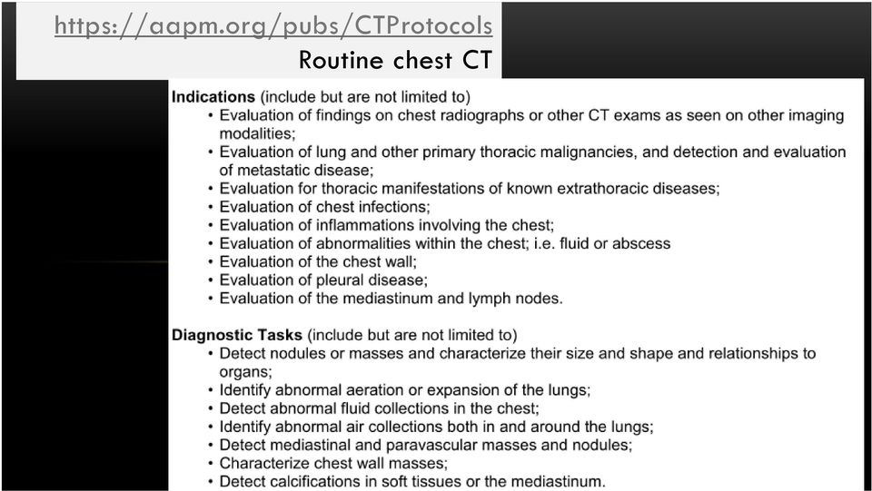
2 Specific principles Routine chest CT Lung nodule follow up CT Review audience sample protocols Review speaker protocols Review AAPM protocols

3 Routine chest CT
4 Institution 1: Routine chest CT protocol Lung nodule Follow up

5 Routine Chest Institution 1: Siemens 64 Scan type Helical Helical Scan range Apex to Adrenals Apex to Adrenals Rotation time 0.5 sec 0.5 sec Beam coll. (Det. Config.) 24 x 1.2 mm 24 x 1.2 mm Pitch KV Quality Ref. mas Image thickness 5mm 3mm and 1.5 mm FBP or IRT (kernel) B35 (no Safire or IRIS) B31 (no Safire or IRIS)

6 Routine Chest Institution 1: Siemens 64 Scan type Helical Helical Scan range Apex to Adrenals Apex to Adrenals Rotation time 0.5 sec 0.5 sec Beam coll. (Det. Config.) 24 x 1.2 mm 64 or 32 x 0.6 mm Pitch KV Quality Ref. mas Image thickness 5mm 5 mm FBP or IRT (kernel) B35 (no Safire or IRIS) B31 (no Safire or IRIS)
 B35 (no Safire or IRIS) B31")
7 Routine Chest Institution 1: Siemens 64 Scan type Helical Helical Helical Scan range Apex to Adrenals Apex to Adrenals Apex to Adrenals Rotation time 0.5 sec 0.5 sec 0.5 sec Beam Coll. (Det. Con) 24 x 1.2 mm 24 x 1.2 mm 64 or 32 x 0.6 mm Pitch KV Quality Ref. mas Image thickness 5mm 1.5 & 3 mm 5 mm FBP or IRT (kernel) B35 (no Safire) B31 (no Safire) B31 (no Safire)

8 Dose: Lung nodule FU = Routine chest Institution 1: Routine Siemens 64 Institution 1: Lung nodule Scan type Helical Helical Scan range Apex to Adrenals Apex to Adrenals Rotation time 0.5 sec 0.5 sec Beam coll. (Det. Config.) 24 x 1.2 mm 24 x 1.2 mm Pitch KV Quality Ref. mas Image thickness 5mm 5mm FBP or IRT (kernel) B35 (no Safire or IRIS) B35 (no Safire or IRIS)
 B35 (no Safire or IRIS) B35 (no Safire or")
9 Lung nodule doses must be much lower Institution 1: Siemens 64 Scan type Helical Helical Scan range Apex to Adrenals Apex to Lung bases only Rotation time 0.5 sec 0.5 sec Beam Coll. (Det. Config.) 24 x 1.2 mm 24 x 1.2 mm Pitch KV Quality Ref. mas mas FIXED Image thickness 5mm 3mm and 1.5 mm FBP or IRT (kernel) B35 (no Safire or IRIS) B31 (no Safire or IRIS)

10 Institution 2: Routine chest CT protocol
11 Routine Chest CT Siemens Definition 128 Institution 2 Scan type Helical Helical Helical Scan range Apex to Adrenals Apex to Adrenals Apex to Adrenals Rotation time 0.28 sec 0.5 sec 0.5 sec Beam Coll. (Det. Con) 128 x 0.6 mm 24 x 1.2 mm 64 or 32 x 0.6 mm Pitch KV Quality Ref. mas mas Image thickness 5mm 1.5 & 3 mm 5 mm FBP or IRT (kernel) I 45 & I 50 Safire I 31 (Safire) I31 (Safire)

12 Institution 3: Doses Routine chest CT = Lung nodule follow up CT

13 Routine Chest CT = Lung nodule FU Institution 3: Routine (Siemens 128 ) Institution 3: Lung nodule Scan type Helical Helical Scan range Apex to Adrenals Apex to Adrenals Rotation time 0.5 sec 0.5 sec Beam Coll. (Det. Config.) 128 * * 0.6 Pitch 1 1 KV Care KV Care KV Quality Ref. mas Image thickness 5mm 5mm FBP or IRT (kernel) B 43 & 80 (no Safire) B 43 & 80 (no Safire)

14 Institution 4: Doses 1. Routine chest CT dose: Too high 2. Routine chest CT = Lung nodule FU CT

15 Routine Chest CT = Lung nodule FU Institution 4: Siemens 64 Scan type Helical (Routine chest) Helical (Lung nodule) Scan range Apex to Adrenals Apex to Adrenals Rotation time 0.5 sec 0.5 sec Beam Coll. (Det. Config.) 24 x 1.2 mm 24 x 1.2 mm Pitch KV Quality Ref. mas Image thickness 3mm 3 mm FBP or IRT (kernel) - -

16 Institution 4: Siemens 64 Routine Chest CT >> Lung nodule FU Scan type Helical (Routine chest) Helical (Lung nodule) Scan range Apex to Adrenals Apex to Adrenals Rotation time 0.5 sec 0.5 sec Beam Coll. (Det. Config.) 24 x 1.2 mm 24 x 1.2 mm Pitch higher better motion 0.75 higher better KV / /120 Quality Ref. mas Image thickness 3mm 3 mm FBP or IRT (kernel) - -

17 Institution 5: Routine chest CT protocol Philips

18 Routine Chest CT Institution 5: Philips Ingenuity 128 Scan type Helical Comments Scan range Rotation time Beam Coll. (Det. Config.) Apex to Adrenals 0.5 sec 64 x mm Pitch Auto 1.1 KV 120 mas/image <150 lbs 90; lbs 120 lbs > (no AEC) Image thickness 3 mm and 1.5 mm 3 mm No AEC FBP or IRT (kernel) Filter B Filter B/C
 Image")
19 Routine Chest CT Institution 5 Scan type Helical Helical Helical Scan range Apex to Adrenals Apex to Adrenals Apex to Adrenals Rotation time 0.5 sec 0.4 sec 0.4 sec Beam Coll. (Det. Config.) 64 x mm 64 x mm 64 x mm Pitch Auto Auto (~ 0.9) 1.1 KV mas/image <150 lbs 90; lbs 120 lbs > (no AEC) Philips Ingenuity (Z-DOM) 140 (??? Z-DOM) Image thickness 3 mm and 1.5 mm 3 mm and 1.5 mm 3 mm FBP or IRT (kernel) Filter B Filter B (idose 2) Filter B/C
 Image thickness 3 mm and 1.5 mm 3 mm and 1.5 mm 3 mm FBP or IRT (kernel) Filter B Filter B (idose 2) Filter B/C")
20 Institution 6: Philips Lung nodule FU << Routine chest CT dose

21 Institution 6: Philips 64 Brilliance Scan type Helical (Routine chest CT) Helical (Lung nodule FU) Scan range Apex to Adrenals Apex to Adrenals Rotation time 0.4 sec 0.4 sec Beam Coll. (Det. Config.) 32 X 1.25 mm 32 X 1.25 mm Pitch KV mas/image 100 (Z-Dom) 40 (Z-Dom) Image thickness 3 mm and 1.5 mm 3 mm FBP or IRT (kernel) Filter B Filter B
22 PHILIPS: ROUTINE CHEST CT: AAPM
23 GE: Routine chest CT (non ASIR): AAPM Do not use noise index for 5mm for thinner slices and vice versa.
24 GE: Routine chest CT (ASIR) AAPM
25 GE 64 Routine Chest CT AAPM MGH Scan type Helical Helical Scan range Apex to Adrenals Apex to Adrenals Rotation time 0.4 sec 0.4 sec Beam Coll. (Det. Config.) 64 * * Pitch KV NI (min max ma) 13 ( ) 16.8 ASIR 50% ( ) ASIR 40% Image thickness 5 mm 2.5 and 1.25 mm FBP or IRT (kernel) Standard Detail
26 Toshiba: Routine chest CT : AAPM (Disclosure)
27 Special caveats Be aggressive with lower kv (<80 kg) or < 30 BMI Prefer use of AEC for routine chest CT Adapt AEC to clinical indication: Dose for lung nodule FU CT must be lower than routine chest CT doses When possible position arms above shoulder When not, position arms on the chest rather than by side
28 Salient points in Chest CT protocols Lung nodule FU CT: KV Fixed low ma or modify AEC Apex to lung bases only CTDI vol mgy IRT: 1-2 mgy or lower Routine chest CT KV (80 kv < 50 kg) Always with AEC Apex to Adrenal coverage Target CTDI vol 3-7 mgy IRT: 2-5 mgy or lower
The disclaimer on page 1 is an integral part of this document. Copyright February 23, 2016 by AAPM. All rights reserved.
 DISCLAIMER: TO THE EXTENT ALLOWED BY LOCAL LAW, THIS INFORMATION IS PROVIDED TO YOU BY THE AMERICAN ASSOCIATION OF PHYSICISTS IN MEDICINE, A NON-PROFIT ORGANIZATION ORGANIZED TO PROMOTE THE APPLICATION
Purchasing a cardiac CT scanner: What the radiologist needs to know
 Purchasing a cardiac CT scanner: What the radiologist needs to know Maria Lewis ImPACT St George s Hospital, London maria.lewis@stgeorges.nhs.uk CT scanner development Slice wars 1998 Increased z-coverage
CT Protocol Optimization over the Range of CT Scanner Types: Recommendations & Misconceptions
 CT Protocol Optimization over the Range of CT Scanner Types: Recommendations & Misconceptions Frank N. Ranallo, Ph.D. Associate Professor of Medical Physics & Radiology University of Wisconsin School of
CT: Size Specific Dose Estimate (SSDE): Why We Need Another CT Dose Index. Acknowledgements
: Why We Need Another CT Dose Index. Acknowledgements") CT: Size Specific Dose Estimate (SSDE): Why We Need Another CT Dose Index Keith J. Strauss, MSc, FAAPM, FACR Clinical Imaging Physicist Cincinnati Children s Hospital University of Cincinnati College of
CT: Size Specific Dose Estimate (SSDE): Why We Need Another CT Dose Index Keith J. Strauss, MSc, FAAPM, FACR Clinical Imaging Physicist Cincinnati Children s Hospital University of Cincinnati College of
Multi-slice Helical CT Scanning of the Chest
 Multi-slice Helical CT Scanning of the Chest Comparison of different low-dose acquisitions Lung cancer is the main cause of deaths due to cancer in human males and the incidence is constantly increasing.
Assessing Radiation Dose: How to Do It Right
 Assessing Radiation Dose: How to Do It Right Michael McNitt-Gray, PhD, DABR, FAAPM Professor, Department of Radiology Director, UCLA Biomedical Physics Graduate Program David Geffen School of Medicine
Low-dose CT for Pulmonary Embolism
 Low-dose CT for Pulmonary Embolism Gautham Gautham P. P. Reddy, Reddy, MD, MD, MPH MPH University University of of Washington Washington Introduction Introduction CT CT accounts accounts for for > 50%
MDCT Technology. Kalpana M. Kanal, Ph.D., DABR Assistant Professor Department of Radiology University of Washington Seattle, Washington
 MDCT Technology Kalpana M. Kanal, Ph.D., DABR Assistant Professor Department of Radiology University of Washington Seattle, Washington ACMP Annual Meeting 2008 - Seattle, WA Educational Objectives Historical
Moving Forward What does this mean for the Medical Physicist and the Imaging Community?
 Moving Forward What does this mean for the Medical Physicist and the Imaging Community? John M. Boone, Ph.D., FAAPM, FACR Professor and Vice Chairman of Radiology University of California Davis Medical
Protocol Management and Review Strategies The MD Anderson Experience
 Protocol Management and Review Strategies The MD Anderson Experience Dianna Cody, Ph.D. Professor Dept. of Imaging Physics Univ. of Texas M.D. Anderson Cancer Center Objectives Setting up a policy for
The disclaimer on page 1 is an integral part of this document. Copyright March 1, 2016 by AAPM. All rights reserved.
 DISCLAIMER: TO THE EXTENT ALLOWED BY LOCAL LAW, THIS INFORMATION IS PROVIDED TO YOU BY THE AMERICAN ASSOCIATION OF PHYSICISTS IN MEDICINE, A NON-PROFIT ORGANIZATION ORGANIZED TO PROMOTE THE APPLICATION
How To Improve Your Ct Image Quality
 Translating Protocols Between Scanner Manufacturer and Model Cynthia H. McCollough, PhD, FACR, FAAPM Professor of Radiologic Physics Director, CT Clinical Innovation Center Department of Radiology Mayo
Acknowledgement. Diagnostic X-Ray Shielding. Nomenclature for Radiation Design Criteria. Shielding Design Goal (Air Kerma):
:") Diagnostic X-Ray Shielding Multi-Slice CT Scanners Using NCRP 47 Methodology Melissa C. Martin, M.S., FAAPM, FACR Therapy Physics Inc., Bellflower, CA AAPM Annual Meeting, Orlando, FL Refresher Course
Diagnostic X-Ray Shielding Multi-Slice CT Scanners Using NCRP 47 Methodology Melissa C. Martin, M.S., FAAPM, FACR Therapy Physics Inc., Bellflower, CA AAPM Annual Meeting, Orlando, FL Refresher Course
CT RADIATION DOSE REPORT FROM DICOM. Frank Dong, PhD, DABR Diagnostic Physicist Imaging Institute Cleveland Clinic Foundation Cleveland, OH
 CT RADIATION DOSE REPORT FROM DICOM Frank Dong, PhD, DABR Diagnostic Physicist Imaging Institute Cleveland Clinic Foundation Cleveland, OH CT Patient comes out... Patient goes in... Big Black Box Radiology
Spiral CT: Single and Multiple Detector Systems. AAPM Refresher Course Nashville, TN July 28,1999
 Spiral CT: Single and Multiple Detector Systems AAPM Refresher Course Nashville, TN July 28,1999 Mike McNitt-Gray, PhD, DABR Assistant Professor UCLA Radiological Sciences mmcnittgray@mednet.ucla.edu X-Ray
The disclaimer on page 1 is an integral part of this document. 1. Copyright December 14, 2015 by AAPM. All rights reserved.
 DISCLAIMER: TO THE EXTENT ALLOWED BY LOCAL LAW, THIS INFORMATION IS PROVIDED TO YOU BY THE AMERICAN ASSOCIATION OF PHYSICISTS IN MEDICINE, A NON-PROFIT ORGANIZATION ORGANIZED TO PROMOTE THE APPLICATION
Patient-centered CT imaging: New methods for patient-specific optimization 1 of image quality and radiation dose
 Patient-centered CT imaging: New methods for patient-specific optimization 1 of image quality and radiation dose ipatient is an advanced platform that delivers focused innovations to facilitate patient-centered
Copyright March 1, 2016 by AAPM. All rights reserved.
 DISCLAIMER: TO THE EXTENT ALLOWED BY LOCAL LAW, THIS INFORMATION IS PROVIDED TO YOU BY THE AMERICAN ASSOCIATION OF PHYSICISTS IN MEDICINE, A NON-PROFIT ORGANIZATION ORGANIZED TO PROMOTE THE APPLICATION
Tracking Radiation Exposure From Medical Diagnostic Procedures: Siemens Perspectives
 Tracking Radiation Exposure From Medical Diagnostic Procedures: Siemens Perspectives Gilbert W. Beebe Symposium The National Academies Katharine Grant, PhD Staff Scientist 8 December 2011 For internal
Chest 1: Pulmonary Nodule Follow-up: Low-Dose Helical CT (Unenhanced) (Non-metastatic) Gantry Rotation Time. mas (Reg-Lg) 40-80
 (Non-metastatic) Gantry Rotation Time. mas (Reg-Lg) 40-80") Revisions Effective January 2012 Chest 1: Pulmonary Nodule Follow-up: Low-Dose Helical CT (Unenhanced) (Non-metastatic) Technologist Instructions Patient must cough several times prior to scan to clear
Revisions Effective January 2012 Chest 1: Pulmonary Nodule Follow-up: Low-Dose Helical CT (Unenhanced) (Non-metastatic) Technologist Instructions Patient must cough several times prior to scan to clear
State-of-the-Art Technology in Cardiac CT
 1 2 Next Step Evolution or Revolution? State-of-the-Art Technology in Cardiac CT Stefan Ulzheimer, PhD Global Director of Collaborations CT Siemens Medical Solutions Major Innovations in CT Head The 80
Table of Contents. Scan acquisition and user interface basics. Dose modulation and reduction tools. Multi-Slice Detector Geometry
 Table of Contents Scan acquisition and user interface basics Dose modulation and reduction tools Multi-Slice Detector Geometry Image Reconstruction and Display Contrast Media Tools Multi-planar formats
The Possibilities Tomosynthesis Brings to Lung Cancer Screening
 Special Report The Possibilities Brings to Lung Cancer Screening Low-dose is a Useful Tool in Lung Cancer Screening A Technical Evaluation of Low-dose with the SONIALVISION safire Joint Industrial-Academic
Clinical Image Gallery: Symbia xspect
 Clinical Image Gallery: Symbia xspect SNMMI 2013 Answers for life. Disclaimer Symbia xspect, xspect Bone and xspect Quant are not licensed according to Canadian law, are pending 510(k) clearance, and are
R/F. Efforts to Reduce Exposure Dose in Chest Tomosynthesis Targeting Lung Cancer Screening. 3. Utility of Chest Tomosynthesis. 1.
 R/F Efforts to Reduce Exposure Dose in Chest Tomosynthesis Targeting Lung Cancer Screening Department of Radiology, National Cancer Center Hospital East Kaoru Shimizu Ms. Kaoru Shimizu 1. Introduction
Rb 82 Cardiac PET Scanning Protocols and Dosimetry. Deborah Tout Nuclear Medicine Department Central Manchester University Hospitals
 Rb 82 Cardiac PET Scanning Protocols and Dosimetry Deborah Tout Nuclear Medicine Department Central Manchester University Hospitals Overview Rb 82 myocardial perfusion imaging protocols Acquisition Reconstruction
Implementation of Cone-beam CT imaging for Radiotherapy treatment localisation.
 Implementation of Cone-beam CT imaging for Radiotherapy treatment localisation. Andrew Bridges Clinical Scientist Diagnostic Radiology & Radiation Protection Physics Overview What is CBCT? Use of CBCT
Role of the Medical Physicist in Clinical Implementation of Breast Tomosynthesis
 Role of the Medical Physicist in Clinical Implementation of Breast Tomosynthesis Bob Liu, Ph.D. Department of Radiology Massachusetts General Hospital And Harvard Medical School Digital Breast Tomosynthesis
Monitoring Patient Radiation Dose in VA. Charles M. Anderson MD, PhD Chief Consultant for Diagnostic Services Veterans Health Administration
 Monitoring Patient Radiation Dose in VA Charles M. Anderson MD, PhD Chief Consultant for Diagnostic Services Veterans Health Administration TWENTY ONE/NOVEMBER 2011 Computed Tomography (CT), Magnetic Resonance
Automated EMR Dose History Extraction and Monitoring
 Automated EMR Dose History Extraction and Monitoring Aaron Sodickson MD, PhD Section Chief, Emergency Radiology Medical Director of CT, Brigham Radiology Network Brigham and Women s Hospital Harvard Medical
Practical exercise: Effective dose estimate in CT
 Practical exercise: Effective dose estimate in CT TRAINING COURCE PROGRAM 19 20 May 2011, Sofia, Bulgaria Virginia Tsapaki Medical Physics Dpt Konstantopoulio General Hospital email: virginia@otenet.gr
scan: : a new tool to analyse
 Gated CT-scan scan: : a new tool to analyse the shape of the aortic annulus. SEROUSSI SAFAR K, JONDEAU G, LANSAC E., SERFATY JM Bichat Hospital, U698, Paris, France Anatomy: 3D + spatial resolution Retrospective
Digital Breast Tomosynthesis QC Requirements
 Digital Breast Tomosynthesis QC Requirements AAPM Spring Clinical Meeting March 8, 2015 Michael S Glaser, MS, DABR Alliance Medical Physics, LLC Learning Objectives 1. GE SenoClaire - Physicist & Technologist
The Whys, Hows and Whats of the Noise Power Spectrum. Helge Pettersen, Haukeland University Hospital, NO
 The Whys, Hows and Whats of the Noise Power Spectrum Helge Pettersen, Haukeland University Hospital, NO Introduction to the Noise Power Spectrum Before diving into NPS curves, we need Fourier transforms
Fundamentals of Cone-Beam CT Imaging
 Fundamentals of Cone-Beam CT Imaging Marc Kachelrieß German Cancer Research Center (DKFZ) Heidelberg, Germany www.dkfz.de Learning Objectives To understand the principles of volumetric image formation
Sistemas de reducción de dosis en CT para pediatría. Nuevos avances Ángela de Pinto Siemens CT Business Manager angela.pinto@siemens.
 Sistemas de reducción de dosis en CT para pediatría. Nuevos avances Ángela de Pinto Siemens CT Business Manager angela.pinto@siemens.com Copyright Siemens AG 2009. All rights reserved. An Innovation Leader
Radiology Workload and Follow-up Considerations
 Radiology Workload and Follow-up Considerations William C. Black, MD Department of Radiology Norris Cotton Cancer Center Dartmouth-Hitchcock Medical Center william.c.black@hitchcock.org No financial disclosures
AAPM Medical Physics Practice Guideline 1.a: CT Protocol Management and Review Practice Guideline
 JOURNAL OF APPLIED CLINICAL MEDICAL PHYSICS, VOLUME 14, NUMBER 5, 2013 AAPM Medical Physics Practice Guideline 1.a: CT Protocol Management and Review Practice Guideline The American Association of Physicists
PET/CT QC/QA. Quality Control in PET. Magnus Dahlbom, Ph.D. Verify the operational integrity of the system. PET Detectors
 Quality Control in PET PET/CT QC/QA Magnus Dahlbom, Ph.D. Division of Nuclear Medicine Ahmanson Biochemical Imaging Clinic David Geffen School of Medicine at UCLA Los Angeles Verify the operational integrity
kv-& MV-CBCT Imaging for Daily Localization: Commissioning, QA, Clinical Use, & Limitations
 kv-& MV-CBCT Imaging for Daily Localization: Commissioning, QA, Clinical Use, & Limitations Moyed Miften, PhD Dept of Radiation Oncology University of Colorado Denver Questions Disease Stage (local, regional,
GE Healthcare. Revolution EVO. More than just high tech. Higher purpose.
 GE Healthcare Revolution EVO More than just high tech. Higher purpose. Revolution EVO. Designed with purpose. Today s healthcare environment is about creating new solutions to pressing needs. It s about
Brilliance CT 64-channel confi guration
 Brilliance CT 64-channel confi guration with Essence technology The Brilliance CT 64-channel confi guration is designed to help you conduct the most advanced multislice CT studies possible. These systems
Computed Tomography Radiation Safety Issues in Ontario
 Computed Tomography Radiation Safety Issues in Ontario Healthcare Human Factors Group Centre for Global ehealth Innovation University Health Network Toronto, ON, Canada June 16, 2006 Table of Contents
Performance testing for Precision 500D Classical R/F System
 Performance testing for Precision 500D Classical R/F System John M. Boudry, Ph.D. Image Quality Systems Engineer GE Healthcare Technologies Outline System background Image Quality Signature Test (IQST)
WHERE IN THE WORLD JILL LIPOTI?
 WHERE IN THE WORLD IS JILL LIPOTI? HELLO FROM NEW JERSEY CRCPD - National Symposium on Fusion Imaging and Multimodalities February 18-20, 2004 Kansas City, Missouri New Jersey s Requirements As They Pertain
Evidence Based And Systems Based Best Practices For Management Of Imaging Utilization
 Evidence Based And Systems Based Best Practices For Management Of Imaging Utilization James H Thrall MD Radiologist-in-Chief Massachusetts General Hospital Juan M Taveras Professor of Radiology Harvard
Development of SCENARIA New Version Software MEDIX VOL. 62 P.32 P.35
 Development of SCENAIA New Version Software Takayuki Kadomura Toshio Sakamoto Kana Tanaka Takatsugu Ito Yuuko Nishimura Naomi Maekawa MEDIX VOL. 62 P.32 P.35 Development of SCENAIA New Version Software
The AAPM does not endorse any products, manufacturers, or suppliers. Nothing in this publication should be interpreted as implying such endorsement.
 DISCLAIMER: This publication is based on sources and information believed to be reliable, but the AAPM, the editors, and the publisher disclaim any warranty or liability based on or relating to the contents
Diagnostic Exposure Tracking in the Medical Record
 Diagnostic Exposure Tracking in the Medical Record J.A. Seibert, Ph.D. Department of Radiology Sacramento, California USA Vancouver. British Columbia Relevant disclosures None Learning objectives Understand
American College of Radiology CT Accreditation Program. Testing Instructions
 American College of Radiology CT Accreditation Program Testing Instructions (Revised July 24, 2015) This guide provides all of the instructions necessary for clinical tests, phantom tests and general submission
Surveying and QC of Stereotactic Breast Biopsy Units for ACR Accreditation
 Surveying and QC of Stereotactic Breast Biopsy Units for ACR Accreditation LORAD Stereotactic Breast Biopsy System AAPM Spring Clinical Meeting Phoenix, AZ March 17, 2013 Melissa C. Martin, M.S., FACR,
SWABIK Project. Software Tools for DICOM Media Exchange in Clinical Research: the. SWABIK Project
 Software Tools for DICOM Media Exchange in Clinical Research: the SWABIK Project Software Tools for DICOM Media Exchange in Clinical Research: the SWABIK Project O. ElGazzar 1, M. Onken 1, M. Eichelberg
Scan Time Reduction and X-ray Scatter Rejection in Dual Modality Breast Tomosynthesis. Tushita Patel 4/2/13
 Scan Time Reduction and X-ray Scatter Rejection in Dual Modality Breast Tomosynthesis Tushita Patel 4/2/13 Breast Cancer Statistics Second most common cancer after skin cancer Second leading cause of cancer
Gestión global de la dosis en TC. Sistema de registro y gestión
 Gestión global de la dosis en TC. Sistema de registro y gestión IV Jornada de Protección Radiológica Hospitalaria SARH FEA Radiofísica Hospital Virgen de las Nieves. Granada jalmansa.lopez@gmail.com 22
Stephen R. Veach, M.D.
 Stephen R. Veach, M.D. Memorial Sloan-Kettering Cancer Center International Oncology Programs 160 E. 53 rd Street New York, NY 10022 212-610 610-08780878 - tel 212-308 308-7063 - fax veachs@mskcc.org SCREENING
Development of pediatric CT protocols to obtain minimal doses in PET/CT studies a physicists approach.
 Development of pediatric CT protocols to obtain minimal doses in PET/CT studies a physicists approach. Wibeke Nordhøy 1, Mehrnaz Sheikhaeri 2, Alise Larsen 1, Otto Glomset 1 Til slutt et utvalg fra foredraget
Low-dose CT Imaging. Edgar Fearnow, M.D. Section Chief, Computed Tomography, Lancaster General Hospital
 Lung Cancer Screening with Low-dose CT Imaging Edgar Fearnow, M.D. Section Chief, Computed Tomography, Lancaster General Hospital Despite recent declines in the incidence of lung cancer related to the
CT QC Under the ACR QC Manual. What? There s a Manual?? Learning Objectives 8/6/12! ! YES!!! Almost. ! Doesn t have a pretty cover yet
 CT QC Under the ACR QC Manual Douglas Pfeiffer, MS, DABR Boulder Community Hospital What? There s a Manual??! YES!!! Almost! Doesn t have a pretty cover yet! Should be out by RSNA! Really seriously I mean
Pediatric Hospitals Bring Low-dose CT to the Middle East
 Pediatric Hospitals ring Low-dose CT to the Middle East For years, radiologists have been cognizant of the importance of limiting pediatric patients exposure to radiation dose. uilding on the LR principle,
Veraview IC5 HD High definition, digital imaging excellence. Thinking ahead. Focused on life.
 Veraview IC5 HD High definition, digital imaging excellence Thinking ahead. Focused on life. Thinking ahead. Focused on life. 2 3 IC5 HD Super High-Speed with High Definition Clarity For dental radiology
Techniques and Applications of Automatic Tube Current Modulation for CT 1
 Review Radiology Mannudeep K. Kalra, MD, DNB Michael M. Maher, MD, FFR (RCSI), FRCR Thomas L. Toth, DSc Bernhard Schmidt, PhD Bryan L. Westerman, PhD Hugh T. Morgan, PhD Sanjay Saini, MD Index terms: Computed
Dose Modulation Technique in CT
 Content Dose Modulation Technique in CT Short Overview - Overview - Dose Saving Features - Take Home Point - Conclusions Senior Application specialist Siemens Limited, Taiwan For internal use only / Copyright
Quality control of CT systems by automated monitoring of key performance indicators: a two-year study
 JOURNAL OF APPLIED CLINICAL MEDICAL PHYSICS, VOLUME 16, NUMBER 4, 2015 Quality control of CT systems by automated monitoring of key performance indicators: a two-year study Patrik Nowik, a Robert Bujila,
QP-2040L CNC VERTICAL MACHINING CENTER
 Page 1 of 8 QP-2040L CNC VERTICAL MACHINING CENTER Page 2 of 8 MACHINE FEATURES: This fully upgradeable VMC is designed to accommodate various upgrades in the future so that it can be reconfigured for
PRACTICAL TIPS IN ENSURING RADIATION SAFETY IN THE USE OF MEDICAL DIAGNOSTIC X-RAY EQUIPMENT
 PRACTICAL TIPS IN ENSURING RADIATION SAFETY IN THE USE OF MEDICAL DIAGNOSTIC X-RAY EQUIPMENT Although the medical uses of X-rays to examine a patient without surgery became an amazing medical breakthrough,
SITE IMAGING MANUAL ACRIN 6698
 SITE IMAGING MANUAL ACRIN 6698 Diffusion Weighted MR Imaging Biomarkers for Assessment of Breast Cancer Response to Neoadjuvant Treatment: A sub-study of the I-SPY 2 TRIAL Version: 1.0 Date: May 28, 2012
M D Anderson Cancer Center Orlando TomoTherapy s Implementation of Image-guided Adaptive Radiation Therapy
 M D Anderson Cancer Center Orlando TomoTherapy s Implementation of Image-guided Adaptive Radiation Therapy Katja Langen, PhD Research supported by TomoTherapy Inc. Today s Lecture Introduction to helical
Thinking ahead. Focused on life. REALIZED: GROUNDBREAKING RESOLUTION OF 80 µm VOXEL
 Thinking ahead. Focused on life. REALIZED: GROUNDBREAKING RESOLUTION OF 80 µm VOXEL X-ray ZOOM RECONSTRUCTION Flat Panel Detector (FPD) Automatic Positioning Function For ø 40 x H 40 mm, ø 60 x H 60 mm,
3/12/2014. Disclosures. Understanding IAC CT Accreditation. Outline. Learning Objectives. Who is the IAC?
 Disclosures Understanding IAC CT Accreditation Serve as one of two AAPM representatives to IAC CT Board of Directors, Serve as IAC representative to AAPM for focus group on accreditations Provide 3-hour
Cynthia H. McCollough b) and Michael R. Bruesewitz Department of Radiology, Mayo Clinic College of Medicine, Rochester, Minnesota 55905
 and Michael R. Bruesewitz Department of Radiology, Mayo Clinic College of Medicine, Rochester, Minnesota 55905") The phantom portion of the American College of Radiology ACR Computed Tomography CT accreditation program: Practical tips, artifact examples, and pitfalls to avoid a Cynthia H. McCollough b) and Michael
The phantom portion of the American College of Radiology ACR Computed Tomography CT accreditation program: Practical tips, artifact examples, and pitfalls to avoid a Cynthia H. McCollough b) and Michael
Institution-wide Training & Educational Program for CT Technologists
 RSNA 2014 Quality Story Boards QSE 110 Institution-wide Training & Educational Program for CT Technologists Christoph Zorich RT, CT; Daisha Marsh, RT CT; Lior Molvin, RT, CT; Jia Wang PhD; Joni Schott,
IGRT. IGRT can increase the accuracy by locating the target volume before and during the treatment.
 DERYA ÇÖNE RADIOTHERAPY THERAPIST ACIBADEM KOZYATAGI HOSPITAL RADIATION ONCOLOGY DEPARTMENT IGRT IGRT (image-guided radiation therapy) is a technique that reduces geometric uncertainties by considering
MYOCARDIAL PERFUSION COMPUTED TOMOGRAPHY PhD course in Medical Imaging. Anne Günther Department of Radiology OUS Rikshospitalet
 MYOCARDIAL PERFUSION COMPUTED TOMOGRAPHY PhD course in Medical Imaging Anne Günther Department of Radiology OUS Rikshospitalet CORONARY CT ANGIOGRAPHY (CTA) Accurate method in the assessment of possible
Head and Neck Treatment Planning: A Comparative Review of Static Field IMRT Rapid Arc Tomotherapy HD
 Good Morning Head and Neck Treatment Planning: A Comparative Review of Static Field IMRT Rapid Arc Tomotherapy HD Barbara Agrimson, BS RT(T)(R), CMD Steve Rhodes, BS RT(T), CMD Disclaimer This presentation
4D Scanning. Image Guided Radiation Therapy. Outline. A Simplified View of the RT Process. Outline. Steve B. Jiang, Ph.D.
 4D Scanning Steve B. Jiang, Ph.D. Department of Radiation Oncology jiang.steve@mgh.harvard.edu http://gray.mgh.harvard.edu/ Outline Problems with free breathing 3D scanning What is 4D CT? How does it work?
Gated Radiotherapy for Lung Cancer
 Gated Radiotherapy for Lung Cancer Steve B. Jiang, Ph.D. Depart Of Radiation Oncology University of California San Diego sbjiang@ucsd.edu radonc.ucsd.edu/research/cart Two Types of Gating Internal gating
Cone Beam Reconstruction Jiang Hsieh, Ph.D.
 Cone Beam Reconstruction Jiang Hsieh, Ph.D. Applied Science Laboratory, GE Healthcare Technologies 1 Image Generation Reconstruction of images from projections. textbook reconstruction advanced acquisition
Tumor. An Brief Introduction to 4D CT Scanning. Outline. Three Types of Motion Artifacts. CT Artifacts in Free Breathing 3D Scan
 An Brief Introduction to 4D CT Scanning Steve B. Jiang, Ph.D. Dept of Radiation Oncology Univ of California San Diego Outline Problems with free breathing 3D scanning What is 4D CT? How does it work? Acknowledgements
Our Department: structure and organization
 EORTC meeting for Radiation Therapy Technologists: RTT s role in the modernization of radiotherapy 10th October 2014, Villejuif (Grand Paris), France Elekta Stereotactic Body Frame: transmission modelled
IMRT for Prostate Cancer. Robert A. Price Jr., Ph.D. Philadelphia, PA
 IMRT for Prostate Cancer Robert A. Price Jr., Ph.D. Philadelphia, PA Number of Patients 16 14 12 1 8 6 4 1481 IMRT Patients at FCCC 293 97 Prostate Breast H&N Other 64 Approximately 13-15 patients per
The Challenge of CT Dose Records
 The Challenge of CT Dose Records Kimberly Applegate, MD, MS, FACR Professor of Radiology and Pediatrics Emory University *financial disclosures: -Springer Textbook contracts -AIM advisory board for patient
X-Mind. Instinct for perfection
 X-Mind Instinct for perfection X-Mind tubes are located at the back of the head which gives the patient better protection because the distance between the focal spot and the skin is 50% greater than in
Clinical Rotation 3: PHYS 705 Fall 2015 (Aug. 25, 2015 to Feb. 25, 2016) COURSE INFORMATION
 COURSE INFORMATION") Clinical Rotation 3: PHYS 705 Fall 2015 (Aug. 25, 2015 to Feb. 25, 2016) COURSE INFORMATION Days: Monday-Friday Times: Full Time Location: One of the participating cancer clinics Program Director: George
Clinical Rotation 3: PHYS 705 Fall 2015 (Aug. 25, 2015 to Feb. 25, 2016) COURSE INFORMATION Days: Monday-Friday Times: Full Time Location: One of the participating cancer clinics Program Director: George
GE Healthcare. DoseWatch. Gathering Radiation Dose Data
 GE Healthcare DoseWatch Gathering Radiation Dose Data Gathering Radiation Dose Data After your organization crosses the bridge of Why? for patient radiation dose management either due to regulation, risk
HIGH PERFORMANCE MOBILE SURGICAL C-ARM KMC-950
 HIGH PERFORMANCE MOBILE SURGICAL C-ARM 1K x 1k CCD Digital Camera System H.F. GENERATOR & ROTATING ANODE TUBE WITH DIGITAL WORKSTATION DESCRIPTION: Mobile Surgical C-arm systems are integrated with a triple
Siemens Computed Tomography
 Siemens Computed Tomography Innovation Leader. The Cosmopolitan Information Topic Waltraud Winter Siemens Copyright Siemens AG 2011. All rights reserved. Page 1 October 2012 Waltraud Winter An Innovation
Chapter 8: Potential Energy and Conservation of Energy. Work and kinetic energy are energies of motion.
 Chapter 8: Potential Energy and Conservation of Energy Work and kinetic energy are energies of motion. Consider a vertical spring oscillating with mass m attached to one end. At the extreme ends of travel
PET/CT-MRI First clinical experience
 20 th April 2013, Barcelona, Sp PET/CT-MRI First clinical experience Philippe Appenzeller, MD Staff Radiologist and Nuclear Medicine Physician Department Medical Imaging, University Hospital Zurich PET/CT-MR
CT Scan Thorax and Upper Abdomen. Respiratory Unit Patient Information Leaflet
 CT Scan Thorax and Upper Abdomen Respiratory Unit Patient Information Leaflet Introduction This leaflet gives you general information about your CT (computerised tomography) scan. It does not replace the
225C-024T-05. Continuous control rotary drive without spring return. Technical data sheet. Description. Technical data. Actuators
 Actuators Technical data sheet 225C-024T-05 Continuous control rotary drive without spring return Description Actuator for adjusting air dampers of 90 angle of rotation to be used in HVAC installations.
Lung Cancer and Pleural Mesothelioma: Cleveland Clinic Multidisciplinary Approaches to Care
 Lung Cancer and Pleural Mesothelioma: Cleveland Clinic Multidisciplinary Approaches to Care Saturday, April 27, 2013 Cleveland Marriott Downtown at Key Center 127 Public Square Cleveland, Ohio Learning
Radiation Therapy for Prostate Cancer: Treatment options and future directions
 Radiation Therapy for Prostate Cancer: Treatment options and future directions David Weksberg, M.D., Ph.D. PinnacleHealth Cancer Institute September 12, 2015 Radiation Therapy for Prostate Cancer: Treatment
3 CT Parameters that Influence the Radiation Dose
 1 3 CT Parameters that Influence the Radiation Dose Hans Dieter Nagel, PhD Philips Medical Systems, Science and Technology, Roentgenstr. 24, D-22335 Hamburg, Germany hans-dieter.nagel@philips.com The radiation
Radiation Protection in Radiotherapy
 Radiation Protection in Radiotherapy Albert Lisbona Medical Physics Department CLCC Nantes Atlantique 44805 Saint-Herblain France a-lisbona@nantes.fnclcc.fr Radiation therapy The lecture is oriented to
Study the Quality Assurance of Conventional X-ray Machines Using Non Invasive KV meter
 Study the Quality Assurance of Conventional X-ray Machines Using Non Invasive KV meter T.M.Taha Radiation Protection Department, Nuclear Research Center, Atomic Energy Authority, Cairo.P.O.13759 Egypt.
Patient Exposure Doses During Diagnostic Radiography
 Patient Exposure Doses During Diagnostic Radiography JMAJ 44(11): 473 479, 2001 Shoichi SUZUKI Associated Professor, Faculty of Radiological Technology, School of Health Sciences, Fujita Health University
DMX protocol. Robin MMX Blade - DMX protocol, version 1.1
 protocol Mode/channel Robin MMX Blade - protocol, version 1.1 1 1 1 Pan 0-255 Pan movement by 540 proportional 2 2 * Pan Fine 0-255 Fine control of pan movement proportional 3 3 2 Tilt 0-255 Tilt movement
Evolve LED Area Light
 GE Lighting Evolve LED Area Light Scalable Area Light (EASB) imagination at work Product Features The next evolution of the GE Evolve LED Area Light continues to deliver outstanding features, while adding
PERFORM-X DIGITAL X-RAY SYSTEM PERFORM-X. with ceiling mounted tube support PERFORM-X
 DIGITAL X-RAY SYSTEM with ceiling mounted tube support Universal Digital Radiographic System Conventional system, full DR components Best-in-class image quality Flexible connectivity to PACS systems General
Patient Dose Tracking for Imaging Studies. David E. Hintenlang, Ph.D., DABR University of Florida
 Patient Dose Tracking for Imaging Studies David E. Hintenlang, Ph.D., DABR University of Florida Conflict of Interest Statement No affiliation or financial interests in any of the commercial products or