GE Medical Systems Training in Partnership. Module 8: IQ: Acquisition Time
|
|
|
- Kelley Davis
- 10 years ago
- Views:
Transcription
1 Module 8: IQ: Acquisition Time

2 IQ : Acquisition Time Objectives...Describe types of data acquisition modes....compute acquisition times for 2D and 3D scans.

3 2D Acquisitions The 2D mode acquires and reconstructs raw image data into two dimensional images whose brightness is proportional to the intensity of the MRI signal from the corresponding protons. This slice excitation method is followed by frequency and phase encoding to produce an image. Because the spatial encoding takes place in two dimensions the images are labeled 2D. Phase Encoding 2D slices Frequency Encoding

4 2D Multi-Planar Acquiring multiple images within a single acquisition or time frame means that the order of slice excitation is odd-numbered first, then even numbered images. All slices are acquired within the TR period Multi-Planar acquisition All 5 slices in the same TR

5 2D Sequential Acquiring an image sequentially means that all the excitation pulses ( # of phase steps x NEX x TR ) are delivered for one location before the process is repeated at another location. 1 Sequential acquisition Each slice is completed before moving on to the next slice

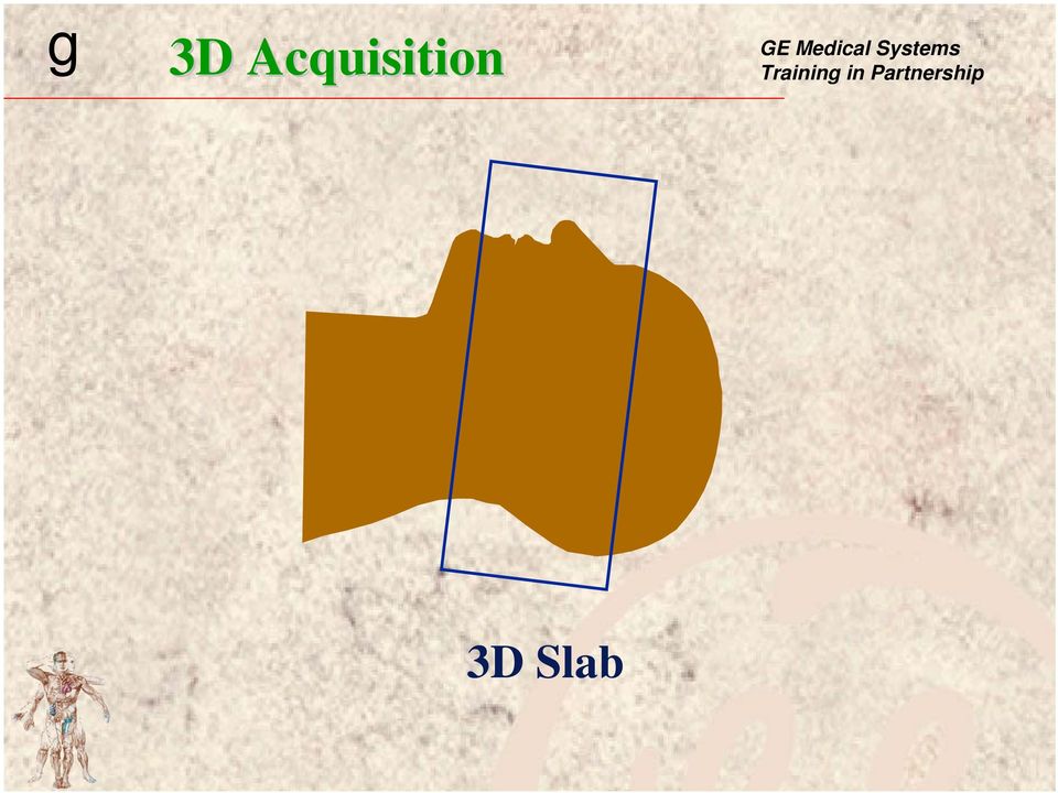
6 3D Acquisition In volume or 3D imaging, a wide RF pulse is delivered to excite an entire scan volume or slab. Spatial encoding must then be done in the phase, frequency and slice axes. Slice Encoding Phase Encoding 3D Volume Frequency Encoding

7 3D Acquisition 3D Slab
8 Progress Check 2D: TR x Phase Matrix x NEX = Acquisition Time 3D: TR x Phase Matrix x NEX x # of Slices = Acq. Time TR- TR is measured in milliseconds and is the time between RF pulses that repeat the pattern. Phase Matrix - The system collects data from the slice once for each phase encoding step. NEX- The number of times each set of phase encoding steps is repeated is called a NEX or Number of EXcitations. Calculate the scan times below using the appropriate formula

9 15. I.Q.: Acquisition Time 16. There are multiple acquisition modes: 2D: sequential and multi-planar 3D / volume MRS/ spectroscopy Cine 2D scan time formula: TR x phase x NEX 60,000 3D scan time formula: TR x phase x NEX x slices 60,000 2D & 3D are standard Go to QLB Pomp and ZIP

10 Module 9: IQ: Spatial Resolution

11 Spatial Resolution Objectives...Recall and explain the scan parameters that affect spatial resolution....compute spatial resolution given specific scan parameters....evaluate images acquired with varying spatial resolution.

12 Spatial Resolution How close two objects can be before they can be distinguished as separate objects.

13 Spatial Resolution Three parameters affect spatial resolution: FOV Matrix Slice Thickness Small FOV s, large matrices and small slice thickness result in high spatial resolution. #Phase Row FOV (cm) #Frequency Column Voxel Slice Thickness (mm)

14 FOV FOV determines the amount of anatomy displayed on the image. The larger the FOV, the larger the pixel size, and therefore the resolution is decreased. FOV can be changed in 1 cm intervals. Pixel size Pixel size 24 cm FOV 16 cm FOV

15 Progress Check Calculating Pixel Size, Area and Volume FOV / # phase steps = phase dimension FOV / # frequency steps = frequency dimension Phase x frequency = pixel area Pixel area x slice thickness = voxel volume Calculate the voxel volume for the image parameters listed below

16 Progress Check Slice Thickness change Image A: Thickness = 3 mm Image B: thickness = 5 mm Image C: Thickness = 10 mm SNR: Spatial resolution: Contrast: Time:

17 Progress Check FOV change Image A: FOV = 18 cm Image B: FOV = 22 cm Image C: FOV= 25 cm SNR: Spatial resolution: Contrast: Time:

18 Module 10: IQ: SNR

19 IQ: SNR Objectives View images for SNR and spatial resolution trade-offs....quantify (calculate) the change in SNR when a parameter has been changed.

20 Signal to Noise Ratio SNR is the ratio of the amplitude of the MR signal to the amplitude of the noise. Noise is the undesirable signal that is generated from the patient, the environment and the system electronics. signal noise

21 Signal to Noise Ratio SNR TIME TR NEX Resolution FOV Matrix Slice Thick Exception to rule : Receive Bandwidth
22 Receive Bandwidth Readout Window + 16 khz X gradient TR Readout Window + 32 khz X gradient TR
23 Receive Bandwidth + 32 khz + 16 khz noise signal
24 Receive Bandwidth Tradeoffs RBW SNR Chemical TE Motion Shift Artifact H 2 0 Chemical shift The frequency difference between protons bound in fat versus water Fat (+) (-) approx. 220 Hz ( 1.5T ) approx. 143 Hz ( 1.0 T ) approx. 74 Hz (.5T ) approx. 35 Hz (.2T )
25 I.Q.: Spatial Resolution/SNR 18. Voxel volume = I.Q. Tradeoffs FOV x FOV x slice phase freq SNR Isotropic voxels occur when all sides (height, width, depth) are equal. Time RBW is an exception Resolution
26 Progress Check NEX change Image A: NEX = 1 Image B: NEX = 2 Image C: NEX= 4 SNR: Spatial resolution: Contrast: Time:
27 Progress Check Receive Bandwidth change Image A: RBW = 6 Image B: RBW = 12.5 Image C: RBW = 15.6 SNR: Contrast: Spatial resolution: Time: # of slices:
28 Module 11 IQ: Contrast
29 I.Q. : Image Contrast Objectives Identify scan timing parameters that determine image contrast....identify changes in image contrast and SNR when scan parameters change.
30 Contrast Weighting Images are labeled according to the internal factors predominately responsible for the variations in signal intensities The goal in MR is to select a pulse sequence and timing parameters that will cause one of the three contrast mechanisms to predominate over the other two.
31 Contrast Weighting T1-weighted images result when the variation in longitudinal regrowth creates transverse magnetization differences which predominate over other contrast mechanisms. T2-weighted images result when the rate of nuclear dephasing predominates over the other contrast mechanisms. Proton density-weighted images result when the number of nuclei in a tissue is predominately responsible for the image contrast.
32 T1 Relaxation T1 relaxation Longitudinal recovery Spin lattice energy exchange
33 T1 Relaxation Factors that affect T1 relaxation rates: Field Strength Lower field strength = faster recovery Higher field strength = slower recovery Tissue Lattice Firm lattice = faster recovery Loose lattice = slower recovery
34 Saturation TR and flip angle control saturation which controls T1 effects S i g n a l 63% 86% 95% 98% 100% fat brain csf 1 T1 2 T1 3 T1 4 T1 5 T1 Time (TR)
35 T1 Relaxation Times Tissue type 1.5T 1.0T 0.5T Gray matter White matter CSF Muscle Kidney Liver Fat Spleen Silicon
36 T2 Relaxation T2 Relaxation Transverse relaxation Spin-spin effect
37 T2* The sum of T2 and T2 effects that influence transverse decay. 1/T2 + 1/T2 = 1/T2* T2 is the transverse decay due to magnetic field inhomogeneities, chemical shift of the second kind and patient induced magnetic susceptibilities. T2 decay echo T2* decay
38 180 degree RF pulse Initial 90 0 pulse. Vectors decay and pulse applied. Fast catch up with slow components. Vectors are rephased.
39 Dephasing TE controls dephasing which controls T2 effects 37% 16% 5% 2% 0% S i g n a l csf fat brain-g brain-w 1 T2 2 T2 3 T2 4 T2 5 T2 Time (TE)
40 Spin Density Hydrogen content Mobility of hydrogen Field strength affects the number of hydrogen protons that are parallel with B O.
41 Tissue Contrast Relaxation recovery ( T1 ) The time it takes the net magnetization to return to B 0. Relaxation dephasing ( T2 ) The time it takes the protons to dephase and thus for the net vector to decay. Nuclear density ( PD ) The number of nuclei that comprise the vectors from various tissues.
42 Scan timing parameter chart TR (T1) TE (T2) Tissue Intensities for the brain T > white/gray = grayer CSF = brighter as TE > fat = dark/bright T : 1.5T : 1.0T : 0.5T : 0.2T 10-15ms minimum (fractional echo) white = light gray gray = gray CSF = dark fat = bright PD 2000 > minimum (fractional echo) gray = light gray white = gray CSF = dark fat = bright
43 Progress Check TR change for T1 Image A: TR = 350 Image B: TR = 500 Image C: TR= 1000 SNR: Contrast: Spatial resolution: Time: # of slices:
44 Progress Check TE change for T1 Image A: TE = Mn Image B: TE = Mn Full Image C: TE = 40 SNR: Spatial resolution: Contrast: Time:
45 Progress Check TR change for PD Image A: TR = 1500 ms, TE =30 Image B: TR = 2500 ms, TE=30 Image C: TR= 4000, TE = 30 SNR: Spatial resolution: Contrast: Time:
46 Progress Check TR change for T2 Image A: TR= 1500, TE = 90 Image B: TR= 2500, TE = 90 Image C: TR= 4000, TE = 90 SNR: Spatial resolution: Contrast: Time:
47 Progress Check TE change for T2 Image A: TE = 25 Image B: TE = 50 Image C: TE= 75 Image D: TE = 100 SNR: Spatial resolution: Contrast: Time:
48 Scan Choices Patient PARAMETERS Image Tissue Contrast Detail Artifacts SNR uniformity Coil Image Options RBW TR/TE TI/FA ETL FOV Matrix ST Scan Time #slices UserCV Nex MR Operator
49 TR Scan Parameter Trade-offs Class Activity SNR Sp. res. Time Cont. Cont. Cont. T1 PD T2 TE T1 PD T2 NEX Slice Thickness FOV Receive Bandwidth Contrast is directly affected by TR, TE, TI, and flip angle. SNR changes can enhance or obscure contrast but cannot change the image weighting from one type of contrast to another. Frequency Phase
50 I.Q.: Contrast Time (TR) T ms PD T1 T1 Saturation Min. Sat. Long TR Max Sat. Short TR T2 Dephase Min dephase Short TE Min. dephase Short TE T2 Time (TE) 300 ms T2 T2* Min sat. Long TR Min sat. Small flip Short TR ( ) Max dephase Long TE Short TE
51 Progress Check Slice Thickness change Image A: Thickness = 3 mm Image B: thickness = 5 mm Image C: Thickness = 10 mm SNR: Spatial resolution: Contrast: Time:
52 Progress Check FOV change Image A: FOV = 18 cm Image B: FOV = 22 cm Image C: FOV= 25 cm SNR: Spatial resolution: Contrast: Time:
53 Progress Check NEX change Image A: NEX = 1 Image B: NEX = 2 Image C: NEX= 4 SNR: Spatial resolution: Contrast: Time:
54 Progress Check Receive Bandwidth change Image A: RBW = 6 Image B: RBW = 12.5 Image C: RBW = 15.6 SNR: Contrast: Spatial resolution: Time: # of slices:
55 Progress Check TR change for T1 Image A: TR = 350 Image B: TR = 500 Image C: TR= 1000 SNR: Contrast: Spatial resolution: Time: # of slices:
56 Progress Check TE change for T1 Image A: TE = Mn Image B: TE = Mn Full Image C: TE = 40 SNR: Spatial resolution: Contrast: Time:
57 Progress Check TR change for PD Image A: TR = 1500 ms, TE =30 Image B: TR = 2500 ms, TE=30 Image C: TR= 4000, TE = 30 SNR: Spatial resolution: Contrast: Time:
58 Progress Check TR change for T2 Image A: TR= 1500, TE = 90 Image B: TR= 2500, TE = 90 Image C: TR= 4000, TE = 90 SNR: Spatial resolution: Contrast: Time:
59 Progress Check TE change for T2 Image A: TE = 25 Image B: TE = 50 Image C: TE= 75 Image D: TE = 100 SNR: Spatial resolution: Contrast: Time:
GE 3.0T NPW,TRF,FAST,F R NPW,TRF,FAST,F R
 GE 3.0T 3.0T WRIST Invivo 8CH Wrist Coil Sequence Ax T2 Cor PD Cor PDFS Cor T1 Cor PD (Small FOV) FOV (mm) 80 80 80 80 40 Matrix 384x224 384x256 320x256 384x320 320x192 Phase Direction RL RL RL RL RL #
GE Medical Systems Training in Partnership. Module 12: Spin Echo
 Module : Spin Echo Spin Echo Objectives Review the SE PSD. Review the concepts of T, T, and T*. Spin Echo PSD RF Gz Gy 90 80 Gx Spin Echo - SE Spin echo is a standard pulse sequence on Signa MRi/LX and
Musculoskeletal MRI Technical Considerations
 Musculoskeletal MRI Technical Considerations Garry E. Gold, M.D. Professor of Radiology, Bioengineering and Orthopaedic Surgery Stanford University Outline Joint Structure Image Contrast Protocols: 3.0T
5 Factors Affecting the Signal-to-Noise Ratio
 5 Factors Affecting the Signal-to-Noise Ratio 29 5 Factors Affecting the Signal-to-Noise Ratio In the preceding chapters we have learned how an MR signal is generated and how the collected signal is processed
MRI SEQUENCES. 1 Gradient Echo Sequence
 5 An MRI sequence is an ordered combination of RF and gradient pulses designed to acquire the data to form the image. In this chapter I will describe the basic gradient echo, spin echo and inversion recovery
Basic Principles of Magnetic Resonance
 Basic Principles of Magnetic Resonance Contents: Jorge Jovicich [email protected] I) Historical Background II) An MR experiment - Overview - Can we scan the subject? - The subject goes into the magnet -
Kap 8 Image quality, signal, contrast and noise
 4/5/ FYS-KJM 474 contrast SNR MR-teori og medisinsk diagnostikk Kap 8 Image qualit, signal, contrast and noise resolution vailable MRparameters speed Main source of noise in MRI: Noise generated within
MGH Adult Diffusion Data Scanning Protocols
 MGH Adult Diffusion Data Scanning Protocols Structural scans SIEMENS MAGNETOM ConnectomA syngo MR D11 \\USER\INVESTIGATORS\Default\AAHScout_64 TA:0:14 PAT:3 Voxel size:1.6 1.6 1.6 mm Rel. SNR:1.00 :fl
32-Channel Head Coil Imaging at 3T
 32-Channel Head Coil Imaging at 3T Thomas Benner Athinoula A. Martinos Center for Biomedical Imaging, Department of Radiology, Massachusetts General Hospital and Harvard Medical School, Boston, MA, USA
SITE IMAGING MANUAL ACRIN 6698
 SITE IMAGING MANUAL ACRIN 6698 Diffusion Weighted MR Imaging Biomarkers for Assessment of Breast Cancer Response to Neoadjuvant Treatment: A sub-study of the I-SPY 2 TRIAL Version: 1.0 Date: May 28, 2012
Medical Imaging. MRI Instrumentation, Data Acquisition, Image Reconstruction. Assistant Professor Department of Radiology, NYU School of Medicine
 G16.4426/EL5823/BE6203 Medical Imaging MRI Instrumentation, Data Acquisition, Image Reconstruction Riccardo Lattanzi, Ph.D. Assistant Professor Department of Radiology, NYU School of Medicine Department
Electronic Supplementary Information
 Electronic Supplementary Material (ESI) for Physical Chemistry Chemical Physics. This journal is the Owner Societies 2016 Electronic Supplementary Information Achieving High Resolution and Controlling
Table 11: Pros and Cons of 1.5 T MRI vs. 3.0 T MRI; Safety and Technical Issues, and Clinical Applications
 Safety Issue 3.0 T MRI Pro 3.0 T MRI Con Immediate fringe field surrounding magnet A ferromagnetic object inadvertently brought into the scan room will experience a sharp increase in attraction toward
LONI De-Identification Policy
 The following defines how different file formats are de-identified with LONI tools. Each metadata attribute of each file can have the following operations performed: Operation keep Remove Replace Description
Toshiba Excelart Vantage 1.5T MRI Tech Specs (Technical Specifications)
") Toshiba Excelart Vantage 1.5T MRI Tech Specs (Technical Specifications) Excelart Vantage Magnet Configuration: Ultra-short-bore Strength (or W x H): 1.5 T Homogeneity, ppm V-RMS: Dimensions of maximum
Toshiba Excelart Vantage 1.5T MRI Tech Specs (Technical Specifications) Excelart Vantage Magnet Configuration: Ultra-short-bore Strength (or W x H): 1.5 T Homogeneity, ppm V-RMS: Dimensions of maximum
Overview. Optimizing MRI Protocols. Image Contrast. Morphology & Physiology. User Selectable Parameters. Tissue Parameters
 Overview Optimizing MRI Protocols Clinical Practice & Compromises Geoffrey D. Clarke, Radiology Department University of Texas Health Science Center at San Antonio Pulse timing parameters for adjusting
MRI for Paediatric Surgeons
 MRI for Paediatric Surgeons Starship David Perry Paediatric Radiologist Starship Children s Hospital CHILDREN S HEALTH What determines the brightness of a pixel in MRI? i.e. What determines the strength
How To Understand The Measurement Process
 April 24, 2015 Exam #3: Solution Key online now! Graded exams by Monday! Final Exam Monday, May 4 th, 10:30 a.m. Room: Perkins 107 1 A Classical Perspective A classical view will help us understand the
Hunting Bats. Diagnostic Ultrasound. Ultrasound Real-time modality
 Diagnostik Ultrasound Basic physics, image reconstruction and signal processing Per Åke Olofsson Dpt of Biomedical Engineering, Malmö University Hospital, Sweden Ultrasound Real-time modality 17-WEEK FETAL
Magnetic Resonance Imaging
 Magnetic Resonance Imaging What are the uses of MRI? To begin, not only are there a variety of scanning methodologies available, but there are also a variety of MRI methodologies available which provide
MRI Physics for Radiologists
 Alfred L. Horowitz MRI Physics for Radiologists A Visual Approach Second Edition With 94 Illustrations Springer-Verlag New York Berlin Heidelberg London Paris Tokyo Hong Kong Barcelona Budapest Alfred
7/16/2010. Pulse Sequences and Acquisition Techniques for Breast MRI. Objectives. ACR Breast MRI Accreditation Program Launched May 2010
 Pulse Sequences and Acquisition Techniques for Breast MRI ACR Breast MRI Accreditation Program Launched May 2010 Ron Price Vanderbilt University Medical Center Nashville, TN 37232 Information available:
NMR Techniques Applied to Mineral Oil, Water, and Ethanol
 NMR Techniques Applied to Mineral Oil, Water, and Ethanol L. Bianchini and L. Coffey Physics Department, Brandeis University, MA, 02453 (Dated: February 24, 2010) Using a TeachSpin PS1-A pulsed NMR device,
Clinical applications of MRI in radiation therapy. Jatta Berberat, PhD Kantonsspital Aarau [email protected]
 Clinical applications of MRI in radiation therapy Jatta Berberat, PhD Kantonsspital Aarau [email protected] Background and introduction Magnetic Resonance Imaging Relaxation mechanisms Imaging gradients
Nuclear Magnetic Resonance (NMR) Spectroscopy cont... Recommended Reading:
 Spectroscopy cont... Recommended Reading:") Applied Spectroscopy Nuclear Magnetic Resonance (NMR) Spectroscopy cont... Recommended Reading: Banwell and McCash Chapter 7 Skoog, Holler Nieman Chapter 19 Atkins, Chapter 18 Relaxation processes We need
Applied Spectroscopy Nuclear Magnetic Resonance (NMR) Spectroscopy cont... Recommended Reading: Banwell and McCash Chapter 7 Skoog, Holler Nieman Chapter 19 Atkins, Chapter 18 Relaxation processes We need
NMR for Physical and Biological Scientists Thomas C. Pochapsky and Susan Sondej Pochapsky Table of Contents
 Preface Symbols and fundamental constants 1. What is spectroscopy? A semiclassical description of spectroscopy Damped harmonics Quantum oscillators The spectroscopic experiment Ensembles and coherence
Etude POPART'MUS MRI Component
 TECHNICAL SURVEY Dear Investigators, This document is a Technical Survey which provides the teams of THERALYS and of the Pierre Wertheimer Neurological Hospital of Lyon with an overview of your site s
In vivo stem cell tracking in the myocardium of small laboratory animals: Feasibility and acquisition strategies on clinical magnetic resonance
 In vivo stem cell tracking in the myocardium of small laboratory animals: Feasibility and acquisition strategies on clinical magnetic resonance imaging (MRI) systems Piotr A. Wielopolski Erasmus MC Department
Diffusione e perfusione in risonanza magnetica. E. Pagani, M. Filippi
 Diffusione e perfusione in risonanza magnetica E. Pagani, M. Filippi DW-MRI DIFFUSION-WEIGHTED MRI Principles Diffusion results from a microspic random motion known as Brownian motion THE RANDOM WALK How
Proton magnetic resonance spectroscopy in the brain: Report of AAPM MR Task Group #9
 Proton magnetic resonance spectroscopy in the brain: Report of AAPM MR Task Group #9 Dick J. Drost a) Nuclear Medicine and MRI Department, St. Joseph s Health Centre, London, Ontario N6A 4L6, Canada William
3/30/2013. Disclosure. Advanced Neuro MRI: Imaging Techniques and Protocol Optimization. MRI, 35 year ago. MRI Today. Outlines
 http://www.magnet.fsu.edu Disclosure Advanced Neuro MRI: Imaging Techniques and Protocol Optimization Research funding provided by Siemens Healthcare. Chen Lin, PhD DABR Indiana University School of Medicine
Generation and Detection of NMR Signals
 Generation and Detection of NMR Signals Hanudatta S. Atreya NMR Research Centre Indian Institute of Science NMR Spectroscopy Spin (I)=1/2h B 0 Energy 0 = B 0 Classical picture (B 0 ) Quantum Mechanical
COST AID ASL post- processing Workshop
 COST AID ASL post- processing Workshop This workshop runs thought the post- processing of ASL data using tools from the FMRIB Software Library (www.fmrib.ox.ac.uk.uk/fsl), we will primarily focus on the
Advances in scmos Camera Technology Benefit Bio Research
 Advances in scmos Camera Technology Benefit Bio Research scmos camera technology is gaining in popularity - Why? In recent years, cell biology has emphasized live cell dynamics, mechanisms and electrochemical
Advanced Physics Labs SEPT 2006. Pulsed NMR
 Advanced Physics Labs SEP006 Pulsed NMR Pulsed NMR is widely used for chemical analysis, in Magnetic Resonance Imaging (MRI), and a number of other applications of magnetic resonance. In this lab you will
Nuclear Magnetic Resonance (NMR) Spectroscopy
 Spectroscopy") April 28, 2016 Exam #3: Graded exams on Tuesday! Final Exam Tuesday, May 10 th, 10:30 a.m. Room: Votey 207 (tentative) Review Session: Sunday, May 8 th, 4 pm, Kalkin 325 (tentative) Office Hours Next week:
April 28, 2016 Exam #3: Graded exams on Tuesday! Final Exam Tuesday, May 10 th, 10:30 a.m. Room: Votey 207 (tentative) Review Session: Sunday, May 8 th, 4 pm, Kalkin 325 (tentative) Office Hours Next week:
C1 Medical Imaging Modalities & Characteristics. 4005-759 Linwei Wang
 C1 Medical Imaging Modalities & Characteristics 4005-759 Linwei Wang Major Types of Medical Imaging Modalities X-ray Imaging Computed Tomography (CT) Magnetic Resonance Imaging (MRI) Nuclear Imaging Positron
THEORY, SIMULATION, AND COMPENSATION OF PHYSIOLOGICAL MOTION ARTIFACTS IN FUNCTIONAL MRI. Douglas C. Noll* and Walter Schneider
 THEORY, SIMULATION, AND COMPENSATION OF PHYSIOLOGICAL MOTION ARTIFACTS IN FUNCTIONAL MRI Douglas C. Noll* and Walter Schneider Departments of *Radiology, *Electrical Engineering, and Psychology University
Cirrus 0.2T. MRI for Everyone. North America, Asia, Europe. contact: [email protected]
 Cirrus 0.2T MRI for Everyone North America, Asia, Europe contact: [email protected] MRI-TECH inc. Cirrus MRI system for all your needs: Low costs Low maintenance High quality Open geometry Imaging of
MRI DATA PROCESSING. Compiled by: Nicolas F. Lori and Carlos Ferreira. Introduction
 MRI DATA PROCESSING Compiled by: Nicolas F. Lori and Carlos Ferreira Introduction Magnetic Resonance Imaging (MRI) is a clinical exam that is safe to the patient. Nevertheless, it s very important to attend
The MRI Study Guide for Technologists
 The MRI Study Guide for Technologists Kenneth S. Meacham The MRI Study Guide for Technologists With 51 Illustrations Springer-Verlag New York Berlin Heidelberg London Paris Tokyo Hong Kong Barcelona Budapest
MDCT Technology. Kalpana M. Kanal, Ph.D., DABR Assistant Professor Department of Radiology University of Washington Seattle, Washington
 MDCT Technology Kalpana M. Kanal, Ph.D., DABR Assistant Professor Department of Radiology University of Washington Seattle, Washington ACMP Annual Meeting 2008 - Seattle, WA Educational Objectives Historical
REVIEW. Magnetic Resonance: An Introduction to Ultrashort TE (UTE) Imaging
 Imaging") REVIEW Magnetic Resonance: An Introduction to Ultrashort TE (UTE) Imaging Matthew D. Robson, PhD, Peter D. Gatehouse, DPhil, Mark Bydder, PhD, and Graeme M. Bydder, MB, ChB Abstract: The background underpinning
REVIEW Magnetic Resonance: An Introduction to Ultrashort TE (UTE) Imaging Matthew D. Robson, PhD, Peter D. Gatehouse, DPhil, Mark Bydder, PhD, and Graeme M. Bydder, MB, ChB Abstract: The background underpinning
Principles of functional Magnetic Resonance Imaging
 1 Principles of functional Magnetic Resonance Imaging Martin A. Lindquist Department of Biostatistics; Johns Hopkins University Tor D. Wager Department of Psychology & Neuroscience; University of Colorado
mri : Physics For anyone who does not have a degree in physics Evert J Blink Application Specialist MRI
 B A S I C mri : Physics For anyone who does not have a degree in physics Evert J Blink Application Specialist MRI 0 Preface Over the years Magnetic Resonance Imaging, hereafter referred to as MRI, has
Particle Surface Area
 Particle Surface Area TM Introducing the Acorn Area TM The Acorn Area TM is a revolutionary instrument designed to measure the surface area of nanoparticles dispersed in a liquid. This patented nuclear
Physiological Basis of the BOLD Signal. Kerstin Preuschoff Social and Neural systems Lab University of Zurich
 Physiological Basis of the BOLD Signal Kerstin Preuschoff Social and Neural systems Lab University of Zurich Source: Arthurs & Boniface, 2002 From Stimulus to Bold Overview Physics of BOLD signal - Magnetic
runl I IUI%I/\L Magnetic Resonance Imaging
 runl I IUI%I/\L Magnetic Resonance Imaging SECOND EDITION Scott A. HuetteS Brain Imaging and Analysis Center, Duke University Allen W. Song Brain Imaging and Analysis Center, Duke University Gregory McCarthy
ParaVision 6. Innovation with Integrity. The Next Generation of MR Acquisition and Processing for Preclinical and Material Research.
 ParaVision 6 The Next Generation of MR Acquisition and Processing for Preclinical and Material Research Innovation with Integrity Preclinical MRI A new standard in Preclinical Imaging ParaVision sets a
Chapter 2 NMR in Inhomogeneous Fields
 Chapter 2 NMR in Inhomogeneous Fields Federico Casanova and Juan Perlo 2.1 Introduction Besides the redesigning of the hardware to excite and detect NMR signals from sample volumes external to the sensor,
Nuclear Magnetic Resonance
 Nuclear Magnetic Resonance Practical Course M I. Physikalisches Institut Universität zu Köln May 15, 2014 Abstract Nuclear magnetic resonance (NMR) techniques are widely used in physics, chemistry, and
E190Q Lecture 5 Autonomous Robot Navigation
 E190Q Lecture 5 Autonomous Robot Navigation Instructor: Chris Clark Semester: Spring 2014 1 Figures courtesy of Siegwart & Nourbakhsh Control Structures Planning Based Control Prior Knowledge Operator
Investigation of Magnetic Resonance Imaging Effects when Using Bone Conduction Implants
 Investigation of Magnetic Resonance Imaging Effects when Using Bone Conduction Implants Master of Science Thesis in Biomedical Engineering, MPBME Karl-Johan Fredén Jansson Department of Signals and Systems
Phantom Test Guidance
 Phantom Test Guidance for the ACR MRI Accreditation Program The American College of Radiology 1891 Preston White Dr Reston, VA 20191-4397 www.acr.org Contents 0.0 INTRODUCTION............................................................................
Automated Quality Assurance for Magnetic Resonance Image with Extensions to Diffusion Tensor Imaging
 Automated Quality Assurance for Magnetic Resonance Image with Extensions to Diffusion Tensor Imaging By Atiba Fitzpatrick Thesis submitted to the graduate faculty of the Virginia Polytechnic Institute
Digital Transmission of Analog Data: PCM and Delta Modulation
 Digital Transmission of Analog Data: PCM and Delta Modulation Required reading: Garcia 3.3.2 and 3.3.3 CSE 323, Fall 200 Instructor: N. Vlajic Digital Transmission of Analog Data 2 Digitization process
BIOMEDICAL ULTRASOUND
 BIOMEDICAL ULTRASOUND Goals: To become familiar with: Ultrasound wave Wave propagation and Scattering Mechanisms of Tissue Damage Biomedical Ultrasound Transducers Biomedical Ultrasound Imaging Ultrasonic
NMR Pulse Spectrometer PS 15. experimental manual
 NMR Pulse Spectrometer PS 15 experimental manual NMR Pulse Spectrometer PS 15 Experimental Manual for MS Windows For: MS Windows software Winner Format: MS Word 2002 File: PS15 Experimental Manual 1.5.1.doc
CHAPTER 3: DIGITAL IMAGING IN DIAGNOSTIC RADIOLOGY. 3.1 Basic Concepts of Digital Imaging
 Physics of Medical X-Ray Imaging (1) Chapter 3 CHAPTER 3: DIGITAL IMAGING IN DIAGNOSTIC RADIOLOGY 3.1 Basic Concepts of Digital Imaging Unlike conventional radiography that generates images on film through
REVIEW. Magnetic Resonance in Medicine 00:00 00 (2014)
") REVIEW Magnetic Resonance in Medicine 00:00 00 (2014) Recommended Implementation of Arterial Spin-Labeled Perfusion MRI for Clinical Applications: A Consensus of the ISMRM Perfusion Study Group and the
REVIEW Magnetic Resonance in Medicine 00:00 00 (2014) Recommended Implementation of Arterial Spin-Labeled Perfusion MRI for Clinical Applications: A Consensus of the ISMRM Perfusion Study Group and the
Single-scan longitudinal relaxation measurements in high-resolution NMR spectroscopy
 Journal of Magnetic Resonance 164 (2003) 321 328 www.elsevier.com/locate/jmr Single-scan longitudinal relaxation measurements in high-resolution NMR spectroscopy Nikolaus M. Loening, a, * Michael J. Thrippleton,
7/22/2011. Breast MRI: Pulse Sequences, Acquisition Protocols, and Analysis. Objectives. Challenges in DCE Breast Imaging
 Breast MRI: Pulse Sequences, Acquisition Protocols, and Analysis Objectives Ron Price Vanderbilt University Medical Center Nashville, TN 37232 1. Review background of MRI breast cancer imaging 2. Present
Resolution enhancement in MRI
 Magnetic Resonance Imaging 24 (2006) 133 154 Resolution enhancement in MRI Eyal Carmi a, T, Siuyan Liu b, Noga Alon a, Amos Fiat a, T, Daniel Fiat c, 4 a School of Computer Science, Sackler Faculty of
Incorporating Internal Gradient and Restricted Diffusion Effects in Nuclear Magnetic Resonance Log Interpretation
 The Open-Access Journal for the Basic Principles of Diffusion Theory, Experiment and Application Incorporating Internal Gradient and Restricted Diffusion Effects in Nuclear Magnetic Resonance Log Interpretation
Magnetic Resonance Imaging Level 1
 MIAP1 Revised July 2015 MEDICAL IMAGING ADVISORY PANEL 1 Course Syllabus Magnetic Resonance Imaging Level 1 Page 1 of 13 MIAP1 MRI Course Syllabus Guide Under Revision 07 July 2015 Please refer to Medical
Functional neuroimaging. Imaging brain function in real time (not just the structure of the brain).
.") Functional neuroimaging Imaging brain function in real time (not just the structure of the brain). The brain is bloody & electric Blood increase in neuronal activity increase in metabolic demand for glucose
Functional neuroimaging Imaging brain function in real time (not just the structure of the brain). The brain is bloody & electric Blood increase in neuronal activity increase in metabolic demand for glucose
MRI SCANNERS: A BUYER S GUIDE
 09 MRI SCANNERS: A BUYER S GUIDE MRI SCANNERS: A BUYER S GUIDE D. Price, I. Delakis, C. Renaud and R. Dickinson Correspondence Centre for Evidence-based Purchasing 152C Skipton House 80 London Road London
Introduction to Magnetic Resonance Imaging Techniques
 Introduction to Magnetic Resonance Imaging Techniques Lars G. Hanson, [email protected] Danish Research Centre for Magnetic Resonance (DRCMR), Copenhagen University Hospital Hvidovre Latest document version:
Nuclear Magnetic Resonance
 Nuclear Magnetic Resonance Author: James Dragan Lab Partner: Stefan Evans Physics Department, Stony Brook University, Stony Brook, NY 794. (Dated: December 5, 23) We study the principles behind Nuclear
Lecture #7 (2D NMR) Utility of Resonance Assignments
 Utility of Resonance Assignments") Lecture #7 (2D NMR) Basics of multidimensional NMR (2D NMR) 2D NOESY, COSY and TOCSY 2/23/15 Utility of Resonance Assignments Resonance Assignments: Assignment of frequency positions of resonances (peaks)
Lecture #7 (2D NMR) Basics of multidimensional NMR (2D NMR) 2D NOESY, COSY and TOCSY 2/23/15 Utility of Resonance Assignments Resonance Assignments: Assignment of frequency positions of resonances (peaks)
RF Coils... They ve Come a Long, Long Way
 GE Healthcare MR Field Notes The sensitivity area relates to the diameter of the coil. By Increasing coil size, the area of sensitivity also increases. But as with most MR princicloser a coil s position
Quality Estimation for Scalable Video Codec. Presented by Ann Ukhanova (DTU Fotonik, Denmark) Kashaf Mazhar (KTH, Sweden)
 Kashaf Mazhar (KTH, Sweden)") Quality Estimation for Scalable Video Codec Presented by Ann Ukhanova (DTU Fotonik, Denmark) Kashaf Mazhar (KTH, Sweden) Purpose of scalable video coding Multiple video streams are needed for heterogeneous
Quality Estimation for Scalable Video Codec Presented by Ann Ukhanova (DTU Fotonik, Denmark) Kashaf Mazhar (KTH, Sweden) Purpose of scalable video coding Multiple video streams are needed for heterogeneous
Pulsed Fourier Transform NMR The rotating frame of reference. The NMR Experiment. The Rotating Frame of Reference.
 Pulsed Fourier Transform NR The rotating frame of reference The NR Eperiment. The Rotating Frame of Reference. When we perform a NR eperiment we disturb the equilibrium state of the sstem and then monitor
Breast MRI Quality Control
 Donna M. Reeve, MS, DABR, DABMP Department of Imaging Physics Educational Objectives Discuss the importance of breast MRI quality control (QC). Provide an overview of the new ACR Breast MRI Accreditation
Wireless Radio Frequency Coil for Magnetic Resonance Image of Knee Joint
 Wireless Radio Frequency Coil for Magnetic Resonance Image of Knee Joint Tomohiro Sahara, Hiroshi Tsutsui, Shigehiro Hashimoto, Shuichi Mochizuki, Kenichi Yamasaki, Hideo Kondo, Teruo Miyazaki Department
Update: MRI in Multiple sclerosis
 Nyt indenfor MS ved MR Update: MRI in Multiple sclerosis Hartwig Roman Siebner Danish Research Centre for Magnetic Resonance (DRCMR) Copenhagen University Hospital Hvidovre Dansk Radiologisk Selskabs 10.
Spin-Lattice Relaxation Times
 Spin-Lattice Relaxation Times Reading Assignment: T. D. W. Claridge, High Resolution NMR Techniques in Organic Chemistry, Chapter 2; E. Breitmaier, W. Voelter, Carbon 13 NMR Spectroscopy,3rd Ed., 3.3.2.
Protein Dynamics by NMR. Why NMR is the best!
 Protein Dynamics by NMR Why NMR is the best! Key Points NMR dynamics divided into 2 regimes: fast and slow. How protein mobons affect NMR parameters depend on whether they are faster or slower than the
ACR AAPM TECHNICAL STANDARD FOR DIAGNOSTIC MEDICAL PHYSICS PERFORMANCE MONITORING OF MAGNETIC RESONANCE IMAGING (MRI) EQUIPMENT
 EQUIPMENT") The American College of Radiology, with more than 30,000 members, is the principal organization of radiologists, radiation oncologists, and clinical medical physicists in the United States. The College
The American College of Radiology, with more than 30,000 members, is the principal organization of radiologists, radiation oncologists, and clinical medical physicists in the United States. The College
Prof.M.Perucca CORSO DI APPROFONDIMENTO DI FISICA ATOMICA: (III-INCONTRO) RISONANZA MAGNETICA NUCLEARE
 RISONANZA MAGNETICA NUCLEARE") Prof.M.Perucca CORSO DI APPROFONDIMENTO DI FISICA ATOMICA: (III-INCONTRO) RISONANZA MAGNETICA NUCLEARE SUMMARY (I/II) Angular momentum and the spinning gyroscope stationary state equation Magnetic dipole
Prof.M.Perucca CORSO DI APPROFONDIMENTO DI FISICA ATOMICA: (III-INCONTRO) RISONANZA MAGNETICA NUCLEARE SUMMARY (I/II) Angular momentum and the spinning gyroscope stationary state equation Magnetic dipole
NMR Measurement of T1-T2 Spectra with Partial Measurements using Compressive Sensing
 NMR Measurement of T1-T2 Spectra with Partial Measurements using Compressive Sensing Alex Cloninger Norbert Wiener Center Department of Mathematics University of Maryland, College Park http://www.norbertwiener.umd.edu
510(k) Summary. This summary of 510(k) safety and effectiveness is being submitted in accordance with the requirements of 21 CFR 807.
 Summary. This summary of 510(k) safety and effectiveness is being submitted in accordance with the requirements of 21 CFR 807.") MAR, 2009 / \MLECH MRI-TECH Sp. z o.o., ul. Zielihska 3, 31-227 Krak6w, Poland MRI-TECH Canada, Inc. 206-3820 Cambie Street Vancouver BC V5Z 2X7 510(k) Summary This summary of 510(k) safety and effectiveness
MAR, 2009 / \MLECH MRI-TECH Sp. z o.o., ul. Zielihska 3, 31-227 Krak6w, Poland MRI-TECH Canada, Inc. 206-3820 Cambie Street Vancouver BC V5Z 2X7 510(k) Summary This summary of 510(k) safety and effectiveness
Nuclear Magnetic Resonance Spectroscopy
 Nuclear Magnetic Resonance Spectroscopy Introduction NMR is the most powerful tool available for organic structure determination. It is used to study a wide variety of nuclei: 1 H 13 C 15 N 19 F 31 P 2
Performance testing for Precision 500D Classical R/F System
 Performance testing for Precision 500D Classical R/F System John M. Boudry, Ph.D. Image Quality Systems Engineer GE Healthcare Technologies Outline System background Image Quality Signature Test (IQST)
13C NMR Spectroscopy
 13 C NMR Spectroscopy Introduction Nuclear magnetic resonance spectroscopy (NMR) is the most powerful tool available for structural determination. A nucleus with an odd number of protons, an odd number
Development and Application of Efficient Strategies for Parallel Magnetic Resonance Imaging
 Development and Application of Efficient Strategies for Parallel Magnetic Resonance Imaging Dissertation zur Erlangung des naturwissenschaftlichen Doktorgrades der Bayerischen Julius-Maximilians-Universität
FDA Guidelines for Magnetic Resonance Equipment Safety
 FDA Guidelines for Magnetic Resonance Equipment Safety Loren A. Zaremba, Ph.D. Center for Devices and Radiological Health Food and Drug Administration Outline I. Introduction II. Static Magnetic Field
Spike-Based Sensing and Processing: What are spikes good for? John G. Harris Electrical and Computer Engineering Dept
 Spike-Based Sensing and Processing: What are spikes good for? John G. Harris Electrical and Computer Engineering Dept ONR NEURO-SILICON WORKSHOP, AUG 1-2, 2006 Take Home Messages Introduce integrate-and-fire
Glossary of MRI Terms
 Glossary of MRI Terms A Absorption mode. Component of the MR signal that yields a symmetric, positive-valued line shape. Acceleration factor. The multiplicative term by which faster imaging pulse sequences
Organic Chemistry Tenth Edition
 Organic Chemistry Tenth Edition T. W. Graham Solomons Craig B. Fryhle Welcome to CHM 22 Organic Chemisty II Chapters 2 (IR), 9, 3-20. Chapter 2 and Chapter 9 Spectroscopy (interaction of molecule with
Spin-lattice and spin-spin relaxation
 Spin-lattice and spin-spin relaation Sequence of events in the NMR eperiment: (i) application of a 90 pulse alters the population ratios, and creates transverse magnetic field components (M () ); (ii)
Accurate Multislice Gradient Echo T 1 Measurement in the Presence of Non-ideal RF Pulse Shape and RF Field Nonuniformity
 Accurate Multislice Gradient Echo T 1 Measurement in the Presence of Non-ideal RF Pulse Shape and RF Field Nonuniformity Geoffrey J.M. Parker,* Gareth J. Barker, and Paul S. Tofts Magnetic Resonance in
Doppler. Doppler. Doppler shift. Doppler Frequency. Doppler shift. Doppler shift. Chapter 19
 Doppler Doppler Chapter 19 A moving train with a trumpet player holding the same tone for a very long time travels from your left to your right. The tone changes relative the motion of you (receiver) and
Atomic and Nuclear Physics
 Atomic and Nuclear Physics Nuclear Physics Nuclear Magnetic Resonance LD Physics Leaflets P6.5.3.1 Nuclear magnetic resonance in polystyrene, glycerine and teflon Objects g Nuclear Magnetic Resonance on
Realtime FFT processing in Rohde & Schwarz receivers
 Realtime FFT in Rohde & Schwarz receivers Radiomonitoring & Radiolocation Application Brochure 01.00 Realtime FFT in Rohde & Schwarz receivers Introduction This application brochure describes the sophisticated
QUANTITATIVE IMAGING IN MULTICENTER CLINICAL TRIALS: PET
 Centers for Quantitative Imaging Excellence (CQIE) LEARNING MODULE QUANTITATIVE IMAGING IN MULTICENTER CLINICAL TRIALS: PET American College of Radiology Clinical Research Center v.1 Centers for Quantitative