ANAT2111 Introductory Anatomy
|
|
|
- Erin Watson
- 9 years ago
- Views:
Transcription
1 ANAT2111 Introductory Anatomy Lecture 4 Articular System Structural Classes of Joints Based on the type of substance between articulating bones: Fibrous joints - least mobile Cartilaginous joints Synovial joints - most mobile

2 Fibrous Joints At a fibrous joint the bones are held together by dense connective tissue. (a) Suture Joint held together with very short, interconnecting fibers, and bone edges interlock. Found only in the skull. (b) Syndesmosis Joint held together by a. Fibrous tissue can vary in length but is longer than in sutures. (c) Gomphosis Peg-in-socket fibrous joint. Periodontal holds tooth in socket. Suture line Fibula Tibia Socket of alveolar process Root of tooth Dense fibrous connective tissue Ligament Periodontal Skull Sutures Coronal suture Parietal bone Squamous suture Lambdoid suture Frontal bone Sphenoid bone (greater wing) Ethmoid bone Lacrimal bone Lacrimal fossa Occipital bone Temporal bone Zygomatic process Occipitomastoid suture Nasal bone Zygomatic bone Maxilla External acoustic meatus Mastoid process Styloid process Mandibular condyle Mandibular notch Mandibular ramus Alveolar margins Mandible Mental foramen Mandibular angle Coronoid process

3 Skull Sutures Posterior fontanelle Parietal bone Ossification center Frontal bone Sphenoidal fontanelle Temporal bone (squamous portion) Mastoid fontanelle Occipital bone Frontal suture Frontal bone Anterior fontanelle Ossification center Parietal bone Posterior fontanelle Occipital bone Cartilagenous Joints At a cartilagenous joint bones are held together by cartilage (a) Synchondroses (Primary cartilaginous joint) Bones united by hyaline cartilage Sternum (manubrium) Epiphyseal plate (temporary hyaline cartilage joint) Joint between first rib & sternum (immovable)
 Bones united by hyaline cartilage Sternum (manubrium) Epiphyseal plate (temporary hyaline cartilage joint) Joint between first rib &")
4 Cartilagenous Joints At a cartilagenous joint bones are held together by cartilage (b) Symphyses (Fibrocartilaginous joint) Bones united by fibrocartilage Body of vertebra Fibrocartilaginous intervertebral disc Hyaline cartilage Pubic symphysis Intervertebral Discs Structure of the annulus fibrosus

5 Intervertebral Discs Herniation or slipping of the intervertebral disc Vertebral spinous process Spinal cord Spinal nerve root Transverse process Herniated portion of disc Nucleus pulposus of disc Anulus fibrosus of disc Characteristic Features: Joint (synovial) cavity Synovial Joints Hyaline (articular) cartilage Fibrous capsule Synovial membrane Ligaments Ligament Joint cavity (contains synovial fluid) Articular (hyaline) cartilage Fibrous capsule Synovial membrane Periosteum Articular capsule
 Articular (hyaline) cartilage Fibrous capsule Synovial membrane Periosteum Articular")
6 Ligaments Prohibit movements in undesirable directions Limit the range of normal movements May exist to protect or hold some important structure in position Acromion Coracoacromial Subacromial bursa Coracohumeral Coracoid process Articular capsule reinforced by glenohumeral s Greater tubercle of humerus Transverse humeral Tendon sheath Subscapular bursa Tendon of the subscapularis muscle Scapula Tendon of long head of biceps brachii muscle Types of Synovial Joints Uniaxial - plane, hinge, pivot Biaxial ellipsoid/condyloid Multiaxial - ball & socket

7 Uniaxial Joints Metacarpals Carpals Plane joint (Intercarpal joints) Gliding Humerus Medial/ lateral axis Ulna Hinge joint (Elbow joint) Flexion and extension Uniaxial Joints Vertical axis Ulna Radius Pivot joint (Proximal radioulnar joint) Rotation
")
8 Biaxial Joints Phalanges Biaxial movement Medial/ lateral axis Anterior/ posterior axis Metacarpals Condyloid joint (2nd-5th Metacarpophalangeal joints) Flexion and extension Adduction and abduction Multiaxial Joints Multiaxial movement Scapula Medial/lateral axis Anterior/posterior axis Vertical axis Humerus Flexion and extension Ball-and-socket joint (Glenohumeral joint) Adduction and abduction Rotation
 Adduction and abduction")
9 Factors Stabilising Joints Shape of articulating surfaces (important in hip joint) Ligaments (important in intervertebral joints & ankle joint) Muscles (important in shoulder joint) Glenohumeral (Shoulder) Joint Articular surfaces of the shoulder joint Note: The shallowness of the socket (glenoid cavity) The glenoid labrum increases the articular surface area for the head of the humerus Acromion Coracoacrominal lig. Coracoid process Fibrous articular capsule Glenoid cavity Glenoid labrum Tendon of long head of biceps brachii muscle Glenohumeral s Tendon of the subscapularis muscle Scapula Posterior Anterior

10 Shoulder Joint: Synovial fluid Acromion of scapula Coracoacromial Subacromial bursa Fibrous articular capsule Tendon sheath Glenoid labrum Synovial cavity of the glenoid cavity containing synovial fluid Hyaline cartilage Tendon of long head of biceps brachii muscle Synovial membrane Fibrous capsule Humerus Shoulder Joint: Ligaments Acromion Coracoacromial Coracoid process Subacromial bursa Coracohumeral Fibrous articular capsule reinforced by Glenohumeral s Greater tubercle of humerus Transverse humeral Tendon sheath Subscapular bursa Tendon of the subscapularis muscle Scapula Tendon of long head of biceps brachii muscle Note: Shoulder Joint Capsule & Coracoacromial arch (formed by the coracoid process, acromion & coracoacromial lig.)

11 Shoulder Joint: Muscles Note humerus removed The capsule is reinforced on three sides by muscles (known collectively as the rotator cuff). The shoulder joint relies primarily on the rotator cuff muscles for its stability. Shoulder: X-Ray & Dislocation Anterior dislocation of head of humerus Elsevier Ltd. Drake et al: Gray s Anatomy for Students

12 Hip Joint Articular cartilage Acetabular labrum Femur Coxal (hip) bone Ligament of the head of the femur (um teres) Synovial cavity Articular capsule Hip Joint: Dissection Acetabular labrum Synovial membrane Ligament of the head of the femur (um teres) Head of femur Articular capsule (cut)
 Head of femur Articular")
13 Hip Joint: Capsule & Ligaments Iliofemoral Ischium Ischiofemoral Greater trochanter of femur Hip Joint: Capsule & Ligaments Anterior inferior iliac spine Iliofemoral Greater trochanter Pubofemoral

14 Knee Joint Articular surfaces - medial & lateral condyles of femur and tibia and the patella Relies on s & muscles for stability Tendon of quadriceps femoris Femur Articular capsule Posterior cruciate Lateral meniscus Anterior cruciate Tibia Suprapatellar bursa Patella Subcutaneous prepatellar bursa Synovial cavity Lateral meniscus Infrapatellar fat pad Deep infrapatellar bursa Patellar Knee Joint: Menisci & Capsule The Menisci act to: deepen the contour of the tibia plateau thereby increasing the area of femoral contact & evening the distribution of compressive load. stabilise the joint by guiding the femoral condyles during flexion/extension & preventing side-to-side rocking of the femur on the tibia. Anterior cruciate Anterior Articular cartilage on medial tibial condyle Articular cartilage on lateral tibial condyle Medial meniscus Posterior cruciate Lateral meniscus

15 Knee Joint: Capsule & Ligs. Quadriceps femoris muscle Tendon of quadriceps femoris muscle Patella Tendon of adductor magnus Medial head of gastrocnemius muscle Femur Articular capsule Oblique popliteal Fibular collateral Patellar Fibula Tibia Tibial collateral Popliteus muscle Tibial collateral Tendon of semimembranosus muscle Lateral head of gastrocnemius muscle Bursa Fibular collateral Arcuate popliteal Tibia Knee Joint: Ligaments Fibular collateral Lateral condyle of femur Lateral meniscus Tibia Fibula Posterior cruciate Medial condyle Tibial collateral Anterior cruciate Medial meniscus Patellar Patella Quadriceps tendon The Collateral ligs. act to: stabilise the knee joint & prevent sideto-side (abduction/adduction) rocking of the femur on the tibia. prevent hyperextension of the knee joint by tightening during extension. The Cruciate ligs. act to: stabilise the knee joint & prevent hyperextension by tightening during extension. prevent posterior sheering of femur relative to tibia (anterior cruciate) or posterior sheering of tibia relative to femur (posterior cruciate).

16 Knee Joint: Cruciate Ligs. 1 During movement of the knee the anterior 2 When the knee is fully extended, both cruciate prevents anterior sliding of the tibia; cruciate s are taut locking the the posterior cruciate prevents posterior knee and preventing hyperextension. sliding of the tibia. Quadriceps muscle Femur Patella Medial condyle Anterior cruciate Anterior cruciate Lateral meniscus Posterior cruciate Tibia Posterior cruciate Knee Joint: Injury Lateral Medial Patella (outline) Lateral force Tibial collateral (torn) Medial meniscus (torn) Anterior cruciate (torn) Anterior view

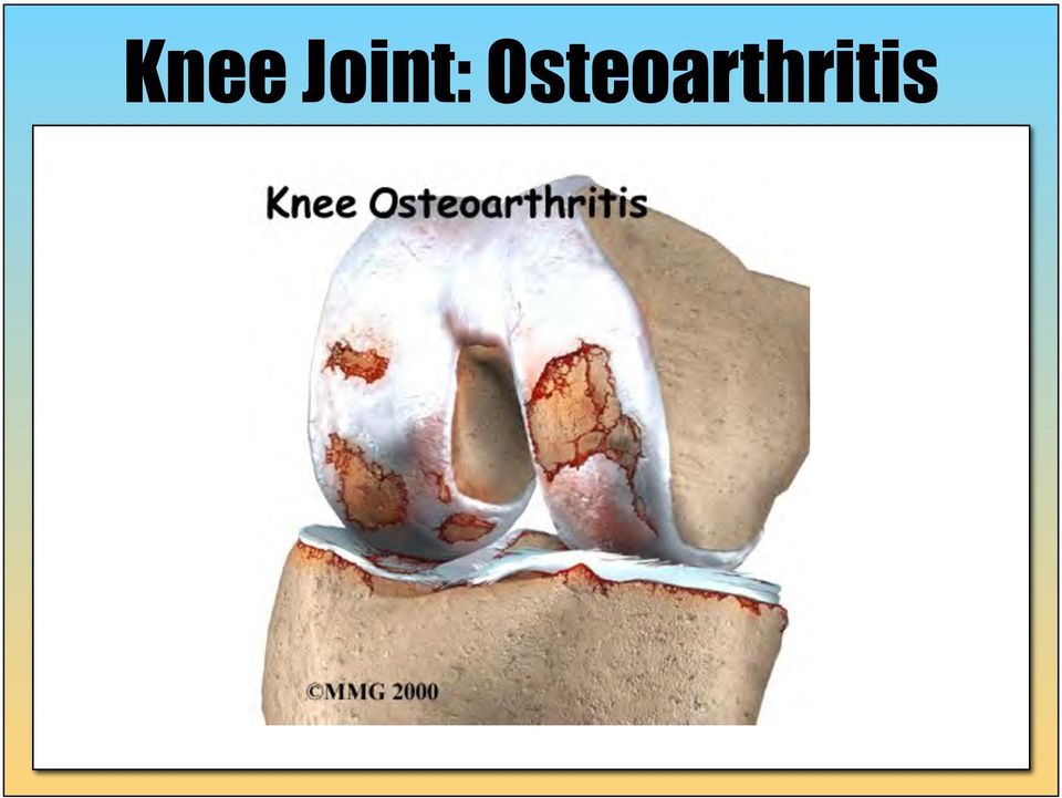
17 Knee Joint: Osteoarthritis
Skeletal system. 2012 Pearson Education, Inc.
 NURS1004 Week 6 Part I Prepared by Didy Button Skeletal system An Introduction to the Skeletal System The Skeletal System Includes: Bones of the skeleton Cartilages, ligaments, and connective tissues 6-1
Definition: A joint or articulation is a place in the body where two bones come together.
 Definition: A joint or articulation is a place in the body where two bones come together. CLASSES OF JOINTS. 1. Joints are classified according to how the bones are held together. 2. The three types of
Chapter 9 Anatomy and Physiology Lecture
 Chapter 9 1 JOINTS Chapter 9 Anatomy and Physiology Lecture Chapter 9 2 JOINTS (Bones are too rigid to bend without causing damage.) (Bones are held together at joints by flexible connective tissue.) (Imagine
LABORATORY EXERCISE 12 BONE STRUCTURE AND CLASSIFICATION
 LABORATORY EXERCISE 12 BONE STRUCTURE AND CLASSIFICATION FIG. 12.1 1. Articular cartilage (hyaline cartilage) 6. Periosteum 2. Spongy bone (red marrow) 7. Proximal epiphysis 3. Medullary cavity 8. Diaphysis
Unit 4: Skeletal System Test Review Test Review
 Name: Period: Unit 4: Skeletal System Test Review Test Review 1. List four functions of the skeletal system: a. b. c. d. 2. Define ossification and identify the roles of the osteoblasts, osteocytes, and
CHAPTER 8: JOINTS OF THE SKELETAL SYSTEM. 4. Name the three types of fibrous joints and give an example of each.
 OBJECTIVES: 1. Define the term articulation. 2. Distinguish between the functional and structural classification of joints, and relate the terms that are essentially synonymous. 3. Compare and contrast
UNIT 4 - SKELETAL SYSTEM LECTURE NOTES
 UNIT 4 - SKELETAL SYSTEM LECTURE NOTES 4.01 FUNCTIONS OF THE SKELETAL SYSTEM A. Support 1. Provides a framework for the body. 2. Supports soft tissue. 3. Serves as a point of attachment for ligaments,
Joints. The graceful movements of ballet dancers and the rough-and-tumble. Classification of Joints
 Classification of Joints (pp. 24 249) Fibrous Joints (pp. 249 250) Sutures (p. 249) Syndesmoses (pp. 249 250) Gomphoses (p. 250) Cartilaginous Joints (pp. 250 251) Synchondroses (pp. 250 251) Symphyses
II. Axial Skeleton (Skull, Thoracic Cage, and Vertebral Column)
") THE SKELETAL SYSTEM Lab Objectives Students should be able to: 1. Recognize bones and bone markings for the axial and appendicular skeleton 2. Recognize bones disarticulated and/or articulated 3. Identify
THE SKELETAL SYSTEM Lab Objectives Students should be able to: 1. Recognize bones and bone markings for the axial and appendicular skeleton 2. Recognize bones disarticulated and/or articulated 3. Identify
Anatomy and Physiology 121: Muscles of the Human Body
 Epicranius Anatomy and Physiology 121: Muscles of the Human Body Covers upper cranium Raises eyebrows, surprise, headaches Parts Frontalis Occipitalis Epicranial aponeurosis Orbicularis oculi Ring (sphincter)
THE SKELETAL & ARTICULAR SYSTEMS. The Bones & Joints
 THE SKELETAL & ARTICULAR SYSTEMS The Bones & Joints CLOSE YOUR POWERPOINT HANDOUTS!! Think-Pair-Share: Why do we need bones? Try to think of 3 reasons. THE SKELETAL SYSTEM Is made up of numerous bones
THE SHOULDER JOINT T H E G L E N O H U M E R A L ( G H ) J O I N T
 J O I N T") THE SHOULDER JOINT T H E G L E N O H U M E R A L ( G H ) J O I N T CLARIFICATION OF TERMS Shoulder girdle = scapula and clavicle Shoulder joint (glenohumeral joint) = scapula and humerus Lippert, p115
THE SHOULDER JOINT T H E G L E N O H U M E R A L ( G H ) J O I N T CLARIFICATION OF TERMS Shoulder girdle = scapula and clavicle Shoulder joint (glenohumeral joint) = scapula and humerus Lippert, p115
The Shoulder Complex & Shoulder Girdle
 The Shoulder Complex & Shoulder Girdle The shoulder complex 4 articulations involving The sternum The clavicle The ribs The scapula and The humerus Bony Landmarks provide attachment points for muscles
Structure & Function of the Knee. One of the most complex simple structures in the human body. The middle child of the lower extremity.
 Structure & Function of the Knee One of the most complex simple structures in the human body. The middle child of the lower extremity. Osteology of the Knee Distal femur (ADDuctor tubercle) Right Femur
Diagnostic MSK Case Submission Requirements
 Diagnostic MSK Case Submission Requirements Note: MSK Ultrasound-Guided Interventional Procedures (USGIP) is considered a separate specialty. Corresponds with 4/21/16 Accred Newsletter* From the main site:
Laerdal' Human Anatomy Manual The Skeleton
 Human Anatomy Manual The Skeleton Laerdal Texas P.O. Box 38.226 EM. 116 Gatesville,Texas U.S.A.76528 U.S.A.1-800-433-5539 IntemationaI1-254-865-7221 24 Hour Fax 254-865-8011 ~ Laerdal' TABLE OF CONTENTS
THE SKELETAL SYSTEM - THE AXIAL SKELETON
 THE SKELETAL SYSTEM - THE AXIAL SKELETON Chapter 7 Anatomy and Physiology Lecture 1 THE SKELETAL SYSTEM THE AXIAL SKELETON Skeletal System forms the framework of the body. TYPES OF BONES: FOUR PRINCIPAL
Skin of eyebrows galea aponeurotica. Muscle and skin of mouth
 : SEE ALSO THE AP SITE FOR OTHER TABLES GROSS ANATOMY OF THE MUSCULAR SYSTEMM Muscles of the Head and Neck: Occipitofrontalis Frontalis Occipitalis Orbicularis oculi Orbicularis oris Buccinator Masseter
Chapter 9 The Hip Joint and Pelvic Girdle
 Copyright The McGraw-Hill Companies, Inc. Reprinted by permission. The Hip Joint and Pelvic Girdle Chapter 9 The Hip Joint and Pelvic Girdle Structural Kinesiology R.T. Floyd, Ed.D, ATC, CSCS Hip joint
Anatomy of Skeletal System
 Anatomy of Skeletal System two main subdivisions of skeletal system: axial : skull, vertebral column, rib cage appendicular: arms and legs and girdles Bone Markings: Foramen: opening in bone passageway
www.ghadialisurgery.com
 P R E S E N T S Dr. Mufa T. Ghadiali is skilled in all aspects of General Surgery. His General Surgery Services include: General Surgery Advanced Laparoscopic Surgery Surgical Oncology Gastrointestinal
9/3/2013 JOINTS. Joints. Axial Skeleton STRUCTURE AND FUNCTION:
 STRUCTURE AND FUNCTION: JOINTS Joints A connection between 2 or more bones A pivot point for bony motion The features of the joint help determine The ROM freedom Functional potential of the joint Axial
Knee Kinematics and Kinetics
 Knee Kinematics and Kinetics Definitions: Kinematics is the study of movement without reference to forces http://www.cogsci.princeton.edu/cgi-bin/webwn2.0?stage=1&word=kinematics Kinetics is the study
The Knee Internal derangement of the knee (IDK) The Knee. The Knee Anatomy of the anteromedial aspect. The Knee
 The Knee. The Knee Anatomy of the anteromedial aspect. The Knee") Orthopedics and Neurology James J. Lehman, DC, MBA, FACO University of Bridgeport College of Chiropractic Internal derangement of the knee (IDK) This a common provisional diagnosis for any patient with
Orthopedics and Neurology James J. Lehman, DC, MBA, FACO University of Bridgeport College of Chiropractic Internal derangement of the knee (IDK) This a common provisional diagnosis for any patient with
STRUCTURE AND FUNCTION: JOINTS
 STRUCTURE AND FUNCTION: JOINTS Joints A connection between 2 or more bones A pivot point for bony motion The features of the joint help determine The ROM Degrees of freedom Functional potential of the
Muscles of Mastication
 Muscles of Mastication Masseter Zygomatic Arch Mandibular angle Elevates mandible Mandibular ramus Temporalis Temporal fossa of the temporal bone Coronoid process of the mandible Elevates mandible Retracts
Biology 105 Human Biology PRACTICE MIDTERM EXAM 1. Essentials of Anatomy and Physiology, 5e (Martini/Nath) Chapter 6 The Skeletal System
 Chapter 6 The Skeletal System") Essentials of Anatomy and Physiology, 5e (Martini/Nath) Chapter 6 The Skeletal System Multiple-Choice Questions 1) Functions of the skeletal system include A) support. B) storage. C) protection. D) blood
Essentials of Anatomy and Physiology, 5e (Martini/Nath) Chapter 6 The Skeletal System Multiple-Choice Questions 1) Functions of the skeletal system include A) support. B) storage. C) protection. D) blood
8/25/2014 JOINTS. The Skeletal System. Axial Skeleton STRUCTURE AND FUNCTION:
 STRUCTURE AND FUNCTION: JOINTS The Skeletal System Made up of the numerous bones of the human body Gives support and framework to the body Protects vital organs Manufactures blood cells Storage of calcium
The Pilates Studio of Los Angeles / PilatesCertificationOnline.com
 Anatomy Review Part I Anatomical Terminology and Review Questions (through pg. 80) Define the following: 1. Sagittal Plane 2. Frontal or Coronal Plane 3. Horizontal Plane 4. Superior 5. Inferior 6. Anterior
LOCOMOTION AND MOVEMENT
 UNIT - HUMAN PHYSIOLOGY Chapter 18 LOCOMOTION AND MOVEMENT Movement is an important feature of living organism. Both the microbes and macrobes show wide range of movements. The movements results in change
Objectives AXIAL SKELETON. 1. Frontal Bone. 2. Parietal Bones. 3. Temporal Bones. CRANIAL BONES (8 total flat bones w/ 2 paired)
") Objectives AXIAL SKELETON SKULL 1. On a skull or diagram, identify and name the bones of the skull 2. Identify the structure and function of the bones of the skull 3. Describe how a fetal skull differs
Objectives AXIAL SKELETON SKULL 1. On a skull or diagram, identify and name the bones of the skull 2. Identify the structure and function of the bones of the skull 3. Describe how a fetal skull differs
Muscles of the Neck and Vertebral Column Sternocleidomastoid (anterior neck) Origin Insertion Action
 Origin Insertion Action") Muscular movements of the head (at the cervical spine/neck) and of the torso (thoracic and lumbar spine/upper, middle, and lower back): flexion, extension, lateral flexion, rotation. Muscles of the Neck
Muscular movements of the head (at the cervical spine/neck) and of the torso (thoracic and lumbar spine/upper, middle, and lower back): flexion, extension, lateral flexion, rotation. Muscles of the Neck
Chapter 5 The Skeletal System
 Essentials of Human Anatomy & Physiology Elaine N. Marieb Seventh Edition Chapter 5 The Skeletal System The Skeletal System Parts of the skeletal system Bones (skeleton) Joints Cartilages Ligaments (bone
Chapter 8. Muscular System: Skeletal Muscles of the Body
 Chapter 8 Muscular System: Skeletal Muscles of the Body INTRODUCTION This chapter continues our study of the muscular system by examining the distribution of muscles throughout the body. We learned in
BODY BODY PEDICLE PEDICLE TRANSVERSE TRANSVERSE PROCESS PROCESS
 Learning Objective Radiology Anatomy of the Spine and Upper Extremity Identify anatomic structures of the spine and upper extremities on standard radiographic and cross-sectional images Timothy J. Mosher,
Chapter 5. The Shoulder Joint. The Shoulder Joint. Bones. Bones. Bones
 Copyright The McGraw-Hill Companies, Inc. Reprinted by permission. Chapter 5 The Shoulder Joint Structural Kinesiology R.T. Floyd, Ed.D, ATC, CSCS Structural Kinesiology The Shoulder Joint 5-1 The Shoulder
Upper Limb QUESTIONS UPPER LIMB: QUESTIONS
 1 Upper Limb QUESTIONS 1.1 Which of the following statements best describes the scapula? a. It usually overlies the 2nd to 9th ribs. b. The spine continues laterally as the coracoid process. c. The suprascapular
Buccinator Presses cheek against molar teeth Facial (CNVII) wrinkles forehead
 wrinkles forehead") Muscles to Identify on the Cadaver and/or Models You are required to identify each of the following muscles or associated structures on the cadavers and/or models in lab. If the box is shaded in a particular
Muscles to Identify on the Cadaver and/or Models You are required to identify each of the following muscles or associated structures on the cadavers and/or models in lab. If the box is shaded in a particular
SPORT AND PHYSICAL ACTIVITY
 2016 Suite Cambridge TECHNICALS LEVEL 3 SPORT AND PHYSICAL ACTIVITY Unit 1 Body systems and the effects of physical activity K/507/4452 Guided learning hours: 90 Version 2 - Revised content - March 2016
Clarification of Terms
 Shoulder Girdle Clarification of Terms Shoulder girdle = scapula and clavicle Shoulder joint (glenohumeral joint) = scapula and humerus What is the purpose (or function) of the shoulder and entire upper
relating to head, neck, and trunk 206 bones organized into structural framework Skeleton relating to limbs and their attachments to the axis
 Axial relating to head, neck, and trunk Skeleton 206 bones organized into structural framework Appendicular relating to limbs and their attachments to the axis Function of Skeletal System 1. Movement 2.
Muscular System. Student Learning Objectives: Identify the major muscles of the body Identify the action of major muscles of the body
 Muscular System Student Learning Objectives: Identify the major muscles of the body Identify the action of major muscles of the body Structures to be identified: Muscle actions: Extension Flexion Abduction
The skeletal and muscular systems
 anatomy and physiology The skeletal and muscular systems CHAPTER 1: Anatomy and physiology LEARNING OBJECTIVES By the end of this chapter, you should be able to: Describe an overview of the skeletal system
Structure and Function of the Hip
 Structure and Function of the Hip Objectives Identify the bones and bony landmarks of the hip and pelvis Identify and describe the supporting structures of the hip joint Describe the kinematics of the
The Axial Skeleton Eighty bones segregated into three regions
 The Axial Skeleton Eighty bones segregated into three regions Skull Vertebral column Bony thorax Bones of the Axial Skeleton Figure 7.1 The Skull The skull, the body s most complex bony structure, is formed
Chapter 4 The Shoulder Girdle
 Chapter 4 The Shoulder Girdle Key Manubrium Clavicle Coracoidprocess Acromionprocess bony landmarks Glenoid fossa Bones Lateral Inferior Medial border angle McGraw-Hill Higher Education. All rights reserved.
UNIT 2 - CHAPTER 7: SKELETAL SYSTEM. 1. Classify bones according to their shapes, and name an example from each group. (p. 200)
") LEARNING OUTCOMES: 7.1 Bone Shape and Structure UNIT 2 - CHAPTER 7: SKELETAL SYSTEM 1. Classify bones according to their shapes, and name an example from each group. (p. 200) 2. Describe the macroscopic
LEARNING OUTCOMES: 7.1 Bone Shape and Structure UNIT 2 - CHAPTER 7: SKELETAL SYSTEM 1. Classify bones according to their shapes, and name an example from each group. (p. 200) 2. Describe the macroscopic
SKELETON AND JOINTS G.C.S.E. PHYSICAL EDUCATION. Unit 1. Factors Affecting Participation and Performance. G.C.S.E. P.E. Teacher:.
 G.C.S.E. PHYSICAL EDUCATION Unit 1 Factors Affecting Participation and Performance SKELETON AND JOINTS Name: G.C.S.E. P.E. Teacher:. By the end of this booklet you should be able to: Understand what the
Unit 8 SPECIFIC SPORTS INJURIES Lecture Notes
 Unit 8 SPECIFIC SPORTS INJURIES Lecture Notes STANDARD 8 Students will explore specific sports injuries. Objective 1: Recognize common injuries to the head and neck to include: concussion, cervical spine
Anterior Superior Iliac Spine. Anterior Inferior Iliac Spine. head neck greater trochanter intertrochanteric line lesser trochanter
 Ilium Bones The Skeleton Ischium Pubis Sacro-iliac Joint Iliac Crest Anterior Superior Superior Pubic Ramus Anterior Inferior Acetabulum Obturator Foramen Ischio-pubic ramus Ischial tuberosity Pubic Crest
Chapter 11. What are the functions of the skeletal system? More detail on bone
 Skeletal System Chapter 11 11.1 Overview of the skeletal system What are the functions of the skeletal system? 1. Supports the body 2. Protects the soft body parts 3. Produces blood cells 4. Stores minerals
Shoulder MRI for Rotator Cuff Tears. Conor Kleweno,, Harvard Medical School Year III Gillian Lieberman, MD
 Shoulder MRI for Rotator Cuff Tears Conor Kleweno,, Harvard Medical School Year III Goals of Presentation Shoulder anatomy Function of rotator cuff MRI approach to diagnose cuff tear Anatomy on MRI images
Functional Anatomy of the Shoulder Complex
 Functional Anatomy of the Shoulder Complex MALCOLM PEAT The shoulder complex, together with other joint and muscle mechanisms of the upper limb, primarily is concerned with the ability to place and control
Elbow & Forearm H O W V I T A L I S T H E E L B O W T O O U R D A I L Y L I V E S?
 Elbow & Forearm H O W V I T A L I S T H E E L B O W T O O U R D A I L Y L I V E S? Clarification of Terms The elbow includes: 3 bones (humerus, radius, and ulna) 2 joints (humeroulnar and humeroradial)
An overview of the anatomy of the canine hindlimb
 An overview of the anatomy of the canine hindlimb Darren Kelly Artwork by Paddy Lennon Original photos courtesy of Mary Ferguson Students at University College Dublin, School of Veterinary Medicine. Video
Good morning, everyone! I m Sue Banta, a coding consultant for Amphion Medical. ambulatory knee and shoulder procedures.
 Copyright 2013 by the American Health Information Management Association. All rights reserved. No part of this publication may be reproduced, stored in a retrieval system, or transmitted in any form or
Anatomy and Pathomechanics of the Sacrum and Pelvis. Charles R. Thompson Head Athletic Trainer Princeton University
 Anatomy and Pathomechanics of the Sacrum and Pelvis Charles R. Thompson Head Athletic Trainer Princeton University Simplify Everything There are actually only three bones: Two innominates, one sacrum.
Review Article. Dislocation Of Shoulder Joint
 Review Article Dislocation Of Shoulder Joint Hafizuddin Ahmed A. H. Introduction: Dislocation means complete loss of contact of the articular surfaces of bony components of a joint. Shoulder joint is the
Human Anatomy & Physiology
 PowerPoint Lecture Slides prepared by Barbara Heard, Atlantic Cape Community College Ninth Edition Human Anatomy & Physiology C H A P T E R 7 The Skeleton: Part B Annie Leibovitz/Contact Press Images Vertebral
Elbow & Forearm. Notes. Notes. Lecture Slides - A.D.A.M. Lab Pics. Bones & Joints: Elbow & Forearm
 Elbow & Forearm Elbow & Forearm Notes Lecture Slides - A.D.A.M. Lab Pics Notes Bones & Joints: The elbow joint is a combination of joints but common reference is usually to the articulation of the distal
LESSON ASSIGNMENT. After completing this lesson, you should be able to: 4-1. Define skeleton.
 LESSON ASSIGNMENT LESSON 4 The Human Skeletal System. TEXT ASSIGNMENT Paragraphs 4-1 through 4-14. LESSON OBJECTIVES After completing this lesson, you should be able to: 4-1. Define skeleton. 4-2. Name
Name the muscle, A: (Action), O: (Origin), and I: (Insertion)
, O: (Origin), and I: (Insertion)") FRONTALIS - A: (Action) Elevates eyebrows in glancing upward and expressions of surprise or fright; draws scalp forward and wrinkles skin of forehead; O: (Origin) Galea aponeurotica; I: Subcutaneous tissue
FRONTALIS - A: (Action) Elevates eyebrows in glancing upward and expressions of surprise or fright; draws scalp forward and wrinkles skin of forehead; O: (Origin) Galea aponeurotica; I: Subcutaneous tissue
Anatomical Terminology: Planes Axes Direction (Text Pg 2 3)
") Anatomical Terminology: Planes Axes Direction (Text Pg 2 3) Anatomy: The structure and organization of the body and its organs. From Greek Origin: Anatome dissection Physiology: The function of the body
Anatomical Terminology: Planes Axes Direction (Text Pg 2 3) Anatomy: The structure and organization of the body and its organs. From Greek Origin: Anatome dissection Physiology: The function of the body
Musculoskeletal Ultrasound Technical Guidelines. V. Knee
 European Society of MusculoSkeletal Radiology Musculoskeletal Ultrasound Technical Guidelines V. Knee Ian Beggs, UK Stefano Bianchi, Switzerland Angel Bueno, Spain Michel Cohen, France Michel Court-Payen,
6 Bones and Skeletal Tissues
 6 Bones and Skeletal Tissues Cartilage Location and basic structure Found throughout adult body Ear and epiglottis Articular cartilages and costal cartilage Larynx, trachea, and nose Intervertebral discs,
Musculoskeletal Ultrasound Technical Guidelines. I. Shoulder
 European Society of MusculoSkeletal Radiology Musculoskeletal Ultrasound Technical Guidelines I. Shoulder Ian Beggs, UK Stefano Bianchi, Switzerland Angel Bueno, Spain Michel Cohen, France Michel Court-Payen,
Lab 5 Overview of the Skeleton: Classification and Structure of Bones and Cartilages Exercise 9 The Axial Skeleton Exercise 10
 Lab 5 Overview of the Skeleton: Classification and Structure of Bones and Cartilages Exercise 9 The Axial Skeleton Exercise 10 Overview of the Skeleton Locate the important cartilages in the human skeleton
The patellofemoral joint and the total knee replacement
 Applied and Computational Mechanics 1 (2007) The patellofemoral joint and the total knee replacement J. Pokorný a,, J. Křen a a Faculty of AppliedSciences, UWB inpilsen, Univerzitní 22, 306 14Plzeň, CzechRepublic
ANATOMY 1 LEARNING TARGETS
 ANATOMY 1 LEARNING TARGETS ORGANIZATION OF THE BODY 1. Define "anatomy" and "physiology." 2. Describe homeostasis. 3. Identify examples of homeostasis 4. Describe the organization of the body according
Scope it Out! Arthroscopic Procedures. Disclaimer
 Scope it Out! Arthroscopic Procedures Julie A. Leu, eu,cpc, C,CPCO, CO,C CPMA, CPC-I 2009-2011 NAB Member, Region 7 Disclaimer Every reasonable effort has been made to ensure that the educational material
Your Practice Online
 P R E S E N T S Your Practice Online Disclaimer This information is an educational resource only and should not be used to make a decision on Knee replacement or arthritis management. All decisions about
Muscle Movements, Types, and Names
 Muscle Movements, Types, and Names A. Gross Skeletal Muscle Activity 1. With a few exceptions, all muscles cross at least one joint 2. Typically, the bulk of the muscle lies proximal to the joint it crossed
Chiropractic ICD 9 Code List
 Use of valid ICD 9 codes, billed with appropriate and corresponding CPT codes, benefits providers by facilitating treatment authorization and claims payment. The use of valid and appropriate codes also
CONTINUING EDUCATION COURSES. for Massage Therapists. Online!
 CONTINUING EDUCATION COURSES for Massage Therapists Online! ccmh Halifax Canadian College of Massage & Hydrotherapy Online Continuing Education Program CCMH Halifax offers a variety of Continuing Education
Introduction. I. Objectives. II. Introduction. A. To become familiar with the terms of direction and location.
 E X E R C I S E Introduction I. Objectives A. To become familiar with the terms of direction and location. B. To become familiar with different types of planes and sections. C. To learn the names and locations
Upper Extremity Special Tests. Cervical Tests. TMJ Dysfunction
 Upper Extremity Special Tests Cervical Tests Vertebral Artery Test: used to test for vertebral artery occlusion or insufficiency. The subject lies supine on the plinth with the examiner seated behind with
Muscle Name Origin Insertion Action Innervation Muscles of Upper Extremity Pectoralis Major Medial half of clavicle, front of sternum, costal
 Muscle Name Origin Insertion Action Innervation Muscles of Upper Extremity Pectoralis Major Medial half of clavicle, front of sternum, costal Crest of greater tubercle (Lateral lip of bicipital groove)
its lifetime. The skeletal system is divided into: 1. Axial Division: bones of the body s axis (skulll, ribs, vertebrae)
") The Axial Skeleton The basic features of the human skeleton have been shaped by evolution, but the detailed characteristics of each bone reflect the stresses put on it. As a result, the skeleton changes
The Axial Skeleton The basic features of the human skeleton have been shaped by evolution, but the detailed characteristics of each bone reflect the stresses put on it. As a result, the skeleton changes
Classification of bones Any bone may be classified into one of the following groups:
 Skeletal system This system is made up of hard tissues like bone and cartilages. This system gives form and shape to animal body The skeleton of a living animal is made up living structures of bones. The
Rotator Cuff Pathophysiology. treatment program that will effectively treat it. The tricky part about the shoulder is that it is a ball and
 Rotator Cuff Pathophysiology Shoulder injuries occur to most people at least once in their life. This highly mobile and versatile joint is one of the most common reasons people visit their health care
NETWORK FITNESS FACTS THE HIP
 NETWORK FITNESS FACTS THE HIP The Hip Joint ANATOMY OF THE HIP The hip bones are divided into 5 areas, which are: Image: www.health.com/health/static/hw/media/medical/hw/ hwkb17_042.jpg The hip joint is
Clients w/ Orthopedic, Injury and Rehabilitation Concerns. Chapter 21
 Clients w/ Orthopedic, Injury and Rehabilitation Concerns Chapter 21 Terminology Macrotrauma A specific, sudden episode of overload injury to a given tissue, resulting in disrupted tissue integrity (Acute)
Acceptable CPT Codes for the ABOS Sports Subspecialty Case List
 20520 Removal of foreign body in muscle or tendon sheath; simple 20525 Removal of foreign body in muscle or tendon sheath; deep or complicated 20670 Removal of implant; superficial, (eg, buried wire, pin
Turnout for Dancers: Hip Anatomy and Factors Affecting Turnout
 Hip Anatomy and Factors Affecting Turnout by the International Association for Dance Medicine and Science www.danceeducation.org Introduction Turnout describes the position of the legs, used in many forms
Avoiding Meniscus Surgery
 Avoiding Meniscus Surgery Robert Tarantino February 9, 2008 2007 NY CTTC-1 1 WARNING THIS MATERIAL DOES NOT CONSTITUTE MEDICAL ADVICE. IT IS INTENDED FOR INFORMATIONAL PURPOSES ONLY. PLEASE CONSULT A PHYSICIAN
Shoulder Instability. Fig 1: Intact labrum and biceps tendon
 Shoulder Instability What is it? The shoulder joint is a ball and socket joint, with the humeral head (upper arm bone) as the ball and the glenoid as the socket. The glenoid (socket) is a shallow bone
TOWN CENTER ORTHOPAEDIC ASSOCIATES P.C. Labral Tears
 Labral Tears The shoulder is your body s most flexible joint. It is designed to let the arm move in almost any direction. But this flexibility has a price, making the joint prone to injury. The shoulder
The human skeleton anterior view
 45 The human skeleton anterior view cranium clavicle mandible scapula sternum rib humerus vertebra radius innominate sacrum ulna carpals metacarpals phalanges femur patella tibia fibula tarsals metatarsals
DEVELOPMENT AND GROWTH OF THE MANDIBLE
 2012-2013 ORAL BIOLOGY DEVELOPMENT AND GROWTH OF THE MANDIBLE Ass. Prof. Dr. Heba M. Elsabaa Development and Growth of the Mandible DEVELOPMENT OF THE MANDIBLE The Mandible Is the largest and strongest
Skeletal System -Training Handout Karen L. Lancour National Rules Committee Chairman Life Science
 Skeletal System -Training Handout Karen L. Lancour National Rules Committee Chairman Life Science INTERACTION OF SKELETAL AND MUSCULAR SYSTEMS: Skeletal and Muscular systems works together to allow movement
Lectures of Human Anatomy
 Lectures of Human Anatomy Vertebral Column-I By DR. ABDEL-MONEM AWAD HEGAZY M.B. with honor 1983, Dipl."Gynecology and Obstetrics "1989, Master "Anatomy and Embryology" 1994, M.D. "Anatomy and Embryology"
SECTION II General Osteopathic Techniques
 SECTION II General Osteopathic Techniques Chapter Four The Lower Extremities 40 Ligamentous Articular Strain The lower extremities are among the most important structures of the body and yet are often
THE SKELETAL SYSTEM FUNCTIONS OF THE SKELETAL SYSTEM
 THE SKELETAL SYSTEM The skeleton is the body s bony framework which consists of 206 bones. The bones are made up of water(45%), calcium and phosphorous(35%) and other organic materials(20%). The calcium
Human Anatomy & Physiology I with Dr. Hubley. Practice Exam #2
 Human Anatomy & Physiology I with Dr. Hubley Practice Exam #2 For questions 1 through 3, select your answers from the following responses: a. stratified squamous epithelium b. reticular connective tissue
Learning IRM. The Knee: lateral ligaments and anatomical quadrants.
 Learning IRM. The Knee: lateral ligaments and anatomical quadrants. Poster No.: C-1733 Congress: ECR 2014 Type: Educational Exhibit Authors: A. Amador Gil, M. D. C. Jurado Gómez, V. de Lara Bendahan ;
EHFA Assessment Strategy (EAD 03) Name of Training Organisation:
 Name of Training Organisation:") EHFA Assessment Strategy (EAD 03) Name of Training Organisation: Introduction European Health and Fitness Association (EHFA) Standards The following EHFA standards are currently available: Fitness Instructor
EHFA Assessment Strategy (EAD 03) Name of Training Organisation: Introduction European Health and Fitness Association (EHFA) Standards The following EHFA standards are currently available: Fitness Instructor