Microscope Lab Introduction to the Microscope Lab Activity
|
|
|
- Kristian May
- 9 years ago
- Views:
Transcription
1 Microscope Lab Introduction to the Microscope Lab Activity Wendy Kim 3B 24 Sep CellDivisionMetaphase.jpeg 1

2 Introduction Microscope is a tool used to enlarge images of small objects that are hard to study with bare eyes. The compound light microscope, which is going to be used in this lab activity, is an instrument with two lenses and various knobs to focus the image. In this lab, we will learn about the proper use and handling of the microscope. Objectives: Demonstrate the appropriate procedures used while using the compound light microscope correctly. Make and use a wet mount. Calculate the total magnification of the microscope. Explain how to handle the microscope properly. Describe changes in the filed of view and the amount of light when going from low to high-power objectives using the compound light microscope. Explain why objects must be centered in the field of view before changing from low to high-power objective. Explain how to control the light intensity when changing the power of objectives. Explain the proper process for focusing under low and high-power using the compound light microscope. Procedures Materials Compound Microscope Glass slides Cover slips Eye dropper Beaker of water The letter e cut from newsprint Scissors Tooth picks Iodine Plant or algae specimens Microscope Handling 1. Microscope should be treated with care; put one hand on the arm and the other under the base of the microscope when carrying it. 2. Carry one microscope carefully and properly from the microscope storage area to the working area. 3. Pick up a pair of scissors, newsprint, a slide, and a coverslip. 4. Remove the dust cover of the microscope and set it properly. 5. Examine the microscope and give the function of each of the parts. 2

3
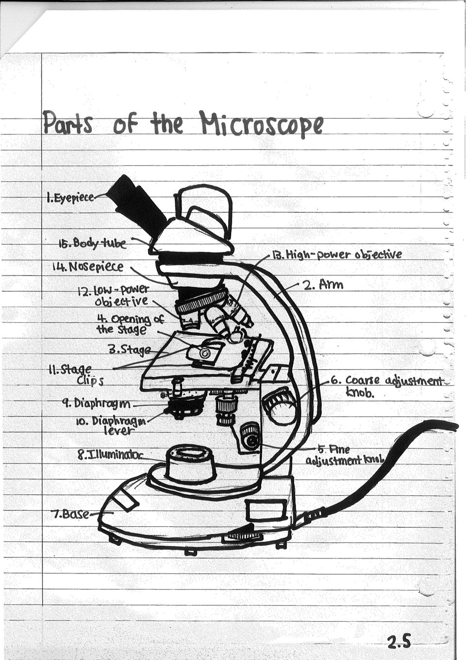
4 Functions of the Microscope No. Name Function 1 Eyepiece Contains a magnifying lens 2 Arm Supports the body tube 3 Stage Supports the slide being observed 4 Opening of the stage Allows light to pass up to the eyepiece 5 Fine adjustment knob Moves the body tube slightly to sharpen the image 6 Coarse adjustment knob Moves the body tube to focus the image 7 Base Supports the microscope 8 Illuminator Produces or reflects light up to the eyepiece 9 Diaphragm Controls the amount of light passing up toward the eyepiece 10 Diaphragm lever Opens and closes the diaphragm 11 Stage clips Holds the slide 12 Low-power objective A shortest objective that magnifies 10X (4X sometimes) 13 High-power objective A longest objective that magnifies 40X (100X sometimes) 14 Nosepiece Holds the objectives and can be rotated 15 Body tube Keeps proper distance between the eyepiece and the objectives. Preparing a wet mount of the letter e. 1. Cut out letter e from the newspaper. 2. Place it on the glass slide. 3. Cover it with a clean cover slip so it looks like : e 4. Place a drop of water on the edge of the cover slip using the eyedropper. 5. Turn on the microscope and place the slide on the stage. 3

5

6
7

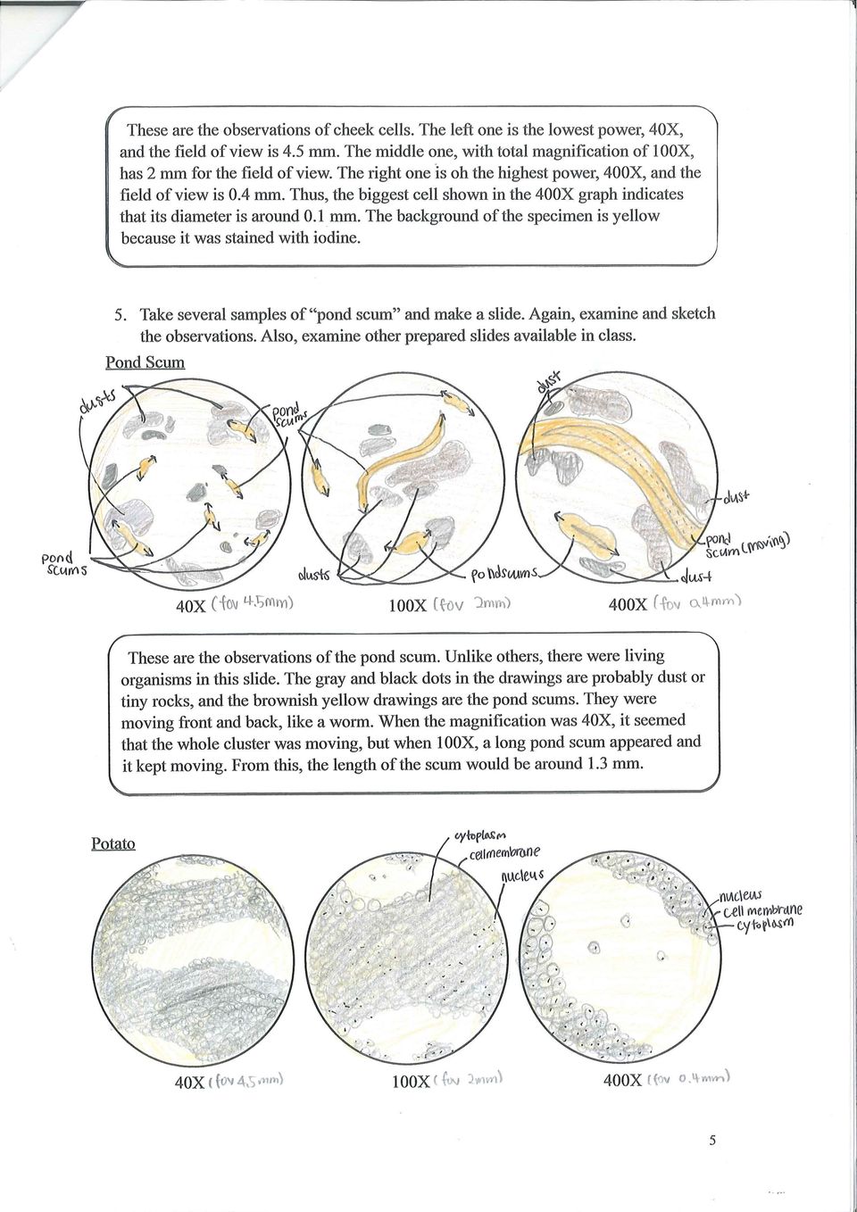
8 These are observations of hydra budding adult. It was also one of the prepared slides. As seen in the 40X view, the specimen seems to be almost 4.5 mm long. It has two heads and several tails at each ends. When it is magnified, its edge is made up of hairs. The entire specimen is red in color. I couldn t get clear image in the 400X view, but I could still recognize the image/specimen. Determining Total Magnification There is a rule for determining total magnification of a compound microscope. To find out the total magnification, multiply the number on the eyepiece magnification and the number on the objective magnification. For instance, the total magnification of a low power objective: Eyepiece magnification (X) Objective magnification = Total Magnification 10 (X) 4 = 40X And for a high power objective: Eyepiece magnification (X) Objective magnification = Total Magnification 10 (X) 100 = 1000X Conclusion (includes answers to the conclusion questions) When handling a light microscope properly, there are two procedures that should be taken carefully. First, we should start observing an object from the low-power objective. To get the image focused when using the low-power objective, we should use coarse adjustment knob to adjust it first, not fine adjustment knob. Fine adjustment knob should be used later, such as when looking at high-power objectives. Otherwise, the slide and the objective can be damaged. Also, we should always treat the microscope with great care. To be specific, the microscope should be carried with two hands; one holding the arm, and another holding the base. Also, when putting down the microscope, it should be placed gently. When not using the microscope, it should be covered with the dust cover and kept. The compound light microscope is the most common microscope that are used in biology classes. The light microscope is also called compound microscope because it contains two lenses. Unlike a simple microscope that uses one lens, a compound microscope uses more than one lens. As shown in the section of observing an e, the images observed under the light microscope are reversed and inverted. This is because when the light passes the lens of the eyepiece, the image gets inverted since those lenses are usually convex lenses. The image is erected again on the retina. The specimen must be centered in the field of view on low power before going to high power because if the specimen is observed on high power from the beginning, it gets very hard to find the specimen. When low-power objective is used from the beginning, we are can start the observation with the entire image of the specimen. Then, it gets much easier to observe the specimen when magnified. 7

9 Let s suppose that there is a microscope that has a 20X ocular (eyepiece) and two objectives of 10X and 43X respectively. Then the low-power magnification of this microscope would be: (Eyepiece magnification) x (Objective magnification) = (Total Magnification) (20) x (10) = (200X) Also, the high-power magnification of this microscope would be: (Eyepiece magnification) x (Objective magnification) = (Total Magnification) (20) x (43) = (860X) There are three steps to make a proper wet mount of the letter e. First, place the letter e from the newspaper on the clean glass slide. Then, put a drop of water on the specimen using a pipette. Finally, place a clean coverslip and remove any excess water at the edge. Make sure when putting a coverslip, the lower one edge of the coverslip so that it touches the side of the drop of water at about a 45 degrees angle. Also, lower the coverslip slowly using a needle or probe. When going from low to high power using the compound microscope, the field of view and the amount of available light changes. The field of view narrows down and gets smaller as the objectives uses higher power. Also, the amount of available light decreases when going from low to high-power objectives. Therefore, the user may have to combat the problems incurred with the microscope when the power changes from low to high. Since the field of view gets smaller, the user may move the stage around and observe the specimen. He may also adjust diaphragm so that there is appropriate amount of light that would not hurt his eye. In addition, the procedure for using the microscope differs slightly under high-power as opposed to low-power. When using a low-power objective, a coarse adjustment knob should be used to focus the image, unless it is very easy to damage both the slide and the objective. On the other hand, when using a high-power objective, use a fine adjustment knob to focus the image sharply, since the specimen is generally focused when using a low-power objective previously. Other than the compound light microscope, there is also a microscope called stereomicroscope. Unlike the compound light microscope which gives two-dimensional views, the stereomicroscope gives three-dimensional image. Therefore, stereomicroscopes are often called dissecting microscopes because they offer the depth of field which is necessary to control the objects while observing them. In addition to light microscopes, there is another type of microscopes - electron microscopes. Electron microscopes use beams of electrons, not light, to produce images. Electron microscopes can present more clear images of even smaller objects compared to the light microscopes. Electron microscopes are classified into two main types: transmission electron microscopes (TEMs) and scanning electron microscopes (SEMs). TEMs shine a beam of electrons through a thin specimen, and thus they reveal very details inside the cell. SEMs scan a narrow beam of electrons back and forth across the surface of a specimen. Therefore, they offer three-dimensional images of the surface of the specimen in a very realistic and dramatic way. 8

10 Timeline for the Various Discoveries of Early Microscopy 1590 Zaccharias Janssen and son Hans Janssen, two Dutch eye glass makers, created the forerunner of the compound microscope and the telescope Robert Hooke, English physicist, looked at a silver of cork through a microscope lens and discovered cells Anton van Leeuwnehoek built a simple microscope with one lens. He used it in observing blood, yeast, insects and other small objects. 18 C Microscopes improved as the technical innovations took place. Scientists found out that combining two lenses reduces chromatic effect, the disturbing halos resulted from differences in refraction of light Richard Zsigmondy invented the ultramicroscope. It enabled scientists to study objects below the wavelength of light Ernst Ruska began to build the electron microscope. This was a TEM Frits Zernike created the phase-contrast microscope. This enabled scientists to study colorless or transparent objects Erwin Wilhelm Muller invented the field emission microscope Erwin Wilhelm Muller invented the field ion microscope, which is the first to see atoms Gerd Bining and Heinrich Rohrer developed the scanning tunneling microscope (STM) Gerd Bining, Quate, and Gerber created the atomic force microscope (AFM). In this lab activity, I observed not only the external features and functions of the microscope, but also the specimens magnified through the microscope. I also made a specimen myself, and drew my observations carefully. Although some fundamental procedures were things that I already learned from the middle school, it was a good opportunity to remind the steps that I should be careful when carrying out, such as placing a coverslip, carrying a microscope, and procedures of observing specimen safely. I remember myself having hard time getting clear image of a specimen. I improved my skills of focusing images through this activity. Now I can use a coarse adjustment knob and a fine adjustment knob without any problems. However, I think I barely controlled the intensity of light using diaphragm. Thus, if I have any chance to use a microscope again next time, I will try to get used to using diaphragm and controlling the amount of light. Also, although I kept reminding myself that I should be careful when treating the microscope and slides, I think I was not still careful when placing the specimen on the stage and changing the objectives from low to high. So, I will also try to improve this next time. Also, I dropped to much iodine when staining the specimen, especially when making a potato slide. Because the specimen was stained too much, it was hard to distinguish the cells. So next time, I should drop less but enough amount of iodine so that I can observe the specimen more clearly. 9

Name Class Date Laboratory Investigation 4B Chapter 4: Cell Structure
 Name Class Date Laboratory Investigation 4B Chapter 4: Cell Structure The Microscope: A Tool of the Scientist You may refer to pages 66-67, 72-73 in your textbook for a general discussion of microscopes.
Care and Use of the Compound Microscope
 Revised Fall 2011 Care and Use of the Compound Microscope Objectives After completing this lab students should be able to 1. properly clean and carry a compound and dissecting microscope. 2. focus a specimen
EXPERIMENT #1: MICROSCOPY
 EXPERIMENT #1: MICROSCOPY Brightfield Compound Light Microscope The light microscope is an important tool in the study of microorganisms. The compound light microscope uses visible light to directly illuminate
Exercise 2. The Compound Light Microscope
 6 Exercise 2 The Compound Light Microscope INTRODUCTION: Student Learning Objectives: After completing this exercise students will: a. Demonstrate proficient use of the microscope using low, high dry,
Compound microscope (Hund)
") 1 2 3 4 5 6 7 8 9 10 11 12 13 14 Compound microscope (Hund) 15 16 17 18 19 20 1) Eyepieces (magnifies 10x), one with diopter adjustment, 2) Interp[upillary adjustment, 3) Head, 4) Revolving nosepiece,
1 2 3 4 5 6 7 8 9 10 11 12 13 14 Compound microscope (Hund) 15 16 17 18 19 20 1) Eyepieces (magnifies 10x), one with diopter adjustment, 2) Interp[upillary adjustment, 3) Head, 4) Revolving nosepiece,
MICROSCOPY. To demonstrate skill in the proper utilization of a light microscope.
 MICROSCOPY I. OBJECTIVES To demonstrate skill in the proper utilization of a light microscope. To demonstrate skill in the use of ocular and stage micrometers for measurements of cell size. To recognize
Chapter 4. Microscopy, Staining, and Classification. Lecture prepared by Mindy Miller-Kittrell North Carolina State University
 Chapter 4 Microscopy, Staining, and Classification 2012 Pearson Education Inc. Lecture prepared by Mindy Miller-Kittrell North Carolina State University Microscopy and Staining 2012 Pearson Education Inc.
Microscopy and Cellular Morphology
 Microscopy and Cellular Morphology As we discussed in class, many organisms on the planet exist as single cells and are referred to as microorganisms bacteria, protozoans, among others. When a single microorganism
MITOSIS IN ONION ROOT TIP CELLS: AN INTRODUCTION TO LIGHT MICROSCOPY
 MITOSIS IN ONION ROOT TIP CELLS: AN INTRODUCTION TO LIGHT MICROSCOPY Adapted from Foundations of Biology I; Lab 6 Introduction to Microscopy Dr. John Robertson, Westminster College Biology Department,
A Fishy Tale. Observing the Circulatory System of a Goldfish with a Compound Light Microscope
 A Fishy Tale Observing the Circulatory System of a Goldfish with a Compound Light Microscope A Fishy Tale About this Lesson In this lesson, students will explore a computer animation of the human body
THE COMPOUND MICROSCOPE
 THE COMPOUND MICROSCOPE In microbiology, the microscope plays an important role in allowing us to see tiny objects that are normally invisible to the naked eye. It is essential for students to learn how
Comparing Plant and Animal Cells
 1.2 Comparing Plant and Animal Cells Here is a summary of what you will learn in this section: Plant and animal cell structures are called organelles. Plant and animal cells perform some similar functions,
Microscopy. MICROSCOPY Light Electron Tunnelling Atomic Force RESOLVE: => INCREASE CONTRAST BIODIVERSITY I BIOL1051 MAJOR FUNCTIONS OF MICROSCOPES
 BIODIVERSITY I BIOL1051 Microscopy Professor Marc C. Lavoie [email protected] MAJOR FUNCTIONS OF MICROSCOPES MAGNIFY RESOLVE: => INCREASE CONTRAST Microscopy 1. Eyepieces 2. Diopter adjustment
Cell Biology Prokaryotic and eukaryotic cells
 Cell Biology Prokaryotic and eukaryotic cells Observation of cells and organelles In this lab you will be looking at an example of a Prokaryotic cell (Bacillus cereus) and a some examples of Eukaryotic
MT-30 & MT-90 Series. Advanced Academic Microscopes/ Advanced Academic Polarizing Microscope INSTRUCTION MANUAL
 Introduction With your purchase of an MT-30/MT-90 series type microscope you have chosen for a quality product. The MT-30/MT-90 series type microscopes are developed for use at schools and laboratories.
BIO 10 Lab 1 Introduction Pre Lab Test
 BIO 10 Lab 1 Introduction Pre Lab Test 1. Why is the microscope in our lab called a compound microscope? 2. How do you calculate total magnification? 3. What is the lowest and the maximal magnification
Revision problem. Chapter 18 problem 37 page 612. Suppose you point a pinhole camera at a 15m tall tree that is 75m away.
 Revision problem Chapter 18 problem 37 page 612 Suppose you point a pinhole camera at a 15m tall tree that is 75m away. 1 Optical Instruments Thin lens equation Refractive power Cameras The human eye Combining
MICROSCOPY OF LIVING MICROBES
 EXPERIMENT 1 MICROSCOPY OF LIVING MICROBES Many students taking microbiology for the first time feel that they are going to have a hard time with the microscope. This lab as an experiment is intended to
7.1 What Are Cells? You are made of cells. A cell is the basic unit of structure and function in a living thing. CHAPTER 7
 CELL STRUCTURE AND FUNCTION 7.1 What Are Cells? Look closely at the skin on your arm. Can you see that it is made of cells? Of course not! Your skin cells are much too small to see with your eyes. Now
The Science of Biology
 Chapter 1 The Science of Biology Section 1 1 What Is Science? (pages 3 7) This section explains what the goal of science is and describes a scientific view of the world. What Science Is and Is Not (page
RAY OPTICS II 7.1 INTRODUCTION
 7 RAY OPTICS II 7.1 INTRODUCTION This chapter presents a discussion of more complicated issues in ray optics that builds on and extends the ideas presented in the last chapter (which you must read first!)
Use of the Microscope and Cytology
 Use of the Microscope and Cytology Introduction: A true study of anatomy not only considers the large, visible structures of an organism, but also the small structures that provide the organism its form
LAB 3 Use of the Microscope
 LAB 3 Use of the Microscope Introduction In this laboratory you will be learning how to use one of the most important tools in biology the compound light microscope to view a variety of specimens. You
7.2. Focusing devices: Unit 7.2. context. Lenses and curved mirrors. Lenses. The language of optics
 context 7.2 Unit 7.2 ocusing devices: Lenses and curved mirrors Light rays often need to be controlled and ed to produce s in optical instruments such as microscopes, cameras and binoculars, and to change
2) A convex lens is known as a diverging lens and a concave lens is known as a converging lens. Answer: FALSE Diff: 1 Var: 1 Page Ref: Sec.
 A convex lens is known as a diverging lens and a concave lens is known as a converging lens. Answer: FALSE Diff: 1 Var: 1 Page Ref: Sec.") Physics for Scientists and Engineers, 4e (Giancoli) Chapter 33 Lenses and Optical Instruments 33.1 Conceptual Questions 1) State how to draw the three rays for finding the image position due to a thin
Physics for Scientists and Engineers, 4e (Giancoli) Chapter 33 Lenses and Optical Instruments 33.1 Conceptual Questions 1) State how to draw the three rays for finding the image position due to a thin
Chapter 17: Light and Image Formation
 Chapter 17: Light and Image Formation 1. When light enters a medium with a higher index of refraction it is A. absorbed. B. bent away from the normal. C. bent towards from the normal. D. continues in the
Forensic Science: The Basics. Microscopy
 Forensic Science: The Basics Microscopy Chapter 6 Jay A. Siegel,Ph.D. Power point presentation by Greg Galardi, Peru State College, Peru Nebraska Presentation by Greg Galardi, Peru State College CRC Press,
Chapter 1 Parts C. Robert Bagnell, Jr., Ph.D., 2012
 Chapter 1 Parts C. Robert Bagnell, Jr., Ph.D., 2012 Figure 1.1 illustrates the parts of an upright compound microscope and indicates the terminology that I use in these notes. Figure 1.1. Parts of a Compound
First let us consider microscopes. Human eyes are sensitive to radiation having wavelengths between
 Optical Differences Between Telescopes and Microscopes Robert R. Pavlis, Girard, Kansas USA icroscopes and telescopes are optical instruments that are designed to permit observation of objects and details
Measuring. User Manual
 0 1 2 3 4 5 6 7 8 9 10 11 Measuring User Manual Accessories for measuring tasks Stage micrometer (1) for calibration Graticules with various measuring pitches (2) in mm and inches Graticule with mesh (3)
Light and its effects
 Light and its effects Light and the speed of light Shadows Shadow films Pinhole camera (1) Pinhole camera (2) Reflection of light Image in a plane mirror An image in a plane mirror is: (i) the same size
LIGHT SECTION 6-REFRACTION-BENDING LIGHT From Hands on Science by Linda Poore, 2003.
 LIGHT SECTION 6-REFRACTION-BENDING LIGHT From Hands on Science by Linda Poore, 2003. STANDARDS: Students know an object is seen when light traveling from an object enters our eye. Students will differentiate
3.1 Cells and cell function
 BTEC s own resources 3.1 Cells and cell function In this section: P1 How you are made Key terms Tissue a group of similar cells acting together to perform a particular function. Epithelial cells one of
Microscopes 2014 Microscopes PB Microscopes 1
 Microscopes 2014 Microscopes 1 Microscopes 2014 Microscopes 2 Just as Celestron s industry-leading telescopes reveal distant galaxies hidden in the night sky, our innovative microscopes give a new perspective
WAVELENGTH OF LIGHT - DIFFRACTION GRATING
 PURPOSE In this experiment we will use the diffraction grating and the spectrometer to measure wavelengths in the mercury spectrum. THEORY A diffraction grating is essentially a series of parallel equidistant
National Optical & Scientific Instruments Inc. 11113 Landmark 35 Drive San Antonio, Texas 78233 Phone (210) 590-9010 Fax (210) 590-1104
 590-9010 Fax (210) 590-1104") National Optical & Scientific Instruments Inc. 11113 Landmark 35 Drive San Antonio, Texas 78233 Phone (210) 590-9010 Fax (210) 590-1104 INSTRUCTIONS FOR MODELS 106, 106-L 107, 107-L 108, 108-L 109-L ELEMENTARY
National Optical & Scientific Instruments Inc. 11113 Landmark 35 Drive San Antonio, Texas 78233 Phone (210) 590-9010 Fax (210) 590-1104 INSTRUCTIONS FOR MODELS 106, 106-L 107, 107-L 108, 108-L 109-L ELEMENTARY
COMPARING PLANT AND ANIMAL CELLS
 COMPARING PLANT AND ANIMAL CELLS OBJECTIVES: Distinguish between plant and animals cells by their structures Demonstrate the benefit of stains Acquire ability to prepare wet mounts SAFETY: Methylene blue
14 The ability of the lenses to distinguish fine detail and structure is called a. Illumination b. Magnification c. Refractive index d.
 1 2 Assume you stain Bacillus by applying malachite green with heat and then counterstain with safranin. Through the microscope, the green structures are a. cell walls. b. capsules. c. endospores. d. flagella.
DETECTION OF BACTERIAL MOTILITY. To demonstrate bacterial motility by microscopic and macroscopic techniques.
 DETECTION OF BACTERIAL MOTILITY I. OBJECTIVES To demonstrate bacterial motility by microscopic and macroscopic techniques. To observe flagella in prepared slides stained by specific flagellar stains. II.
The illustrations below reflect other scientists results in identifying and counting the stages of the onion root tip and the whitefish blastula.
 Abstract: The purpose of this laboratory experiment was to identify in what stage of mitosis viewed cells were in. The stages of mitosis include prophase, metaphase, anaphase and telophase. Although the
Solution Derivations for Capa #14
 Solution Derivations for Capa #4 ) An image of the moon is focused onto a screen using a converging lens of focal length (f = 34.8 cm). The diameter of the moon is 3.48 0 6 m, and its mean distance from
Using a Microscope to See Different Types of Cells
 Using a Microscope to See Different Types of Cells copyright 2003 by Dr. Vivianne Nachmias, University of Pennsylvania All organisms are made up of cells - a cell is the simplest collection of matter that
Geometric Optics Converging Lenses and Mirrors Physics Lab IV
 Objective Geometric Optics Converging Lenses and Mirrors Physics Lab IV In this set of lab exercises, the basic properties geometric optics concerning converging lenses and mirrors will be explored. The
Lecture 17. Image formation Ray tracing Calculation. Lenses Convex Concave. Mirrors Convex Concave. Optical instruments
 Lecture 17. Image formation Ray tracing Calculation Lenses Convex Concave Mirrors Convex Concave Optical instruments Image formation Laws of refraction and reflection can be used to explain how lenses
Where is Mitosis Most Common in the Onion Root?
 Where is Mitosis Most Common in the Onion Root? Faith Loyd Biology Miss Carpenter February 20, 2013 Problem, Hypothesis, and Prediction The problem in this lab is: To analyze data to see whether mitosis
Pre-Lab Questions. 1. What is cell theory? 2. What do all cells contain? 3. What is a prokaryote? 4. What is a eukaryote? 5. What is an organelle?
 Name: TOC# Background Ever since the first microscope was used, biologists have been interested in studying the cellular organization of all living things. After hundred s of years of observations by many
OBJECTIVES PROCEDURE. Lab 2- Bio 160. Name:
 Lab 2- Bio 160 Name: Prokaryotic and Eukaryotic Cells OBJECTIVES To explore cell structure and morphology in prokaryotes and eukaryotes. To gain more experience using the microscope. To obtain a better
GRADE 7: Life science 1. UNIT 7L.1 7 hours. Specialised cells. Resources. About this unit. Previous learning. Key vocabulary and technical terms
 GRADE 7: Life science 1 Specialised cells UNIT 7L.1 7 hours About this unit This unit is the first of six units on life science for Grade 7. This unit is designed to guide your planning and teaching of
Lenses and Apertures of A TEM
 Instructor: Dr. C.Wang EMA 6518 Course Presentation Lenses and Apertures of A TEM Group Member: Anup Kr. Keshri Srikanth Korla Sushma Amruthaluri Venkata Pasumarthi Xudong Chen Outline Electron Optics
Section 13.3 Telescopes and Microscopes
 Glass correcting plate Secondary Finder scope ive Diagonal prism Equatorial drive Equatorial mount Section 13.3 Telescopes and Microscopes Tripod Not everything that we wish to see is visible to the naked
Chapter 27 Optical Instruments. 27.1 The Human Eye and the Camera 27.2 Lenses in Combination and Corrective Optics 27.3 The Magnifying Glass
 Chapter 27 Optical Instruments 27.1 The Human Eye and the Camera 27.2 Lenses in Combination and Corrective Optics 27.3 The Magnifying Glass Figure 27 1 Basic elements of the human eye! Light enters the
EXPERIMENT O-6. Michelson Interferometer. Abstract. References. Pre-Lab
 EXPERIMENT O-6 Michelson Interferometer Abstract A Michelson interferometer, constructed by the student, is used to measure the wavelength of He-Ne laser light and the index of refraction of a flat transparent
Microbiology Laboratory Safety and Basic Procedures Safety in a microbiology laboratory is important in the prevention of infection that might be
 Microbiology Laboratory Safety and Basic Procedures Safety in a microbiology laboratory is important in the prevention of infection that might be caused by the microorganisms being studied. This laboratory
waves rays Consider rays of light from an object being reflected by a plane mirror (the rays are diverging): mirror object
: mirror object") PHYS1000 Optics 1 Optics Light and its interaction with lenses and mirrors. We assume that we can ignore the wave properties of light. waves rays We represent the light as rays, and ignore diffraction.
PHYS1000 Optics 1 Optics Light and its interaction with lenses and mirrors. We assume that we can ignore the wave properties of light. waves rays We represent the light as rays, and ignore diffraction.
Making a reflector telescope
 Making a reflector telescope telescope built by Sir Isaac Newton Replica of the first reflector Nowadays, professional astronomers use another type of telescope that is different to the first telescope
FIFTH GRADE TECHNOLOGY
 FIFTH GRADE TECHNOLOGY 3 WEEKS LESSON PLANS AND ACTIVITIES SCIENCE AND MATH OVERVIEW OF FIFTH GRADE SCIENCE AND MATH WEEK 1. PRE: Interpreting data from a graph. LAB: Estimating data and comparing results
CALIBRATION FOR LAL20X & LAL24X
 CALIBRATION AND FAULT FINDING FOR LAL20X & LAL24X DUMPY LEVELS MEASURING EXPERTS SINCE 1869 How The LAL20X & LAL24X Works The Automatic level is called Automatic because it requires only a simple basic
Students will identify these animal cell structures: Students should properly answer the pre-activity cell membrane, nucleus. questions.
 WHAT DO PLANT & ANIMAL CELLS LOOK LIKE? Grade Levels: 10-12 Time Frame: 2 periods Big Idea: Students will compare various plant epithelial cells (onion and elodea) with human epithelial cells (cheek lining
The Basics of Scanning Electron Microscopy
 The Basics of Scanning Electron Microscopy The small scanning electron microscope is easy to use because almost every variable is pre-set: the acceleration voltage is always 15kV, it has only a single
Animal & Plant Cell Slides
 Animal & Plant Cell Slides Category: Biology Type: Class Experiment, 60 min class Materials: 2 Glass Slides 2 Cover Slips 1 Bottle of methylene blue (optional) 1 Plastic tray 1 Bottle of iodine 1 Plastic
Prokaryotic and Eukaryotic Cells
 Lab 2- Bio 201 Prokaryotic and Eukaryotic Cells Name: OBJECTIVES To explore cell structure and morphology in prokaryotes and eukaryotes. To gain more experience using the microscope, and in particular,
THE BOHR QUANTUM MODEL
 THE BOHR QUANTUM MODEL INTRODUCTION When light from a low-pressure gas is subject to an electric discharge, a discrete line spectrum is emitted. When light from such a low-pressure gas is examined with
CHAPTER 3 OBSERVING MICROORGANISMS THROUGH A MICROSCOPE. I. UNITS OF MEASUREMENT - See Table 3.1 in text. + Fig. 3.2
 CHAPTER 3 OBSERVING MICROORGANISMS THROUGH A MICROSCOPE I. UNITS OF MEASUREMENT - See Table 3.1 in text. + Fig. 3.2 II. MICROSCOPY: THE INSTRUMENTS A. COMPOUND LIGHT MICROSCOPY Figure 3.3 1. Have ocular
Atomic Force Microscopy. July, 2011 R. C. Decker and S. Qazi
 Atomic Force Microscopy July, 2011 R. C. Decker and S. Qazi Learning through Visualization Visualization of physical phenomena can confirm hypothesis Observation provides opportunities for study without
Study Guide for Exam on Light
 Name: Class: Date: Study Guide for Exam on Light Multiple Choice Identify the choice that best completes the statement or answers the question. 1. Which portion of the electromagnetic spectrum is used
Thin Lenses Drawing Ray Diagrams
 Drawing Ray Diagrams Fig. 1a Fig. 1b In this activity we explore how light refracts as it passes through a thin lens. Eyeglasses have been in use since the 13 th century. In 1610 Galileo used two lenses
Biology. STANDARD II: Objective 3. Osmosis Inquiry Labs
 Biology STANDARD II: Objective 3 Osmosis Inquiry Labs Background Knowledge: Students should have used a microscope before and be familiar with the parts. They should also know how to make a wet mount slide.
Nanoelectronics 09. Atsufumi Hirohata Department of Electronics. Quick Review over the Last Lecture
 Nanoelectronics 09 Atsufumi Hirohata Department of Electronics 12:00 Wednesday, 4/February/2015 (P/L 006) Quick Review over the Last Lecture ( Field effect transistor (FET) ): ( Drain ) current increases
Science In Action 8 Unit C - Light and Optical Systems. 1.1 The Challenge of light
 1.1 The Challenge of light 1. Pythagoras' thoughts about light were proven wrong because it was impossible to see A. the light beams B. dark objects C. in the dark D. shiny objects 2. Sir Isaac Newton
1051-232 Imaging Systems Laboratory II. Laboratory 4: Basic Lens Design in OSLO April 2 & 4, 2002
 05-232 Imaging Systems Laboratory II Laboratory 4: Basic Lens Design in OSLO April 2 & 4, 2002 Abstract: For designing the optics of an imaging system, one of the main types of tools used today is optical
CHAPTER 2 : CELL AS THE BASIC UNIT OF LIFE
 CHAPTER 2 : CELL AS THE BASIC UNIT OF LIFE Parts of microscope : An instrument that magnifies minute objects so they can be seen easily. It is one of the most important tools of science. Physicians and
9/16 Optics 1 /11 GEOMETRIC OPTICS
 9/6 Optics / GEOMETRIC OPTICS PURPOSE: To review the basics of geometric optics and to observe the function of some simple and compound optical devices. APPARATUS: Optical bench, lenses, mirror, target
The microscope is an important tool.
 KEY CONCEPT Microscopes allow us to see inside the cell. BEFORE, you learned Some organisms are unicellular and some are multicellular A microscope is necessary to study most cells The cell theory describes
Biology 3A Laboratory MITOSIS Asexual Reproduction
 Biology 3A Laboratory MITOSIS Asexual Reproduction OBJECTIVE To study the cell cycle and understand how, when and why cells divide. To study and identify the major stages of cell division. To relate the
Microscopes and the Metric System
 Microscopes and the Metric System BIO162 Fall 2007 Sizes of Microorganisms: -Viruses: 0.01 0.3 um -Bacteria: 1 3 um -Fungi: 3 30 um -Protozoa: 5 1000 um 1 Measuring Microorganisms Ocular Micrometer The
How To Use An Asbestos Microscope
 Asbestos Microscopes and Accessories Pyser-SGI has been supplying microscopes and accessories into Asbestos Laboratories for over 40 years PS12 Stage Micrometer with UKAS Certificate of Calibration - For
National Optical & Scientific Instruments Inc. 11113 Landmark 35 Drive San Antonio, Texas 78233 Phone (210) 590-9010 Fax (210) 590-1104
 590-9010 Fax (210) 590-1104") National Optical & Scientific Instruments Inc. 11113 Landmark 35 Drive San Antonio, Texas 78233 Phone (210) 590-9010 Fax (210) 590-1104 INSTRUCTIONS FOR MODELS 156, 156-S, 157 COMPOUND BIOLOGICAL MICROSCOPES
National Optical & Scientific Instruments Inc. 11113 Landmark 35 Drive San Antonio, Texas 78233 Phone (210) 590-9010 Fax (210) 590-1104 INSTRUCTIONS FOR MODELS 156, 156-S, 157 COMPOUND BIOLOGICAL MICROSCOPES
VISM Evolution Scope Series
 1 VISM Evolution Scope Series Congratulations on the purchase of your New VISM Evolution (EVO) Series Scope! The EVO Series of Scopes give you many great high end features and various magnification ranges
1. You stand two feet away from a plane mirror. How far is it from you to your image? a. 2.0 ft c. 4.0 ft b. 3.0 ft d. 5.0 ft
 Lenses and Mirrors 1. You stand two feet away from a plane mirror. How far is it from you to your image? a. 2.0 ft c. 4.0 ft b. 3.0 ft d. 5.0 ft 2. Which of the following best describes the image from
Osmosis Demonstration Lab
 Osmosis Demonstration Lab Objectives The student will: 1) Observe the effects of different concentrations of salt solutions on potato cores. 2) Infer the relationship between weight loss and rate of osmosis.
Protocol for Microscope Calibration
 Protocol for Microscope Calibration A properly calibrated system is essential for successful and efficient software use. The following are step by step instructions on how to calibrate the hardware using
Understanding astigmatism Spring 2003
 MAS450/854 Understanding astigmatism Spring 2003 March 9th 2003 Introduction Spherical lens with no astigmatism Crossed cylindrical lenses with astigmatism Horizontal focus Vertical focus Plane of sharpest
How to make a Galileian Telescope
 How to make a Galileian Telescope I. THE BASICS THE PRINCIPLES OF OPTICS A Galileian telescope uses just two lenses. The objective lens is convergent (plano-convex), the ocular lens is divergent (plano-concave).
Mitosis in Onion Root Tip Cells
 Mitosis in Onion Root Tip Cells A quick overview of cell division The genetic information of plants, animals and other eukaryotic organisms resides in several (or many) individual DNA molecules, or chromosomes.
Living things: Cells Living things:
 unit 1 The cell is the smallest unit capable of living an independent existence. Most cells contain a nucleus which controls the way they work; the only cells in the human body with no nuclei are the red
Lenses and Telescopes
 A. Using single lenses to form images Lenses and Telescopes The simplest variety of telescope uses a single lens. The image is formed at the focus of the telescope, which is simply the focal plane of the
FirstView 3 Reflector Telescope Owner s Manual
 FirstView 3 Reflector Telescope Owner s Manual 1. Horizontal Locking Auxiliary Screw 2. Main Mount 3. Pitching Auxiliary Knob 4. Pitching Shaft Screw 5. Rack and Pinion Focusing Knob 6. Thumb Nut for Finder
CONFOCAL LASER SCANNING MICROSCOPY TUTORIAL
 CONFOCAL LASER SCANNING MICROSCOPY TUTORIAL Robert Bagnell 2006 This tutorial covers the following CLSM topics: 1) What is the optical principal behind CLSM? 2) What is the spatial resolution in X, Y,
OLYMPUS STUDENT MICROSCOPES INSTRUCTIONS ST. HS&HSC MODELS OLYMPUS
 OLYMPUS STUDENT MICROSCOPES INSTRUCTIONS MODELS ST. HS&HSC OLYMPUS OLYMPUS MICROSCOPES MODELS ST, HS, HSB, HSC This booklet describes the common features and functions of Models ST, HS. HSB and HSC of
Discovering cells. The fi rst drawing of cells
 Discovering cells Living things are made of cells. Just as bricks and planks of wood are the building blocks for houses, cells are the building blocks of living things. Cells, although they are very small,
CSCI 4974 / 6974 Hardware Reverse Engineering. Lecture 8: Microscopy and Imaging
 CSCI 4974 / 6974 Hardware Reverse Engineering Lecture 8: Microscopy and Imaging Data Acquisition for RE Microscopy Imaging Registration and stitching Microscopy Optical Electron Scanning Transmission Scanning
GRID AND PRISM SPECTROMETERS
 FYSA230/2 GRID AND PRISM SPECTROMETERS 1. Introduction Electromagnetic radiation (e.g. visible light) experiences reflection, refraction, interference and diffraction phenomena when entering and passing
Laboratory Observing the Cell Cycle of Onion Root Tip Cells
 Laboratory Observing the Cell Cycle of Onion Root Tip Cells Background: Because of their rapid growth, the cells of the root tips of plants undergo rapid cell division. Ornamental onion root tips cells
Procedure: Geometrical Optics. Theory Refer to your Lab Manual, pages 291 294. Equipment Needed
 Theory Refer to your Lab Manual, pages 291 294. Geometrical Optics Equipment Needed Light Source Ray Table and Base Three-surface Mirror Convex Lens Ruler Optics Bench Cylindrical Lens Concave Lens Rhombus
Information. From the LowVision Specialists. Guidelines for the fitting of telescopic systems
 Information From the LowVision Specialists Guidelines for the fitting of telescopic systems About a successful fitting Eye care professionals dispensing telescopic spectacles must ensure they have successfully
Amazing Cells A Cell Biology Unit for Grades 5 through 7 Developed by Washington MESA and University of Washington Genome Sciences Education Outreach
 Amazing Cells A Cell Biology Unit for Grades 5 through 7 Developed by Washington MESA and University of Washington Genome Sciences Education Outreach Authors Megan T. Brown, Ph.D., Maureen Munn, Ph.D.,
Measuring the Point Spread Function of a Fluorescence Microscope
 Frederick National Laboratory Measuring the Point Spread Function of a Fluorescence Microscope Stephen J Lockett, PhD Principal Scientist, Optical Microscopy and Analysis Laboratory Frederick National
ST-80 MICROSCOPE With Electronic Eyepiece #6810. User Guide
 ST-80 MICROSCOPE With Electronic Eyepiece #6810 User Guide The ST-80 Microscope Thank you for purchasing your student microscope from ioptron. The ST-80 Microscope is both versatile and easy to use with