Unravelling the cell-type dependent radiosensitising effects of gold through the development of a multifunctional gold nanoparticle
|
|
|
- Winfred Heath
- 4 years ago
- Views:
Transcription
. Unravelling the celltype dependent radiosensitising effects of gold through the development of a multifunctional gold nanoparticle.")
1 Unravelling the cell-type dependent radiosensitising effects of gold through the development of a multifunctional gold nanoparticle Nicol, J. R., Harrison, E., O'Neill, S. M., Dixon, D., McCarthy, H. O., & Coulter, J. A. (2018). Unravelling the celltype dependent radiosensitising effects of gold through the development of a multifunctional gold nanoparticle. Nanomedicine: Nanotechnology, Biology and Medicine, 14(2), Published in: Nanomedicine: Nanotechnology, Biology and Medicine Document Version: Peer reviewed version Queen's University Belfast - Research Portal: Link to publication record in Queen's University Belfast Research Portal Publisher rights Copyright 2017 Elsevier. This manuscript is distributed under a Creative Commons Attribution-NonCommercial-NoDerivs License ( which permits distribution and reproduction for non-commercial purposes, provided the author and source are cited. General rights Copyright for the publications made accessible via the Queen's University Belfast Research Portal is retained by the author(s) and / or other copyright owners and it is a condition of accessing these publications that users recognise and abide by the legal requirements associated with these rights. Take down policy The Research Portal is Queen's institutional repository that provides access to Queen's research output. Every effort has been made to ensure that content in the Research Portal does not infringe any person's rights, or applicable UK laws. If you discover content in the Research Portal that you believe breaches copyright or violates any law, please contact openaccess@qub.ac.uk. Download date:14. Jan. 2020
2 Unravelling the cell-type dependent radiosensitising effects of gold through the development of a multifunctional gold nanoparticle James R. Nicol 1, Emma Harrison 2, Shannon M. O Neill 1, Dorian Dixon 2, Helen O. McCarthy 1, Jonathan A. Coulter 1. 1 School of Pharmacy, Queen's University Belfast, 97 Lisburn Road, Belfast, BT9 7AE, United Kingdom. 2 School of Engineering, Ulster University, Jordanstown campus, Shore Road, Newtonabbey, BT37 0QB, United Kingdom. Corresponding Author: Dr. Jonathan A. Coulter Tel: +44 (0) j.coulter@qub.ac.uk Conflict of interest: The authors report no conflict of interest Financial support: This work was supported by funding from the EPSRC, grant number EP/K039342/1. Abstract word count: 148 Manuscript word count: 4977 References: 50 Number of figures: 7 Number of tables: 0 Number of Supplementary figures: 3
3 Abstract The radiosensitising efficacy of gold is well established, however, there remain several significant barriers to the successful clinical translation of nano-sized gold particles (AuNPs). These barriers include: retaining stability in relevant biological sera, demonstrating effectiveness at clinically relevant AuNP concentrations and identifying the biological context where significant benefit is most likely to be achieved. Herein we have developed a AuNP preparation, stress-tested to provide effective protection from salt and serum mediated agglomeration. Furthermore, the core AuNP is co-functionalised with two biologically derived peptides designed to enhance endocytosis and promote endosomal escape, thus maximising intracellular AuNP surface area. In summary, these investigations demonstrate restored AuNP internalisation using the co-functionalised preparation that generated significant radiosensitisation, in both in vitro and in vivo models, at clinically viable treatment concentrations. Furthermore, we have identified an underpinning biological mechanism in the inherent radical scavenging capacity that could be used to predict radiosensitising efficacy. Keywords: Gold nanoparticles, endocytosis, radiosensitisation, reactive oxygen species, DNA damage
4 Introduction Gold nanoparticles (AuNPs) are proven radiosensitisers at kilovoltage (kev) and megavoltage (MV) photon energies, an effect attributed, in part, to the high mass absorption coefficient of gold.(1-3) To date, most studies have focused on the impact of nanoparticle size, shape, charge and elasticity on internalisation efficacy, with several authors indicating 50 nm AuNPs are most avidly endocytosed.(4-6) Endocytosis of AuNPs exceeding 10 nm predominantly occurs via the endosomal-lysosomal degradation pathway, while direct pinocytosis dominates for sub 10 nm particles.(4,7) Achieving sufficient target cell AuNP uptake represents a major barrier to clinical translation. With this in mind we have developed a novel multi-functional AuNP, designed to promote intracellular uptake and overcome endosomal entrapment, thereby improving the radiosensitising capability.(4,8) To date, the majority of radiosensitising AuNP studies rely on treatment up to 1% wt/wt AuNP/tumour.(9) However, the practical implications of up-scaling to these concentrations for clinical use would prove prohibitive on a cost and potentially toxicity basis.(10) Furthermore, as-synthesized AuNPs aggregate at physiological conditions, requiring the addition of stabilizing moieties. Polyethylene glycol (PEG) is the most widely studied due to existing regulatory approval (MHRA and EMA) and a proven ability to increase systemic circulation time. This stabilising effect is possible as PEG limits nanoparticle opsonisation through the inhibition of non-specific absorption.(11) Despite these obvious advantages, PEG also severely hinders internalization, an effect referred to as the PEG dilemma. (12) No optimal PEG chain length has been definitively established, but 5000 MW PEG is thought to provide the required stability while minimising the negative impact on endocytosis.(13) Herein, AuNPs were synthesised and co-functionalized with 5000 MW PEG, conferring stability and improved in vivo circulation times. Furthermore, synthetic derivatives of two natural peptides were used to overcome biological barriers.(14) The peptide RME derived from viral origins, contains a string of cationic lysine residues that promote internalization. Furthermore, the free thiol group within the N-terminal cysteine residue permits robust chemisorption (enthalpy ~126 kj/mol) of the RME peptide to the AuNP surface.(15,16) A second peptide H5WYG remains unprotonated at physiological ph, but undergoes profound conformational restructuring due to histidyl protonation under mildly acidic conditions found within the endosome.(17) This remodeling effect is further enhanced by the presence of a tryptophan-histidine bridge (WH) that confers strong alpha-helicity capable of destabilizing the endosomal membrane in a process known as the proton sponge effect.(18,19) Combining
5 these functional groups, we aimed to develop a AuNP capable of conferring significant radiation enhancement at a clinically relevant concentration. Breast cancer was chosen as an exemplar tumour model based on the results of the EORTC trial and the recommendations of the Early Breast Cancer Trialists Collaborative Group (EBCTCG).(20,21) The EORTC trial assessed the clinical benefit of a radiation boost (16 Gy) following conventional breast conserving therapy plus radiotherapy (50 Gy).(21) The results indicate that increasing the total administered dose by 30%, results in 39.2% reduction in the 10-year recurrence rate, significantly reducing the requirement for salvage mastectomies. While encouraging, patients receiving the radiotherapy boost had a 2.6 fold greater risk of developing severe fibrosis and an elevated risk of ischemic heart disease, particularly for patients with cancer of the left breast. Darby et al (2013) reported that the mean dose to the heart was 6.6 Gy for women with tumours in the left breast, and that with each subsequent 1 Gy increase to the heart the risk of a major coronary event increased by 7.4%.(22) In this context AuNPs hold real potential, delivering increased radiosensitivity, while limiting physical radiation dose delivered. Further evidence for this was recently presented in vivo where breast tumours doped with 0.5 mg AuNP generated radiation dose enhancement equivalent to three doses of cisplatin (IC25 4 mg/kg) without inducing intrinsic toxicity.(23) While our study focused on breast cancer, most tumours routinely treated with IR (ionising radiation) could potentially benefit, with AuNP radiosensitisation previously demonstrated in prostate, glioma and head and neck cancers.(24-26) Herein we provide a robust characterisation of AuNP synthesis and physical properties, as well as comprehensive assessment of stability and biological efficacy in vitro and in vivo including an insight into key radiosensitising mechanisms. Methods Materials Chloroauric acid (HAuCl4.3H2O), PEG-SH (5000 MW) and sodium citrate were obtained from Sigma Aldrich (UK). The 0.01 wt.% chlorauric acid and 1 wt.% sodium citrate solutions were prepared using distilled water. RME peptide (sequence CKKKKKKSEDEYPYVPN) and H5WYG peptide (sequence GCGLFHAIAHFIHGGWHGHHGWYG) were purchased from BIOMATIK (USA). Peptides were synthesised to >99% purity, lyophilized in acetate salt with trifluoracetic acid constituting <1%.
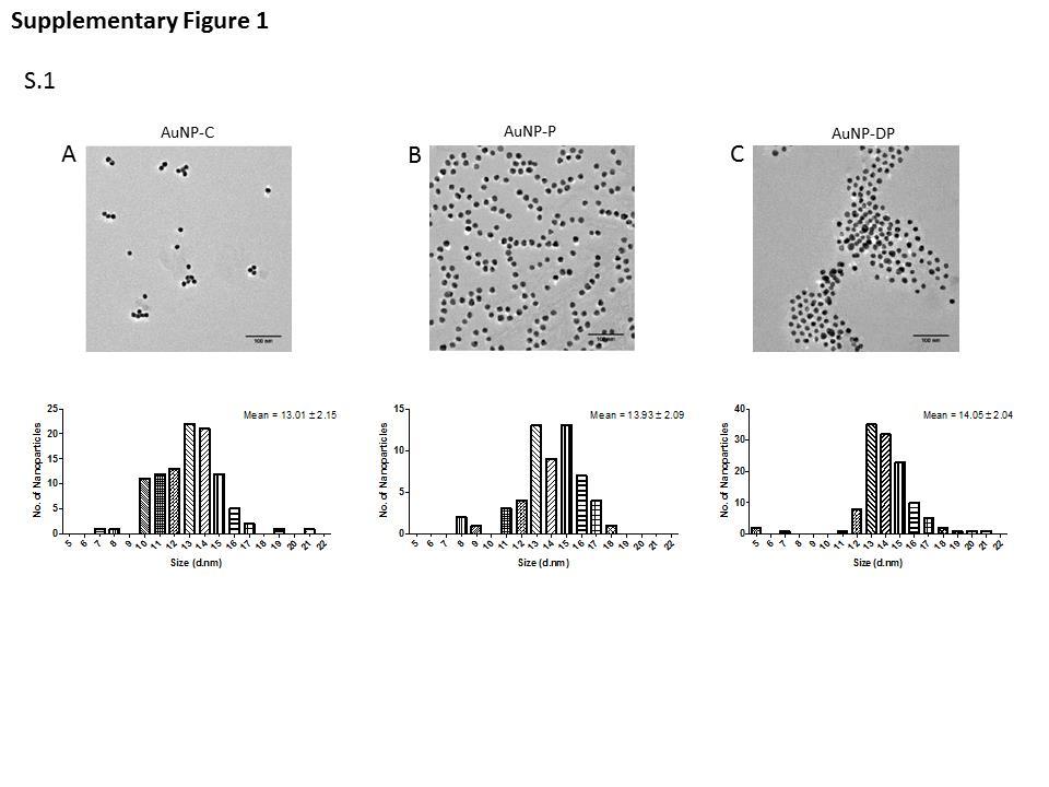
6 Synthesis and Functionalization of gold nanoparticles The Turkevitch/Frens AuNP synthesis method has been widely employed since the 1950 s and a schematic is available in Zhoa et al.(27,28) Briefly, 13 ± 2.15 nm (Supplementary Figure 1) citrate capped nanoparticles (AuNP-C) were produced by reducing 400 ml of boiling 0.01 wt.% chloroauric acid using 9 ml of 1 wt.% sodium citrate solution, as described in our previous publication.(29) To prepare 50% PEG capped AuNPs (AuNP-P), μg/ml 5000MW PEG- SH was dissolved in 1 ml distilled water and added to the citrate reduced AuNPs.(30) The colloid/peg solution was vigorously mixed for 8 h at room temperature allowing PEG/citrate exchange. Excess unconjugated PEG was removed by centrifugation (12000 RCF for 90 min). Single peptide preparations (50% PEG / 50% peptide) functionalised with RME (AuNP-R) or H5WYG (AuNP-H5) were produced by adding 4.4 and 7.86 μg/ml of free peptide respectively. Finally the co-functionalised preparation (termed dual peptide, AuNP-DP) was produced by adding 2.23 μg/ml of free RME peptide (dissolved in 1 ml of distilled water) and 3.93 μg/ml of H5WYG peptide to AuNP-P, mixed for 8 h, then washed to remove excess unconjugated peptide. AuNP characterization Nanoparticle size, charge and polydispersity were determined by dynamic light scattering (DLS) using a Malvern Nano Zetasizer ZS Series. Fourier transform infrared spectroscopy (FTIR) spectra were recorded in the range of cm -1 at a resolution of 4 cm -1 on a Varian 640-IR FTIR spectrometer. UV-Visible spectra were recorded using a Perkin Elmer Lambda 35 in the range nm at 0.1 nm increments. Stress testing AuNP stability in high ionic solutions (1 M NaCl) was conducted by UV/Vis measurement of the characteristic plasmon peak over 24 h. Serum stably was established using DLS following a 24 h incubation medium containing 10% or 40% foetal calf serum. Cell culture and AuNP delivery MDA-MB-231 and MCF-7 human breast cancer cell lines were cultured in Dulbecco s modified eagle s medium (DMEM) supplemented with 10% fetal bovine serum (FBS). Normal breast epithelial cell line MCF-10A were cultured in DMEM/F-12 supplemented with 5% horse serum, 20 ng/ml epidermal growth factor, 0.5 mg/ml hydrocortisone, 100 ng/ml cholera toxin and 10 µg/ml insulin. Cells were maintained at 37 C, 95% air/5% CO2 and sub-cultured every
7 3-4 days to maintain exponential growth. Unless specifically stated, all AuNP treatments were performed at 25 µg/ml for 24 h. Inductively coupled plasma atomic absorption spectroscopy (ICP-AAS) AuNP uptake was quantified using ICP-AAS as previously described.(24) Data is normalized to AuNP-P, to represent the fold-increase in nanoparticle internalisation following peptide conjugation. In Vitro Cytotoxicity AuNP induced toxicity was assessed using both the WST-1 assay and xcelligence platform. Briefly, 2x10 4 AuNP treated (25 µg/ml) cells were washed twice with PBS prior to the addition of 10% WST-1 for 4 h. Cell viability was determined by changes in absorbance at ʎ 440 nm relative to untreated controls. Concentration dependent toxicity was assessed using the xcelligence platform as previously described (ACEA, US). (31) CytoViva analysis of cells with internalized AuNP-DP Cytoviva enhanced darkfield/hyperspectral microscopy permits the optical observation and spectral confirmation of nanoparticles, where AuNPs appear bright due to their high scattering cross-section. Hyperspectral imaging permits capture and recording of unique reflectance spectra (compared against untreated controls) on a pixel-by-pixel basis, providing confirmation of internalisation and intracellular localization. 3x10 4 cells were seeded into a 4-well chamber slide (Corning-US), treated with AuNPs, then fixed with 10% Formalin. AuNP induced radiosensitivity Survival fractions were generated by clonogenic assay, as previously described.(24) Samples were irradiated with doses between 0-6 Gy using a 160 kvp X-ray cabinet source. Sensitiser enhancement ratio (SER) was calculated by fitting the surviving fraction data to the linear quadratic model, establishing the area under curve (mean inactivation dose - MID) at a surviving fraction of 0.1, then dividing the MID of AuNP treated cells against corresponding controls. DNA Damage Analysis 7.5 x10 4 cells were seeded, left to adhere, then treated with AuNPs for 24 h. AuNP treated cells were irradiated (1 Gy), then returned to the incubator for 4 h allowing partial DNA repair. Cells
8 were fixed and permeabilised using 100% methanol at -20 C, then blocked using 2% FBS in PBS at room temperature. Primary H2aX (1:300 in PBS) and secondary antibody (1:400 in PBS) incubations were performed at room temperature prior to mounting using VECTASHIELD plus DAPI at 0.75 µg/ml. H2aX foci for 100 cells per condition were scored. Radiation and AuNP induced Reactive Oxygen Species (ROS) In the presence of ROS 2,7 dichlorofluorescin diacetate (DCFDA) is oxidised to fluorescent 2, 7 dichlorofluorescein (DCF). 1.5 x 10 4 cells were seeded and allowed to adhere for 24 h. Medium containing 5 μm DCFDA was added for 40 min allowing internalisation, and replaced with fresh media. Cells were irradiated with a 2 Gy dose and ROS levels measured using a fluorescent plate reader at 495/529 nm. To detect ROS levels following AuNP and IR treatment, cells were first pre-treated with AuNP for 2 h, during the last 40 min 5 μm DCFDA was added. Western Blotting for SOD2 Whole cell lysates were collected by adding 150 μl Laemmli buffer (Sigma) to adherent cells following AuNP and/or 2 Gy IR treatments. Western blotting was then performed as previously described.(32) Primary antibodies were used at 1:1000 (anti-sod2 and anti-β-actin, Thermo- Fisher Scientific) in 5% blocking buffer at 4 C. Secondary antibody was used at 1:5000 in 1% blocking buffer for 1 h. Densitometry analysis was performed using Image J. In vivo tumour growth delay model All animal experiments were carried out in accordance with QUB institutional regulations and the Animal (Scientific Procedures) Act of 1986, conforming to the current United Kingdom Co-ordinating Committee on Cancer Research (UKCCCR) guidelines. BALB/C SCID mice (6 8 week-old) were used to develop a sub-cutaneous (s.c.) MDA-MB- 231 tumour xenograft model. Mice were anesthetized using isoflurance (3% in O2) and 1.5x10 6 MDA-MB-231 cells injected into the rear dorsum. Tumours were grown to approximately 100 mm 3, at which point animals were randomly assigned to one of four experimental groups; untreated, AuNP-DP only, IR only (4 Gy), AuNP-DP + 4 Gy. AuNP-DP (6 mg/kg) was delivered by direct intra-tumoural injection equating to 150 g Au per 25 g animal. In vivo radiation treatments were delivered using a small animal radiation research platform (SARRP - Xstrahl). Tumours were imaged by CT and treated using two parallel beams delivering 4 Gy
9 225 kvp x-rays. Upon recruitment to the study animal weights and tumour volume were recorded three times weekly. Tumour volume was measured in three orthogonal dimensions. Experimental endpoints for individual animals were defined by tumor volume exceeding 400 mm 3 or animals losing more than 10% of their maximum body weight. Results AuNP Characterization DLS was used to establish size and surface charge (Figures 1A&B) characteristics. Following PEG conjugation hydrodynamic size increases to 28.7 nm ± 0.89 nm, towards the optimal range for uptake.(5) Nanoparticle size increased slightly (AuNP-DP: 36.6 nm ± 3.07) following peptide conjugation with the AuNP-H5 preparation exhibiting the most significant increase to 45.9 ± 0.92 nm. Comparable sizing between nanoparticle preparations indicate a mixed monolayer configuration between PEG and the bioactive peptides.(29) Citrate AuNPs possess a strong negative charge at mv, which reduces to -7.2 mv after PEG conjugation. Both the RME and H5WYG peptides alone further reduce the negative surface charge, remaining anionic. However, the combination of both peptides (AuNP-DP) yields an overall cationic particle with a net charge of 6.5 mv. The position of surface plasmon peak shifts from 518 nm for the AuNP-C, to for AuNP- P and further to nm after conjunction with both peptides (Figure 1C). This measured upshift in peak position of 2-3 nm is consistent with AuNP functionalization.(33) Samples functionalized with PEG together with only the RME or H5WYG peptides displayed ʎmax positions of 524 and nm respectively. The FTIR Spectra of washed and dried, AuNP-P, RME and H5 peptides alone and AuNP-DP are shown in Figure 1D. A comparison of the FTIR spectra of AuNPs before and after PEGylation is detailed in our previous publications.(29,30) The presence of PEG on the AuNP- P sample is confirmed by a peak at 1,106cm -1 (C-O-C stretching) and narrowing of the C-H2 peak (2,850cm -1 ) indicative of ethylene glycol moieties (29, 30, 34, 35) Peptide groups elicit characteristic amide bands in several regions. Characteristic Amide A resulting from N-H stretching vibrations at ~3500 cm -1 are evident on the spectra of the free H5WYG peptide and on the AuNP-DP sample, indicating the presence of H5WYG on the surface. A small broad peak is also visible in the spectra of the RME sample in this region. The Amide band I resonates between cm -1, is associated with C=O stretching and is sensitive to conformation.(36) Amide I peaks are observed in the H5WYG and RME spectra
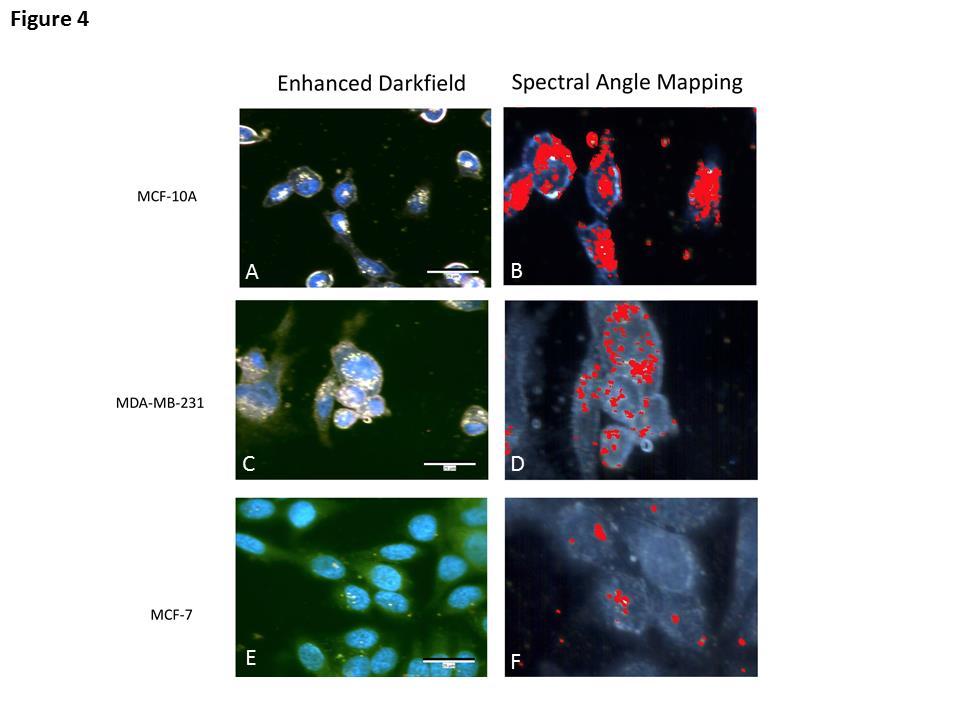
10 at 1666 and 1680 cm -1 respectively. The series of features in the region on the dual peptide samples is consistent with the presence of the peptides. The spectra of the AuNP sample capped only with PEG shows no activity in this region. The presence of the RME peptide on the dual peptide sample is further supported by peaks at 982 and 850 cm -1 (N-H wagging), which are visible on the spectra of both the RME peptide and dual peptide samples. The FTIR data combined with the UV-Vis and Zeta measurements strongly supports the successful conjunction of both peptides to the surface. This is further reinforced by Figure 3A illustrating superior internalization using AuNP-DP. UV-vis was also used to demonstrate nanoparticle stability in strong ionic fluids (1 M NaCL) (Figure 2A). Importantly, both AuNP-P and AuNP-DP show no change in their UV-spectral profile over time, indicating stability in excess of physiologically relevant concentrations. In contrast AuNP-C shows a rapid reduction in absorbance almost immediately, indicative of irreversible agglomeration. Serum stability (Figure 2B) was assessed by changes in particle size. DLS measurements indicate significant (p<0.001) particle agglomeration of AuNP-C in medium containing 10 % serum, an effect totally abrogated following PEG conjugation. Indeed, protection against non-specific protein absorption was also conferred peptide conjugated variants including AuNP-DP. AuNP cellular uptake While PEG provides effective protection against agglomeration, it is widely acknowledged to impair cell internalisation.(37) Figure 3A represents ICP-AAS data, used to quantify intracellular Au per cell following AuNP treatment. In all cells, intracellular AuNP levels failed to exceed 0.3 pg Au/cell following AuNP-P treatment. Conjugation of either the RME or H5WYG improved AuNP internalisation; however, nanoparticle uptake was maximally restored following conjugation with both peptides, yielding an 12.08, 16.3 and 8.3 fold increase over AuNP-P in MCF-10A, MDA-MB-231 and MCF-7 cells respectively, corresponding to 4.14 pg/cell in MDA-MB-231 cells. AuNP Induced Cytotoxicity WST-1 assay (Figure 3B) was used to assess the short-term impact of AuNP treatment on viability. At 25 μg/ml, no significant cytotoxicity was observed from any AuNP preparations (Supplementary Figure 2 AuNP-R and AuNP-H5). Using xcelligence, concentration dependent cytotoxicity of AuNP-DP was assessed over 72 h (Figure 3C and 3D).
11 Biocompatibility was observed up to 300 μg/ml, after which higher exposure concentrations significantly reduced viability. CytoViva Imaging Intracellular distribution was investigated using enhanced darkfield and hyperspectral imaging (HSI) (Figure 4). Enhanced darkfield images (Panels A,C,E) of high intensity bright areas potentially represents AuNPs, absent from control samples. Clusters are located within the perinuclear region of the cell and more diffusely distributed throughout the cytoplasm. Using pixelby-pixel hyperspectral filtering against untreated controls, false positives are eliminated, producing a spectral map confirming the intracellular locality of AuNP-DP (mapped in red). Hyperspectral images generate maximum intensity projected images which compress z-plane data, therefore AuNPs which appear to co-localise with the cell nucleus are likely cytoplasmic, located above or below the nucleus. In vitro AuNP Induced Radiosensitivity Colony forming assays were used to determine the radiosensitising potential of the AuNP preparations (Figure 5 A-C). In normal MCF-10A cells, no radiation dose enhancement was observed with AuNP-P, and a modest, but significant (p=0.003) SER of 1.19 recorded using the fully functionalised AuNP-DP. Conversely, highly significant sensitisation was observed in MDA-MB-231 cells with AuNP-P and AuNP-DP generating average SERs of 1.25 (p=0.01) and 1.54 (p<0.001) respectively. Importantly, radiosensitisation was significantly increased at higher radiation doses, where a 4 Gy SER of 3.19 was obtained. In MCF-7 cells, no significant radiosensitisation was observed using the MID which incorporates data from the full survival curve. However, like the MDA-MB-231 cell, AuNP-DP significantly sensitised MCF-7 cells to higher dose fractions producing a SER of 2.57 at 6 Gy. Similar radiosensitisation survival effects, although less pronounced at higher radiation doses, were obtained using the single peptide (AuNP-R and AuNP-H5) preparations (Supplementary Figure 3A-C). Underpinning mechanisms As AuNP-DP resulted in maximum nanoparticle internalisation, and the greatest radiation dose modifying effects, subsequent mechanistic and in vivo studies focused on the AuNP-DP formulation. To establish if AuNP-DP radiosensitisation is due to elevated DNA double strand damage (DSB), γ-h2ax foci were quantified (Figure 6A). In both cancer cell lines, incorporation of AuNP-DP significantly (p<0.01) increased DNA damage yields by 1.7 fold
12 over radiation alone. Interestingly, despite greater AuNP internalisation AuNP-C induced less DNA DSB, producing a 1.2 and 1.4 fold increase in DNA lesions in MDA-MB-231 and MCF- 7 cells respectively. DCFDA, a fluorescent dye was used to measure AuNP stimulated ROS following radiation treatment. Unsurprisingly, radiation alone (2 Gy) elevated ROS levels in both MDA-MB-231 and MCF-7 cells (Figure 6B and 6C). However, the extent of the response markedly differed between cell lines. MDA-MB-231 saw a rapid spike in ROS yields (5.4 fold) within 30 min, while the response was less pronounced (2.4 fold) in MCF-7 cells. A similar response was observed in cells pre-treated with AuNP-DP prior to radiation treatment. ROS experiments were expanded to assess both the endogenous and treatment induced levels of key ROS scavenging proteins including catalase, glutathione and superoxide disumatase (SOD-2). Basal and treatment induced expression of catalase and glutathione remained unchanged between both MDA-MB-231 and MCF-7 cells, however, significant differences in the expression of SOD-2 were observed (Figure 6 D-F). Normalised against MDA-MB-231 cells, MCF-7 have on average 2.45 fold high basal expression of SOD-2. However, following AuNP treatment SOD-2 expression levels were significantly (AuNP-DP p<0.01) increased in MDA-MB-231 cells. No AuNP related upregulation in SOD-2 was observed in MCF-7 cells where the basal level expression was already high. To determine the timing of this response, samples from MDA-MB-231 cells were collected at various time points over a 24 h period (Figure 6 G-I). Expression levels of SOD-2 following AuNP-DP treatment gradually increased from 4 h post treatment, to a maximum 4.6 fold (p<0.01) increase 24 h post exposure (Figure 6H), with a similar response observed in irradiated cells doped with AuNP-DP (Figure 6I). Xenograft tumour delay model An MDA-MB-231 xenograft tumour model was used to assess the in vivo radiosensitising ability of AuNP-DP (Figure 7 A-D). This tumour model was selected due to the underlying biology highlighted in figures 4 and 5, which indicate a likely benefit from including AuNPs. Figure 7A represents the mean animal weight per treatment group. Despite slight daily fluctuations, no animal dropped below the 10% threshold, indicating minimal treatment toxicity. Once recruited, untreated control tumours reached the experimental endpoint in just 9.8 days (Figure 7B). Direct inject of AuNP-DP alone had no impact on tumour growth. Analysis of the mean tumour growth data indicate that while there was a significant difference between control and both IR only (p<0.05) and AuNP-DP + IR (p<0.001) treated animals, no significant difference between IR only and AuNP-DP +IR was observed. Similarly, Kaplan-
13 Meier survival analysis supported this finding, with a trend (p=0.081) towards increased survival, evidenced by a separation of the IR and AuNP-DP + IR curves (Figure 7C). Analysis of tumour growth to 300 mm 3 reveals a different picture (Figure 7D). Unsurprisingly, IR significantly (p<0.05) delayed tumour growth by 3.39 days compared to untreated controls, however, in this instance AuNP-DP in combination with IR highly significantly (p<0.001) impaired tumour progression by 6.7 days relative to animals receiving 4 Gy only (Figure 7D). Discussion While the biocompatibility, tunable surface chemistry and therapeutic potential of AuNPs are well established; the number of multi-functional (>2 moieties) therapeutic particles remains low.(4,38) Physical AuNP characterisation provides evidence of successful conjugation (Figure 1). AuNP-DP size was measured as ~40 nm, close to the optimal size range for clathrinmediated endocytosis.(5,39) An important benefit of PEG is increased nanoparticle homogeneity and stability, of which polydispersity index (PDI) is a direct measure. AuNP-P and peptide conjugated derivatives had low PDI measurements ( ), indicative of a stable colloid.(40) PEG also affords protection against ionic and serum mediated agglomeration (Figure 2 A&B).(41) Despite the obvious advantage of incorporating PEG, stealth molecules negatively affect cell interaction, attributed to steric hindrance.(42) This is demonstrated with the near abolition of intracellular Au using AuNP-P. RME and H5WYG peptides were grafted in an attempt to restore AuNP uptake, while retaining the stabilizing effect from PEG. The net positive charge conferred by the six-lysine residues of RME peptide promotes internalisation through non-specific electrostatic interaction with phospholipid cell membranes, enhancing uptake up to 17-fold over AuNP-P. This data was further supported by evidence of intense peri-nuclear AuNP-DP accumulation using hyperspectral imaging (Figure 4). AuNP-DP (+6.5 mv) exhibited good biocompatibility producing little to no reduction in viability in MCF-10A and MCF-7 cells, using concentrations < 300 μg/ml. In general, AuNP radiosensitisation studies correlate higher intracellular Au levels with greater radiosensitisation.(43) However, despite comparable AuNP-DP internalisation minimal radiosensitisation was observed in MCF-7 and MCF-10A cells until higher (>4 Gy) radiation doses. This suggests that a positive correlation between intracellular Au and radiosensitisation represents an overly simplistic view of the underlying mechanisms. Subsequent functional assays sought to establish this through probing mechanistic and cell line variations. Of further interest is the effect of AuNP-DP on the α/β ratio; the dose that represents an equal contribution towards cell death from both direct DNA damage (α) and indirect damage (β). Clinical studies
14 indicate that many breast cancers could benefit from hypofractioned treatment plans due to its relatively low α/β ratio estimated at 3.8 Gy.(44) Importantly, AuNP-DP reduces the α/β ratio of MDA-MB-231 and MCF-7 cells by 2.5 and 6.1 fold respectively, implying that AuNP-DP enhances cell death primarily via indirect, biologically mediated mechanisms. Non-resolved DNA double strand break damage is the main cause of radiation-induced death. The recruitment and phosphorylation of DNA damage repair protein histone H2XA acts as a marker of DNA DSB lesions.(45) Four hours post irradiation (1 Gy), cells treated +/- AuNP- DP were scored for DSB lesions, permitting non-complex lesion repair.(46) Consistantly AuNP-DP treated cells yield more unresolved γh2ax foci compared to IR only and AuNP-C. Citrate capped AuNPs are avidly endocytosed, more efficiently that any PEG modified preparation, however, persistent DSBs are lower than recorded with AuNP-DP treated cells. This effect is attributed to the H5WYG peptide, which promotes endosomal escape.(47) McMahon et al (2011) modelled nanoscale secondary electron deposition following AuNP ionisation.(48) Internal nanoparticle ionisation events lack sufficient penetrance to confer Auger mediated effects, while surface ionisations deposit high doses in very close proximity to the nanoparticle (~10 nm). It was hypothesised that the combined endosomal escape and PEG stabilising properties of AuNP-DP increase the internal AuNP surface area, driving interactions with cell cytoplasmic organelles such as the mitochondria, inducing redox related stress, and thus enhancing nanoparticle efficacy. To test this hypothesis, DCFDA was used to measure AuNP induced ROS, with expression levels of key ROS scavenging proteins also quantified. Both MDA-MB-231 and MCF-7 cells saw a time dependent increase in ROS levels post exposure, with the magnitude of effect greatest in MDA-MB-231 cells. SOD-2 basal levels were variable, with MCF-7 cells expressing higher initial levels. Interestingly, both AuNP and radiation treatment promoted SOD-2 expression in MDA-MB-231 cells. The low basal levels of SOD-2 in MDA-MB-231 cells renders them sensitive to the effects of elevated superoxide. Assessing SOD-2 expression levels over 24 h revealed that it took up to 8 h post AuNP or IR treatment for SOD-2 elevation. Therefore, it is likely that the time lag between nanoparticleinduced ROS production (early event 30 min 1 h) and the cells biological response (elevated SOD-2) is sufficient to allow significant ROS driven damage. In the clinical context, AuNPs could be directly administered to the tumour bed after lumpectomy or mastectomy, prior to intraoperative radiotherapy. As such we elected to directly inject AuNP-DP to determine if the radiosensitising efficacy identified in vitro translated to the in vivo setting. Landmark studies highlighting the radiosensitisation potential of AuNPs have typically used in vivo dosing in excess of 2 g/kg Au.(49,50) However, due to the higher
15 endocytotic potential AuNP-DP was administered at 6 mg/kg. This concentration was based on our previous study using small (<4 nm) DTDTPA-grafted AuNPs, which exhibited a lower internalisation potential.(24) A pre-determined experimental end-point of tumour volume developing to four times initial treatment volume was applied. Solely using this criteria suggests that AuNP-DP confers no statistically significant (p=0.13) advantage in terms of tumour growth delay compared to radiation alone, a result mirrored by the Kaplan Meyer survival curve (Figure 7C: IR verses IR + AuNP-DP p=0.081 Mantel-Cox test). This result is strongly influenced by the treatment scheduling. A fixed experimental endpoint fails to account for the pronounced tumour delay observed during the first 12 days post treatment and the data presented in Figure 7D. Extrapolating from the radiation sensitivity data (Figure 5B) at 4 Gy, AuNP-DP generates a dose enhancement factor (DEF) of 3.2, indicating that the combined effect of IR +AuNP-DP treatment is equivalent to an approximate dose of 12 Gy. On the basis that a single 4 Gy fraction resulted in a 3.4 day tumour growth delay compared to the untreated tumours, applying the estimated dose fraction of 12 Gy in the presence of AuNP-DP, a growth delay of 10.2 days should result. This was indeed the observed outcome with a measured combined growth delay of days. This encouraging result outlines the potential for AuNPs to prove effective within a specific cohort of tumours that exhibit lower basal ROS scavenging capacity. Furthermore, as the impact of the combined treatment was front loaded post treatment, the full potential of AuNP-DP is yet to be realised through scheduling and radiation dose fractionation studies. These investigations report the successful synthesis of a novel multifunctional AuNP that demonstrates significant internalisation and radiosensitisation. Furthermore, SOD-2 is identified as a key mediator of inherent radioresistance between cell lines. Importantly, in vitro observations translated to effective tumour growth delay in vivo. Using the example of breast cancer, the significance of the in vivo radiosensitisation should not be underestimated, given the known doubled edged sword effects conferred by a radiotherapy boost for patients with left sided breast cancer. References
16 1. Jain S, Coulter JA, Hounsell AR, Butterworth KT, McMahon SJ, Hyland WB, et al. Cellspecific radiosensitization by gold nanoparticles at megavoltage radiation energies. Int J Radiat Oncol Biol Phys 2011;79(2): (2) Miladi I, Alric C, Dufort S, Mowat P, Dutour A, Mandon C, et al. The In Vivo Radiosensitizing Effect of Gold Nanoparticles Based MRI Contrast Agents. Small 2014;10(6): (3) Butterworth KT, McMahon SJ, Currell FJ, Prise KM. Physical basis and biological mechanisms of gold nanoparticle radiosensitization. Nanoscale (16): (4) Yang C, Uertz J, Yohan D, Chithrani BD. Peptide modified gold nanoparticles for improved cellular uptake, nuclear transport, and intracellular retention. Nanoscale (20): (5) Chithrani BD, Ghazani AA, Chan WC. Determining the size and shape dependence of gold nanoparticle uptake into mammalian cells. Nano Lett (4): (6) Yi X, Shi X, Gao H. Cellular uptake of elastic nanoparticles. Phys Rev Lett 2011; 107(9): (7) Chithrani BD, Chan WC. Elucidating the mechanism of cellular uptake and removal of protein-coated gold nanoparticles of different sizes and shapes. Nano Lett 2007;7(6): (8) Taggart LE, McMahon SJ, Currell FJ, Prise KM, Butterworth KT. The role of mitochondrial function in gold nanoparticle mediated radiosensitisation. Cancer Nanotechnol 2014;5(1):5. (9) Coulter JA, Hyland WB, Nicol J, Currell FJ. Radiosensitising nanoparticles as novel cancer therapeutics--pipe dream or realistic prospect? Clin Oncol 2013; 25(10): (10) Lasagna-Reeves C, Gonzalez-Romero D, Barria MA, Olmedo I, Clos A, Sadagopa Ramanujam VM, et al. Bioaccumulation and toxicity of gold nanoparticles after repeated administration in mice. Biochem Biophys Res Commun 2010; 393(4): (11) Gref R, Luck M, Quellec P, Marchand M, Dellacherie E, Harnisch S, et al. 'Stealth' coronacore nanoparticles surface modified by polyethylene glycol (PEG): influences of the corona (PEG chain length and surface density) and of the core composition on phagocytic uptake and plasma protein adsorption. Colloids Surf B Biointerfaces 2000; 18(3-4): (12) Wang M, Thanou M. Targeting nanoparticles to cancer. Pharmacol Res 2010; 62(2): (13) Hao Y, Yang X, Song S, Huang M, He C, Cui M, et al. Exploring the cell uptake mechanism of phospholipid and polyethylene glycol coated gold nanoparticles. Nanotechnology 2012; 23(4): (14) McCaffrey J, McCrudden CM, Ali AA, Massey AS, McBride JW, McCrudden MT, et al. Transcending epithelial and intracellular biological barriers; a prototype DNA delivery device. J Control Release 2016; 226:
17 (15) Liu Y, Shipton MK, Ryan J, Kaufman ED, Franzen S, Feldheim DL. Synthesis, stability, and cellular internalization of gold nanoparticles containing mixed peptide-poly(ethylene glycol) monolayers. Anal Chem 2007; 79(6): (16) Engel S, Fritz EC, Ravoo BJ. New trends in the functionalization of metallic gold: from organosulfur ligands to N-heterocyclic carbenes. Chem Soc Rev 2017; 46(8): (17) Midoux P, Kichler A, Boutin V, Maurizot JC, Monsigny M. Membrane permeabilization and efficient gene transfer by a peptide containing several histidines. Bioconjug Chem 1998; 9(2): (18) Blanco E, Shen H, Ferrari M. Principles of nanoparticle design for overcoming biological barriers to drug delivery. Nat Biotechnol 2015; 33(9): (19) Fernandez-Recio J, Vazquez A, Civera C, Sevilla P, Sancho J. The tryptophan/histidine interaction in alpha-helices. J Mol Biol 1997; 267(1): (20) Early Breast Cancer Trialists' Collaborative Group (EBCTCG), Darby S, McGale P, Correa C, Taylor C, Arriagada R, et al. Effect of radiotherapy after breast-conserving surgery on 10-year recurrence and 15-year breast cancer death: meta-analysis of individual patient data for 10,801 women in 17 randomised trials. Lancet 2011; 378(9804): (21) Bartelink H, Horiot JC, Poortmans PM, Struikmans H, Van den Bogaert W, Fourquet A, et al. Impact of a higher radiation dose on local control and survival in breast-conserving therapy of early breast cancer: 10-year results of the randomized boost versus no boost EORTC trial. J Clin Oncol 2007; 25(22): (22) Darby SC, Ewertz M, McGale P, Bennet AM, Blom-Goldman U, Bronnum D, et al. Risk of ischemic heart disease in women after radiotherapy for breast cancer. N Engl J Med 2013; 368(11): (23) Cui L, Her S, Dunne M, Borst GR, De Souza R, Bristow RG, et al. Significant Radiation Enhancement Effects by Gold Nanoparticles in Combination with Cisplatin in Triple Negative Breast Cancer Cells and Tumor Xenografts. Radiat Res 2017; 187(2): (24) Butterworth KT, Nicol JR, Ghita M, Rosa S, Chaudhary P, McGarry CK, et al. Preclinical evaluation of gold-dtdtpa nanoparticles as theranostic agents in prostate cancer radiotherapy. Nanomedicine (Lond) 2016; 11(16): (25) Joh DY, Sun L, Stangl M, Al Zaki A, Murty S, Santoiemma PP, et al. Selective targeting of brain tumors with gold nanoparticle-induced radiosensitization. PLoS One 2013; 8(4):e (26) Hainfeld JF, Dilmanian FA, Zhong Z, Slatkin DN, Kalef-Ezra JA, Smilowitz HM. Gold nanoparticles enhance the radiation therapy of a murine squamous cell carcinoma. Phys Med Biol 2010; 55(11): (27) Turkevich J, Stevenson PC, Hillier J. A Study of the nucleation and growth processes in the synthesis of colloidal gold. Discuss. faraday Soc 1951; 11:55-75.
18 (28) Zhao P, Li N, Astruc D. State of the art in gold nanoparticle synthesis. Coord. Chem. Rev 2013; 257: (29) Harrison E, Nicol JR, Macias-Montero M, Burke GA, Coulter JA, Meenan BJ, et al. A comparison of gold nanoparticle surface co-functionalization approaches using Polyethylene Glycol (PEG) and the effect on stability, non-specific protein adsorption and internalization. Mater Sci Eng C Mater Biol Appl 2016; 62: (30) Manson J, Kumar D, Meenan BJ, Dixon D. Polyethylene glycol functionalized gold nanoparticles: the influence of capping density on stability in various media. Gold Bulletin 2011;44(2): (31) Butterworth KT, Coulter JA, Jain S, Forker J, McMahon SJ, Schettino G, et al. Evaluation of cytotoxicity and radiation enhancement using 1.9 nm gold particles: potential application for cancer therapy. Nanotechnology 2010; 21(29): (32) Coulter JA, Jain S, Butterworth KT, Taggart LE, Dickson GR, McMahon SJ, et al. Cell type-dependent uptake, localization, and cytotoxicity of 1.9 nm gold nanoparticles. Int J Nanomedicine 2012;7: (33) Harrison E, Hamilton JWJ, Macias-Montero M, Dixon D. Peptide functionalized gold nanoparticles: the influence of ph on binding efficiency. Nanotechnology 2017; 28(29): (34) Shi W, Sahoo Y, Swihart MT. Gold nanoparticles surface-terminated with bifunctional ligands. Colloids Surf A Physicochem Eng Asp 2004; 246(1): (35) Park JW, Shumaker-Parry JS. Structural study of citrate layers on gold nanoparticles: role of intermolecular interactions in stabilizing nanoparticles. J Am Chem Soc. 2014;136(5): (36) Barth A. Infrared spectroscopy of proteins. Biochim Biophys Acta. 2007; 1767(9): (37) Liu T, Thierry B. A solution to the PEG dilemma: efficient bioconjugation of large gold nanoparticles for biodiagnostic applications using mixed layers. Langmuir 2012; 28(44): (38) Wang YH, Chen SP, Liao AH, Yang YC, Lee CR, Wu CH, et al. Synergistic delivery of gold nanorods using multifunctional microbubbles for enhanced plasmonic photothermal therapy. Sci Rep 2014; 4:5685. (39) Nicol JR, Dixon D, Coulter JA. Gold nanoparticle surface functionalization: a necessary requirement in the development of novel nanotherapeutics. Nanomedicine (Lond) 2015;10(8): (40) Okita,K., Teramoto,K., Kawahara,H., Fujita,H. Light scattering and refractometry of a monodisperse polymer in binary mixed solvents. J. Phys. Chem. 1968;72(1):
19 (41) Terry CA, Fernandez MJ, Gude L, Lorente A, Grant KB. Physiologically relevant concentrations of NaCl and KCl increase DNA photocleavage by an N-substituted 9- aminomethylanthracene dye. Biochemistry 2011; 50(47): (42) Kumar D, Meenan BJ, Dixon D. Glutathione-mediated release of Bodipy(R) from PEG cofunctionalized gold nanoparticles. Int J Nanomedicine 2012; 7: (43) Schuemann J, Berbeco R, Chithrani DB, Cho SH, Kumar R, McMahon SJ, et al. Roadmap to Clinical Use of Gold Nanoparticles for Radiation Sensitization. Int J Radiat Oncol Biol Phys 2016; 94(1): (44) Ray KJ, Sibson NR, Kiltie AE. Treatment of Breast and Prostate Cancer by Hypofractionated Radiotherapy: Potential Risks and Benefits. Clin Oncol (R Coll Radiol) 2015; 27(7): (45) Redon CE, Dickey JS, Bonner WM, Sedelnikova OA. gamma-h2ax as a biomarker of DNA damage induced by ionizing radiation in human peripheral blood lymphocytes and artificial skin. Adv Space Res 2009; 43(8): (46) Lobrich M, Shibata A, Beucher A, Fisher A, Ensminger M, Goodarzi AA, et al. gammah2ax foci analysis for monitoring DNA double-strand break repair: strengths, limitations and optimization. Cell Cycle 2010; 9(4): (47) Huotari J, Helenius A. Endosome maturation. EMBO J 2011; 30(17): (48) McMahon SJ, Hyland WB, Muir MF, Coulter JA, Jain S, Butterworth KT, et al. Nanodosimetric effects of gold nanoparticles in megavoltage radiation therapy. Radiother Oncol 2011; 100(3): (49) Hainfeld JF, Slatkin DN, Smilowitz HM. The use of gold nanoparticles to enhance radiotherapy in mice. Phys Med Biol 2004; 49(18): (50) Hainfeld JF, Smilowitz HM, O'Connor MJ, Dilmanian FA, Slatkin DN. Gold nanoparticle imaging and radiotherapy of brain tumors in mice. Nanomedicine (Lond) 2013; 8(10): Figure 1 AuNP physical characterization. A) Hydrodynamic size and B) zeta potential of the various AuNP preparations. C) UV/Vis spectra of the AuNP preparations D) FTIR spectra of free peptide compared against AuNP-P and AuNP-DP indicating successful conjugation. Figure 2 A) Stability of nanoparticles over time in 1 M NaCl. Experimental replicates were sampled from multiple batches (n=4). B) Stability of AuNP preparations, assessed by change in size following incubation in foetal calf serum (10% or 40%) (n=3).
20 Figure 3 AuNP internalisation and cytotoxicity. A) Uptake data quantified using ICP- AAS and normalised against AuNP-P (n = 5). B) WST-1 assay indicates minimal cytotoxicity following a 24 h incubation with AuNPs at 25 µg/ml. C & D) Dose dependant cytotoxicity of AuNP-DP in cancer cell lines MDA-MB-231 (C) and MCF-7 (D) (n=3). Statistical analysis was performed using a one-way ANOVA with Bonferroni-correction. Figure 4 CytoViva imaging of AuNP-DP in breast cells. Enhanced darkfield images (A,C & E) show cells co-stained with DAPI. Clusters of AuNP-DP appear as bright regions due to their high cross-scattering. Spectral angle mapping (B, D & F) paints AuNP-DP containing pixels red confirming AuNP uptake. Enhanced darkfield were acquired using a x100 objective, with HSI digitally focused on the central field of view. Figure 5 In vitro radiosensitisation of AuNP-DP. Clonogenic survival assays of AuNP-DP in (A) MCF-10A, (B) MDA-MB-231 and (C) MCF-7 cells following incubation at 25 µg/ml for 24 h. Annotated sensitizer enhancement ratios (SER) were calculated using the mean inactivation method and compared against IR alone (n=3). Statistical significance determined by two-way ANOVA. Figure 6 AuNP induced oxidative stress. A) DNA double strand breaks were quantified by scoring γh2ax foci following a 1 Gy dose. B&C) ROS quantified using the fluorescent dye DCFDA over time following IR (2 Gy) alone or AuNP-DP plus IR in (B) MDA-MB-231 and (C) MCF-7 cells. D-F) Western blotting for SOD-2 in cancer cell lines, show the basal level and AuNP induced expression in (D&E) MDA-MB-231 and (D&F) MCF-7 cells. G-I) MDA-MB-231 cells were selected due to the SOD-2 induced response and probed to establish time (G&H) and radiation (2 Gy; G&I) dependent effects. Data represent the mean of three independent experiments. Statistical analysis was performed using a one-way ANOVA with Bonferroni-correction. Figure 7 Xenograft tumour delay model with AuNP-DP. Animals were randomly allocated into four treatment groups: untreated (n=6), AuNP-DP only (n=5), IR only (4 Gy; n=5) and AuNP-DP + IR (4 Gy; n=6). A) Mean animal weights. B) Mean tumour growth characteristics of the four treatment groups until the experimental endpoint of 400 mm. B) Kaplan Meyer survival curve based on tumour growth to experimental endpoint. Statistical
21 analysis determined using the log-rank test. D) Treatment efficacy defined by the delay in time taken for tumours to develop to 300 mm 3. Statistical analysis was performed using a one-way ANOVA with Bonferroni-correction.
22
23
24
25
26
27
28
29
30
31
Supporting Information. Rational design of Au nanorods assemblies for highly sensitive and selective SERS detection of prostate specific antigen
 Electronic Supplementary Material (ESI) for RSC Advances. This journal is The Royal Society of Chemistry 2015 Supporting Information Rational design of Au nanorods assemblies for highly sensitive and selective
Pharmaceutical Nanotechnology
 Pharmaceutical Nanotechnology Pharmaceutical Nanotechnology Development of Gold-Chitosan-coated PLGA Nanoparticles Marius Hittinger 28.04.2011 Supervisor: Juniorprofessor Dr. Marc Schneider Pharmaceutical
UV/VIS/IR SPECTROSCOPY ANALYSIS OF NANOPARTICLES
 UV/VIS/IR SPECTROSCOPY ANALYSIS OF NANOPARTICLES SEPTEMBER 2012, V 1.1 4878 RONSON CT STE K SAN DIEGO, CA 92111 858-565 - 4227 NANOCOMPOSIX.COM Note to the Reader: We at nanocomposix have published this
Effect of ph on the Size of Gold Nanoparticles
 International Journal of Electronic and Electrical Engineering. ISSN 0974-2174, Volume 7, Number 2 (2014), pp. 159-164 International Research Publication House http://www.irphouse.com Effect of ph on the
Covalent Conjugation to Cytodiagnostics Carboxylated Gold Nanoparticles Tech Note #105
 Covalent Conjugation to Cytodiagnostics Carboxylated Gold Nanoparticles Tech Note #105 Background Gold nanoparticle conjugates have been widely used in biological research and biosensing applications.
DNA Assembly and Enzymatic Cutting in Solutions: A Gold Nanoparticle Based SERS Detection Strategy
 Supporting Information DNA Assembly and Enzymatic Cutting in Solutions: A Gold Nanoparticle Based SERS Detection Strategy Elizabeth Crew 1, Hong Yan 1, Liqin Lin 1, Jun Yin 1, Zakiya Skeete 1, Timur Kotlyar
Safe Nano Design Molecule Manufacturing Market
 August 14 16, 212 College of Nanoscale Science & Engineering (CNSE) of the University at Albany National Institute for Occupational Safety and Health (NIOSH) Prevention through Design Program Safe Nano
The Need for a PARP in vivo Pharmacodynamic Assay
 The Need for a PARP in vivo Pharmacodynamic Assay Jay George, Ph.D., Chief Scientific Officer, Trevigen, Inc., Gaithersburg, MD For further infomation, please contact: William Booth, Ph.D. Tel: +44 (0)1235
[Image removed due to copyright concerns]
![[Image removed due to copyright concerns]](/thumbs/40/21531271.jpg "[Image removed due to copyright concerns]") Radiation Chemistry Ionizing radiation produces abundant secondary electrons that rapidly slow down (thermalize) to energies below 7.4 ev, the threshold to produce electronic transitions in liquid water.
Radiation Chemistry Ionizing radiation produces abundant secondary electrons that rapidly slow down (thermalize) to energies below 7.4 ev, the threshold to produce electronic transitions in liquid water.
Molecular Spectroscopy
 Molecular Spectroscopy UV-Vis Spectroscopy Absorption Characteristics of Some Common Chromophores UV-Vis Spectroscopy Absorption Characteristics of Aromatic Compounds UV-Vis Spectroscopy Effect of extended
ZETA POTENTIAL ANALYSIS OF NANOPARTICLES
 ZETA POTENTIAL ANALYSIS OF NANOPARTICLES SEPTEMBER 2012, V 1.1 4878 RONSON CT STE K SAN DIEGO, CA 92111 858-565 - 4227 NANOCOMPOSIX.COM Note to the Reader: We at nanocomposix have published this document
Prussian blue nanoparticles for laser-induced photothermal therapy of tumors
 Electronic Supplementary Material (ESI) for RSC Advances. This journal is The Royal Society of Chemistry 2014 Supporting Information Prussian blue nanoparticles for laser-induced photothermal therapy of
Human serum albumin (HSA) nanoparticles stabilized with. intermolecular disulfide bonds. Supporting Information
 nanoparticles stabilized with. intermolecular disulfide bonds. Supporting Information") Human serum albumin (HSA) nanoparticles stabilized with intermolecular disulfide bonds Wentan Wang, Yanbin Huang*, Shufang Zhao, Ting Shao and Yi Cheng* Department of Chemical Engineering, Tsinghua University,
Human serum albumin (HSA) nanoparticles stabilized with intermolecular disulfide bonds Wentan Wang, Yanbin Huang*, Shufang Zhao, Ting Shao and Yi Cheng* Department of Chemical Engineering, Tsinghua University,
Nanotechnology in Cancer Treatment and Detection. Richard Acosta
 Nanotechnology in Cancer Treatment and Detection Richard Acosta Motivation Ineffectiveness of many Cancer treatments Numerous side effects Difficulties in early Cancer detection No immunization Scale and
Theranostics: A combination of diagnostics and therapy
 Theranostics: A combination of diagnostics and therapy Professor Peter Dobson Academic Director Oxford University Begbroke Science Park, Oxford, England Theranostics: the possible scenarios Therapeutic
Modulating Glucose Uptake in Skeletal Myotubes:
 icell Skeletal Myoblasts Application Protocol Introduction Modulating Glucose Uptake in Skeletal Myotubes: Insulin Induction with Bioluminescent Glucose Uptake Analysis The skeletal muscle is one of the
INFRARED SPECTROSCOPY (IR)
") INFRARED SPECTROSCOPY (IR) Theory and Interpretation of IR spectra ASSIGNED READINGS Introduction to technique 25 (p. 833-834 in lab textbook) Uses of the Infrared Spectrum (p. 847-853) Look over pages
INFRARED SPECTROSCOPY (IR) Theory and Interpretation of IR spectra ASSIGNED READINGS Introduction to technique 25 (p. 833-834 in lab textbook) Uses of the Infrared Spectrum (p. 847-853) Look over pages
STUDENT BACKGROUND READING FOR EXPERIMENT C: COLORIMETRIC GOLD NANOSENSOR
 STUDENT BACKGROUND READING FOR EXPERIMENT C: In this experiment you will synthesize and test a plasmonic colorimetric nanosensor made of nanoparticles of gold. Here we provide you with some background
Medical Tourism - Role of Synchrotron in Biophysical Research
 Scienze mediche e raggi X Dr. Alberto Bravin, PhD European Synchrotron Radiation Facility Grenoble, France bravin@esrf.fr Page 1 Outline - The role of synchrotrons in biomedical research - Highlights of
TITLE: Comparison of the dosimetric planning of partial breast irradiation with and without the aid of 3D virtual reality simulation (VRS) software.
 software.") SAMPLE CLINICAL RESEARCH APPLICATION ABSTRACT: TITLE: Comparison of the dosimetric planning of partial breast irradiation with and without the aid of 3D virtual reality simulation (VRS) software. Hypothesis:
SAMPLE CLINICAL RESEARCH APPLICATION ABSTRACT: TITLE: Comparison of the dosimetric planning of partial breast irradiation with and without the aid of 3D virtual reality simulation (VRS) software. Hypothesis:
Atomic and Nuclear Physics Laboratory (Physics 4780)
") Gamma Ray Spectroscopy Week of September 27, 2010 Atomic and Nuclear Physics Laboratory (Physics 4780) The University of Toledo Instructor: Randy Ellingson Gamma Ray Production: Co 60 60 60 27Co28Ni *
Gamma Ray Spectroscopy Week of September 27, 2010 Atomic and Nuclear Physics Laboratory (Physics 4780) The University of Toledo Instructor: Randy Ellingson Gamma Ray Production: Co 60 60 60 27Co28Ni *
Managing Joint Venture Arrangements Between Two Biotech Companies: A Case Report. Dr. Jan Mous Midatech / PharMida May 21 st 2014
 Managing Joint Venture Arrangements Between Two Biotech Companies: A Case Report Dr. Jan Mous Midatech / PharMida May 21 st 2014 1 Midatech Ltd. Corporate verview Founded 2000 in xford, England Drug development
PUMPED Nd:YAG LASER. Last Revision: August 21, 2007
 PUMPED Nd:YAG LASER Last Revision: August 21, 2007 QUESTION TO BE INVESTIGATED: How can an efficient atomic transition laser be constructed and characterized? INTRODUCTION: This lab exercise will allow
Nucleic Acid Purity Assessment using A 260 /A 280 Ratios
 Nucleic Acid Purity Assessment using A 260 /A 280 Ratios A common practice in molecular biology is to perform a quick assessment of the purity of nucleic acid samples by determining the ratio of spectrophotometric
Method Development for Size-Exclusion Chromatography of Monoclonal Antibodies and Higher Order Aggregates
 Method Development for Size-Exclusion Chromatography of Monoclonal Antibodies and Higher Order Aggregates Paula Hong and Kenneth J. Fountain Waters Corporation, 34 Maple St., Milford, MA, USA APPLICATION
AMBERLITE IRP64 Pharmaceutical Grade Cation Exchange Resin (Polacrilex Resin)
") AMBERLITE IRP64 Pharmaceutical Grade Cation Exchange Resin (Polacrilex Resin) Description AMBERLITE IRP64 [1] resin is an insoluble, weakly acidic, hydrogen form, cation exchange resin supplied as a dry,
AMBERLITE IRP64 Pharmaceutical Grade Cation Exchange Resin (Polacrilex Resin) Description AMBERLITE IRP64 [1] resin is an insoluble, weakly acidic, hydrogen form, cation exchange resin supplied as a dry,
Absorption of Drugs. Transport of a drug from the GI tract
 Absorption of Drugs Absorption is the transfer of a drug from its site of administration to the bloodstream. The rate and efficiency of absorption depend on the route of administration. For IV delivery,
Unfolding and Aggregation of mabs Application Note NT-PR-005
 Unfolding and Aggregation of mabs Application Note NT-PR-005 Analysis of formulation-dependent colloidal and conformational stability of monoclonal antibodies Franziska Söltl 1, Jonathan Derix 1, Michaela
296 cohort patient study. May 2015. Spirometry-monitored deep breathing technique to increase the accuracy of radiotherapy treatment
 breath-hold radiotherapy for breast cancer: Cancer Partners UK s approach to improving outcomes in left-sided breast cancer radiotherapy - an evidence - based review 296 cohort patient study May 2015 Overview
Cell Cycle Phase Determination Kit
 Cell Cycle Phase Determination Kit Item No. 10009349 Customer Service 800.364.9897 * Technical Support 888.526.5351 www.caymanchem.com TABLE OF CONTENTS GENERAL INFORMATION 3 Materials Supplied 3 Safety
Measuring Cell Viability/Cytotoxicity: Cell Counting Kit-F
 Introduction The Cell Counting Kit-F is a fluorometic assay for the determination of viable cell numbers. Calcein-AM in this kit passes through the cell membrane and is hydrolized by the esterase in the
Spectroscopic Characterization of Cytochrome C Entrapped in AOT Reverse Micelles. Kristen Grinstead
 Spectroscopic Characterization of Cytochrome C Entrapped in AOT Reverse Micelles Kristen Grinstead Chemistry Department Marshall University One John Marshall Drive Huntington, WV 25755 grinstead1@marshall.edu
TITLE: Treatment of Prostate Cancer with a DBP-MAF-Vitamin D Complex to Target Angiogenesis and Tumorigenesis
 AD AWARD NUMBER: W81XWH-04-1-0010 TITLE: Treatment of Prostate Cancer with a DBP-MAF-Vitamin D Complex to Target Angiogenesis and Tumorigenesis PRINCIPAL INVESTIGATOR: Michael W. Fannon, Ph.D. CONTRACTING
ArC Amine Reactive Compensation Bead Kit
 ArC Amine Reactive Compensation Bead Kit Catalog no. A1346 Table 1. Contents and storage information. Material Amount Composition Storage Stability ArC reactive beads (Component A) ArC negative beads (Component
BIOLOGICAL MEMBRANES: FUNCTIONS, STRUCTURES & TRANSPORT
 BIOLOGICAL MEMBRANES: FUNCTIONS, STRUCTURES & TRANSPORT UNIVERSITY OF PNG SCHOOL OF MEDICINE AND HEALTH SCIENCES DISCIPLINE OF BIOCHEMISTRY AND MOLECULAR BIOLOGY BMLS II / B Pharm II / BDS II VJ Temple
Supporting Information
 Supporting Information Simple and Rapid Synthesis of Ultrathin Gold Nanowires, Their Self-Assembly and Application in Surface-Enhanced Raman Scattering Huajun Feng, a Yanmei Yang, a Yumeng You, b Gongping
COMMITTEE FOR MEDICINAL PRODUCTS FOR HUMAN USE (CHMP)
") European Medicines Agency Evaluation of Medicines for Human Use London, 10 October 2007 Doc. Ref. COMMITTEE FOR MEDICINAL PRODUCTS FOR HUMAN USE (CHMP) GUIDELINE ON POTENCY TESTING OF CELL BASED IMMUNOTHERAPY
European Medicines Agency Evaluation of Medicines for Human Use London, 10 October 2007 Doc. Ref. COMMITTEE FOR MEDICINAL PRODUCTS FOR HUMAN USE (CHMP) GUIDELINE ON POTENCY TESTING OF CELL BASED IMMUNOTHERAPY
Biomaterials in tissue engineering
 Biomaterials in tissue engineering S. Swaminathan Director Centre for Nanotechnology & Advanced Biomaterials School of Chemical & Biotechnology SASTRA University Thanjavur 613 401 Tamil Nadu Page 1 of
Ionization of amino acids
 Amino Acids 20 common amino acids there are others found naturally but much less frequently Common structure for amino acid COOH, -NH 2, H and R functional groups all attached to the a carbon Ionization
MISCIBILITY AND INTERACTIONS IN CHITOSAN AND POLYACRYLAMIDE MIXTURES
 MISCIBILITY AND INTERACTIONS IN CHITOSAN AND POLYACRYLAMIDE MIXTURES Katarzyna Lewandowska Faculty of Chemistry Nicolaus Copernicus University, ul. Gagarina 7, 87-100 Toruń, Poland e-mail: reol@chem.umk.pl
PRODUCT INFORMATION...
 VIROMER RED In vitro plasmid DNA and mrna Standard Transfection PRODUCT INFORMATION... 2 GENERAL... 2 RED OR YELLOW?... 3 PROTOCOL GUIDELINES... 4 GENERAL REMARKS... 4 CELL CULTURE AND PLATING... 4 FORWARD/REVERSE
Controlling Gold Nanoparticles with Atomic Precision: Synthesis and Structure Determination
 Controlling Gold Nanoparticles with Atomic Precision: Synthesis and Structure Determination Huifeng Qian Department of Chemistry, Carnegie Mellon University, USA Advisor: Prof. Rongchao Jin Background
Problem Set 6 UV-Vis Absorption Spectroscopy. 13-1. Express the following absorbances in terms of percent transmittance:
 Problem Set 6 UV-Vis Absorption Spectroscopy 13-1. Express the following absorbances in terms of percent transmittance: a 0.051 b 0.918 c 0.379 d 0.261 e 0.485 f 0.072 A = log P o /P = log1/t = - log T
Predict Reactivity Note Chicken (100%), Sheep (100%), Rhesus Monkey (100%), Chimpanzee (100%), Bovine (100%), Guinea pig (100%)
, Sheep (100%), Rhesus Monkey (100%), Chimpanzee (100%), Bovine (100%), Guinea pig (100%)") Datasheet GeneTex, Inc : Toll Free 1-877-GeneTex (1-877-436-3839) Fax:1-949-309-2888 info@genetex.com GeneTex International Corporation : Tel:886-3-6208988 Fax:886-3-6208989 infoasia@genetex.com Date :
Datasheet GeneTex, Inc : Toll Free 1-877-GeneTex (1-877-436-3839) Fax:1-949-309-2888 info@genetex.com GeneTex International Corporation : Tel:886-3-6208988 Fax:886-3-6208989 infoasia@genetex.com Date :
The validation of a tumour control probability (TCP) model for supporting radiotherapy treatment planning.
 model for supporting radiotherapy treatment planning.") The validation of a tumour control probability (TCP) model for supporting radiotherapy treatment planning. KAROLINA KOKUREWICZ S U P E R V I S O R S : D I N O J A R O S Z Y N S K I, G I U S E P P E S C
The validation of a tumour control probability (TCP) model for supporting radiotherapy treatment planning. KAROLINA KOKUREWICZ S U P E R V I S O R S : D I N O J A R O S Z Y N S K I, G I U S E P P E S C
CD3/TCR stimulation and surface detection Determination of specificity of intracellular detection of IL-7Rα by flow cytometry
 CD3/TCR stimulation and surface detection Stimulation of HPB-ALL cells with the anti-cd3 monoclonal antibody OKT3 was performed as described 3. In brief, antibody-coated plates were prepared by incubating
Validation & Assay Performance Summary
 Validation & Assay Performance Summary GeneBLAzer RAR alpha DA Assay Kit GeneBLAzer RAR alpha-uas-bla GripTite Cells Cat. no. K1387, K1685 Target Description The retinoic acid receptor alpha (RAR alpha)
ICH Topic S 1 A The Need for Carcinogenicity Studies of Pharmaceuticals. Step 5
 European Medicines Agency July 1996 CPMP/ICH/140/95 ICH Topic S 1 A The Need for Carcinogenicity Studies of Pharmaceuticals Step 5 NOTE FOR GUIDANCE ON THE NEED FOR CARCINOGENICITY STUDIES OF PHARMACEUTICALS
Calculating Nucleic Acid or Protein Concentration Using the GloMax Multi+ Microplate Instrument
 Calculating Nucleic Acid or Protein Concentration Using the GloMax Multi+ Microplate Instrument Technical Note INTRODUCTION Direct measurements of nucleic acid samples at OD 260 or protein samples at OD
Compartmentalization of the Cell. Objectives. Recommended Reading. Professor Alfred Cuschieri. Department of Anatomy University of Malta
 Compartmentalization of the Cell Professor Alfred Cuschieri Department of Anatomy University of Malta Objectives By the end of this session the student should be able to: 1. Identify the different organelles
BBSRC TECHNOLOGY STRATEGY: TECHNOLOGIES NEEDED BY RESEARCH KNOWLEDGE PROVIDERS
 BBSRC TECHNOLOGY STRATEGY: TECHNOLOGIES NEEDED BY RESEARCH KNOWLEDGE PROVIDERS 1. The Technology Strategy sets out six areas where technological developments are required to push the frontiers of knowledge
Application Information Bulletin: Set-Up of the CytoFLEX Set-Up of the CytoFLEX* for Extracellular Vesicle Measurement
 Application Information Bulletin: Set-Up of the CytoFLEX Set-Up of the CytoFLEX* for Extracellular Vesicle Measurement Andreas Spittler, MD, Associate Professor for Pathophysiology, Medical University
Application Note No. 2 / July 2012. Quantitative Assessment of Cell Quality, Viability and Proliferation. System
 Application Note No. 2 / July 2012 Quantitative Assessment of Cell Quality, Viability and Proliferation System Quantitative Assessment of Cell Quality, Viability and Proliferation Introduction In vitro
CHEMISTRY STANDARDS BASED RUBRIC ATOMIC STRUCTURE AND BONDING
 CHEMISTRY STANDARDS BASED RUBRIC ATOMIC STRUCTURE AND BONDING Essential Standard: STUDENTS WILL UNDERSTAND THAT THE PROPERTIES OF MATTER AND THEIR INTERACTIONS ARE A CONSEQUENCE OF THE STRUCTURE OF MATTER,
ISOLATION AND PROPERTIES OF SECRETORY GRANULES FROM RAT ISLETS OF LANGERHANS. II. Ultrastructure of the Beta Granule
 ISOLATION AND PROPERTIES OF SECRETORY GRANULES FROM RAT ISLETS OF LANGERHANS II Ultrastructure of the Beta Granule MARIE H GREIDER, S L HOWELL, and P E LACY From the Department of Pathology, Washington
NMR and other Instrumental Techniques in Chemistry and the proposed National Curriculum.
 NMR and other Instrumental Techniques in Chemistry and the proposed National Curriculum. Dr. John Jackowski Chair of Science, Head of Chemistry Scotch College Melbourne john.jackowski@scotch.vic.edu.au
Radiation Oncology Nursing Care. Helen Lusby Radiation Oncology Nurse BAROC 2012
 Radiation Oncology Nursing Care Helen Lusby Radiation Oncology Nurse BAROC 2012 Definitions Radiation Therapy: Treatment of cancer using x- ray particles that cause ionisation within the cells. External
AAGPs TM Anti-Aging Glyco Peptides. Enhancing Cell, Tissue and Organ Integrity Molecular and biological attributes of lead AAGP molecule
 AAGPs TM Anti-Aging Glyco Peptides Enhancing Cell, Tissue and Organ Integrity Molecular and biological attributes of lead AAGP molecule 1 Acknowledgements This presentation was prepared by Dr. Samer Hussein
MLX BCG Buccal Cell Genomic DNA Extraction Kit. Performance Characteristics
 MLX BCG Buccal Cell Genomic DNA Extraction Kit Performance Characteristics Monolythix, Inc. 4720 Calle Carga Camarillo, CA 93012 Tel: (805) 484-8478 monolythix.com Page 2 of 9 MLX BCG Buccal Cell Genomic
ab139418 Propidium Iodide Flow Cytometry Kit for Cell Cycle Analysis
 ab139418 Propidium Iodide Flow Cytometry Kit for Cell Cycle Analysis Instructions for Use To determine cell cycle status in tissue culture cell lines by measuring DNA content using a flow cytometer. This
INTERNATIONAL CONFERENCE ON HARMONISATION OF TECHNICAL REQUIREMENTS FOR REGISTRATION OF PHARMACEUTICALS FOR HUMAN USE S1A. Current Step 4 version
 INTERNATIONAL CONFERENCE ON HARMONISATION OF TECHNICAL REQUIREMENTS FOR REGISTRATION OF PHARMACEUTICALS FOR HUMAN USE ICH HARMONISED TRIPARTITE GUIDELINE GUIDELINE ON THE NEED FOR CARCINOGENICITY STUDIES
Novel Method for Solid Phase Peptide Synthesis Using Microwave Energy
 Novel Method for Solid Phase Peptide Synthesis Using Microwave Energy Jonathan M. Collins, Michael J. Collins, Rebecca C. Steorts CEM Corporation, Matthews, NC 28106-0200, U.S.A. Presented at American
MATHEMATICAL MODELS OF TUMOR GROWTH INHIBITION IN XENOGRAFT MICE AFTER ADMINISTRATION OF ANTICANCER AGENTS GIVEN IN COMBINATION
 MATHEMATICAL MODELS OF TUMOR GROWTH INHIBITION IN XENOGRAFT MICE AFTER ADMINISTRATION OF ANTICANCER AGENTS GIVEN IN COMBINATION Nadia Terranova UNIVERSITÀ DI PAVIA New model development: course of action
Supplemental Information. McBrayer et al. Supplemental Data
 1 Supplemental Information McBrayer et al. Supplemental Data 2 Figure S1. Glucose consumption rates of MM cell lines exceed that of normal PBMC. (A) Normal PBMC isolated from three healthy donors were
Nursing 113. Pharmacology Principles
 Nursing 113 Pharmacology Principles 1. The study of how drugs enter the body, reach the site of action, and are removed from the body is called a. pharmacotherapeutics b. pharmacology c. pharmacodynamics
Introduction to Flow Cytometry
 Outline Introduction to Flow Cytometry Basic Concept of Flow Cytometry Introduction to Instrument Subsystems Daisy Kuo Assistant Product Manager E-mail: daisy_kuo@bd.com BDBiosciences Application Examples
Time out states and transitions
 Time out states and transitions Spectroscopy transitions between energy states of a molecule excited by absorption or emission of a photon hn = DE = E i - E f Energy levels due to interactions between
STANDARD OPERATING PROCEDURE
 STANDARD OPERATING PROCEDURE Title: Antibody Production at Strategic Diagnostics Inc. SOP#: M-119 Version #: 1 Date Approved: August 6, 2009 Author: Strategic Diagnostic Inc. Date Modified: 1. PURPOSE
Q4 2015 PRESENTATION February 9, 2016 Per Walday, CEO Ronny Skuggedal, CFO
 Q4 2015 PRESENTATION February 9, 2016 Per Walday, CEO Ronny Skuggedal, CFO 1 PCI BIOTECH Important notice and disclaimer This presentation may contain certain forward-looking statements and forecasts based
ab185915 Protein Sumoylation Assay Ultra Kit
 ab185915 Protein Sumoylation Assay Ultra Kit Instructions for Use For the measuring in vivo protein sumoylation in various samples This product is for research use only and is not intended for diagnostic
Development and validation of neutralising anti-drug antibody (Nabs) assays
 assays") Development and validation of neutralising anti-drug antibody (Nabs) assays Clare Kingsley Sector Manager, Bioanalytical Sciences, Quotient Bioresearch EBF 2012 Overview Anti-drug antibody and neutralising
Development and validation of neutralising anti-drug antibody (Nabs) assays Clare Kingsley Sector Manager, Bioanalytical Sciences, Quotient Bioresearch EBF 2012 Overview Anti-drug antibody and neutralising
Introduction to flow cytometry
 Introduction to flow cytometry Flow cytometry is a popular laser-based technology. Discover more with our introduction to flow cytometry. Flow cytometry is now a widely used method for analyzing the expression
Laser-induced modification of metal nanoparticles formed by laser ablation technique in liquids
 Applied Surface Science 247 (2005) 418 422 www.elsevier.com/locate/apsusc Laser-induced modification of metal nanoparticles formed by laser ablation technique in liquids N.V. Tarasenko *, A.V. Butsen,
Bottlenecks in Clinical Source Material Acquisition. Aby J. Mathew, PhD May 5, 2009 ISCT Annual Meeting San Diego, CA amathew@biolifesolutions.
 Bottlenecks in Clinical Source Material Acquisition Aby J. Mathew, PhD May 5, 2009 ISCT Annual Meeting San Diego, CA amathew@biolifesolutions.com Biopreservation What s the issue? Biopreservation considerations
1.1.2. thebiotutor. AS Biology OCR. Unit F211: Cells, Exchange & Transport. Module 1.2 Cell Membranes. Notes & Questions.
 thebiotutor AS Biology OCR Unit F211: Cells, Exchange & Transport Module 1.2 Cell Membranes Notes & Questions Andy Todd 1 Outline the roles of membranes within cells and at the surface of cells. The main
Copyright 1999 2010 by Mark Brandt, Ph.D. 12
 Introduction to Absorbance Spectroscopy A single beam spectrophotometer is comprised of a light source, a monochromator, a sample holder, and a detector. An ideal instrument has a light source that emits
Transfection reagent for visualizing lipofection with DNA. For ordering information, MSDS, publications and application notes see www.biontex.
 METAFECTENE FluoR Transfection reagent for visualizing lipofection with DNA For ordering information, MSDS, publications and application notes see www.biontex.com Description Cat. No. Size METAFECTENE
Review of Chemical Equilibrium 7.51 September 1999. free [A] (µm)
![Review of Chemical Equilibrium 7.51 September 1999. free [A] (µm)](/thumbs/40/21435474.jpg "Review of Chemical Equilibrium 7.51 September 1999. free [A] (µm)") Review of Chemical Equilibrium 7.51 September 1999 Equilibrium experiments study how the concentration of reaction products change as a function of reactant concentrations and/or reaction conditions. For
Review of Chemical Equilibrium 7.51 September 1999 Equilibrium experiments study how the concentration of reaction products change as a function of reactant concentrations and/or reaction conditions. For
DYNAMIC LIGHT SCATTERING COMMON TERMS DEFINED
 DYNAMIC LIGHT SCATTERING COMMON TERMS DEFINED Abstract: There are a number of sources of information that give a mathematical description of the terms used in light scattering. However, these will not
Routine Assessment of Cancer Cell Cytotoxicity in a Novel Three Dimensional Culture Assay. Introduction
 Routine Assessment of Cancer Cell Cytotoxicity in a Novel Three Dimensional Culture Assay Application Note 2 Introduction Cancer is a leading cause of death worldwide, accounting for around 7.6 million
How To Make A Molecular Encoder And Decoder
 Electronic Supplementary Material (ESI) for ChemComm. This journal is The Royal Society of Chemistry 2014 Supporting information Molecular Encoder - Decoder Based on Assembly of Graphene with Dye-labeled
FIDA for Rapid Detection of Protein Based Biomarkers
 FIDA for Rapid Detection of Protein Based Biomarkers Guisheng Zhuang 1, Nicklas N. Poulsen 1, Nina Z. Andersen 1, Jesper Østergaard 1,2, Jørgen Schøller 2 and Henrik Jensen 1,2 1 Department of Pharmacy,
GenScript Antibody Services
 GenScript Antibody Services Scientific experts, innovative technologies, proven performance We specialize in custom antibody production to empower your research Custom Polyclonal Antibody Production Custom
SYNTHESIS AND ANALYSIS OF SILVER/GOLD NANOPARTICLES
 SYNTHESIS AND ANALYSIS OF SILVER/GOLD NANOPARTICLES Background Shelby Hatch and George Schatz Northwestern University, Evanston, IL 60208 All physical and chemical properties are size dependent, and the
Cellulär och molekylär respons på låga doser av joniserande strålning
 Cellulär och molekylär respons på låga doser av joniserande strålning Bo Stenerlöw Biomedicinsk strålningsvetenskap Rudbecklaboratoriet Uppsala universitet Bo Stenerlöw Biomedical Radiation Sciences Uppsala
Scanning Near-Field Optical Microscopy for Measuring Materials Properties at the Nanoscale
 Scanning Near-Field Optical Microscopy for Measuring Materials Properties at the Nanoscale Outline Background Research Design Detection of Near-Field Signal Submonolayer Chemical Sensitivity Conclusions
Vacuum Evaporation Recap
 Sputtering Vacuum Evaporation Recap Use high temperatures at high vacuum to evaporate (eject) atoms or molecules off a material surface. Use ballistic flow to transport them to a substrate and deposit.
PROTOCOL. Immunocytochemistry (ICC) MATERIALS AND EQUIPMENT REQUIRED
 MATERIALS AND EQUIPMENT REQUIRED") PROTOCOL Immunocytochemistry (ICC) 1850 Millrace Drive, Suite 3A Eugene, Oregon 97403 11-07 MATERIALS AND EQUIPMENT REQUIRED Materials: MitoSciences primary monoclonal antibody/antibodies Fluorophore-conjugated
PROTOCOL Immunocytochemistry (ICC) 1850 Millrace Drive, Suite 3A Eugene, Oregon 97403 11-07 MATERIALS AND EQUIPMENT REQUIRED Materials: MitoSciences primary monoclonal antibody/antibodies Fluorophore-conjugated
How TARGIT Intra-operative Radiotherapy can help Older Patients with Breast cancer
 How TARGIT Intra-operative Radiotherapy can help Older Patients with Breast cancer Jeffrey S Tobias, Jayant S Vaidya, Frederik Wenz and Michael Baum, University College Hospital, London, UK - on behalf
Quantitation of Peptides and Amino Acids with a Synergy HT using UV Fluorescence
 Quantitation of Peptides and Amino Acids with a Synergy HT using UV Fluorescence Introduction Eukaryotic and prokaryotic cells contain a number of compounds that are fluorescent with UV light excitation.
Fighting the Battles: Conducting a Clinical Assay
 Fighting the Battles: Conducting a Clinical Assay 6 Vocabulary: In Vitro: studies in biology that are conducted using components of an organism that have been isolated from their usual biological surroundings
Supporting Information
 Electronic Supplementary Material (ESI) for RSC Advances. This journal is The Royal Society of Chemistry 2016 Supporting Information Nanosilver Rainbow: A rapid and facile method to tune different colors
ASMS Regulated Bioanalysis Interest Group (RBIG) Workshop. Antibody-Drug Conjugates (ADC) A Complex Problem in Regulated Bioanalysis.
 Workshop. Antibody-Drug Conjugates (ADC) A Complex Problem in Regulated Bioanalysis.") ASMS Regulated Bioanalysis Interest Group (RBIG) Workshop Antibody-Drug Conjugates (ADC) A Complex Problem in Regulated Bioanalysis June 17, 2014 Presiding: Dr. Keyang Xu (Genentech) and Dr. Fabio Garofolo
ASMS Regulated Bioanalysis Interest Group (RBIG) Workshop Antibody-Drug Conjugates (ADC) A Complex Problem in Regulated Bioanalysis June 17, 2014 Presiding: Dr. Keyang Xu (Genentech) and Dr. Fabio Garofolo
Infrared Spectroscopy: Theory
 u Chapter 15 Infrared Spectroscopy: Theory An important tool of the organic chemist is Infrared Spectroscopy, or IR. IR spectra are acquired on a special instrument, called an IR spectrometer. IR is used
Chemical Sputtering. von Kohlenstoff durch Wasserstoff. W. Jacob
 Chemical Sputtering von Kohlenstoff durch Wasserstoff W. Jacob Centre for Interdisciplinary Plasma Science Max-Planck-Institut für Plasmaphysik, 85748 Garching Content: Definitions: Chemical erosion, physical
Figure 5. Energy of activation with and without an enzyme.
 Biology 20 Laboratory ENZYMES & CELLULAR RESPIRATION OBJECTIVE To be able to list the general characteristics of enzymes. To study the effects of enzymes on the rate of chemical reactions. To demonstrate
Routine Assessment of Cancer Cell Cytotoxicity in a Novel Three Dimensional Culture Assay. Introduction
 Routine Assessment of Cancer Cell Cytotoxicity in a Novel Three Dimensional Culture Assay Application Note Introduction Cancer is a leading cause of death worldwide, accounting for around 7.6 million deaths
CFSE Cell Division Assay Kit
 CFSE Cell Division Assay Kit Item No. 10009853 Customer Service 800.364.9897 * Technical Support 888.526.5351 www.caymanchem.com TABLE OF CONTENTS GENERAL INFORMATION 3 Materials Supplied 4 Precautions
Non Specific Binding (NSB) in Antigen-Antibody Assays
 in Antigen-Antibody Assays") Non Specific Binding (NSB) in Antigen-Antibody Assays Chem 395 Spring 2007 Instructor : Dr. James Rusling Presenter : Bhaskara V. Chikkaveeraiah OUTLINE Immunoassays Introduction Factors contributing to
Non Specific Binding (NSB) in Antigen-Antibody Assays Chem 395 Spring 2007 Instructor : Dr. James Rusling Presenter : Bhaskara V. Chikkaveeraiah OUTLINE Immunoassays Introduction Factors contributing to
Protease Peptide Microarrays Ready-to-use microarrays for protease profiling
 Protocol Protease Peptide Microarrays Ready-to-use microarrays for protease profiling Contact us: InfoLine: +49-30-97893-117 Order per fax: +49-30-97893-299 Or e-mail: peptide@jpt.com www: www.jpt.com