Focused assessment of sonography in trauma
|
|
|
- Leonard Elwin Lewis
- 10 years ago
- Views:
Transcription
1 Chapter1 Focused assessment of sonography in trauma Patricia Fermin and John Christian Fox Epicardial fat pad When imaging the heart, careful attention must be made in identifying any surrounding fluid. The presence of epicardial fat should be ruled out to have a clear determination of the presence of fluid. Hemopericardium An examination of the heart is crucial during trauma to the chest, such as a stabbing, gunshot wound, or motor vehicle collision. The identification of blood surrounding the heart is critical in order to prevent or Epicardial fat pad: In this subxiphoid view, the four chambers of the heart (H) are difficult to visualize. However, a layer of fat (arrows) surrounding the heart may be seen. It is important to distinguish epicardial fat from hemopericardium, which is more echolucent. Atlas of Emergency Ultrasound, ed. John Christian Fox. Published by Cambridge University Press. J.C. Fox
 are difficult to visualize.")
2 Hemopericardium: In this subxiphoid view, blood (arrows) surrounding the heart is found in a patient stabbed through the chest. There is a clear view of the echolucent space between the visceral and parietal pericardium and the right ventricle (RV) and left ventricle (LV). Hemopericardium: In this subxiphoid view, blood (arrows) surrounding the heart is found in a patient stabbed through the chest. There is a clear view of the echolucent space between the visceral and parietal pericardium and the right ventricle (RV), left ventricle (LV), right atrium (RA), and left atrium (LA). Hemopericardium: In this subxiphoid view, blood (asterisks) surrounding the heart is found in a patient stabbed through the chest. There is a clear view of the echolucent space between the visceral and parietal pericardium and the right ventricle (RV) and left ventricle (LV). The arrow refers to the anterior pericardium. Hemopericardium: In this subxiphoid view, blood (asterisks) surrounding the heart is found in a patient stabbed through the chest. There is a clear view of the echolucent space between the visceral and parietal pericardium and the right ventricle (RV), left ventricle (LV), right atrium (RA), and left atrium (LA). The arrow refers to the anterior pericardium. 2 treat cardiac tamponade. The contractility of the heart will also assist in determining the severity of the hemopericardium. Left chest view The view of the left chest is additionally essential with trauma to the chest, such as in a stabbing, gunshot wound, or motor vehicle collision. The diaphragm is a significant marker, which will have the fluid superior to it. Fluid found in this area reveals a traumatic hemothorax.
 surrounding the heart is found in a patient stabbed through the chest. The arrow refers to the anterior pericardium.")
3 Fluid in the left chest: Placing the transducer on the left axillary line superior to the rib margin with the indicator facing up, reveals fluid (F) in the patient s left chest. The spleen (S) and diaphragm (arrows) are clearly visualized. This view is useful when there is trauma to the chest, such as in a motor vehicle collision, stabbing, or gunshot wound. 3

4 Fluid in the left chest: Placing the transducer on the left axillary line superior to the rib margin with the indicator facing up, reveals fluid (F) in the patient s left chest. The spleen (S), kidney (K), and diaphragm (arrows) are clearly visualized. This view is useful when there is a trauma to the chest, such as in a motor vehicle collision, stabbing, or gunshot wound. Morrison s pouch The examination of the Morrison s pouch, the recess between the liver and kidney is crucial during trauma to the abdomen or pelvis. Damage to internal organs and blood vessels will result in fluid settling in this area primarily. A clear view of the liver and kidney should be obtained, which normally would not present with fluid in between. 4 Normal view of Morrison s pouch: In this normal view of the Morrison s pouch, the recess between the liver and kidney, the transducer is placed in the right axillary line at the rib margin with the indicator facing up. There is a clear view of the liver (L) and kidney (K) with no presence of fluid.

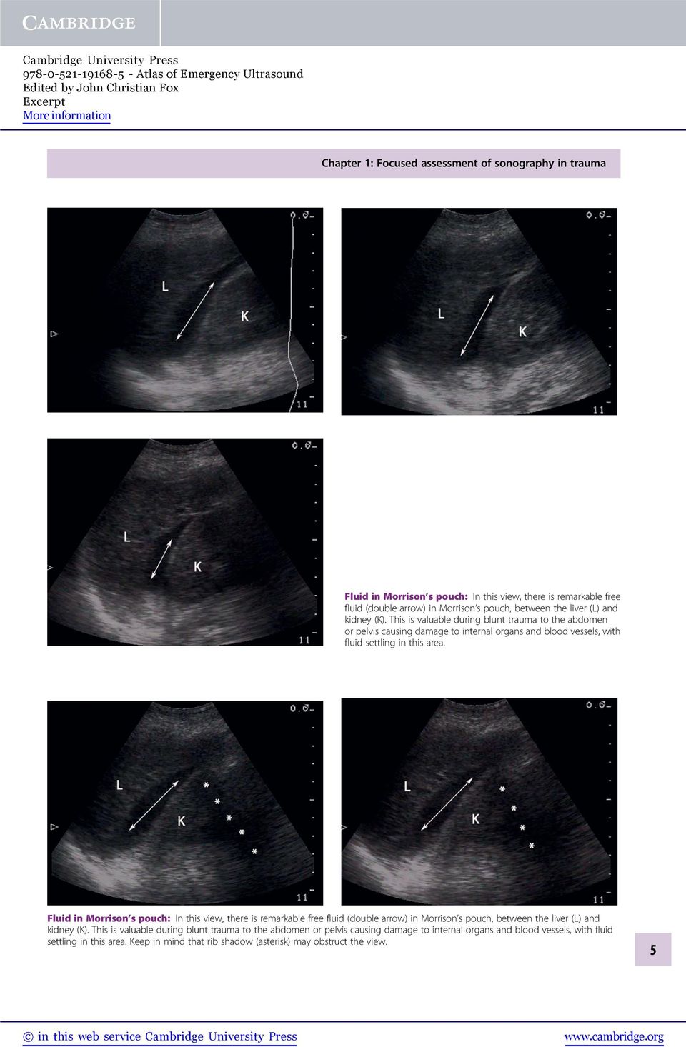
5 Fluid in Morrison s pouch: In this view, there is remarkable free fluid (double arrow) in Morrison s pouch, between the liver (L) and kidney (K). This is valuable during blunt trauma to the abdomen or pelvis causing damage to internal organs and blood vessels, with fluid settling in this area. Fluid in Morrison s pouch: In this view, there is remarkable free fluid (double arrow) in Morrison s pouch, between the liver (L) and kidney (K). This is valuable during blunt trauma to the abdomen or pelvis causing damage to internal organs and blood vessels, with fluid settling in this area. Keep in mind that rib shadow (asterisk) may obstruct the view. 5
6 Fluid in Morrison s pouch: In this view, there is remarkable free fluid (arrows) in Morrison s pouch, between the liver (L) and kidney (K). This is valuable during blunt trauma to the abdomen or pelvis causing damage to internal organs and blood vessels, with fluid settling in this area. Fluid around loops of bowel: In this view, the transducer is placed on the lower abdomen of the patient. There is a clear view of free fluid (F) around the loops of bowel (arrows). Pelvis Examining the lower abdomen of a patient after a trauma may reveal free fluid around bowel. This is a sign that major injury has occurred in the abdomen or pelvis. Pericardial clot While examining the heart, a pericardial clot may be visualized instead of newly escaped blood. This reveals that the trauma has occurred for a significant period and careful examination of heart activity should be performed. 6

7 Pericardial clot: This subxiphoid view was obtained on a patient with a gunshot wound to the chest. The chambers of the heart (H) are not clearly visualized; however, an echodense clot (C) between the visceral and parietal pericardium (arrows) may be seen. Pericardial clot: This subxiphoid view was obtained on a patient with a gunshot wound to the chest. All four chambers of the heart are clearly visualized the right ventricle (RV), left ventricle (LV), right atrium (RA) and the left atrium (LA). Additionally, an echodense clot (asterisk) between the visceral and parietal pericardium (arrow) may be seen. Pericardial clot: This subxiphoid view was obtained on a patient with a gunshot wound to the chest. The right ventricle (RV) and left ventricle (LV) of the heart are clearly visualized. Additionally, an echodense clot (asterisks) between the visceral and parietal pericardium (arrow) may be seen. 7
 between the visceral and parietal pericardium (arrow) may be seen. The right ventricle (RV) and left ventricle (LV) of the heart are clearly visualized.")
8 Perinephric fat While visualizing Morrison s pouch and the splenorenal recess, careful consideration of perinephric fat should be obtained. This should not be mistaken for free fluid, which has contrasting echogenicities. Right chest The view of the right chest is essential as well when performing an exam on the left side with trauma to the chest. The diaphragm is a significant marker, which will have the fluid superior and the liver inferior to it. Fluid found in this area reveals a traumatic hemothorax. Perinephric fat: This view reveals fat (asterisks) surrounding the kidney (K), which should not be mistaken for free fluid, with its increased echogenicity. Perinephric fat: This view reveals fat (asterisks) surrounding the kidney (K) and a clear view of the adjacent liver (L). This should not be mistaken for free fluid, with its increased echogenicity. 8 Fluid in the right chest: Placing the transducer on the right axillary line, superior to the rib with the indicator facing up reveals fluid (F) in the patient s right chest. This fluid is bordered by the diaphragm (arrows) below and with a clear view of the liver (L). This view is also useful when there is trauma to the chest, such as in a motor vehicle collision, stabbing, or gunshot wound.

9 Fluid in the right chest: (cont.) Fluid in the right chest: Placing the transducer on the right axillary line, superior to the rib with the indicator facing up reveals fluid (F) in the patient s right chest. This fluid is bordered by the diaphragm (arrows) below and with a clear view of the kidney (K). This view is also useful when there is trauma to the chest, such as in a motor vehicle collision, stabbing, or gunshot wound. Splenorenal view The examination of the splenorenal recess is additionally crucial during trauma to the abdomen or pelvis. Damage to internal organs and blood vessels will result in fluid settling in this area primarily. A clear view of the spleen and kidney should be obtained, which normally would not present with fluid in between. Fluid found in the stomach which is superior to the spleen and kidney should not be mistaken for fluid in the chest, which is adjacent to the diaphragm. 9

10 Normal splenorenal view: In this normal view, there is no fluid present in the recess between the spleen (S) and kidney (K). The transducer is placed in the left axillary line at the rib margin with the indicator facing up. Keep in mind that rib shadow (asterisks) may obstruct the view. Normal splenorenal view: In this normal view, there is no fluid present in the recess between the spleen (S) and kidney (K). The transducer is placed in the left axillary line at the rib margin with the indicator facing up. 10 Fluid in the stomach with the splenorenal view: In this view of the splenorenal recess, the stomach which is superior to the spleen (S) and kidney (K) is found to have fluid (FS). This should not be mistaken for fluid in the chest, which is adjacent to the diaphragm. Keep in mind that rib shadow (asterisks) may obstruct the view.
 and kidney (K) is found to have fluid (FS).")
Emergency Ultrasound Course
 Emergency Ultrasound Course Dr Justin Bowra ED Course Manual 2: EFAST Extended Focused Assessment with Sonography in Trauma JUSTIN BOWRA 1 The Questions: 1. Is there free fluid (FF) a. In the pleural space?
Sonography for Trauma
 Sonography for Trauma Christine Butts and Justin Cook 9 KEY POINTS Focused abdominal sonography for trauma (FAST) is sensitive and specific for the detection of intraperitoneal free fluid, but it has poor
Traumatic Cardiac Tamponade. Shane KF Seal 19 November 2003 POS
 Traumatic Cardiac Tamponade Shane KF Seal 19 November 2003 POS Objectives Definition Pathophysiology Diagnosis Treatment Cardiac Tamponade The decompensated phase of cardiac compression resulting from
How To Teach An Integrated Ultrasound
 University of South Carolina School of Medicine Integrated Ultrasound Curriculum iusc Richard Hoppmann The Integrated Ultrasound Curriculum Initiated 2006 First (M1) and Second (M2) Year Medical Students
The RUSH Exam: Rapid Ultrasound in SHock in the Evaluation of the Critically lll
 The RUSH Exam: Rapid Ultrasound in SHock in the Evaluation of the Critically lll Phillips Perera, MD, RDMS, FACEP a, *, Thomas Mailhot, MD, RDMS b, David Riley, MD, MS, RDMS a, Diku Mandavia, MD, FACEP,
RACE I Rapid Assessment by Cardiac Echo. Intensive Care Training Program Radboud University Medical Centre NIjmegen
 RACE I Rapid Assessment by Cardiac Echo Intensive Care Training Program Radboud University Medical Centre NIjmegen RACE Goal-directed study with specific questions Excludes Doppler ultrasound Perform 50
Focused Assessment With Sonography for Trauma (FAST) Examination
 Examination") AIUM Practice Parameter for the Performance of the Focused Assessment With Sonography for Trauma (FAST) Examination Parameter developed in collaboration with the American College of Emergency Physicians
AIUM Practice Parameter for the Performance of the Focused Assessment With Sonography for Trauma (FAST) Examination Parameter developed in collaboration with the American College of Emergency Physicians
An Introduction to Anatomy and Physiology
 An Introduction to Anatomy and Physiology Objectives Define anatomy and physiology Identify the levels of organization in organisms from simplest to most complex Identify the organ systems of the human
Administrative. Patient name Date compare with previous Position markers R-L, upright, supine Technical quality
 CHEST X-RAY Administrative Patient name Date compare with previous Position markers R-L, upright, supine Technical quality AP or PA ( with x-ray beam entering from back of patient, taken at 6 feet) Good
TRAUMA SURGERY Dr. Michal Cheatham Orlando Regional Health PGY-4
 ROTATION LIAISON: INSTITUTION: LEVEL(S): TRAUMA SURGERY Dr. Michal Cheatham Orlando Regional Health PGY-4 I. GENERAL INFORMATION The General Surgery Department at Orlando Regional Health has three full
Case III. Disscussion. the UHP ultrasound protocol. Novel Ultrasound Approach to the Empiric Evaluation of the Undifferentiated Hypotensive Patient
 The UHP Ultrasound Protocol: A Novel Ultrasound Approach to the Empiric Evaluation of the Undifferentiated Hypotensive Patient JOHN S. ROSE, MD,* AARON E. BAIR, MD,* DIKU MANDAVIA, MD, AND DONNA J. KINSER,
Autonomous Diagnostic Imaging Performed by Untrained Operators using Augmented Reality as a Form of Just in Time Training
 Autonomous Diagnostic Imaging Performed by Untrained Operators using Augmented Reality as a Form of Just in Time Training PROPOSAL TEAM PI: David S. Martin, MS, Wyle Science, Technology, and Engineering
How To Learn To Perform An Ultrasound
 CAE ICCU E-Learning CAE VIMEDIX Ultrasound Simulator Master Ultrasonography of the Thoracic, Abdominal and Pelvic Cavities An Engaging Learning Solution for Ultrasound Hands-on Simulation, Multimedia Content,
Introduction to A&P (Chapter 1) Lecture Materials for Amy Warenda Czura, Ph.D. Suffolk County Community College. Eastern Campus
 Lecture Materials for Amy Warenda Czura, Ph.D. Suffolk County Community College. Eastern Campus") Introduction to A&P (Chapter 1) Lecture Materials for Amy Warenda Czura, Ph.D. Suffolk County Community College Primary Sources for figures and content: Eastern Campus Marieb, E. N. Human Anatomy & Physiology
Introduction to A&P (Chapter 1) Lecture Materials for Amy Warenda Czura, Ph.D. Suffolk County Community College Primary Sources for figures and content: Eastern Campus Marieb, E. N. Human Anatomy & Physiology
BLUNT TORSO INJURY FROM IMPACT OR DECELERATION HAS ABDOMINAL SOLID ORGAN TRAUMA UNTIL PROVEN OTHERWISE
 Abdominal and Pelvic Trauma In the primary survey, the circulation part includes thinking about the abdomen as a source of occult hemorrhage The OBVIOUS THING is a penetrating abdominal injury Generally
STORM MANUAL CONTENTS
 CONTENTS 1. Introduction 3 2. Accreditation 4 3. Sources and acknowledgements 5 4. Physics 6 5. Instrumentation 24 6. E-FAST 31 7. Echocardiography in Life Support 52 8. Resources & further learning 88
Introduction to A&P (Chapter 1) Lecture Materials for Amy Warenda Czura, Ph.D. Suffolk County Community College Eastern Campus
 Lecture Materials for Amy Warenda Czura, Ph.D. Suffolk County Community College Eastern Campus") Introduction to A&P (Chapter 1) Lecture Materials for Amy Warenda Czura, Ph.D. Suffolk County Community College Eastern Campus Primary Sources for figures and content: Marieb, E. N. Human Anatomy & Physiology
Introduction to A&P (Chapter 1) Lecture Materials for Amy Warenda Czura, Ph.D. Suffolk County Community College Eastern Campus Primary Sources for figures and content: Marieb, E. N. Human Anatomy & Physiology
Practical class 3 THE HEART
 Practical class 3 THE HEART OBJECTIVES By the time you have completed this assignment and any necessary further reading or study you should be able to:- 1. Describe the fibrous pericardium and serous pericardium,
CHAPTER 1: THE LUNGS AND RESPIRATORY SYSTEM
 CHAPTER 1: THE LUNGS AND RESPIRATORY SYSTEM INTRODUCTION Lung cancer affects a life-sustaining system of the body, the respiratory system. The respiratory system is responsible for one of the essential
Mechanism of Injury 1
 Mechanism of Injury 1 Objectives At the end of this lecture the participant will be able to: Describe the importance of the mechanism of injury in history taking of the trauma patient. Identify patterns
Welcome to Anatomy & Physiology
 Welcome to Anatomy & Physiology Chapter 1 -Human Organization What do you need to do to pass this class? MEMORIZE! The Scope of Human Anatomy Human anatomy is the study of the structure of the human body.
Scott Hubbell, MHSc, RRT-NPS, C-NPT, CCT Clinical Education Coordinator/Flight RRT EagleMed
 Scott Hubbell, MHSc, RRT-NPS, C-NPT, CCT Clinical Education Coordinator/Flight RRT EagleMed Identify the 12-Lead Views Explain the vessels of occlusion Describe the three I s Basic Interpretation of 12-Lead
Aehlert: Paramedic Practice Today PowerPoint Lecture Notes Chapter 50: Abdominal Trauma
 Aehlert: Paramedic Practice Today PowerPoint Lecture Notes Chapter 50: Abdominal Trauma Chapter 50 Abdominal Trauma 1 Describe the epidemiology, including morbidity, mortality rates, and prevention strategies,
United States Department of Transportation National Highway Traffic Safety Administration Paramedic: National Standard Curriculum 1
 UNIT TERMINAL OBJECTIVE 4-8 At the completion of this unit, the paramedic student will be able to integrate pathophysiologic principles and the assessment findings to formulate a field impression and implement
NEEDLE THORACENTESIS Pneumothorax / Hemothorax
 NEEDLE THORACENTESIS Pneumothorax / Hemothorax By: Steven Jones, NREMT-P Pneumothorax Pneumothorax is a collection of air or gas in the pleural space of the lung, causing the lung to collapse. Pneumothorax
Medical Terminology, Anatompy & Physiology
 1. Which of the following BEST describes the anatomical position? a. Supine with arms crossed over the chest and knees slightly bent b. Standing, facing forward, with arms raised above the head c. Standing,
Human Anatomy & Physiology II with Dr. Hubley
 Human Anatomy & Physiology II with Dr. Hubley Exam #1 Name: Instructions This exam consists of 40 multiple-choice questions. Each multiple-choice question answered correctly is worth one point, and the
Objectives. Mylene T. Truong, MD. Malignant Pleural Mesothelioma Background
 Imaging of Pleural Tumors Mylene T. Truong, MD Imaging of Pleural Tumours Mylene T. Truong, M. D. University of Texas M.D. Anderson Cancer Center, Houston, TX Objectives To review tumors involving the
CHEST TUBES AND CHEST DRAINAGE SYSTEMS
 CHEST TUBES AND CHEST DRAINAGE SYSTEMS Central Nursing Orientation April 2008 Revised September 2011 OBJECTIVES Describe common tubes and indications for use at LHSC Review indications and contraindications,
Chapter 2 Cardiac Interpretation of Pediatric Chest X-Ray
 Chapter 2 Cardiac Interpretation of Pediatric Chest X-Ray Ra-id Abdulla and Douglas M. Luxenberg Key Facts The cardiac silhouette occupies 50 55% of the chest width on an anterior posterior chest X-ray
How To Understand How Cancer Works
 Mesothelioma Understanding your diagnosis Mesothelioma Understanding your diagnosis When you first hear that you have cancer, you may feel alone and afraid. You may be overwhelmed by the large amount of
MOLLOY COLLEGE DIVISION OF NURSING NURSE PRACTITIONER PROGRAMS. Study Guide for the Basic Physical Assessment Exam
 DIVISION OF NURSING S Study Guide for the Basic Physical Assessment Exam Questions will be based on following chapters in, Bickley, L.S. (2009). (10 th ed). Bates guide to physical examination and history
INTERNATIONAL TRAUMA LIFE SUPPORT
 INTERNATIONAL TRAUMA LIFE SUPPORT NEEDLE DECOMPRESSION OF TENSION PNEUMOTHORAX Roy Alson, MD, PhD, FACEP, FAAEM and Sabina Braithwaite, MD, MPH, FACEP INTRODUCTION The purpose of this document is to update
Retrospective Review of Intra Abdominal Injuries Sustained in a Tertiary Teaching Hospital
 American Journal of Medicine and Medical Sciences 2015, 5(1): 26-30 DOI: 10.5923/j.ajmms.20150501.06 Retrospective Review of Intra Abdominal Injuries Sustained in a Tertiary Teaching Hospital Azhar Amir
Injury Law Center OTHER INJURIES
 Injury Law Center Note: This information is provided to give you a basic understanding of the injury. It is not intended as medical advice. You should consult a qualified medical provider. OTHER INJURIES
Diseases. Inflammations Non-inflammatory pleural effusions Pneumothorax Tumours
 Pleura Visceral pleura covers lungs and extends into fissures Parietal pleura limits mediastinum and covers dome of diaphragm and inner aspect of chest wall. Two layers between them (pleural cavity) contains
REGIONAL INJURIES (LECTURE III) INJURIES TO SPINE, NECK, CHEST, ABDOMEN AND PELVIS.
 INJURIES TO SPINE, NECK, CHEST, ABDOMEN AND PELVIS.") REGIONAL INJURIES (LECTURE III) INJURIES TO SPINE, NECK, CHEST, ABDOMEN AND PELVIS. LEARNING OBJECTIVES At the end of this lecture, last in the series of regional injuries, the students will be knowing
REGIONAL INJURIES (LECTURE III) INJURIES TO SPINE, NECK, CHEST, ABDOMEN AND PELVIS. LEARNING OBJECTIVES At the end of this lecture, last in the series of regional injuries, the students will be knowing
PATIENT CONSENT TO PROCEDURE - ROUX-EN-Y GASTRIC BYPASS
 As a patient you must be adequately informed about your condition and the recommended surgical procedure. Please read this document carefully and ask about anything you do not understand. Please initial
404 Section 5 Shock and Resuscitation. Scene Size-up. Primary Assessment. History Taking
 404 Section 5 and Resuscitation Scene Size-up Scene Safety Mechanism of Injury (MOI)/ Nature of Illness (NOI) Ensure scene safety and address hazards. Standard precautions should include a minimum of gloves
2161-1 - Page 1. Name: 1) Choose the disease that is most closely related to the given phrase. Questions 10 and 11 refer to the following:
 Choose the disease that is most closely related to the given phrase. Questions 10 and 11 refer to the following:") Name: 2161-1 - Page 1 1) Choose the disease that is most closely related to the given phrase. a disease of the bone marrow characterized by uncontrolled production of white blood cells A) meningitis B)
Name: 2161-1 - Page 1 1) Choose the disease that is most closely related to the given phrase. a disease of the bone marrow characterized by uncontrolled production of white blood cells A) meningitis B)
1.INTRODUCTION 2. MATERIALS AND METHODS
 PATTERN OF INJURIES TO MOTORCYCLISTS IN FATAL ROAD TRAFFIC ACCIDENTS Dr. Srinivasulu Pothireddy* 1, Dr. Naresh Karukutla 2 1. Associate Professor 2. Assistant Professor Dept of Forensic Medicine, Katuri
Distance Learning Program Anatomy of the Human Heart/Pig Heart Dissection Middle School/ High School
 Distance Learning Program Anatomy of the Human Heart/Pig Heart Dissection Middle School/ High School This guide is for middle and high school students participating in AIMS Anatomy of the Human Heart and
Urinary Diversion: Ileovesicostomy/Ileal Loop/Colon Loop
 Urinary Diversion: Ileovesicostomy/Ileal Loop/Colon Loop Why do I need this surgery? A urinary diversion is a surgical procedure that is performed to allow urine to safely pass from the kidneys into a
Peritoneal Carcinosis
 Peritoneal Carcinosis What is it and how to cure it Peritoneum Peritoneum is a thin and transparent membrane that covers the internal part of the abdominal and pelvic cavity and all the viscera contained
New Cardiothoracic Surgery CPT Codes for 2013
 New Cardiothoracic Surgery CPT Codes for 2013 There were several changes to the cardiothoracic surgery CPT codes for 2013. There are five new codes in the general thoracic surgery section, with one revised
Teaching Med-5 Students Point-of-Care Transthoracic Echocardiography
 Teaching Med-5 Students Point-of-Care Transthoracic Echocardiography Anthony M.-H. Ho, Lester A. H. Critchley, Patricia Kan, Sylvia Au, Siu Keung Ng, Simon K. C. Chan, Philip Lam, Gordon Choi, Alex Lee,
Heart and Vascular System Practice Questions
 Heart and Vascular System Practice Questions Student: 1. The pulmonary veins are unusual as veins because they are transporting. A. oxygenated blood B. de-oxygenated blood C. high fat blood D. nutrient-rich
Mr GH: Pericardial Window. Anaesthetic Management of Cardiac Tamponade
 Mr GH: Pericardial Window Anaesthetic Management of Cardiac Tamponade Mr GH 56 yo M HOPCx Asbestosis, adenoca R lung 8/52 6/52 cisplatin/ taxol chemo Weekly pleural taps for effusions Sent from Bendigo
LINCOLN UNIVERSITY DI 281 B Practicum / Externship II in Sonography Summer 2015 Course Syllabus
 LINCOLN UNIVERSITY DI 281 B Practicum / Externship II in Sonography Summer 2015 Course Syllabus Course Number: DI 281 B Course Title: Practicum / Externship II in Sonography Course Credit: 3 units = 135
Understanding Pleural Mesothelioma
 Understanding Pleural Mesothelioma UHN Information for patients and families Read this booklet to learn about: What is pleural mesothelioma? What causes it? What are the symptoms? What tests are done to
Mesothelioma: Questions and Answers
 CANCER FACTS N a t i o n a l C a n c e r I n s t i t u t e N a t i o n a l I n s t i t u t e s o f H e a l t h D e p a r t m e n t o f H e a l t h a n d H u m a n S e r v i c e s Mesothelioma: Questions
Anatomy and Physiology: Understanding the Importance of CPR
 Anatomy and Physiology: Understanding the Importance of CPR Overview This document gives you more information about the body s structure (anatomy) and function (physiology). This information will help
Chapter 19 Ci C r i cula l t a i t o i n
 Chapter 19 Circulation A closed system Circulatory System Consisting of Heart, Arteries, Veins, Capillaries, Blood & the Lymphatic system Blood Make up The blood is made up of Plasma and three main types
Blood vessels. transport blood throughout the body
 Circulatory System Parts and Organs Blood vessels transport blood throughout the body Arteries blood vessels that carry blood AWAY from the heart Pulmonary arteries carry the deoxygenated blood from heart
Ultrasound Simulators
 Ultrasound Simulators Tripp Bell, MD University of South Carolina School of Medicine Objectives Give a brief history of simulation in medicine Describe the types of ultrasound simulators Show how ultrasound
Pericardial disease. Usually secondary to systemic or other cardiac diseases. Pericardial fluid accumulations Pericarditis
 Pericardial disease Usually secondary to systemic or other cardiac diseases Pericardial fluid accumulations Pericarditis Pericardial Anatomy Two major components visceral pericardium mesothelial monolayer
Cardiology. Anatomy and Physiology of the Heart.
 Cardiology Self Learning Package Module 1: Anatomy and Physiology of the Heart. Module 1: Anatomy and Physiology of the Heart Page 1 CONTENT Introduction Page 3 How to use the ECG Self Learning package.page
.org. Fractures of the Thoracic and Lumbar Spine. Cause. Description
 Fractures of the Thoracic and Lumbar Spine Page ( 1 ) Spinal fractures can vary widely in severity. While some fractures are very serious injuries that require emergency treatment, other fractures can
Recanalized Umbilical Vein in the Presence of Cirrhosis-Induced Portal Hypertension
 Recanalized Umbilical Vein in the Presence of Cirrhosis-Induced Portal Hypertension Audrey Galey RDMS, RVT, Mary Grace Renfro RDSM, RVT, Lindsey Simon, RVT March 22, 2013 2 Abstract A recanalized umbilical
Abdomen X-Ray (AXR) Collimation is ideally from diaphragms to lower border of the symphysis pubis and the lateral skin margins.
 Collimation is ideally from diaphragms to lower border of the symphysis pubis and the lateral skin margins.") Abdomen X-Ray (AXR) Collimation is ideally from diaphragms to lower border of the symphysis pubis and the lateral skin margins. LMP of child-bearing age female patients should be checked. 1. Acute abdomen
Abdomen X-Ray (AXR) Collimation is ideally from diaphragms to lower border of the symphysis pubis and the lateral skin margins. LMP of child-bearing age female patients should be checked. 1. Acute abdomen
The Body s Transport System
 Circulation Name Date Class The Body s Transport System This section describes how the heart, blood vessels, and blood work together to carry materials throughout the body. Use Target Reading Skills As
Pericardium. Pericardial Diseases. Function of Pericardium 10/1/2012
 NO LASIX, PLEASE! PERICARDIAL DISEASE IN THE DOG Pericardium Michael Luethy, DVM Diplomate ACVIM Cardiology September 13 th, 2012 Tough, outer, parietal pericardium Delicate, serous, visceral pericardium
AUTONOMIC NERVOUS SYSTEM
 AUTONOMIC NERVOUS SYSTEM Somatic efferent and ANS Somatic Efferent Control is over skeletal muscles. External environment This division of the PNS responds to some change in the external environment. single
Thoracic Cavity. Photo: This normal canine lung collapsed when the thorax was opened and the negative pressure was lost in the thorax.
 Thoracic Cavity There are significant anatomical differences in the mediastinum of domestic animals. For instance, bovines, like humans, have well-developed mediastinal separation between the left and
The Circulatory System. Chapter 17 Lesson 1
 The Circulatory System Chapter 17 Lesson 1 Functions of the Circulatory System Your circulatory system maintains an internal environment in which all the cells in your body are nourished. As your heart
Chapter 15. Sympathetic Nervous System
 Chapter 15 Sympathetic Nervous System Somatic versus Autonomic Pathways Somatic efferent innervation ACh Myelinated fiber Somatic effectors (skeletal muscles) Autonomic efferent innervation ACh ACh or
Nerve Tissue. Muscle Tissue. Connective Tissue
 Human Body Tissues Levels of Organization 1. Cells 2. = groups of similar cells that perform a 3. Organ = 4. = group of organs Four Major Tissues 1. 2. 3. 4. Epithelial Tissue Nerve Tissue Muscle Tissue
Anatomi & Fysiologi 060301. The cardiovascular system (chapter 20) The circulation system transports; What the heart can do;
 The circulation system transports; What the heart can do;") The cardiovascular system consists of; The cardiovascular system (chapter 20) Principles of Anatomy & Physiology 2009 Blood 2 separate pumps (heart) Many blood vessels with varying diameter and elasticity
The cardiovascular system consists of; The cardiovascular system (chapter 20) Principles of Anatomy & Physiology 2009 Blood 2 separate pumps (heart) Many blood vessels with varying diameter and elasticity
The Whipple Procedure. Sally Hodges, Ph.D.(c) Given the length and difficulty of the procedure, regardless of the diagnosis, certain
 Given the length and difficulty of the procedure, regardless of the diagnosis, certain") The Whipple Procedure Sally Hodges, Ph.D.(c) Preoperative procedures Given the length and difficulty of the procedure, regardless of the diagnosis, certain assurances must occur prior to offering a patient
The Whipple Procedure Sally Hodges, Ph.D.(c) Preoperative procedures Given the length and difficulty of the procedure, regardless of the diagnosis, certain assurances must occur prior to offering a patient
Cardiovascular diseases. pathology
 Cardiovascular diseases pathology Atherosclerosis Vascular diseases A disease that results in arterial wall thickens as a result of build- up of fatty materials such cholesterol, resulting in acute and
FALLS-protocol: lung ultrasound in hemodynamic assessment of shock
 Review article Heart, Lung and Vessels. 2013; 5(3): 142-147 142 FALLS-protocol: lung ultrasound in hemodynamic assessment of shock D. Lichtenstein Service de Réanimation Médicale, Hôpital Ambroise-Paré,
Laparoscopic Cholecystectomy
 Laparoscopic Cholecystectomy Removal of Gall Bladder Page 12 Patient Information Further Information We endeavour to provide an excellent service at all times, but should you have any concerns please,
Blood Vessels and Circulation
 13 Blood Vessels and Circulation FOCUS: Blood flows from the heart through the arterial blood vessels to capillaries, and from capillaries back to the heart through veins. The pulmonary circulation transports
Ultrasound Credentialing & Curriculum for CCRMC FM Residency
 1. Purpose: Ultrasound Credentialing & Curriculum for CCRMC FM Residency To establish a credentialing process and a curriculum for an ultrasound program in the family medicine residency program at Contra
Spleen. Anatomy. (Effective February 2007) (1%-5%) Normal. Related Anatomy Anterior to spleen. Medial border. Posteriorly
 (1%-5%) Normal. Related Anatomy Anterior to spleen. Medial border. Posteriorly") Spleen (Effective February 2007) (1%-5%) Anatomy Normal Intraperitoneal, except hilum Left hypochondrium Left hemidiaphragm superior generally considered to be ovoid, with a convex superior and a concave
Spleen (Effective February 2007) (1%-5%) Anatomy Normal Intraperitoneal, except hilum Left hypochondrium Left hemidiaphragm superior generally considered to be ovoid, with a convex superior and a concave
Human Body Vocabulary Words Week 1
 Vocabulary Words Week 1 1. arteries Any of the blood vessels that carry blood away from the heart to all parts of the body 2. heart The muscular organ inside the chest that pumps blood through the body
Cardiac Masses and Tumors
 Cardiac Masses and Tumors Question: What is the diagnosis? A. Aortic valve myxoma B. Papillary fibroelastoma C. Vegetation from Infective endocarditis D. Thrombus in transit E. None of the above Answer:
UW MEDICINE PATIENT EDUCATION. Aortic Stenosis. What is heart valve disease? What is aortic stenosis?
 UW MEDICINE PATIENT EDUCATION Aortic Stenosis Causes, symptoms, diagnosis, and treatment This handout describes aortic stenosis, a narrowing of the aortic valve in your heart. It also explains how this
Measuring central venous pressure
 Elaine Cole Senior lecturer ED/Trauma, City University Barts and the London NHS Trust 1 Learning outcomes That the clinician can: Describe the sites of central venous catheterisation Understand why central
Office of the Medical Examiner in the County of Dallas State of Texas AFFIDAVIT
 This affidavit is in compliance with Texas Rules of Criminal Evidence, Rule 902 (10b). Case No. 1811-96 in the matter of Routier, Devon, deceased. Office of the in the County of Dallas State of Texas AFFIDAVIT
THE COLLEGE OF EMERGENCY MEDICINE. Core (Level 1) Ultrasound Curriculum
 Ultrasound Curriculum") THE COLLEGE OF EMERGENCY MEDICINE Core (Level 1) Ultrasound Curriculum Contents! Ultrasound in Emergency Medicine... 3 1. Curriculum for EMUS 2009... 3 2. Equipment - basics... 9 3. Core knowledge... 12
THE COLLEGE OF EMERGENCY MEDICINE Core (Level 1) Ultrasound Curriculum Contents! Ultrasound in Emergency Medicine... 3 1. Curriculum for EMUS 2009... 3 2. Equipment - basics... 9 3. Core knowledge... 12
Laboratory 1 Anatomical Planes and Regions
 Laboratory 1 Anatomical Planes and Regions Goals: Define the anatomical position, including the application of the terms right and left. List and correctly use the major directional terms used in anatomy.
By Casey Schmidt and Wendy Ford
 By Casey Schmidt and Wendy Ford Body systems Digestive System Circulatory System Respiratory System Excretory System Immune System Reproductive System Nervous System Muscular System Skeletal System Endocrine
Ovarian Cystectomy / Oophorectomy
 Cystectomy and Ovarian Cysts Ovarian cysts are sacs filled with fluids or pockets located on or in an ovary. In some cases, these cysts need to be removed surgically. Types of Cysts Ovarian cysts are quite
Dr Dan Gill Department of Radiology Windsor Regional Hospital Oxford Medical Imaging Erie St Clair Radiology
 Dr Dan Gill Department of Radiology Windsor Regional Hospital Oxford Medical Imaging Erie St Clair Radiology -5000 Olympic athletes and team officials -1350 Paralympic athletes and team officials -82
UNIT 1 BODY PLAN AND ORGANIZATION LECTURE
 UNIT 1 BODY PLAN AND ORGANIZATION LECTURE 1.03 CONTRAST THE SCIENCES OF ANATOMY AND PHYSIOLOGY A. Anatomy Anatomy is the scientific study of structures and the relationship of.. structures to each other.
Digestive System AKA. GI System. Overview. GI Process Process Includes. G-I Tract Alimentary Canal
 Digestive System AKA G-I Tract Alimentary Canal Overview GI System Consists of Mouth, pharynx, esophagus, stomach, small intestine, large intestine, anus About 30 in length Accessory Organs Teeth, tongue,
Mesothelioma. 1995-2013, The Patient Education Institute, Inc. www.x-plain.com ocft0101 Last reviewed: 03/21/2013 1
 Mesothelioma Introduction Mesothelioma is a type of cancer. It starts in the tissue that lines your lungs, stomach, heart, and other organs. This tissue is called mesothelium. Most people who get this
Chapter 7. Expose the Injured Area
 Chapter 7 GUNSHOT WOUNDS KEY FIGURES: Entrance/exit wounds This chapter describes how to treat the external, surface wounds caused by a bullet. The evaluation for underlying injury related to gunshot wounds
Human Digestive System Anatomy
 Human Digestive System Anatomy Biology 104 Objectives: 1. Learn the anatomy of the digestive system. You should be able to find all terms in bold on the human torso models. 2. Relate structure of the system
CHAPTER 9 BODY ORGANIZATION
 CHAPTER 9 BODY ORGANIZATION Objectives Identify the meaning of 10 or more terms relating to the organization of the body Describe the properties of life Describe the function for the structures of the
Preparing for your laparoscopic pyeloplasty
 Preparing for your laparoscopic pyeloplasty Welcome We look forward to welcoming you to The Royal London Hospital. You have been referred to us for a laparoscopic pyeloplasty, which is an operation using
PERCUTANOUS TUBE THORACOSTOMY
 PERCUTANOUS TUBE THORACOSTOMY Suveer Singh BSc MBBS FRCP PhD EDIC BDICM Consultant Pulmonary and Critical Care Chelsea and Westminster Hospital London, UK [email protected] September 2007 Anatomy