Skeletal Development Multiple Cellular Origins
|
|
|
- Ezra Kennedy
- 9 years ago
- Views:
Transcription
1 Skeletal Development Multiple Cellular Origins 1 - Paraxial Mesoderm Somite, Sclerotome Axial Skeleton (e.g. vertebra) 2 - Lateral Plate Mesoderm Appendicular Skeleton (e.g. limb) 3 - Neural Crest Head Skeleton Established as 1 - Hyaline Cartilage replaced by Endochondrial Ossification 2 Intramembranous Bone Formation - direct ossification
 3 - Neural Crest Head Skeleton Established as 1 - Hyaline Cartilage replaced by")
2 Intramembranous Bone Intramembranous bone = dermal bone (e.g. skull, clavicle) Mesenchymal condensation, becomes vascularized Osteoid Tissue (prebone) - cells differentiate into osteoblasts - matrix deposition - Calcium Phosphate Osteoblast Osteocytes - trapped in matrix Bone Spicules organized around blood vessels - concentric layers = Haversian system.

3

4 Compact Bone - Osteoblast in periphery lay down layers of compact bone Spongy bone - beneath bony plates - osteoclasts breaks down bone Continual bone remodeling via action of osteoblasts and osteoclast Bone marrow differentiates from mesenchyme in spongy bone

5 Endochondrial Bone Endochondral ossification Hyaline cartilage template of bone forms Cartilage - differentiates from mesenchyme cells Chondroblasts - condenses - become rounded and deposit matrix - collagen fibers or elastic fiber Three types of cartilage - hyaline (most common), fibrocartilage, elastic cartilage Perichondrium - outer layer of cells
, fibrocartilage, elastic cartilage Perichondrium - outer layer")
6 Cartilage template of the limb in the Chick wing

7 Endochondrial Bone Primary ossification center - initiation of ossification Perichondrial cells differentiate into Osteoblasts - deposit matrix as a collar in center of long bone diaphysis

8 Endochondrial Bone Perichondrium becomes Periostium Ossification spreads towards ends of bone Osteoclasts differentiate and begin to breakdown bone Chondrocytes die off center is invaded by vascular system the bone marrow. Cells also invade and differentiate into osteoblasts - forming bone spicules that are remodeled by osteoclasts

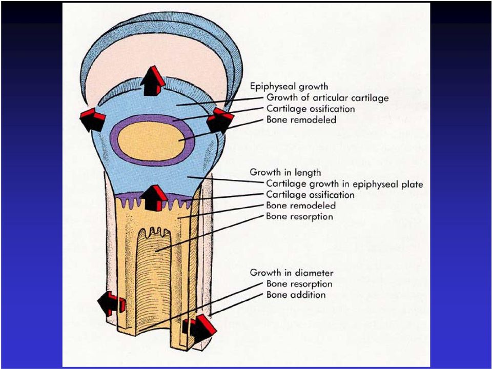
9 Bone Growth Bone lengthening occurs at diaphyseal-epiphyseal junction - epiphyseal cartilage plate (growth plate) Epiphysis - chondrogenic Secondary ossification centers in the epiphysis after birth After growth termination the epiphyseal cartilage plate is replaced with spongy bone

10
11 Skeletal Development Multiple Cellular Origins 1 - Paraxial Mesoderm Somite, Sclerotome Axial Skeleton (e.g. vertebra) 2 - Lateral Plate Mesoderm Appendicular Skeleton (e.g. limb) 3 - Neural Crest Head Skeleton Established as 1 - Hyaline Cartilage replaced by Endochondrial Ossification 2 Intramembranous Bone Formation - direct ossification
 3 - Neural Crest Head Skeleton Established as 1 - Hyaline Cartilage replaced by")
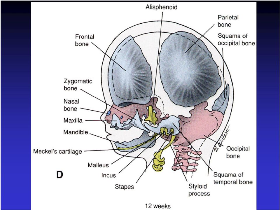
12 Skull / Head Neurocranium skeleton around the brain Viscerocranium skeleton of the face Both consist of two components: Membranous (Intramembranous ossification) Cartilaginous (Endochondrial ossification)
 Cartilaginous")
13 Neurocranium Membranous neurocranium cranial vault = calvaria flat bones of skull Sutures - fibrous joints between flat bones Fontanelles - where several sutures meet Moldling - bones are soft, sutures are loose allows for changes during birth Cartilaginous neurocranium bones at the base of the skull

14

15 Viscerocranium Cartilaginous viscerocranium middle ear bones - incus, malleus, stapes reichert s cartilage hyoid bone Membranous viscerocranium Jaw Bones maxilla, zygomatic, squamous temporal bones, mandible

16
17 Apert syndrome Crouzon s syndrome Pfeiffer s syndrome Craniosynostosis Premature closure of sutures Abnormal skull shape Multiple causes: FGF signaling Msx gene function

18 FGF Receptor (FGFR) mutations cause craniosynostosis Autosomal dominant abnormal dimer function

19 Skeletal Development Multiple Cellular Origins 1 - Paraxial Mesoderm Somite, Sclerotome Axial Skeleton (e.g. vertebra) 2 - Lateral Plate Mesoderm Appendicular Skeleton (e.g. limb) 3 - Neural Crest Head Skeleton Established as 1 - Hyaline Cartilage replaced by Endochondrial Ossification 2 Intramembranous Bone Formation - direct ossification
 3 - Neural Crest Head Skeleton Established as 1 - Hyaline Cartilage replaced by")
20

21 Vertebral Column Three parts to each vertebra - body, vertebral arch, ribs
22 Sclerotome cells form a mesenchyme that chondrofies around the notochord to form the centrum
23 Development of Vertebra Sclerotome - cells surround notochord on both sides cranial - loosely arranged cells caudally - densely packed cells Each vertebra is derived from two sclerotome segments Caudal (dense) cells from a cranial sclerotome Cranial (loose) cells from the next caudal sclerotome Intervertebral disc between vertebra Intervertebral disc forms at the interface between loose and dense cells (center of sclerotome)
24
25 The nucleus pulposus is surrounded by fibrous tissue (concentric) - anulus fibrosus The centrum is the primordium of the body Notochord degenerates in the center of body Notochord expands in the intervertebral disc region forms the nucleus pulposus = gelatinous disc center
26 Development of Vertebra Sclerotome cells surround the neural tube - forms the vertebral arch - fuses ventrally with the centrum Sclerotome cells in the body wall form the costal processes, the ribs
27 Primary ossification centers 1 - Surrounding the notochord in the centrum 2 - Lateral to the neural tube in the vertebral arch Secondary ossification centers 1 - anular epiphyses - between body and intervertebral disc) 2 - tip of spinous process 3 tips of transverse processes
28 Joints: neurocentral joint - centrum / vertebral arch - allows for growth of the spinal cord until 5 years Costovertebral synchondrosis - vertebral arch / ribs synovial joint
29 Thoracic Cervical
30 Lumbar Sacrum Coccyx
31 Hox Genes Regional characteristics of vertebrae are specified by unique combinatorial expression of Hox genes Homeotic transformations of vertebrae have been described Retinoic Acid can cause cranial to caudal segment shifts
32 Ribs / Sternum Sclerotome cells in the body wall form the costal processes that form the ribs The Sternum forms from a pair of ventral cartilagenous bands that converge at the ventral midline Converged sternal bands undergo secondary segmentation similar to joint formation Sternal segments later fuse
33
34 Muscle Development Muscle types Skeletal, Cardiac, Smooth Smooth muscle : Derived from splanchnic mesoderm surrounding gut. Cellular elongation without cell fusion Cardiac muscle Derived - splanchnic mesoderm Myoblasts adhere but do not fuse Form intercalated discs
35
36 Skeletal Muscle Head region skeletal musculature Derived from head mesenchyme Migration from the cranial somitomeres Trunk region skeletal musculature Myoblasts derived from somites Migration - FGF controlled Spindle shaped cells - line up and fuse Multinucleated syncitium Myofibrils with cross-striations - actin-myosin
37 Region-Specific myoblast behavior Limb Region myoblast migration into limb primordia, Differentiation is delayed Thoracic Region myotubes form at the somite then invade the body wall to form the intercostal muscles Lumbar Region myoblast migrate to form the abdominal muscles Myoblast behavior is controlled by their environment
38
39 Myotome: two parts Epimere Dorsomedial Extensors of Vertebral column Hypomere Ventrolateral limb/body wall Innervating nerves Dorsal ramus; Ventral ramus
40 Thoracic level 3 myogenic layers external intercostal, internal intercostal, transversus abdominis muscles Ribs maintain segmented musculature, elsewhere fusion large muscle sheets
41
42 Determination of myoblast occurs very early Key regulators Myf-5, Pax3, MyoD
Chapter 11. What are the functions of the skeletal system? More detail on bone
 Skeletal System Chapter 11 11.1 Overview of the skeletal system What are the functions of the skeletal system? 1. Supports the body 2. Protects the soft body parts 3. Produces blood cells 4. Stores minerals
Unit 4: Skeletal System Test Review Test Review
 Name: Period: Unit 4: Skeletal System Test Review Test Review 1. List four functions of the skeletal system: a. b. c. d. 2. Define ossification and identify the roles of the osteoblasts, osteocytes, and
1. outer fibrous layer contains fibroblasts that secrete collagen
 I. cartilage A. perichondrium 1. outer fibrous layer contains fibroblasts that secrete collagen 2. inner chondrogenic layer contains cells that can proliferate and turn into chondroblasts B. cartilage
BIOL 4260 Human Evolu3onary Anatomy Lecture 5: Bone Development & Trunk Anatomy. Lecture 2: Fossil Record
 BIOL 4260 Human Evolu3onary Anatomy Lecture 5: Bone Development & Trunk Anatomy Lecture 2: Fossil Record Segmentation Cyclic genescreate segme ntation clock for somite production Final #s 4 occipital 8
Osseous Tissue & Structure. The skeletal system includes: Storage of minerals: calcium salts
 Chapter 15 Lecture The Skeletal System: Osseous Tissue & Skeletal Structure The Skeletal System The skeletal system includes: Bones, cartilages, ligaments Bone tissue = osseous tissue Includes living cells
Lab 5 Overview of the Skeleton: Classification and Structure of Bones and Cartilages Exercise 9 The Axial Skeleton Exercise 10
 Lab 5 Overview of the Skeleton: Classification and Structure of Bones and Cartilages Exercise 9 The Axial Skeleton Exercise 10 Overview of the Skeleton Locate the important cartilages in the human skeleton
E. Blood cells production. Blood cells are produced within the cavities of bones.
 FUNCTIONS OF THE SKELETAL SYSTEM 1. The skeletal system consists of bone, cartilage, ligaments (attach bone to another bone), and tendons (attach muscle to bone). 2. Functions. A. Support. Bone helps to
CHS 06-07 BONES AND SKELETAL TISSUES
 CHS 06-07 BONES AND SKELETAL TISSUES This chapter provides a review of bone and skeletal tissue. The human skeleton is composed primarily of two connective tissues: (1) cartilage and (2) bone. CHARACTERISTICS
BONES AND BONE TISSUE
 BIO 2401 BONES & BONE TISSUE page 1 BONES AND BONE TISSUE Organization of the Skeletal System components: 1) bone 2) skeletal cartilage: surrounded by dense irregular connective tissue which acts to girdle
Function of Bones. Bone Tissue and Bones BONE CLASSIFICATION. Long Bones Gross Anatomy. Lab Activity 1 - Gross Anatomy of a Long Bone
 Function of Bones Bone Tissue and Bones Bones form the framework of the skeleton. Anatomy and Physiology Text and Laboratory Workbook, Stephen G. Davenport, Copyright 2006, All Rights Reserved, no part
7. Skeletal System: Bone Structure and Function
 7. Skeletal System: Bone Structure and Function For the next two chapters (7 and 9) we will study the skeletal system. Although the major feature of this system is the bones, the skeletal system also consists
Divisions of the Skeletal System
 OpenStax-CNX module: m46344 1 Divisions of the Skeletal System OpenStax College This work is produced by OpenStax-CNX and licensed under the Creative Commons Attribution License 3.0 By the end of this
UNIT 4 - SKELETAL SYSTEM LECTURE NOTES
 UNIT 4 - SKELETAL SYSTEM LECTURE NOTES 4.01 FUNCTIONS OF THE SKELETAL SYSTEM A. Support 1. Provides a framework for the body. 2. Supports soft tissue. 3. Serves as a point of attachment for ligaments,
OBJECTIVE: List and describe the functions of the skeletal system. D. Storage depot for minerals and homeostasis
 Text Reference: Chapters 6 and 7 Pages 1-6 are mostly a review from lab Know this material before you come to lecture. Lecture will begin on page 7 Read text and complete packet before lecture. I. Introduction
Chapter 6: The Skeletal System: Bone Tissue
 Chapter 6: The Skeletal System: Bone Tissue Chapter Objectives FUNCTIONS OF THE SKELETAL SYSTEM 1. Discuss the functions of support, protection, assistance in movement, mineral homeostasis, blood cell
5. MESODERM FORMATION / SEGMENTATION
 5. MESODERM FORMATION / SEGMENTATION Dr. Ann -Judith Silverman Department of Anatomy & Cell Biology Telephone: 305-3540 E-mail: [email protected] READING ASSIGNMENT: Larsen Human Embryology, 3rd Edition,
Vertebral anatomy study guide. Human Structure Summer 2015. Prepared by Daniel Schmitt, Angel Zeininger, and Karyne Rabey.
 Vertebral anatomy study guide. Human Structure Summer 2015 Prepared by Daniel Schmitt, Angel Zeininger, and Karyne Rabey. 1. Plan of Action: In this guide you will learn to identify these structures: Cervical
its lifetime. The skeletal system is divided into: 1. Axial Division: bones of the body s axis (skulll, ribs, vertebrae)
") The Axial Skeleton The basic features of the human skeleton have been shaped by evolution, but the detailed characteristics of each bone reflect the stresses put on it. As a result, the skeleton changes
The Axial Skeleton The basic features of the human skeleton have been shaped by evolution, but the detailed characteristics of each bone reflect the stresses put on it. As a result, the skeleton changes
Chapter 5 The Skeletal System
 Essentials of Human Anatomy & Physiology Elaine N. Marieb Seventh Edition Chapter 5 The Skeletal System The Skeletal System Parts of the skeletal system Bones (skeleton) Joints Cartilages Ligaments (bone
10/12/2011. Classification of Bones Every adult skeleton contains 206 bones which can be arranged into six broad categories according to shape
 Primary Functions of Skeletal System 1. support 2. storage of minerals & lipids -calcium salts provide vital minerals -lipids are in stored yellow marrow 3. blood cell production -RBC s, WBC s, and other
SAMPLE LECTURE EXAM 1 -- HUMAN ANATOMY
 SAMPLE LECTURE EXAM 1 -- HUMAN ANATOMY 1. The subcutaneous layer consists mostly of. a. smooth muscle c. areolar and adipose connective tissues d. melanin e. keratin 2. Which of the following statements
THE SKELETAL & ARTICULAR SYSTEMS. The Bones & Joints
 THE SKELETAL & ARTICULAR SYSTEMS The Bones & Joints CLOSE YOUR POWERPOINT HANDOUTS!! Think-Pair-Share: Why do we need bones? Try to think of 3 reasons. THE SKELETAL SYSTEM Is made up of numerous bones
Classification of bones Any bone may be classified into one of the following groups:
 Skeletal system This system is made up of hard tissues like bone and cartilages. This system gives form and shape to animal body The skeleton of a living animal is made up living structures of bones. The
Module F SKELETAL SYSTEM & ARTICULATIONS
 Module F SKELETAL SYSTEM & ARTICULATIONS Topic from General functions of bone & the skeletal system Structural components microscopic anatomy Structural components gross anatomy Physiology of embryonic
LESSON ASSIGNMENT. After completing this lesson, you should be able to: 4-1. Define skeleton.
 LESSON ASSIGNMENT LESSON 4 The Human Skeletal System. TEXT ASSIGNMENT Paragraphs 4-1 through 4-14. LESSON OBJECTIVES After completing this lesson, you should be able to: 4-1. Define skeleton. 4-2. Name
THE SKELETAL SYSTEM: BONE TISSUE. Chapter 6. Anatomy and Physiology Lecture
 Chapter 6 1 THE SKELETAL SYSTEM: BONE TISSUE Chapter 6 Anatomy and Physiology Lecture Chapter 6 2 THE SKELETAL SYSTEM: BONE TISSUE Bone (Osseous) Tissue forms most of the skeleton: Skeletal System - the
Human Anatomy & Physiology
 PowerPoint Lecture Slides prepared by Barbara Heard, Atlantic Cape Community College Ninth Edition Human Anatomy & Physiology C H A P T E R 7 The Skeleton: Part B Annie Leibovitz/Contact Press Images Vertebral
LOCOMOTION AND MOVEMENT
 UNIT - HUMAN PHYSIOLOGY Chapter 18 LOCOMOTION AND MOVEMENT Movement is an important feature of living organism. Both the microbes and macrobes show wide range of movements. The movements results in change
9/3/2013 JOINTS. Joints. Axial Skeleton STRUCTURE AND FUNCTION:
 STRUCTURE AND FUNCTION: JOINTS Joints A connection between 2 or more bones A pivot point for bony motion The features of the joint help determine The ROM freedom Functional potential of the joint Axial
DEVELOPMENT AND GROWTH OF THE MANDIBLE
 2012-2013 ORAL BIOLOGY DEVELOPMENT AND GROWTH OF THE MANDIBLE Ass. Prof. Dr. Heba M. Elsabaa Development and Growth of the Mandible DEVELOPMENT OF THE MANDIBLE The Mandible Is the largest and strongest
Brunswick School Department: Grades 11-12. Essential Understandings
 Understandings Questions Knowledge Vocabulary Skills Bones provide invaluable structure and support for the body. There are a variety of bone shapes. Joints connect bones. The skeletal system is divided
Thoracic Spine Anatomy
 A Patient s Guide to Thoracic Spine Anatomy 228 West Main, Suite C Missoula, MT 59802 Phone: [email protected] DISCLAIMER: The information in this booklet is compiled from a variety of sources.
Structure and Function of the Musculoskeletal System Professor Alan Hedge DEA 3250/6510
 Structure and Function of the Musculoskeletal System Professor Alan Hedge DEA 3250/6510 Functions of the Musculoskeletal System Support and protect the body and its organs. Provide motion. Musculoskeletal
Bone Tissue Chapter 7
 Bone Tissue Chapter 7 Tissues and organs of the skeletal system Histology of osseous tissue Bone development Physiology of osseous tissue Bone disorders Bone Tissue Bone is alive and continuously changing.
8/25/2014 JOINTS. The Skeletal System. Axial Skeleton STRUCTURE AND FUNCTION:
 STRUCTURE AND FUNCTION: JOINTS The Skeletal System Made up of the numerous bones of the human body Gives support and framework to the body Protects vital organs Manufactures blood cells Storage of calcium
SKELETON AND JOINTS G.C.S.E. PHYSICAL EDUCATION. Unit 1. Factors Affecting Participation and Performance. G.C.S.E. P.E. Teacher:.
 G.C.S.E. PHYSICAL EDUCATION Unit 1 Factors Affecting Participation and Performance SKELETON AND JOINTS Name: G.C.S.E. P.E. Teacher:. By the end of this booklet you should be able to: Understand what the
THE SKELETAL SYSTEM - THE AXIAL SKELETON
 THE SKELETAL SYSTEM - THE AXIAL SKELETON Chapter 7 Anatomy and Physiology Lecture 1 THE SKELETAL SYSTEM THE AXIAL SKELETON Skeletal System forms the framework of the body. TYPES OF BONES: FOUR PRINCIPAL
THE SKELETAL SYSTEM FUNCTIONS OF THE SKELETAL SYSTEM
 THE SKELETAL SYSTEM The skeleton is the body s bony framework which consists of 206 bones. The bones are made up of water(45%), calcium and phosphorous(35%) and other organic materials(20%). The calcium
Biology 141 Anatomy and Physiology I
 Fall 2016 Biology 141 Anatomy and Physiology I COURSE OUTLINE Faculty Name: Enter Faculty Name Here Program Head: Enter Program Head Here Dean s Review: Dean s Signature: Date Reviewed: / / Revised: Fall
STRUCTURE AND FUNCTION: JOINTS
 STRUCTURE AND FUNCTION: JOINTS Joints A connection between 2 or more bones A pivot point for bony motion The features of the joint help determine The ROM Degrees of freedom Functional potential of the
Limb Development. Limb Development. Limb Axes. Limb Development. Clinical Terms
 Limb Development Overview of Limb Formation Initiation of Limb Development Limb Field Outgrowth of the Mesoderm Morphogenetic Signaling Development of Limb Tissues Skeleton Musculature Innervation Vasculature
North Bergen School District Benchmarks
 Grade: 10,11, and 12 Subject: Anatomy and Physiology First Marking Period Define anatomy and physiology, and describe various subspecialties of each discipline. Describe the five basic functions of living
Lumbar Spine Anatomy. eorthopod.com 228 West Main St., Suite D Missoula, MT 59802-4345 Phone: 406-721-3072 Fax: 406-721-2619 info@eorthopod.
 A Patient s Guide to Lumbar Spine Anatomy 228 West Main St., Suite D Missoula, MT 59802-4345 Phone: 406-721-3072 Fax: 406-721-2619 [email protected] DISCLAIMER: The information in this booklet is compiled
Vertebrate Body Organization
 Vertebrate Body Organization Digestive tube suspended in coelom from mouth to anus Body supported by internal skeleton of jointed bones Vertebrae and Cranium protects nervous system Diaphragm divides coelom
9. PHARYNGEAL ARCHES. READING ASSIGNMENT: Larsen 3 rd Edition. Chapter 12: pp. 352; 358-365; 405-412 SUMMARY: LEARNING OBJECTIVES:
 9. PHARYNGEAL ARCHES Letty Moss-Salentijn DDS, PhD Dr. Edwin S. Robinson Professor of Dentistry (in Anatomy and Cell Biology) E-mail: [email protected] READING ASSIGNMENT: Larsen 3 rd Edition. Chapter
LABORATORY EXERCISE 12 BONE STRUCTURE AND CLASSIFICATION
 LABORATORY EXERCISE 12 BONE STRUCTURE AND CLASSIFICATION FIG. 12.1 1. Articular cartilage (hyaline cartilage) 6. Periosteum 2. Spongy bone (red marrow) 7. Proximal epiphysis 3. Medullary cavity 8. Diaphysis
Skeletal, Muscular, and Integumentary Systems
 Chapter 36 Skeletal, Muscular, and Integumentary Systems Section 36 1 The Skeletal System (pages 921 925) This section describes the skeletal system and its functions. Introduction (page 921) 1. What forms
Laerdal' Human Anatomy Manual The Skeleton
 Human Anatomy Manual The Skeleton Laerdal Texas P.O. Box 38.226 EM. 116 Gatesville,Texas U.S.A.76528 U.S.A.1-800-433-5539 IntemationaI1-254-865-7221 24 Hour Fax 254-865-8011 ~ Laerdal' TABLE OF CONTENTS
Ground substance is the component of connective tissue between the cells and the fibers
 Connective Tissues Directions: Insert and install your Interactions: Foundations CD. a. Click the "Contents" button. b. Open the Tissue Level of Organization file. c. Click on Anatomy Overviews. d. Work
Human Anatomy & Physiology I with Dr. Hubley. Practice Exam #2
 Human Anatomy & Physiology I with Dr. Hubley Practice Exam #2 For questions 1 through 3, select your answers from the following responses: a. stratified squamous epithelium b. reticular connective tissue
Anatomy of Skeletal System
 Anatomy of Skeletal System two main subdivisions of skeletal system: axial : skull, vertebral column, rib cage appendicular: arms and legs and girdles Bone Markings: Foramen: opening in bone passageway
Skeletal system. 2012 Pearson Education, Inc.
 NURS1004 Week 6 Part I Prepared by Didy Button Skeletal system An Introduction to the Skeletal System The Skeletal System Includes: Bones of the skeleton Cartilages, ligaments, and connective tissues 6-1
Definition: A joint or articulation is a place in the body where two bones come together.
 Definition: A joint or articulation is a place in the body where two bones come together. CLASSES OF JOINTS. 1. Joints are classified according to how the bones are held together. 2. The three types of
Animal Tissues. I. Epithelial Tissue
 Animal Tissues There are four types of tissues found in animals: epithelial tissue, connective tissue, muscle tissue, and nervous tissue. In this lab you will learn the major characteristics of each tissue
THE SKELETAL AND MUSCULAR SYSTEMS
 i A Wealth of Information. A World of Ideas. Instructor s Guide The Human Body: How It Works Introduction This program is part of the nine-part series The Human Body: How It Works. The series uses physiologic
Lectures of Human Anatomy
 Lectures of Human Anatomy Vertebral Column-I By DR. ABDEL-MONEM AWAD HEGAZY M.B. with honor 1983, Dipl."Gynecology and Obstetrics "1989, Master "Anatomy and Embryology" 1994, M.D. "Anatomy and Embryology"
Skeletal System. Axial Skeleton: Vertebral Column and Ribs
 Skeletal System Axial Skeleton: Vertebral Column and Ribs Functions Regions Cervical Thoracic Lumbar Sacral Primary & secondary curvatures There are three major functions of the vertebral column. First,
Objectives AXIAL SKELETON. 1. Frontal Bone. 2. Parietal Bones. 3. Temporal Bones. CRANIAL BONES (8 total flat bones w/ 2 paired)
") Objectives AXIAL SKELETON SKULL 1. On a skull or diagram, identify and name the bones of the skull 2. Identify the structure and function of the bones of the skull 3. Describe how a fetal skull differs
Objectives AXIAL SKELETON SKULL 1. On a skull or diagram, identify and name the bones of the skull 2. Identify the structure and function of the bones of the skull 3. Describe how a fetal skull differs
Chetek-Weyerhaeuser High School
 Chetek-Weyerhaeuser High School Anatomy and Physiology Units and Anatomy and Physiology A Unit 1 Introduction to Human Anatomy and Physiology (6 days) Essential Question: How do the systems of the human
Skeletal System -Training Handout Karen L. Lancour National Rules Committee Chairman Life Science
 Skeletal System -Training Handout Karen L. Lancour National Rules Committee Chairman Life Science INTERACTION OF SKELETAL AND MUSCULAR SYSTEMS: Skeletal and Muscular systems works together to allow movement
Anatomical Terminology: Planes Axes Direction (Text Pg 2 3)
") Anatomical Terminology: Planes Axes Direction (Text Pg 2 3) Anatomy: The structure and organization of the body and its organs. From Greek Origin: Anatome dissection Physiology: The function of the body
Anatomical Terminology: Planes Axes Direction (Text Pg 2 3) Anatomy: The structure and organization of the body and its organs. From Greek Origin: Anatome dissection Physiology: The function of the body
Tissues (Histology) Ch. 3 Human Anatomy lecture
 Ch. 3 Human Anatomy lecture") I. Histology the study of tissues A. 4 basic tissue types epithelial connective muscle nervous Tissues (Histology) Ch. 3 Human Anatomy lecture B. Usually found in combinations to form organs. C. As you
I. Histology the study of tissues A. 4 basic tissue types epithelial connective muscle nervous Tissues (Histology) Ch. 3 Human Anatomy lecture B. Usually found in combinations to form organs. C. As you
Anatomy and Physiology of Domestic Animals
 Anatomy and Physiology of Domestic Animals 6 Bones and skeletal system Contents Bones Introduction Classification of Bones Bone Structure Gross Anatomy Microscopic Anatomy of Bone Chemical Composition
Anatomy and Physiology 121: Muscles of the Human Body
 Epicranius Anatomy and Physiology 121: Muscles of the Human Body Covers upper cranium Raises eyebrows, surprise, headaches Parts Frontalis Occipitalis Epicranial aponeurosis Orbicularis oculi Ring (sphincter)
Anatomy and Terminology of the Spine. Bones of the Spine (Vertebrae)
") Anatomy and Terminology of the Spine The spine, also called the spinal column, vertebral column or backbone, consists of bones, intervertebral discs, ligaments, and joints. In addition, the spine serves
Anatomy and Terminology of the Spine The spine, also called the spinal column, vertebral column or backbone, consists of bones, intervertebral discs, ligaments, and joints. In addition, the spine serves
The Petrylaw Lawsuits Settlements and Injury Settlement Report
 The Petrylaw Lawsuits Settlements and Injury Settlement Report BACK INJURIES How Minnesota Juries Decide the Value of Pain and Suffering in Back Injury Cases The Petrylaw Lawsuits Settlements and Injury
Section B: Epithelial Tissue 1. Where are epithelial tissues found within the body? 2. What are the functions of the epithelial tissues?
 Tissue worksheet Name Section A: Intro to Histology Cells are the smallest units of life. In complex organisms, cells group together with one another based on similar structure and function to form tissues.
Animal Systems: The Musculoskeletal System
 Animal Systems: The Musculoskeletal System Tissues, Organs, and Systems of Living Things Cells, Cell Division, and Animal Systems and Plant Systems Cell Specialization Human Systems The Digestive The Circulatory
Welcome to Anatomy & Physiology
 Welcome to Anatomy & Physiology Chapter 1 -Human Organization What do you need to do to pass this class? MEMORIZE! The Scope of Human Anatomy Human anatomy is the study of the structure of the human body.
Anatomy and Physiology Warm up questions Fall 2013
 Anatomy and Physiology Warm up questions Fall 2013 QUESTION POSSIBLE ANSWERS : Chapter 1 Introduction and Regions Chapter 2 Chemistry I can name systems of the I can identify regions of the I can describe
Biology 105 Human Biology PRACTICE MIDTERM EXAM 1. Essentials of Anatomy and Physiology, 5e (Martini/Nath) Chapter 6 The Skeletal System
 Chapter 6 The Skeletal System") Essentials of Anatomy and Physiology, 5e (Martini/Nath) Chapter 6 The Skeletal System Multiple-Choice Questions 1) Functions of the skeletal system include A) support. B) storage. C) protection. D) blood
Essentials of Anatomy and Physiology, 5e (Martini/Nath) Chapter 6 The Skeletal System Multiple-Choice Questions 1) Functions of the skeletal system include A) support. B) storage. C) protection. D) blood
Spinal Anatomy. * MedX research contends that the lumbar region really starts at T-11, based upon the attributes of the vertebra.
 Spinal Anatomy Overview Neck and back pain, especially pain in the lower back, is one of the most common health problems in adults. Fortunately, most back and neck pain is temporary, resulting from short-term
6 BONE TISSUE AND THE SKELETAL SYSTEM
 CHAPTER 6 BONE TISSUE AND THE SKELETAL SYSTEM 203 6 BONE TISSUE AND THE SKELETAL SYSTEM Figure 6.1 Child Looking at Bones Bone is a living tissue. Unlike the bones of a fossil made inert by a process of
Introduction to Anatomy and Physiology: Tissues and Integumentary System. Biology 105 Lecture 7 Chapter 4
 Introduction to Anatomy and Physiology: Tissues and Integumentary System Biology 105 Lecture 7 Chapter 4 Outline I. Tissues A. Epithelial B. Connective C. Muscle D. Nervous tissues II. Cell-to-cell contact
THE LANGUAGE OF ANATOMY
 THE LANGUAGE OF ANATOMY Know all the terms identified in these sections of this exercise: Correct anatomical position: When the body is erect, with feet only slightly apart, head and toes pointing forward,
6 Bones and Skeletal Tissues
 6 Bones and Skeletal Tissues Cartilage Location and basic structure Found throughout adult body Ear and epiglottis Articular cartilages and costal cartilage Larynx, trachea, and nose Intervertebral discs,
CHAPTER 8: JOINTS OF THE SKELETAL SYSTEM. 4. Name the three types of fibrous joints and give an example of each.
 OBJECTIVES: 1. Define the term articulation. 2. Distinguish between the functional and structural classification of joints, and relate the terms that are essentially synonymous. 3. Compare and contrast
Skeletal System. Skeletal Anatomy. bones, cartilage and ligaments are tightly joined to form a strong, flexible framework
 Skeletal System bones, cartilage and ligaments are tightly joined to form a strong, flexible framework bone is active tissue:!5-7% bone mass/week Functions of Skeletal System: 1. Support strong and relatively
Mobility and Exercise for Health and Social Care
 Unit 32: Mobility and Exercise for Health and Social Care Unit code: QCF Level 3: Credit value: 10 Guided learning hours: 60 Aim and purpose R/600/8987 BTEC National This unit aims to enable learners to
Patient Information. Lateral Lumbar Interbody Fusion Surgery (LLIF).
.") Patient Information. Lateral Lumbar Interbody Fusion Surgery (LLIF). Understanding your spine Disc Between each pair of vertebrae there is a disc that acts as a cushion to protect the vertebra, allows
Patient Information. Lateral Lumbar Interbody Fusion Surgery (LLIF). Understanding your spine Disc Between each pair of vertebrae there is a disc that acts as a cushion to protect the vertebra, allows
Problems. Knowing. back of the
 Introduction Welcome to BodyZone Physiotherapy patient resource about Cervical Spine Problems. Knowing the main parts of your neck and how these parts work is important as you learn to care for your neck
UNIT 2 - CHAPTER 7: SKELETAL SYSTEM. 1. Classify bones according to their shapes, and name an example from each group. (p. 200)
") LEARNING OUTCOMES: 7.1 Bone Shape and Structure UNIT 2 - CHAPTER 7: SKELETAL SYSTEM 1. Classify bones according to their shapes, and name an example from each group. (p. 200) 2. Describe the macroscopic
LEARNING OUTCOMES: 7.1 Bone Shape and Structure UNIT 2 - CHAPTER 7: SKELETAL SYSTEM 1. Classify bones according to their shapes, and name an example from each group. (p. 200) 2. Describe the macroscopic
The Tissue Level of Organization
 The Tissue Level of Organization Tissues A groups of similar cells, usually having similar embryonic origin and specialized function Histology: the study of tissues Four general types Epithelial Muscle
If you or a loved one have suffered because of a negligent error during spinal surgery, you will be going through a difficult time.
 If you or a loved one have suffered because of a negligent error during spinal surgery, you will be going through a difficult time. You may be worried about your future, both in respect of finances and
Level 2 Certificate in Fitness Instructing Unit 1: Anatomy and Physiology
 Level 2 Certificate in Fitness Instructing Unit 1: Anatomy and Physiology These questions have been compiled based on the information available for the above qualification and unit. This mock should be
Patient Information. Anterior Cervical Discectomy and Fusion Surgery (ACDF).
.") Patient Information. Anterior Cervical Discectomy and Fusion Surgery (ACDF). Understanding your spine Disc Between each pair of vertebrae there is a disc that acts as a cushion to protect the vertebra,
Patient Information. Anterior Cervical Discectomy and Fusion Surgery (ACDF). Understanding your spine Disc Between each pair of vertebrae there is a disc that acts as a cushion to protect the vertebra,
Development of Teeth
 Development of Teeth Dr. Khaldoun Darwich Specialist in Oral and Maxillo-Facial Surgery Hamburg University PhD Hamburg University Academic Teacher - Department of OMF Surgery in Damascus University Instructor
Secondary dentition permanent teeth - 32. Primary dentition deciduous teeth - 20
 Department of Histology and Embryology, P. J. Šafárik University, Medical Faculty, Košice DEVELOPMENT OF TEETH: Sylabus for foreign students Dental medicine Author: doc. MVDr. Iveta Domoráková, PhD. Primary
UNIT COVER PAGE. 6. Classes of joints 7. 6 types of synovial joints 8. diseases and disorders --osteoporosis --arthritis, etc.
 Unit 4 UNIT COVER PAGE School District: Bremen Dist. 228 Department: Science Course: Anatomy Unit Title: Skeletal System Grade Levels: Topic Areas: Time Frame: 3-4 Weeks Date Created: 2/13/06 Date Modified:
Anatomy of the Spine. Figure 1. (left) The spine has three natural curves that form an S-shape; strong muscles keep our spine in alignment.
 The spine has three natural curves that form an S-shape; strong muscles keep our spine in alignment.") 1 2 Anatomy of the Spine Overview The spine is made of 33 individual bony vertebrae stacked one on top of the other. This spinal column provides the main support for your body, allowing you to stand upright,
1 2 Anatomy of the Spine Overview The spine is made of 33 individual bony vertebrae stacked one on top of the other. This spinal column provides the main support for your body, allowing you to stand upright,
Skeletal System. Skeletal Anatomy. bones, cartilage and ligaments are tightly joined to form a strong, flexible framework
 Skeletal System bones, cartilage and ligaments are tightly joined to form a strong, flexible framework bone is active tissue:!5-7% bone mass/week daily Calcium requirement: 1200mg/day Functions of Skeletal
CONTINUING EDUCATION COURSES. for Massage Therapists. Online!
 CONTINUING EDUCATION COURSES for Massage Therapists Online! ccmh Halifax Canadian College of Massage & Hydrotherapy Online Continuing Education Program CCMH Halifax offers a variety of Continuing Education
II. Axial Skeleton (Skull, Thoracic Cage, and Vertebral Column)
") THE SKELETAL SYSTEM Lab Objectives Students should be able to: 1. Recognize bones and bone markings for the axial and appendicular skeleton 2. Recognize bones disarticulated and/or articulated 3. Identify
THE SKELETAL SYSTEM Lab Objectives Students should be able to: 1. Recognize bones and bone markings for the axial and appendicular skeleton 2. Recognize bones disarticulated and/or articulated 3. Identify
SPINE ANATOMY AND PROCEDURES. Tulsa Spine & Specialty Hospital 6901 S. Olympia Avenue Tulsa, Oklahoma 74132
 SPINE ANATOMY AND PROCEDURES Tulsa Spine & Specialty Hospital 6901 S. Olympia Avenue Tulsa, Oklahoma 74132 SPINE ANATOMY The spine consists of 33 bones called vertebrae. The top 7 are cervical, or neck
Clarification of Terms
 Shoulder Girdle Clarification of Terms Shoulder girdle = scapula and clavicle Shoulder joint (glenohumeral joint) = scapula and humerus What is the purpose (or function) of the shoulder and entire upper
BIO 201 ANATOMY AND PHYSIOLOGY I with LAB
 BIO 201 ANATOMY AND PHYSIOLOGY I with LAB (Title change ONLY Oct. 2013) Presented and Approved: January 12, 2012 Effective: 2012-13 FA Prefix & Number BIO 201 Course Title: Anatomy and Physiology I Purpose
LUMBAR LAMINECTOMY AND DISCECTOMY. Basic Anatomical Landmarks: Posterior View Lumbar Spine
 Lumbar Relating to the loins or the section of the back and sides between the ribs and the pelvis. In the spinal column, the last five vertebrae (from superior to inferior, L1-L5) Laminectomy Surgical
Tissue Types. 1. Epithelial Tissue (or epithelium) is the lining, covering, and glandular tissue of the body
 is the lining, covering, and glandular tissue of the body") Tissue Types A. Tissues 1. Tissues: groups of cells similar in structure and function 2. Four Types of Tissues: a. Epithelium: for covering b. Connective Tissue: for support c. Muscle: for movement d.
Tissue Types A. Tissues 1. Tissues: groups of cells similar in structure and function 2. Four Types of Tissues: a. Epithelium: for covering b. Connective Tissue: for support c. Muscle: for movement d.
BIO 2401 MUSCLE TISSUE page 1 MUSCLES AND MUSCLE TISSUE. Striations Present or Absent?
 BIO 2401 MUSCLE TISSUE page 1 Types of Muscle MUSCLES AND MUSCLE TISSUE Type of Muscle Skeletal Location of Muscle attaches to and covers bony skeleton Striations Present or Absent? present Control of