Lu L m u b m a b r a rta T p a Figure 12.30
|
|
|
- Joseph Townsend
- 9 years ago
- Views:
Transcription
1 SPINAL CORD ANATOMY AND DANIL HAMMOUDI.MD FUNCTION

2

3 SPINAL CORD ANATOMY

4 Lumbar Tap Figure 12.30

5 Spinal Cord Figure 12.29a

6 There are 31 spinal cord segments: 8 cervical segments 12 thoracic segments 5 lumbar segments 5 sacral segments 1 coccygeal segment There are two regions where the spinal cord enlarges: Cervical enlargement - corresponds roughly to the brachial plexus nerves, which innervate the upper limb. It includes spinal cord segments from about C4 to T1. The vertebral levels of the enlargement are roughly the same (C4 to T1). Lumbosacral enlargement - corresponds to the lumbosacral plexus nerves, which innervate the lower limb. It comprises the spinal cord segments from L2 to S3, and is found about the vertebral levels of T9 to T12.

7

8 Spinal Cord Conus medullaris terminal portion of the spinal cord Filum terminale fibrous extension of the pia mater; anchors the spinal cord to the coccyx Denticulate ligaments delicate shelves of pia mater; attach the spinal cord to the vertebrae

9 The dermatomes are somatic or musculocutaneous areas served by fibers from specific spinal nerves. Referred pain is caused when the sensory fibers from an internal organ enter the spinal cord in the same root as fibers from a dermatome. The brain is poor at interpreting visceral pain and instead interprets it as pain from the somatic area of the dermatome.

10 Cervical Plexus - the phrenic nerve travels through the thorax to innervate the diaphragm. Brachial Plexus - Axillary nerve - innervates the deltoid muscle and shoulder, along with the posterior aspect of the upper arm. Musculocutaneous nerve - innervates anterior skin of upper arm and elbow flexors. Radial nerve - innervates dorsal aspect of the arm and extensors of the elbow, wrist, and fingers, abduction of thumb. Median nerve - innervates the middle elbow, wrist and finger flexors, adducts the thumb. Ulnar nerve - innervates the medial aspect wrist and finger flexors. Lumbar Plexus genitofemoral - to the external genitalia obturator - to the adductor muscles femoral - innervates the skin and muscles of upper thigh, including the quadriceps. Sacral Plexus gluteal nerves (superior and inferior) - superior innervates the gluteus medius and minimus, inferior innervates the gluteus maximus. sciatic nerve - the body's largest nerve, consisting of two major branches, the tibial and common peroneal. Together they innervate most all of leg including the flexors of the knee, part of adductor magnus, muscles for plantar flexion, dorsiflexion, and other movements of the foot and toes.

11 ganglion - a collection of cell bodies located outside the Central Nervous System. The spinal ganglia or dorsal root ganglia contain the cell bodies of sensory neurons entering the cord at that region. nerve - a group of fibers (axons) outside the CNS. The spinal nerves contain the fibers of the sensory and motor neurons. A nerve does not contain cell bodies. They are located in the ganglion (sensory) or in the gray matter (motor). tract - a group of fibers inside the CNS. The spinal tracts carry information up or down the spinal cord, to or from the brain. Tracts within the brain carry information from one place to another within the brain. Tracts are always part of white matter. gray matter - an area of unmyelinated neurons where cell bodies and synapses occur. In the spinal cord the synapses between sensory and motor and interneurons occurs in the gray matter. The cell bodies of the interneurons and motor neurons also are found in the gray matter. white matter - an area of myelinated fiber tracts. Myelination in the CNS differs from that in nerves.

12 The spinal cord proper begins at the level of the foramen magnum of the skull and ends at the level of the L1ÐL2 intervertebral joint

13

14

15
16

17

18 Myelinatedfibers

19

20

21
22
23
24 a pia mater b subarachnoid space c dura mater d myelinated axon e unipolar neuron of the dorsal root ganglion surrounded by satellite cells (neuroglia).
25 a Pia mater b Subarachnoid space filled with cerebral spinal fluid, wastes and various cells. c Fibrocyte mixed in the blue collagen fibers of the dura mater. d Nucleus & nucleolus of unipolar neuron e Nucleus of one of many tiny satellite cells surrounding the large unipolar neuron. f Myelinated axon g Node of Ranvier h Nucleus of white Schwann cell
26 a Synaptic bulbs over the motor end plate - neuromuscular junction b Neuron axon terminal - black fibers
27 The central canal is the cerebrospinal fluid-filled space that runs longitudinally through the length of the entire spinal cord. The central canal is contiguous with the ventricular system of the brain.
28 PNS in the Nervous System Figure 13.1
29
30
31 Scheuermann kyphosis In some rare cases, the bones of the back (vertebrae) do not grow correctly. In Scheuermann kyphosis, the front part of the vertebrae does not grow as well as the back part.
32 Structure of a Nerve Nerve cordlike organ of the PNS consisting of peripheral axons enclosed by connective tissue Connective tissue coverings include: Endoneurium loose connective tissue that surrounds axons Perineurium coarse connective tissue that bundles fibers into fascicles Epineurium tough fibrous sheath around a nerve
33
34
35 Structure of a Nerve Figure 13.3b
36 Classification of Nerves Sensory and motor divisions Sensory (afferent) carry impulse to the CNS Motor (efferent) carry impulses from CNS Mixed sensory and motor fibers carry impulses to and from CNS; most common type of nerve
37 Peripheral Nerves Mixed nerves carry somatic and autonomic (visceral) impulses The four types of mixed nerves are: Somatic afferent and somatic efferent Visceral afferent and visceral efferent Peripheral nerves originate from the brain or spinal column
38 Regeneration of Nerve Fibers Damage to nerve tissue is serious because mature neurons are amitotic If the soma of a damaged nerve remains intact, damage can be repaired Regeneration involves coordinated activity among: Macrophages remove debris Schwann cells form regeneration tube and secrete growth factors Axons regenerate damaged part
39 Regeneration of Nerve Fibers Figure 13.4
40 Regeneration of Nerve Fibers Figure 13.4
41 Cranial Nerves Twelve pairs of cranial nerves arise from the brain They have sensory, motor, or both sensory and motor functions Each nerve is identified by a number (I through XII) and a name Four cranial nerves carry parasympathetic fibers that serve muscles and glands
42 Peripheral Nervous System 31 spinal nerves We ve already discussed their structure 12 cranial nerves How do they differ from spinal nerves? We need to learn their: Names Locations Functions
43
44 Cranial Nerves Figure 13.5a
45 Summary of Function of Cranial Nerves Figure 13.5b
46 12 Cranial Nerves How do you remember which nerve is which number? Here is a G-rated mnemonic devices: Old Opie occasionally tries trigonometry and feels very gloomy, vague, and hypoactive. There are also several R-rated ones Some cranial nerves are sensory, some motor, and some are both (mixed)? Some say marry money but my brother says big butts matter more.
47
48 Cranial Nerve I: Olfactory Arises from the olfactory epithelium Passes through the cribriform plate of the ethmoid bone Fibers run through the olfactory bulb and terminate in the primary olfactory cortex Functions solely by carrying afferent impulses for the sense of smell
49 Cranial Nerve I: Olfactory Figure I from Table 13.2
50 How many noses do you have? Sensory, motor, or mixed? Run from the nasal mucosa to the olfactory bulb. Extend thru the cribriform plate. Lesion to these nerves or cribriform plate fracture may yield anosmia loss of smell. CN1 Olfactory nerves
51 Cranial Nerve II: Optic Figure II from Table 13.2
52 How many eyes do you have? Sensory, motor, or mixed? Begin at the retina, run to the optic chiasm, cross over, continue as the optic tract and synapse in the thalamus. Optic nerve damage yields blindness in the eye served by the nerve. Optic tract damage yields partial visual loss. Visual defects = anopsias CN2 Optic Nerves
53
54 Cranial Nerve III: Oculomotor Fibers extend from the ventral midbrain, pass through the superior orbital fissure, and go to the extrinsic eye muscles Functions in raising the eyelid, directing the eyeball, constricting the iris, and controlling lens shape Parasympathetic cell bodies are in the ciliary ganglia
55 Cranial Nerve III: Oculomotor Figure III from Table 13.2
56 CN3 Eye mover Sensory, motor, or mixed? Originate at the ventral midbrain. Synapse on: Extraocular muscles Inferior oblique; Inferior, medial, and superior rectus Iris constrictor muscle Ciliary muscle Disorders can result in eye paralysis, diplopia or ptosis. Oculomotor Nerves
57 Cranial Nerve IV: Trochlear Fibers emerge from the dorsal midbrain and enter the orbits via the superior orbital fissures; innervate the superior oblique muscle Primarily a motor nerve that directs the eyeball
58
59 Controls the superior oblique muscle which depresses the eye via pulling on the superior oblique tendon which loops over a ligamentous pulley known as the trochlea. Originates on the dorsal midbrain and synapses on the superior oblique Sensory, motor, or mixed? Trauma can result in double vision. Why? CN4 Trochlear Nerves
60
61 Cranial Nerve V: Trigeminal Three divisions: ophthalmic (V 1 ), maxillary (V 2 ), and mandibular (V 3 ) Fibers run from the face to the pons via the superior orbital fissure (V 1 ), the foramen rotundum (V 2 ), and the foramen ovale (V 3 ) Conveys sensory impulses from various areas of the face (V 1 ) and (V 2 ), and supplies motor fibers (V 3 ) for mastication
62 Cranial Nerve V: Trigeminal Figure V from Table 13.2
63 CN5 Trigeminal Nerves Sensory, motor, or mixed? Biggest cranial nerve Originates in the pons and eventually splits into 3 divisions: Ophthalmic (V1), Maxillary (V2), & Mandibular (V3). Sensory info (touch, temp., and pain) from face. Motor info to muscles of mastication Damage?
64
65
66 The spinal trigeminal nucleus represents pain/temperature sensation from the face. Pain/temperature fibers from peripheral nociceptors are carried in cranial nerves V, VII, IX and X. On entering the brainstem, sensory fibers are grouped together and sent to the spinal trigeminal nucleus. This bundle of incoming fibers can be identified in cross sections of the pons and medulla as the spinal tract of the trigeminal nucleus, which parallels the spinal trigeminal nucleus itself. The spinal tract of V is analogous to, and continuous with, Lissauer s tract in the spinal cord.
67 Cranial Nerve VI: Abdcuens Fibers leave the inferior pons and enter the orbit via the superior orbital fissure Primarily a motor nerve innervating the lateral rectus muscle Figure VI from Table 13.2
68 Sensory, motor, or mixed? Runs between inferior pons and lateral rectus. CN6 Abducens Nerves
69
70 Cranial Nerve VII: Facial Cranial Nerve VII: Facial Fibers leave the pons, travel through the internal acoustic meatus, and emerge through the stylomastoid foramen to the lateral aspect of the face Mixed nerve with five major branches Motor functions include facial expression, and the transmittal of autonomic impulses to lacrimal and salivary glands Sensory function is taste from the anterior twothirds of the tongue
71 The facial nerve has four compo nents with distinc t functio ns: Branchial motor (special visceral efferent) Visceral motor (general visceral efferent) Special sensory (special afferent) Supplies the muscles of facial expression; posterior belly of digastric muscle; stylohyoid, and stapedius. Parasympathetic innervation of the lcrimal, submandibular, and sublingual glands, as well as mucous membranes of nasopharynx, hard and soft palate. Taste sensation from the anterior 2/3 of tongue; hard and soft palates. General sensory (general somatic afferent) General sensation from the skin of the concha of the auricle and from a small area behind the ear.
72
73
74 Branches of facial nerve in the face
75
76 Cranial Nerve VII: Facial Figure VII from Table 13.2
77 Sensory, motor, or mixed? Originates at the pons Convey motor impulses to facial skeletal muscles except for chewing muscles. Convey parasympathetic motor impulses to tear, nasal, and some salivary glands. Convey sensory info from taste buds on anterior 2/3 of the tongue. Facial nerve damage may yield Bell s palsy, total ipsilateral hemifacial paralysis CN7 Facial Nerves
78
79 CN8 Auditory/Vestibulocochlear Nerves Sensory, motor, or mixed? Originates at the pons 2 divisions: Cochlear Afferent fibers from cochlea in the inner ear HEARING Vestibular Afferent fibers from equilibrium receptors in inner ear BALANCE Functional impairment?
80 CN9 Glossopharyngeal Nerves Sensory, motor, or mixed? Fibers run emerge from medulla and run to the throat. Motor Functions: Motor fibers to some swallowing muscles Parasympathetic fibers to some salivary glands Sensory Functions: Taste, touch, heat from pharynx and posterior tongue. Info from chemoreceptors on the level of O 2 and CO 2 in the blood. Info from baroreceptors on BP. Chemoreceptors and baroreceptors are located in the carotid sinus a dilation in the internal carotid artery.
81 CN10 Vagus Nerves Sensory, motor, or mixed? Only cranial nerves to extend beyond head and neck. Fibers emerge from medulla, leave the skull, and course downwards into the thorax and abdomen. Motor Functions: Parasympathetic efferents to the heart, lungs, and abdominal organs. Sensory Functions: Input from thoracic and abdominal viscera; from baro- and chemoreceptors in the carotid sinus; from taste buds in posterior tongue and pharynx
82
83 CN11 Sensory, motor, or mixed? Formed by the union of a cranial root and a spinal root. CR arises from medulla while SR arises from superior spinal cord. SR passes thru the FM and joins with CR to form the accessory nerve. They then leave the skull via the jugular foramen. Cranial division then joins vagus and innervates larynx, pharynx, and soft palate. Spinal division innervates sternocleidomastoids and trapezius. Accessory Nerves
84
85 CN12 Hypoglossal Nerves Sensory, motor, or mixed? Arise from the medulla and exit the skull via the hypoglossal canal and innervate the tongue. Innervate the intrinsic & extrinsic muscles of the tongue. Swallowing, speech, food manipulation. Damage?
86 Peripheral Nervous System Now that we ve looked at spinal and cranial nerves, we can examine the divisions of the PNS. The PNS is broken down into a sensory and a motor division. We ll concentrate on the motor division which contains the somatic nervous system and the autonomic nervous system.
Peripheral Nervous System
 Nervous system consists of: Peripheral Nervous System CNS = brain and spinal cord ~90% (90 Bil) of all neurons in body are in CNS PNS = Cranial nerves and spinal nerves, nerve plexuses & ganglia ~10% (10
The Nervous System: The Spinal Cord and Spinal Nerves
 14 The Nervous System: The Spinal Cord and Spinal Nerves PowerPoint Lecture Presentations prepared by Steven Bassett Southeast Community College Lincoln, Nebraska Introduction The Central Nervous System
Human Anatomy & Physiology Spinal Cord, Spinal Nerves and Somatic Reflexes 13-1
 Human Anatomy & Physiology Spinal Cord, Spinal Nerves and Somatic Reflexes 13-1 Spinal Cord, Spinal Nerves and Somatic Reflexes Spinal cord Spinal nerves Somatic reflexes 13-2 Overview of Spinal Cord Information
Cranial Nerve I Name: Foramen: Fiber Type: Function: Branches: Embryo:
 Cranial Nerve I Olfactory nerve Cribiform plate Special sensory Olfactory bulbs, Smell Olfactory filaments CNS (ectoderm) cribiform plate sensory = Cranial Nerve II Other: Optic nerve Optic canal Special
CHAPTER 11: NERVOUS SYSTEM II: DIVISIONS OF THE NERVOUS SYSTEM OBJECTIVES: 1. Outline the major divisions of the nervous system.
 CHAPTER 11: NERVOUS II: DIVISIONS OF THE NERVOUS OBJECTIVES: 1. Outline the major divisions of the nervous system. NERVOUS CENTRAL NERVOUS (BRAIN & SPINAL CORD) (INTERNEURONS) PERIPHERAL NERVOUS (CRANIAL
3) Cerebral Cortex & Functions of the 4 LOBES. 5) Cranial Nerves (Nerves In the Cranium, i.e., Head)
 Cerebral Cortex & Functions of the 4 LOBES. 5) Cranial Nerves (Nerves In the Cranium, i.e., Head)") Lecture 5 (Oct 8 th ): ANATOMY and FUNCTION OF THE NERVOUS SYSTEM Lecture Outline 1) Basic Divisions (CNS vs. PNS, Somatic vs. Autonomic) and Directional Terms 2) The Brain (Hindbrain/ Midbrain/ Forebrain)
Lecture 5 (Oct 8 th ): ANATOMY and FUNCTION OF THE NERVOUS SYSTEM Lecture Outline 1) Basic Divisions (CNS vs. PNS, Somatic vs. Autonomic) and Directional Terms 2) The Brain (Hindbrain/ Midbrain/ Forebrain)
Chapter 13: The Spinal Cord, Spinal Nerves, and Spinal Reflexes
 Chapter 13: The Spinal Cord, Spinal Nerves, and Spinal Reflexes I. General Organization of the Nervous System, p. 422 Objectives 1. Describe the basic structural and organizational characteristics of the
Nervous System: Spinal Cord and Spinal Nerves (Chapter 13) Lecture Materials for Amy Warenda Czura, Ph.D. Suffolk County Community College
 Lecture Materials for Amy Warenda Czura, Ph.D. Suffolk County Community College") Nervous System: Spinal Cord and Spinal Nerves (Chapter 13) Lecture Materials for Amy Warenda Czura, Ph.D. Suffolk County Community College Primary Sources for figures and content: Eastern Campus Marieb,
Nervous System: Spinal Cord and Spinal Nerves (Chapter 13) Lecture Materials for Amy Warenda Czura, Ph.D. Suffolk County Community College Primary Sources for figures and content: Eastern Campus Marieb,
Peripheral nervous system (PNS) Consists of: 12 pairs of cranial nerves 31 pairs of spinal nerves The autonomic nervous system
 Consists of: 12 pairs of cranial nerves 31 pairs of spinal nerves The autonomic nervous system") Peripheral nervous system (PNS) Consists of: 12 pairs of cranial nerves 31 pairs of spinal nerves The autonomic nervous system Nerves and neurons Cranial nerves There are 12 pairs They are numbered according
Peripheral nervous system (PNS) Consists of: 12 pairs of cranial nerves 31 pairs of spinal nerves The autonomic nervous system Nerves and neurons Cranial nerves There are 12 pairs They are numbered according
Lab Exercise 9. Nervous Tissue. Brain. Cranial Nerves. Spinal Cord. Spinal Nerves
 Lab Exercise 9 Nervous Tissue Brain Cranial Nerves Spinal Cord Spinal Nerves Textbook Reference: See Chapter 11 for histology of nerve tissue and spinal cord See Chapter 12 for brain and spinal cord anatomy
Chapter 15. Autonomic Nervous System (ANS) and Visceral Reflexes. general properties Anatomy. Autonomic effects on target organs
 and Visceral Reflexes. general properties Anatomy. Autonomic effects on target organs") Chapter 15 Autonomic Nervous System (ANS) and Visceral Reflexes general properties Anatomy Autonomic effects on target organs Central control of autonomic function 15-1 Copyright (c) The McGraw-Hill Companies,
Chapter 15 Autonomic Nervous System (ANS) and Visceral Reflexes general properties Anatomy Autonomic effects on target organs Central control of autonomic function 15-1 Copyright (c) The McGraw-Hill Companies,
General A&P Nervous Tissues, Nerves, Spinal Cord and Reflexes Lab Exercises
 1 General A&P Nervous Tissues, Nerves, Spinal Cord and Reflexes Lab Exercises Have someone in your group read the following out loud, while the others read along: In this "Lab Guide", we will be looking
Sheep Brain Dissection
 Sheep Brain Dissection http://www.carolina.com/product/preserved+organisms/preserved+animals+%28mammal s%29/sheep+organs/preserved+sheep+dissection.do Michigan State University Neuroscience Program Brain
THE BRAIN, SPINAL CORD, AND CRANIAL NERVES
 THE BRAIN, SPINAL CORD, AND CRANIAL NERVES I. BRAIN ANATOMY A. Meninges (coverings) of the brain and spinal cord (Fig. [13.120 p. 452 [457]) Use text illustrations to study these. Note that the singular
Autonomic Nervous System Dr. Ali Ebneshahidi
 Autonomic Nervous System Dr. Ali Ebneshahidi Nervous System Divisions of the nervous system The human nervous system consists of the central nervous System (CNS) and the Peripheral Nervous System (PNS).
Chapter 15 Anatomy and Physiology Lecture
 1 THE AUTONOMIC NERVOUS SYSTEM Chapter 15 Anatomy and Physiology Lecture 2 THE AUTONOMIC NERVOUS SYSTEM Autonomic Nervous System (ANS) regulates the activity of smooth muscles, cardiac muscles, and certain
CSE511 Brain & Memory Modeling. Lect04: Brain & Spine Neuroanatomy
 CSE511 Brain & Memory Modeling CSE511 Brain & Memory Modeling Lect02: BOSS Discrete Event Simulator Lect04: Brain & Spine Neuroanatomy Appendix of Purves et al., 4e Larry Wittie Computer Science, StonyBrook
Chapter 7: The Nervous System
 Chapter 7: The Nervous System I. Organization of the Nervous System Objectives: List the general functions of the nervous system Explain the structural and functional classifications of the nervous system
Nervous System: PNS and CNS
 Nervous System: PNS and CNS Biology 105 Lecture 10 Chapter 8 Outline I. Central Nervous System vs Peripheral Nervous System II. Peripheral Nervous System A. Somatic Nervous System B. Autonomic Nervous
Parts of the Brain. Chapter 1
 Chapter 1 Parts of the Brain Living creatures are made up of cells. Groups of cells, similar in appearance and with the same function, form tissue. The brain is a soft mass of supportive tissues and nerve
Basic Cranial Nerve Examination
 Basic Cranial Nerve Examination WIPE Wash hands Introduce yourself Permission Position (Patient sitting facing you, maintain comparable eye level) Exposure (Face exposed only, i.e. remove hats etc) Identify
Chapter 15. Sympathetic Nervous System
 Chapter 15 Sympathetic Nervous System Somatic versus Autonomic Pathways Somatic efferent innervation ACh Myelinated fiber Somatic effectors (skeletal muscles) Autonomic efferent innervation ACh ACh or
Autonomic Nervous System of the Neck. Adam Koleśnik, MD Department of Descriptive and Clinical Anatomy Center of Biostructure Research, MUW
 Autonomic Nervous System of the Neck Adam Koleśnik, MD Department of Descriptive and Clinical Anatomy Center of Biostructure Research, MUW Autonomic nervous system sympathetic parasympathetic enteric Autonomic
Chapter 9 - Nervous System
 Chapter 9 - Nervous System 9.1 Introduction (p. 215; Fig. 9.1) A. The nervous system is composed of neurons and neuroglia. 1. Neurons transmit nerve impulses along nerve fibers to other neurons. 2. Nerves
Student Academic Learning Services Page 1 of 8 Nervous System Quiz
 Student Academic Learning Services Page 1 of 8 Nervous System Quiz 1. The term central nervous system refers to the: A) autonomic and peripheral nervous systems B) brain, spinal cord, and cranial nerves
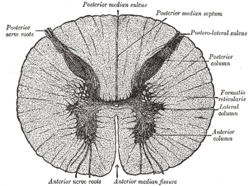
Transverse Sections of the Spinal Cord
 Transverse Sections of the Spinal Cord The spinal cord is perhaps the most simply arranged part of the CNS. Its basic structure, indicated in a schematic drawing of the eighth cervical segment (Figure
Please read chapter 15, The Autonomic Nervous System, complete this study guide, and study this material BEFORE coming to the first class.
 Please read chapter 15,, complete this study guide, and study this material BEFORE coming to the first class. I. Introduction to the autonomic nervous system: Briefly describe the autonomic nervous system.
Integration and Coordination of the Human Body. Nervous System
 I. General Info Integration and Coordination of the Human Body A. Both the and system are responsible for maintaining 1. Homeostasis is the process by which organisms keep internal conditions despite changes
Chapter 3 The Anatomy of the Nervous System
 Chapter 3 The Anatomy of the Nervous System Systems, Structures, and Cells That Make Up Your Nervous System 1 General Layout of the Nervous System Central Nervous System (CNS) Brain (in the skull) Spinal
Reflex Physiology. Dr. Ali Ebneshahidi. 2009 Ebneshahidi
 Reflex Physiology Dr. Ali Ebneshahidi Reflex Physiology Reflexes are automatic, subconscious response to changes within or outside the body. a. Reflexes maintain homeostasis (autonomic reflexes) heart
Orbit & Cranial Nerves II, III, IV, & VI
 Orbit & Cranial Nerves II, III, IV, & VI PCC Year 1, Spring Quarter Lawrence M. Witmer, PhD Life Sciences Building 123 OBJECTIVES: to understand the anatomy of the bony orbit and its contents, in particular,
Chapter 9 Nervous System
 Chapter 9 Nervous System Nervous System function: The nervous system is composed of neurons and neuroglia. at the ends of peripheral nerves gather information and convert it into nerve impulses. When sensory
The Anatomy of Spinal Cord Injury (SCI)
") The Anatomy of Spinal Cord Injury (SCI) What is the Spinal Cord? The spinal cord is that part of your central nervous system that transmits messages between your brain and your body. The spinal cord has
The Anatomy of Spinal Cord Injury (SCI) What is the Spinal Cord? The spinal cord is that part of your central nervous system that transmits messages between your brain and your body. The spinal cord has
Anatomy of the Spine. Figure 1. (left) The spine has three natural curves that form an S-shape; strong muscles keep our spine in alignment.
 The spine has three natural curves that form an S-shape; strong muscles keep our spine in alignment.") 1 2 Anatomy of the Spine Overview The spine is made of 33 individual bony vertebrae stacked one on top of the other. This spinal column provides the main support for your body, allowing you to stand upright,
1 2 Anatomy of the Spine Overview The spine is made of 33 individual bony vertebrae stacked one on top of the other. This spinal column provides the main support for your body, allowing you to stand upright,
Welcome to Lesson 11 of the Basic Human Anatomy Course. Today, we ll be studying the Human Nervous System.
 Basic Human Anatomy Lesson 11: Nervous System Welcome to Lesson 11 of the Basic Human Anatomy Course. Today, we ll be studying the Human Nervous System. I have 19 goals for you in this lesson: 1. Name
Anatomy and Terminology of the Spine. Bones of the Spine (Vertebrae)
") Anatomy and Terminology of the Spine The spine, also called the spinal column, vertebral column or backbone, consists of bones, intervertebral discs, ligaments, and joints. In addition, the spine serves
Anatomy and Terminology of the Spine The spine, also called the spinal column, vertebral column or backbone, consists of bones, intervertebral discs, ligaments, and joints. In addition, the spine serves
Chapter 7: The Nervous System
 Chapter 7: The Nervous System Objectives Discuss the general organization of the nervous system Describe the structure & function of a nerve Draw and label the pathways involved in a withdraw reflex Define
Chapter 13. The Nature of Somatic Reflexes
 Chapter 13 The Nature of Somatic Reflexes Nature of Reflexes (1 of 3) A reflex is an involuntary responses initiated by a sensory input resulting in a change in a gland or muscle tissue occur without our
Anatomy & Physiology Bio 2401 Lecture. Instructor: Daryl Beatty Nervous System Introduction Part 1
 Anatomy & Physiology Bio 2401 Lecture Instructor: Daryl Beatty Nervous System Introduction Part 1 Nervous System Introduction Chapter 11 Section A Sequence 4.1 DB Nervous system 1 Intro Presentations 4.2,
THE BRAIN AND CRANIAL NERVES
 THE BRAIN AND CRANIAL NERVES The Brain - made up of a trillion neurons - weighs about 3 lbs - has four principle parts 1. Brain stem - medulla oblongata, pons, midbrain (mesencephalon) 2. Diencephalon
The Spinal Cord, Spinal Nerves, and Somatic Reflexes
 Cross section through two fascicles (bundles) of nerve fibers in a nerve CHAPTER 13 The Spinal Cord, Spinal Nerves, and Somatic Reflexes CHAPTER OUTLINE The Spinal Cord 482 Functions 482 Gross Anatomy
CENTRAL NERVOUS SYSTEM. Sensory Pathway (PNS) OVERVIEW OF SPINAL CORD ANATOMY OF THE SPINAL CORD FUNCTIONS OF THE SPINAL CORD
 OVERVIEW OF SPINAL CORD ANATOMY OF THE SPINAL CORD FUNCTIONS OF THE SPINAL CORD") CENTRAL NERVOUS SYSTEM Central nervous system (CNS) brain and spinal cord enclosed in bony coverings Functions of the spinal cord spinal cord reflexes integration ti (summation of inhibitory and excitatory)
CENTRAL NERVOUS SYSTEM Central nervous system (CNS) brain and spinal cord enclosed in bony coverings Functions of the spinal cord spinal cord reflexes integration ti (summation of inhibitory and excitatory)
D.U.C. Assist. Lec. Faculty of Dentistry General Physiology Ihsan Dhari. The Autonomic Nervous System
 The Autonomic Nervous System The portion of the nervous system that controls most visceral functions of the body is called the autonomic nervous system. This system helps to control arterial pressure,
THE SPINAL CORD AND THE INFLUENCE OF ITS DAMAGE ON THE HUMAN BODY
 THE SPINAL CORD AND THE INFLUENCE OF ITS DAMAGE ON THE HUMAN BODY THE SPINAL CORD. A part of the Central Nervous System The nervous system is a vast network of cells, which carry information in the form
1. Which of the following is NOT part of the diencephalon? a. Pineal gland b. Tectum c. Interthalamic adhesion d. Hypothalamus e.
 1. Which of the following is NOT part of the diencephalon? a. Pineal gland b. Tectum c. Interthalamic adhesion d. Hypothalamus e. Thalamus 2. The is the primary relay station for sensory information coming
Nervous System sensor input integration motor output sensory organs central nervous system
 Nervous System Nervous system performs three overlapping functions of sensor input, integration, and motor output. This process is generally the same even at a very primitive level of nervous system, but
2401 : Anatomy/Physiology
 Dr. Chris Doumen Week 7 2401 : Anatomy/Physiology The Brain Central Nervous System TextBook Readings Pages 431 through 435 and 463-467 Make use of the figures in your textbook ; a picture is worth a thousand
NEURONS NEUROGLIAL CELLS.
 1 THE NERVOUS TISSUE Definition: The nervous tissue is an assemblage of cells and supportive elements (materials) in which there is a predominance of cells which are highly specialized in the property
AUTONOMIC NERVOUS SYSTEM
 AUTONOMIC NERVOUS SYSTEM Somatic efferent and ANS Somatic Efferent Control is over skeletal muscles. External environment This division of the PNS responds to some change in the external environment. single
Human Body Vocabulary Words Week 1
 Vocabulary Words Week 1 1. arteries Any of the blood vessels that carry blood away from the heart to all parts of the body 2. heart The muscular organ inside the chest that pumps blood through the body
Diagram 2(i): Structure of the Neuron
: Structure of the Neuron") Diagram 2(i): Structure of the Neuron Generally speaking, we can divide the nervous system into different parts, according to location and function. So far we have mentioned the central nervous system
Diagram 2(i): Structure of the Neuron Generally speaking, we can divide the nervous system into different parts, according to location and function. So far we have mentioned the central nervous system
Common visual symptoms and findings in MS: Clues and Identification
 Common visual symptoms and findings in MS: Clues and Identification Teresa C Frohman, PA-C, MSCS Neuro-ophthalmology Research Manager, UT Southwestern Medical Center at Dallas Professor Biomedical Engineering,
1 Cornea 6 Macula 2 Lens 7 Vitreous humor 3 Iris 8 Optic disc 4 Conjunctiva 9 Ciliary muscles 5 Sclera 10 Choroid
 Anatomy and Physiology Quiz 1 Sample Question Answers Use the following table to answer Questions 1 2. 1 Cornea 6 Macula 2 Lens 7 Vitreous humor 3 Iris 8 Optic disc 4 Conjunctiva 9 Ciliary muscles 5 Sclera
Nerves and Nerve Impulse
 Nerves and Nerve Impulse Terms Absolute refractory period: Period following stimulation during which no additional action potential can be evoked. Acetylcholine: Chemical transmitter substance released
Ex. 7-1: Skeletal Muscle Anatomy & Muscle Tissue, p. 161
 116 Lab 10: Muscle Tissue and Selected Muscles Unit 7: Muscle Tissue & Muscular System (p. 153-180) Ex. 7-1: Skeletal Muscle Anatomy & Muscle Tissue, p. 161 Muscle Tissue Sketch View of Muscle Tissue Under
組 織 學 實 驗 : 神 經 系 統 Histology Lab : Nervous system
 組 織 學 實 驗 : 神 經 系 統 Histology Lab : Nervous system 實 驗 講 義 : 謝 侑 霖 老 師 Yu-Lin Hsieh, PhD. 劉 俊 馳 Chun-Chih Liu 李 怡 琛 Yi-Chen Lee 張 昭 元 Chao-Yuah Chang 張 瀛 双 Ying-Shuang Chang :07-3121101 ext 2144-18 :[email protected]
18. What is limbic system? A. The inner parts of cerebral hemispheres associated with deep structures and from a complex structure. 19.
 CHAPTER 21 NEURAL CONTROL AND COORDINATION One mark Questions: 1. Name the structural and functional unit of nervous system? A. Neuron. 2. What does central Nervous System consists of? A. Brain and spinal
Contact your Doctor or Nurse for more information.
 A spinal cord injury is damage to your spinal cord that affects your movement, feeling, or the way your organs work. The injury can happen by cutting, stretching, or swelling of the spinal cord. Injury
Objectives AXIAL SKELETON. 1. Frontal Bone. 2. Parietal Bones. 3. Temporal Bones. CRANIAL BONES (8 total flat bones w/ 2 paired)
") Objectives AXIAL SKELETON SKULL 1. On a skull or diagram, identify and name the bones of the skull 2. Identify the structure and function of the bones of the skull 3. Describe how a fetal skull differs
Objectives AXIAL SKELETON SKULL 1. On a skull or diagram, identify and name the bones of the skull 2. Identify the structure and function of the bones of the skull 3. Describe how a fetal skull differs
1 PYRAMIDS - CORTICOSPINAL FIBERS
 151 Brain stem Pyramids/Corticospinal Tract 1 PYRAMIDS - CORTICOSPINAL FIBERS The pyramids are two elongated swellings on the ventral aspect of the medulla. Each pyramid contains approximately 1,000,000
PUPILS AND NEAR VISION. Akilesh Gokul PhD Research Fellow Department of Ophthalmology
 PUPILS AND NEAR VISION Akilesh Gokul PhD Research Fellow Department of Ophthalmology Iris Anatomy Two muscles: Radially oriented dilator (actually a myo-epithelium) - like the spokes of a wagon wheel Sphincter/constrictor
Human Anatomy & Physiology Reflex Physiology lab. Objectives: To understand what reflexes are, the processes involved, and purpose of reflexes.
 Human Anatomy & Physiology Reflex Physiology lab Objectives: To understand what reflexes are, the processes involved, and purpose of reflexes. Introduction: A reflex is an involuntary neural response to
A. function: supplies body with oxygen and removes carbon dioxide. a. O2 diffuses from air into pulmonary capillary blood
 A. function: supplies body with oxygen and removes carbon dioxide 1. ventilation = movement of air into and out of lungs 2. diffusion: B. organization a. O2 diffuses from air into pulmonary capillary blood
Name Date Hour. Nerve Histology Microscope Lab
 Name Date Hour Nerve Histology Microscope Lab PRE-LAB: Answer the following questions using your reading and class notes before starting the microscope lab. 1. What is the difference between the functions
Brain Tumor 101. Shanna Armstrong, RN Neuro Oncology Nurse Clinician UC Brain Tumor Center
 Brain Tumor 101 Shanna Armstrong, RN Neuro Oncology Nurse Clinician UC Brain Tumor Center Objectives Identify the different parts of the brain Describe how each part of the brain works Connect each part
AP Biology I. Nervous System Notes
 AP Biology I. Nervous System Notes 1. General information: passage of information occurs in two ways: Nerves - process and send information fast (eg. stepping on a tack) Hormones - process and send information
Spine Anatomy and Spine General The purpose of the spine is to help us stand and sit straight, move, and provide protection to the spinal cord.
 Spine Anatomy and Spine General The purpose of the spine is to help us stand and sit straight, move, and provide protection to the spinal cord. Normal List Kyphosis The human spine has 7 Cervical vertebra
Biology 141 Anatomy and Physiology I
 Fall 2016 Biology 141 Anatomy and Physiology I COURSE OUTLINE Faculty Name: Enter Faculty Name Here Program Head: Enter Program Head Here Dean s Review: Dean s Signature: Date Reviewed: / / Revised: Fall
Anatomicalintroduction
 3rd INTERVENTIONAL HANDS-ON PAIN RELIEF & NEUROMODULATION CADAVER WORKSHOP Anatomicalintroduction Gdansk-Poland Programmeofthelecture Shortintroductionto theanatomyof: 1.Vertebralcolumn 2.Intervertebraldiscs
Chapter 15. Neurotransmitters of the ANS
 Chapter 15 Neurotransmitters of the ANS Neurotransmitters and Receptors How can the same ANS neurons create different effects on different target tissue? Variety of neurotransmitters Secondly, different
12. Nervous System: Nervous Tissue
 12. Nervous System: Nervous Tissue I. Introduction to the Nervous System General functions of the nervous system The nervous system has three basic functions: 1. Gather sensory input from the environment
Thoracic Spine Anatomy
 A Patient s Guide to Thoracic Spine Anatomy 228 West Main, Suite C Missoula, MT 59802 Phone: [email protected] DISCLAIMER: The information in this booklet is compiled from a variety of sources.
Divisions of the Skeletal System
 OpenStax-CNX module: m46344 1 Divisions of the Skeletal System OpenStax College This work is produced by OpenStax-CNX and licensed under the Creative Commons Attribution License 3.0 By the end of this
MOLLOY COLLEGE DIVISION OF NURSING NURSE PRACTITIONER PROGRAMS. Study Guide for the Basic Physical Assessment Exam
 DIVISION OF NURSING S Study Guide for the Basic Physical Assessment Exam Questions will be based on following chapters in, Bickley, L.S. (2009). (10 th ed). Bates guide to physical examination and history
What role does the nucleolus have in cell functioning? Glial cells
 Nervous System Lab The nervous system of vertebrates can be divided into the central nervous system, which consists of the brain and spinal cord, and the peripheral nervous system, which contains nerves,
Laboratory 1 Anatomical Planes and Regions
 Laboratory 1 Anatomical Planes and Regions Goals: Define the anatomical position, including the application of the terms right and left. List and correctly use the major directional terms used in anatomy.
Nervous System Organization. PNS and CNS. Nerves. Peripheral Nervous System. Peripheral Nervous System. Motor Component.
 Nervous System Organization PNS and CNS Chapters 8 and 9 Peripheral Nervous System (PNS) connects CNS to sensory receptors, muscles and glands Central Nervous System (CNS) control/integrating center brain
Human Neuroanatomy. Grades 9-12. Driving Question: How did the evolution of the human brain impact the structure and function it has today?
 Human Neuroanatomy Grades 9-12 Driving Question: How did the evolution of the human brain impact the structure and function it has today? Objectives: Students will be able to Describe the basic parts and
Cranial Nerves CHAPTER 15 CLINICAL CASE
 CHAPTER 15 Cranial Nerves CLINICAL CASE OLFACTORY NERVE (CN I) OPTIC NERVE (CN II) OCULOMOTOR NERVE (CN III) TROCHLEAR NERVE (CN IV) TRIGEMINAL NERVE (CN V) ABDUCENT NERVE (CN VI) FACIAL NERVE (CN VII)
CHAPTER 9 BODY ORGANIZATION
 CHAPTER 9 BODY ORGANIZATION Objectives Identify the meaning of 10 or more terms relating to the organization of the body Describe the properties of life Describe the function for the structures of the
DISSECTION OF THE SHEEP'S BRAIN
 DISSECTION OF THE SHEEP'S BRAIN Introduction The purpose of the sheep brain dissection is to familiarize you with the threedimensional structure of the brain and teach you one of the great methods of studying
Nervous Tissue Dr. Archana Rani Associate Professor Department of Anatomy KGMU UP, Lucknow
 13.01.2015 Nervous Tissue Dr. Archana Rani Associate Professor Department of Anatomy KGMU UP, Lucknow Introduction Property of irritability and conductivity Respond to various types of stimuli Distributed
CONTINUING EDUCATION COURSES. for Massage Therapists. Online!
 CONTINUING EDUCATION COURSES for Massage Therapists Online! ccmh Halifax Canadian College of Massage & Hydrotherapy Online Continuing Education Program CCMH Halifax offers a variety of Continuing Education
BIOL 1108 Vertebrate Anatomy Lab
 BIOL 1108 Vertebrate Anatomy Lab This lab explores major organs associated with the circulatory, excretory, and nervous systems of mammals. Circulatory System Vertebrates are among the organisms that have
North Bergen School District Benchmarks
 Grade: 10,11, and 12 Subject: Anatomy and Physiology First Marking Period Define anatomy and physiology, and describe various subspecialties of each discipline. Describe the five basic functions of living
MUSCULAR SYSTEM REVIEW. 1. Identify the general functions of the muscular system
 MUSCULAR SYSTEM REVIEW 1. Identify the general functions of the muscular system 2. Define the four characteristics of muscular tissue a. irritability (excitability) - b. extensibility- c. contractibility
Anatomy of the Brain > 1. Figure 1. Eight bones form the skull and fourteen bones form the face.
 Anatomy of the Brain Overview The human brain is an amazing three-pound organ that controls all functions of the body, interprets information from the outside world, and embodies the essence of the mind
IV. DEFINITION OF LYMPH NODE GROUPS (FIGURE 1) Level IA: Submental Group
 Level IA: Submental Group") IV. DEFINITION OF LYMPH NODE GROUPS (FIGURE 1) Fig. 1 The level system is used for describing the location of lymph nodes in the neck: Level I, submental and submandibular group; Level II, upper jugular
IV. DEFINITION OF LYMPH NODE GROUPS (FIGURE 1) Fig. 1 The level system is used for describing the location of lymph nodes in the neck: Level I, submental and submandibular group; Level II, upper jugular
Chapter 15. The Autonomic Nervous. The Autonomic Nervous System. Autonomic Motor Pathways. ANS vs. SNS
 The Autonomic Nervous System Chapter 15 The subconscious involuntary nervous system Regulates activity of smooth muscle, cardiac muscle & certain glands The Autonomic Nervous System 1 2 ANS vs. SNS Somatic
The Trigeminal and Facial Nerves The Facial and Blink
 The Trigeminal and Facial Nerves The Facial and Blink Introduction We commonly perform nerve conduction studies on three cranial nerves. Two of these, the trigeminal nerve (CN V) and the facial nerve (CN
Human Anatomy and Physiology The Respiratory System
 Human Anatomy and Physiology The Respiratory System Basic functions of the respiratory system: as a Gas exchange supply oxygen to aerobic tissues in the body and remove carbon dioxide waste product. in-
The Axial Skeleton Eighty bones segregated into three regions
 The Axial Skeleton Eighty bones segregated into three regions Skull Vertebral column Bony thorax Bones of the Axial Skeleton Figure 7.1 The Skull The skull, the body s most complex bony structure, is formed
Level 2 Certificate in Fitness Instructing Unit 1: Anatomy and Physiology
 Level 2 Certificate in Fitness Instructing Unit 1: Anatomy and Physiology These questions have been compiled based on the information available for the above qualification and unit. This mock should be
About Brain Injury: A Guide to Brain Anatomy Information from http://www.waiting.com, 1997-2002, Becca, Ltd.
 About Brain Injury: A Guide to Brain Anatomy Information from http://www.waiting.com, 1997-2002, Becca, Ltd. Brain Anatomy Definitions Brainstem: The lower extension of the brain where it connects to the
Welcome to Anatomy & Physiology
 Welcome to Anatomy & Physiology Chapter 1 -Human Organization What do you need to do to pass this class? MEMORIZE! The Scope of Human Anatomy Human anatomy is the study of the structure of the human body.
Mini-atlas of the Marmoset Brain
 Mini-atlas of the Marmoset Brain http://marmoset-brain.org Aya Senoo Tokyo University of Agriculture and Technology Hironobu Tokuno Tokyo Metropolitan Institute of Medical Science Charles Watson Curtin
o Understand the anatomy of the covered areas. This includes bony, muscular and ligamentous anatomy.
 COURSE TITLE Kin 505 Activities, Injuries Disease in the Larger Society On-Line offering Instructor Dr. John Miller [email protected] Course Description. Sports and exercise are a part of American society