EKG Recognition: When to Worry
|
|
|
- Allison Booker
- 9 years ago
- Views:
Transcription
1 EKG Recognition: When to Worry Roslinde M. Collins, MD, D,ABSM, FCCP, FAASM Medical Director, Center for Sleep Disorders at Rutland Regional Medical Center in Vermont NEPS Conference September 14, 2012
2 Goals of this lecture To gain basic understanding of the electricity of the heart To review patterns of arrhythmias To know when a rhythm is life threatening or potentially life threatening Review special cases such as artificial pacemakers and AICDs
3 Case #1: Spike Jonze

 room Don t")
4 Lessons Learned from Case #1: Keep an eye on your monitors It is helpful to hear your alarms No singing and dancing and fooling around in the trauma (control) room Don t shock asystole
5 AASM Cardiac Scoring Rules Score sinus tachycardia during sleep for a sustained sinus heart rate of greater than 90 beats per minute (bpm) for adults Score wide complex tachycardia for a rhythm lasting a minimum of 3 consecutive beats at a rate of greater than 100 bpm with QRS duration of greater than or equal to 120 msec Score narrow complex tachycardia for a rhythm lasting a minimum of 3 consecutive beats at a rate of greater than 100 bpm with QRS duration of less than 120 msec
6 AASM Cardiac Scoring Rules (cont.) Score bradycardia during sleep for a sustained heart rate of less than 40/minute for ages 6 years through adult Score asystole for cardiac pauses greater than 3 seconds for ages 6 years through adult Score atrial fibrillation if there is an irregularly irregular ventricular rhythm associated with replacement of consistent P waves by rapid oscillations that vary in size, shape, and timing
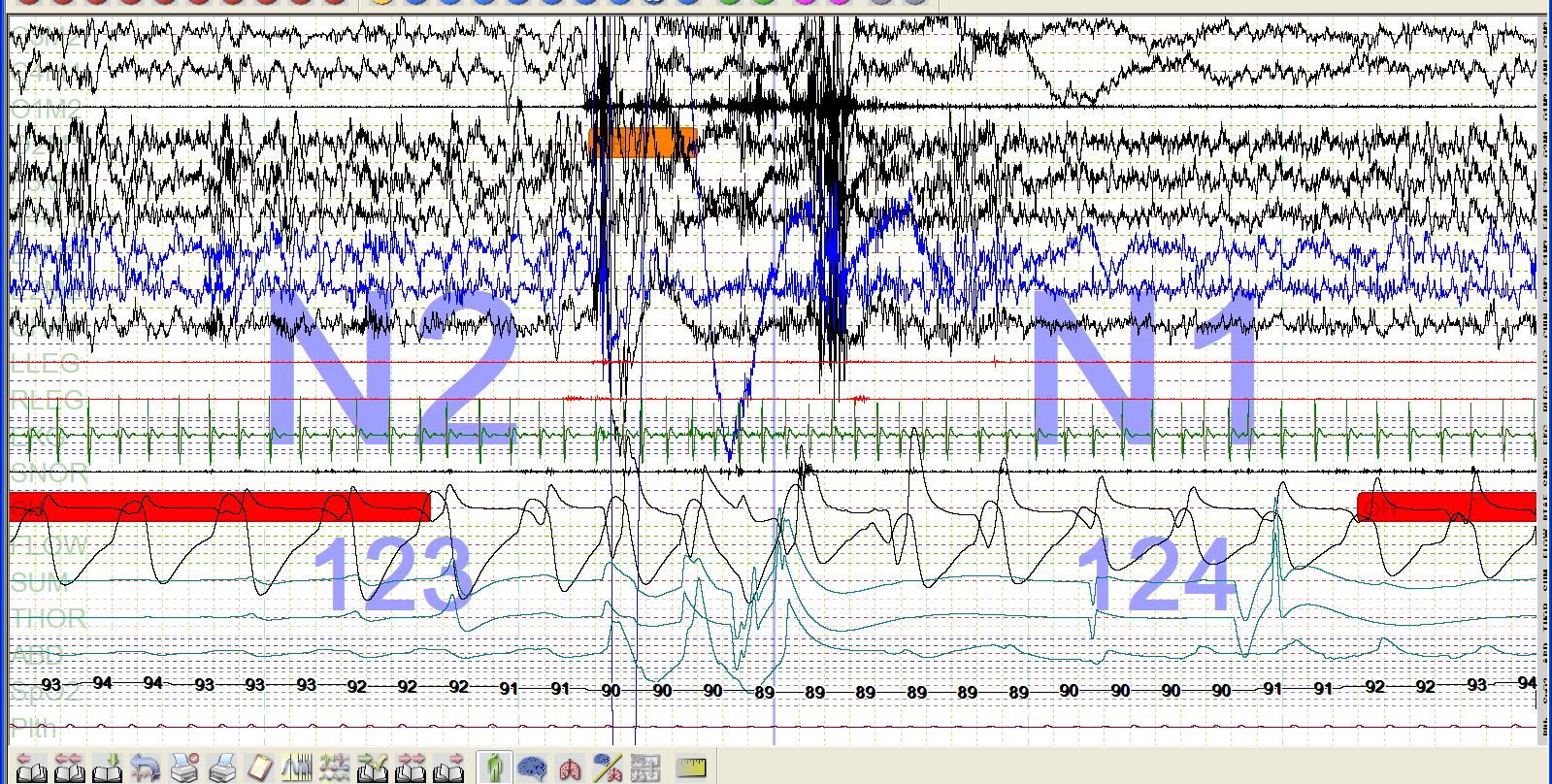
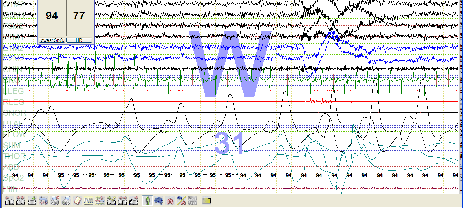
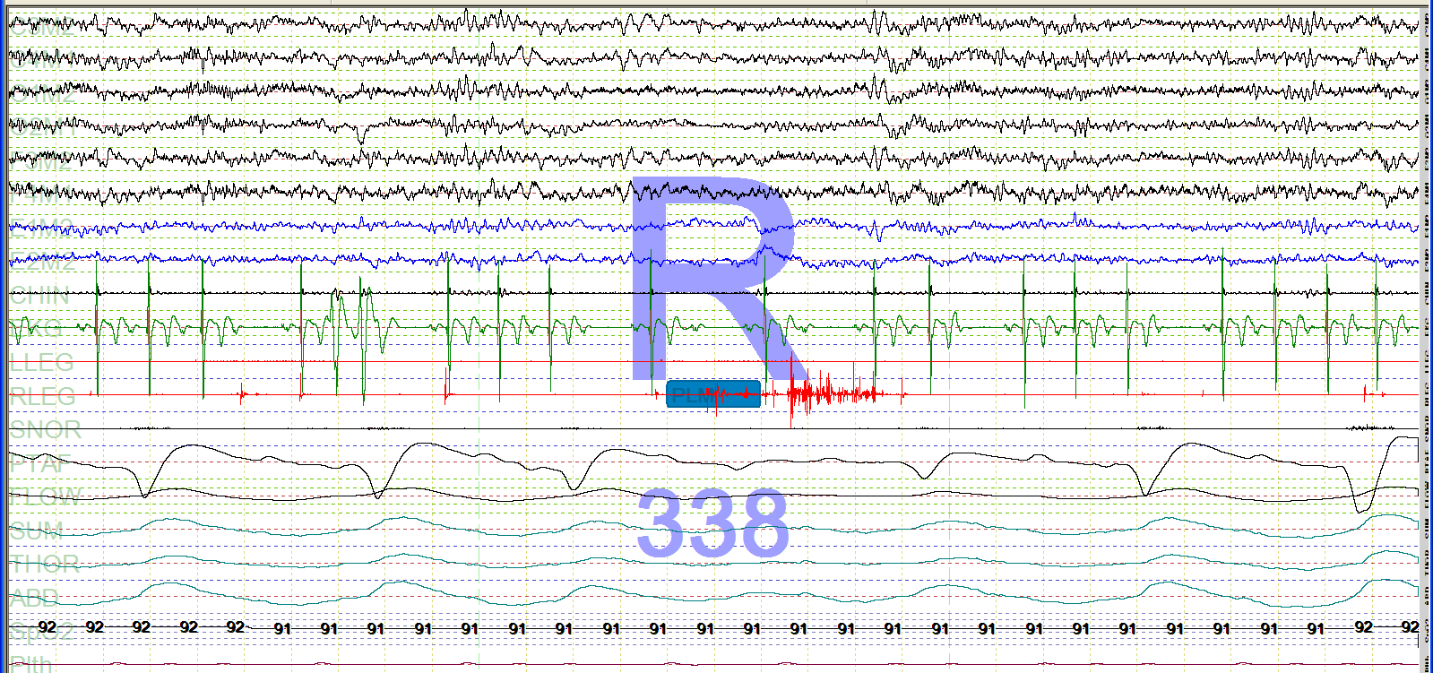
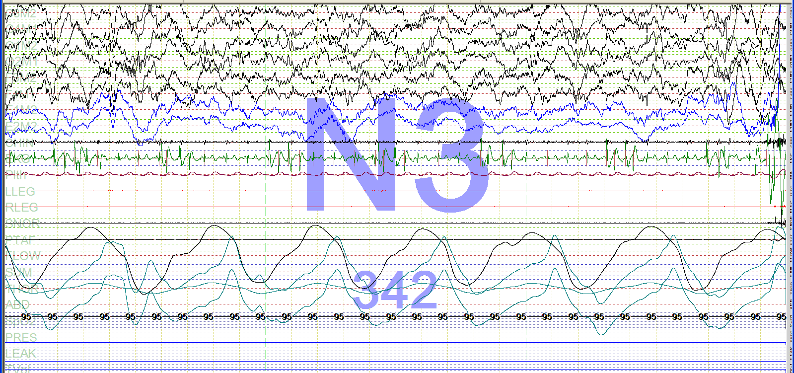
7
8 EKG Basics
9 EKG= Electrocardiogram= ECG
10 ECG Intervals and Waves-KH Frank G. Yanowitz, M.D., copyright 1997 The EKG complex
11 Cardiac Conduction System Diagram - Marquette-KH Marquette Electronics Copyright 1996
12 Depolarization = muscle contraction = Systole Repolarization = muscle relaxation = Diasystole

13 12 lead EKG Rate Rhythm Axis Ischemia, hypertrophy, etc. Frontal and Horizontal Plane Lead Diagram-KH Frank G. Yanowitz, M.D.
14 Rate Rhythm? Holter monitor (3 channels) Channel/lead Rhythm Strip
15 (a) 25 seconds snap shot from sleep study in patient number 2 during wakefulness. White arrows show the beats that were initially reported as non-conducted P-waves. Upper channels (C3-A1) (01-A1): electroencephalogram; third and fourth channel (L-EOG-A1) (R-EOG- A1): oculogram; fifth channel (EMG1) (EMG2): electromyogram; sixth channel (EKG1) (EKG2): electrocardiogram; seventh channel (SaO2): oxygen saturation; eighth channel (LEG1) (LEG2): leg movement; ninth channel: airflow; tenth channel: chest movement; eleventh channel: abdomen movement. (Panel (b)) Amplification of the area under the highlighted rectangle in panel (a). White arrows show the PVCs followed by a post-extrasystolic pause. (Panel (c)) 12-lead ECG shows right bundle branch block. White arrows show PVCs arising probably from the left outflow tract. Truths and Lies from the Polysomnography ECG Recording: An Electrophysiologist Perspective Case Report Med May 4; 2009:675078
16 Communication of an abnormal EKG There is a (wide or narrow) complex (regular or irregular) rhythm with a heart rate of. There is a beat run of (wide or narrow) complex (regular or irregular) rhythm with a heart rate of. There is/are second pause(s).
17 EKG Interpretation Here s what you need to determine/describe Heart rate Morphology of QRS complex (narrow v. wide) Rhythm: regular or irregular Identify Premature beats Tachycardia Bradycardia Pause/arrest/asystole Atrial fibrillation
18 EKG Interpretation 1. Heart rate 2. Morphology of QRS complex (narrow v. wide) 3. Rhythm: regular or irregular
19 First: Determine Heart Rate (Beats Per Minute) Do you believe your PSG software? (it counts QRS complexes) Do you believe your pulse oximeter? (it uses plethysmography)
20 Determine Heart Rate (Beats Per Minute: there are 60 seconds in a minute!) Count the QRS complexes in your EKG: # QRS complexes in 60 seconds = BPM # QRS complexes in 30 seconds X2 = BPM # QRS complexes in 20 seconds X3 = BPM # QRS complexes in 15 seconds X4 = BPM # QRS complexes in 10 seconds X6 = BPM # QRS complexes in 6 seconds X10 = BPM # QRS complexes in 5 seconds X12 = BPM # QRS complexes in 4 seconds X15 = BPM # QRS complexes in 3 seconds X20 = BPM # QRS complexes in 2 seconds X30 = BPM # QRS complexes in 1 seconds X60 = BPM
21 Determine Heart Rate: Eyeballing It One second One compex per second = 60 BPM (1X60) More than one complex per second is >60 BPM Less than one complex per second is <60 BPM
22 Practice heart rate (5 seconds) (one second) Heart rate of the whole strip: 6 complexes x 12 = 72 Heart rate of the triplet: 3 complexes x 60 = 180
23 EKG Interpretation 1. Heart rate 2. Morphology of QRS complex (narrow v. wide) 3. Rhythm: regular or irregular
24 QRS Morphology Narrow = Normal Wide = Abnormal
25 Narrow (QRS) Complex vs. Wide (QRS) Complex Narrow complex QRS interval is < 0.12 sec Originating from above or at the AV node Wide complex QRS interval is > 0.12 sec Intraventricular conduction delay
26 Wide QRS Complexes Are Abnormal Represent Abnormal conduction through the ventricles LBBB, RBBB, fascicular blocks Intraventricular conduction delay Primary (pacemaker) of ventricular origin Premature Ventricular Contraction (PVC) Idioventricular rhythm Ventricular tachycardia (VT) Ventricular fibrillation (VF) Artificial pacemaker (ventricular)
27 Example of wide complex QRS


28 How can I tell? Narrow Complex EEG spike Wide Complex EEG K complex
29 EKG Interpretation 1. Heart rate 2. Morphology of QRS complex (narrow v. wide) 3. Rhythm: regular or irregular
30 Regular rhythm: R-R interval is constant (measure distance or time)
31 Irregular rhythm (includes premature beats)
32 Regular v. Irregular Rhythm
33 Sinus Rhythm or Not? Look for the P waves! Regular v. Irregular Rhythm
34 Identify Premature beats Tachycardia Bradycardia Pause/arrest/asystole Atrial fibrillation
35 Premature Beats: occur earlier than your next predicted QRS complex Premature Atrial Contractions (PACs) Narrow (usually) Look like EEG spikes Premature Ventricular Contractions (PVCs) Wide complex Look like K complexes
36 Premature Atrial Contractions (PACs)
37 Premature Ventricular Contractions (PVCs)
38 PVC patterns are important to recognize: abnormal PVC rhythms increase the risk of having deadly ventricular rhythms such as Venticular Tachycardia (VT) and Ventricular Fibrillation (VF)
39 Bigeminy (every other beat is a PVC) Trigeminy (every third beat is a PVC)
40 Couplet More than 3 PVCs = Ventricular tachycardia (VT)
41

42

43

44
45
46 Identify Premature beats Tachycardia (heart rate greater than 100) Bradycardia Pause/arrest/asystole Atrial fibrillation
47 Wide complex tachycardias are VERY BAD
48 Sustained Ventricular Tachycardia (VT)
49 Non-Sustained Ventricular Tachycardia (NSVT) 6 second strip 3 seconds Heart rate of whole strip: 120 (12 x 10) Heart rate of burst of NSVT: 160 (8 x 20)
50 Ventricular Fibrillation (VF or Vfib) (often looks like sawtooth waves!)
51 VF: Torsades de Pointe (think of CSR)
52 Holter monitor recording showing ventricular tachycardia degenerating to ventricular fibrillation. HR, heart rate. Cleveland Clinic
53 Ventricular Tachycardia (VT) and Ventricular Fibrillation (VF)

54 Vfib Shocked
55 AICD: Automatic Implantable Cardioverter Defibrillator Indicated in patients with previous VT, VF, sudden cardiac death or increased risk of ventricular arrhythmias Often has pacemaker ability Safe for health care providers to perform CPR, etc. Send the patient to the ED if the device discharges
56 Narrow Complex Tachycardia
57 Sinus Tachycardia v. SVT (Supraventricular Tachycardia) (10 second strip) Heart rate is 25 x 6 = 150
58 Burst of SVT: often seen in association with arousals following respiratory events 10 second strip 3 seconds Heart rate of whole strip: 78 (13 x 6) Heart rate of SVT: 200 (10 x 20)

59
60 Identify Premature beats Tachycardia Bradycardia (heart rate less than 60) Pause/arrest/asystole Atrial fibrillation
61 Bradycardia: wide or narrow? P waves or not? (6 second strip) 4 QRS complexes X 10 = 40 BPM
62 Identify Premature beats Tachycardia Bradycardia Pause/arrest/asystole Atrial fibrillation
63 One Second Pause
64 PAUSE/ARREST/ASYSTOLE (3.5 seconds) Greater than 3 second pause is considered asystole
65
66 Sick Sinus Syndrome Narrow complex irregular rhythm with frequent long pauses and escape beats.
67 Artificial Pacemakers

68
69 Atrial Pacemaker: spike instead of P wave
70 Electronic Ventricular Pacemaker Rhythm - Marquette-KH Marquette Electronics Copyright 1996
71 AV Sequential Pacemaker - Marquette-KH Marquette Electronics Copyright 1996
72 Identify Premature beats Tachycardia Bradycardia Pause/arrest/asystole Atrial fibrillation
73 Atrial Fibrillation (Afib) Atrial Fibrillation With Moderate Ventricular Response - Marquette-KH Marquette Electronics Copyright 1996
74 Atrial Fibrillation with Rapid Ventricular Response (RVR) is a type of Supraventricular Tachycardia (SVT)
75 Abnormal EKG? Make sure that it s real! Artifact can look like arrhythmias and even asystole Lead on the patient? Look in another channel/lead Asystole artifact: look for your pleth wave tracing or be happy that you have EKG artifact in your EEG
76

77
78

79
80
81 When to Worry: The Threatening Threes Heart rate in the triple digits (> 100) Tachycardia of more than 3 beats in a row Heart rate in the 30s (or less!) Pause of greater than 3 seconds More than 6 PVCs per minute (or more than 3 PVCs per 30 second epoch) (Irregularly irregular heart rhythm)
82 What do I do now? Confirm that the arrhythmia is real Check on the patient (ABCs) Call a code (or 911) if unstable If stable and unsymptomatic, call physician coverage if unsure about severity or transfer to ED Know your emergency policies and procedures Document well and sign out your findings
83 Communication of an abnormal EKG There is a (wide or narrow) complex (regular or irregular) rhythm with a heart rate of. There is a beat run of (wide or narrow) complex (regular or irregular) rhythm with a heart rate of. There is/are second pause(s).
84 (Free!) References The Alan E. Lindsay ECG Learning Center in Cyberspace ECGPedia LearnTheHeart.com
85 (6 seconds) Narrow or wide complex? Regular or irregular? Heart rate? What is it? Narrow Irregular 80 NSR with pauses
86 (6 seconds) Narrow or wide complex? Regular or irregular? Heart rate? What is it? Wide complex Regular 140 VT until proven otherwise
87 (6 seconds) Narrow or wide complex? Regular or irregular? Heart rate? What is it? Narrow Irregular 50 Atrial fibrillation (Afib)
88 (6 second strip) Narrow or wide complex? Regular or irregular? Heart rate? What is it? Wide Regular 50 Wide complex bradycardia without P waves (likely idioventricular)
89 (6 second strip) Narrow or wide complex? Regular or irregular? Heart rate? What is it? Narrow Irregular 110 Sinus tachycardia with two unifocal PVCs
NEONATAL & PEDIATRIC ECG BASICS RHYTHM INTERPRETATION
 NEONATAL & PEDIATRIC ECG BASICS & RHYTHM INTERPRETATION VIKAS KOHLI MD FAAP FACC SENIOR CONSULATANT PEDIATRIC CARDIOLOGY APOLLO HOSPITAL MOB: 9891362233 ECG FAX LINE: 011-26941746 THE BASICS: GRAPH PAPER
ACLS RHYTHM TEST. 2. A 74-year-old woman with chest pain. Blood pressure 192/90 and rates her pain 9/10.
 ACLS RHYTHM TEST Name Date Choose the best answer for each of the following questions. Each of the following strips is 6 seconds in length. 1. Identify the following rhythm a. Sinus bradycardia with 2
By the end of this continuing education module the clinician will be able to:
 EKG Interpretation WWW.RN.ORG Reviewed March, 2015, Expires April, 2017 Provider Information and Specifics available on our Website Unauthorized Distribution Prohibited 2015 RN.ORG, S.A., RN.ORG, LLC Developed
INTRODUCTORY GUIDE TO IDENTIFYING ECG IRREGULARITIES
 INTRODUCTORY GUIDE TO IDENTIFYING ECG IRREGULARITIES NOTICE: This is an introductory guide for a user to understand basic ECG tracings and parameters. The guide will allow user to identify some of the
QRS Complexes. Fast & Easy ECGs A Self-Paced Learning Program
 6 QRS Complexes Fast & Easy ECGs A Self-Paced Learning Program Q I A ECG Waveforms Normally the heart beats in a regular, rhythmic fashion producing a P wave, QRS complex and T wave I Step 4 of ECG Analysis
MULTIPLE CHOICE. Choose the one alternative that best completes the statement or answers the question.
 Exam Name MULTIPLE CHOICE. Choose the one alternative that best completes the statement or answers the question. 1) What term is used to refer to the process of electrical discharge and the flow of electrical
the basics Perfect Heart Institue, Piyavate Hospital
 ECG INTERPRETATION: the basics Damrong Sukitpunyaroj MD Damrong Sukitpunyaroj, MD Perfect Heart Institue, Piyavate Hospital Overview Conduction Pathways Systematic Interpretation Common abnormalities in
Normal Sinus Rhythm. Sinus Bradycardia. Sinus Tachycardia. Rhythm ECG Characteristics Example (NSR) & consistent. & consistent.
 & consistent. & consistent.") Normal Sinus Rhythm (NSR) Rate: 60-100 per minute Rhythm: R- R = P waves: Upright, similar P-R: 0.12-0.20 second & consistent P:qRs: 1P:1qRs Sinus Tachycardia Exercise Hypovolemia Medications Fever Hypoxia
Normal Sinus Rhythm (NSR) Rate: 60-100 per minute Rhythm: R- R = P waves: Upright, similar P-R: 0.12-0.20 second & consistent P:qRs: 1P:1qRs Sinus Tachycardia Exercise Hypovolemia Medications Fever Hypoxia
Atrial & Junctional Dysrhythmias
 Atrial & Junctional Dysrhythmias Atrial & Junctional Dysrhythmias Atrial Premature Atrial Complex Wandering Atrial Pacemaker Atrial Tachycardia (ectopic) Multifocal Atrial Tachycardia Atrial Flutter Atrial
Understanding the Electrocardiogram. David C. Kasarda M.D. FAAEM St. Luke s Hospital, Bethlehem
 Understanding the Electrocardiogram David C. Kasarda M.D. FAAEM St. Luke s Hospital, Bethlehem Overview 1. History 2. Review of the conduction system 3. EKG: Electrodes and Leads 4. EKG: Waves and Intervals
BASIC CARDIAC ARRHYTHMIAS Revised 10/2001
 BASIC CARDIAC ARRHYTHMIAS Revised 10/2001 A Basic Arrhythmia course is a recommended prerequisite for ACLS. A test will be given that will require you to recognize cardiac arrest rhythms and the most common
PRO-CPR. 2015 Guidelines: PALS Algorithm Overview. (Non-AHA supplementary precourse material)
") PRO-CPR 2015 Guidelines: PALS Algorithm Overview (Non-AHA supplementary precourse material) Please reference Circulation (from our website), the ECC Handbook, or the 2015 ACLS Course Manual for correct
PRO-CPR 2015 Guidelines: PALS Algorithm Overview (Non-AHA supplementary precourse material) Please reference Circulation (from our website), the ECC Handbook, or the 2015 ACLS Course Manual for correct
Diagnosis Code Crosswalk : ICD-9-CM to ICD-10-CM Cardiac Rhythm and Heart Failure Diagnoses
 Diagnosis Code Crosswalk : to 402.01 Hypertensive heart disease, malignant, with heart failure 402.11 Hypertensive heart disease, benign, with heart failure 402.91 Hypertensive heart disease, unspecified,
Activity 4.2.3: EKG. Introduction. Equipment. Procedure
 Activity 4.2.3: EKG The following is used with permission of Vernier Software and Technology. This activity is based on the experiment Analyzing the Heart with EKG from the book Human Physiology with Vernier,
RAPID INTERPRETATION OF. EKG s
 Personal Quick Reference Sheets 333 (pages 333 to 346) There is no need to remove these reference pages from your book. To download and print them in full color, go to: www.themdsite.com Reference Sheets
How to read the ECG in athletes: distinguishing normal form abnormal
 How to read the ECG in athletes: distinguishing normal form abnormal Antonio Pelliccia, MD Institute of Sport Medicine and Science www.antoniopelliccia.it Cardiac adaptations to Rowing Vagotonia Sinus
Tachyarrhythmias (fast heart rhythms)
") Patient information factsheet Tachyarrhythmias (fast heart rhythms) The normal electrical system of the heart The heart has its own electrical conduction system. The conduction system sends signals throughout
Patient information factsheet Tachyarrhythmias (fast heart rhythms) The normal electrical system of the heart The heart has its own electrical conduction system. The conduction system sends signals throughout
Introduction to Electrocardiography. The Genesis and Conduction of Cardiac Rhythm
 Introduction to Electrocardiography Munther K. Homoud, M.D. Tufts-New England Medical Center Spring 2008 The Genesis and Conduction of Cardiac Rhythm Automaticity is the cardiac cell s ability to spontaneously
The abbreviation EKG, for electrocardiogram,
 CLIN PEDIATR OnlineFirst, published on January 28, 2010 as doi:10.1177/0009922809336206 Simplified Pediatric Electrocardiogram Interpretation Clinical Pediatrics Volume XX Number X Month XXXX xx-xx 2009
Electrophysiology Heart Study - EPS -
 Electrophysiology Heart Study - EPS - What is an EPS? EPS is short for ElectroPhysiology heart Study. This procedure looks at the electrical system of your heart. An EPS will show if you have a heart rhythm
HTEC 91. Topic for Today: Atrial Rhythms. NSR with PAC. Nonconducted PAC. Nonconducted PAC. Premature Atrial Contractions (PACs)
") HTEC 91 Medical Office Diagnostic Tests Week 4 Topic for Today: Atrial Rhythms PACs: Premature Atrial Contractions PAT: Paroxysmal Atrial Tachycardia AF: Atrial Fibrillation Atrial Flutter Premature Atrial
HTEC 91 Medical Office Diagnostic Tests Week 4 Topic for Today: Atrial Rhythms PACs: Premature Atrial Contractions PAT: Paroxysmal Atrial Tachycardia AF: Atrial Fibrillation Atrial Flutter Premature Atrial
Electrophysiology Daymar College. Lisa H. Young, RN, BSN, MAE 2011
 Electrophysiology Daymar College Lisa H. Young, RN, BSN, MAE 2011 Electrical Conduction Pathway Chemical Basis for Impulse Formation Cardiac Action Potential Phases http://www.youtube.com/watch?v=oqpffilde0e
1 Meet Your AliveCor Heart Monitor
 GETTING STARTED HOW TO RECORD YOUR FIRST ECG 1 Meet Your AliveCor Heart Monitor UNPACK Take your AliveCor out of the box. The device is already connected to an attachment plate that can adhere to the back
Premature Ventricular Contractions. Ralph Augostini, MD FACC FHRS
 Premature Ventricular Contractions Ralph Augostini, MD FACC FHRS Orlando, Florida October 7-9, 2011 Premature Ventricular Contractions: ACC/AHA/ESC 2006 Guidelines for Management of Patients With Ventricular
Basics of Pacing. Ruth Hickling, RN-BSN Tasha Conley, RN-BSN
 Basics of Pacing Ruth Hickling, RN-BSN Tasha Conley, RN-BSN The Cardiac Conduction System Cardiac Conduction System Review Normal Conduction Conduction QRS QRS Complex Complex RR PP ST ST segment segment
Electrodes placed on the body s surface can detect electrical activity, APPLIED ANATOMY AND PHYSIOLOGY. Circulatory system
 4 READING AND INTERPRETING THE ELECTROCARDIOGRAM Electrodes placed on the body s surface can detect electrical activity, which occurs in the heart. The recording of these electrical events comprises an
Arrhythmia Monitoring Algorithm
 Arrhythmia Monitoring Algorithm Application Note Introduction The ST/AR (ST and Arrhythmia) algorithm is an ECG algorithm that the HeartStart MRx and XL+ monitor/defibrillators utilize for basic and cardiotach
ACLS Chapter 3 Rhythm Review Instructor Lesson Plan to Accompany ACLS Study Guide 3e
 ACLS Chapter 3 Rhythm Review Lesson Plan Required reading before this lesson: ACLS Study Guide 3e Textbook Chapter 3 Materials needed: Multimedia projector, computer, ACLS Chapter 3 Recommended minimum
How To Understand What You Know
 Heart Disorders Glossary ABG (Arterial Blood Gas) Test: A test that measures how much oxygen and carbon dioxide are in the blood. Anemia: A condition in which there are low levels of red blood cells in
12-Lead EKG Interpretation. Judith M. Haluka BS, RCIS, EMT-P
 12-Lead EKG Interpretation Judith M. Haluka BS, RCIS, EMT-P ECG Grid Left to Right = Time/duration Vertical measure of voltage (amplitude) Expressed in mm P-Wave Depolarization of atrial muscle Low voltage
School of Health Sciences
 School of Health Sciences Cardiology Teaching Package A Beginners Guide to Normal Heart Function, Sinus Rhythm & Common Cardiac Arrhythmias Welcome This document extends subjects covered in the Cardiology
Evaluation copy. Analyzing the Heart with EKG. Computer
 Analyzing the Heart with EKG Computer An electrocardiogram (ECG or EKG) is a graphical recording of the electrical events occurring within the heart. In a healthy heart there is a natural pacemaker in
ECG Measurments and Interpretation Programs
 ECG Measurments and Interpretation Programs Physician s Guide Distributed by Welch Allyn 4341 State Street Road, PO Box 220 Skaneateles Falls, NY 13153-0220 www.welchallyn.com Sales and Service information:
Table of Contents Error! Bookmark not defined.
 Table of Contents EKG TRACING...1 Figure 1 - EKG Tracing... Error! Bookmark not defined. STEP 1...1 Rate... 1 Figure 2 - Determining the Rate... 1 Step 2...2 Rhythm... 2 Figure 3 - Determining the Rhythm
ECG made extra easy. medics.cc
 ElectroCardioGraphyraphy ECG made extra easy Overview Objectives for this tutorial What is an ECG? Overview of performing electrocardiography on a patient Simple physiology Interpreting the ECG Objectives
Wide-Complex Tachycardias in the ED: Myths and Pitfalls
 Wide-Complex Tachycardias in the ED: Myths and Pitfalls, FACEP, FAAEM Professor and Vice Chair Director, Emergency Cardiology Fellowship Department of Emergency Medicine University of Maryland School of
Electrocardiography Review and the Normal EKG Response to Exercise
 Electrocardiography Review and the Normal EKG Response to Exercise Cardiac Anatomy Electrical Pathways in the Heart Which valves are the a-v valves? Closure of the a-v valves is associated with which heart
Electrocardiography I Laboratory
 Introduction The body relies on the heart to circulate blood throughout the body. The heart is responsible for pumping oxygenated blood from the lungs out to the body through the arteries and also circulating
An Introduction to Tachyarrhythmias R. A. Seyon MN, NP, CCN(C) & Dr. R. G. Williams
 & Dr. R. G. Williams") Arrhythmias 1 An Introduction to Tachyarrhythmias R. A. Seyon MN, NP, CCN(C) & Dr. R. G. Williams Things to keep in mind when analyzing arrhythmias: Electrical activity recorded in 12 and 15 leads Examine
Arrhythmias 1 An Introduction to Tachyarrhythmias R. A. Seyon MN, NP, CCN(C) & Dr. R. G. Williams Things to keep in mind when analyzing arrhythmias: Electrical activity recorded in 12 and 15 leads Examine
Copyright 2006 Blaufuss Multimedia. All rights reserved. Page 1
 Copyright 2006 Blaufuss Multimedia. All rights reserved. Page 1 002 Sinus Rhythm, atrial rate 90 Mobitz II AVB, Ventricular rate 50 Left Atrial Enlargement Left Ventricular Hypertrophy RBBB a) Long R-R
Equine Cardiovascular Disease
 Equine Cardiovascular Disease 3 rd most common cause of poor performance in athletic horses (after musculoskeletal and respiratory) Cardiac abnormalities are rare Clinical Signs: Poor performance/exercise
Feature Vector Selection for Automatic Classification of ECG Arrhythmias
 Feature Vector Selection for Automatic Classification of ECG Arrhythmias Ch.Venkanna 1, B. Raja Ganapathi 2 Assistant Professor, Dept. of ECE, G.V.P. College of Engineering (A), Madhurawada, A.P., India
Sleep Heart Health Study (SHHS) ECG Protocol
 ECG Protocol") Sleep Heart Health Study (SHHS) ECG Protocol SHHS 1 Electrocardiography (ECG) Baseline ECG is performed in all parent study clinic visits preceding the PSG. All sites perform a standard resting 12-lead
Sleep Heart Health Study (SHHS) ECG Protocol SHHS 1 Electrocardiography (ECG) Baseline ECG is performed in all parent study clinic visits preceding the PSG. All sites perform a standard resting 12-lead
Basic Cardiac Rhythms Identification and Response
 Basic Cardiac Rhythms Identification and Response Module 1 ANATOMY, PHYSIOLOGY, & ELECTRICAL CONDUCTION Objectives Describe the normal cardiac anatomy and physiology and normal electrical conduction through
BIPOLAR LIMB LEADS UNIPOLAR LIMB LEADS PRECORDIAL (UNIPOLAR) LEADS VIEW OF EACH LEAD INDICATIVE ECG CHANGES
 LEADS VIEW OF EACH LEAD INDICATIVE ECG CHANGES") BIPOLAR LIMB LEADS Have both a distinctive positive and negative pole. Lead I LA (positive) RA (negative) Lead II LL (positive) RA (negative) Lead III LL (positive) LA (negative) UNIPOLAR LIMB LEADS Have
BIPOLAR LIMB LEADS Have both a distinctive positive and negative pole. Lead I LA (positive) RA (negative) Lead II LL (positive) RA (negative) Lead III LL (positive) LA (negative) UNIPOLAR LIMB LEADS Have
Banner Staff Service ECG Study Guide
 Banner Staff Service ECG Study Guide Edited by Larry H. Lybbert, MS, RN Table of Contents ECG STUDY GUIDE... 3 ECG INTERPRETATION BASICS... 4 EKG GRAPH PAPER...4 RATE MEASUREMENT...9 The Six Second Method...9
HEART HEALTH WEEK 3 SUPPLEMENT. A Beginner s Guide to Cardiovascular Disease HEART FAILURE. Relatively mild, symptoms with intense exercise
 WEEK 3 SUPPLEMENT HEART HEALTH A Beginner s Guide to Cardiovascular Disease HEART FAILURE Heart failure can be defined as the failing (insufficiency) of the heart as a mechanical pump due to either acute
VCA Veterinary Specialty Center of Seattle
 An electrocardiogram (ECG) is a graph of the heart`s electrical current, which allows evaluation of heart rate, rhythm and conduction. Identification of conduction problems within the heart begins with
Systematic Approach to 12 Lead EKG Interpretation
 Systematic Approach to 12 Lead EKG Interpretation Maureen Knechtel MPAS, PA-C Wellmont CVA Heart Institute Disclosure Statement of Financial Interest I, Maureen Knechtel, do not have a financial interest/arrangement
Signal-averaged electrocardiography late potentials
 SIGNAL AVERAGED ECG INTRODUCTION Signal-averaged electrocardiography (SAECG) is a special electrocardiographic technique, in which multiple electric signals from the heart are averaged to remove interference
Current Management of Atrial Fibrillation DISCLOSURES. Heart Beat Anatomy. I have no financial conflicts to disclose
 Current Management of Atrial Fibrillation Mary Macklin, MSN, APRN Concord Hospital Cardiac Associates DISCLOSURES I have no financial conflicts to disclose Book Women: Fit at Fifty. A Guide to Living Long.
Pacemakers 12/04. Pacemakers. 1. What is a pacemaker?
 Pacemakers 12/04 1-What is a pacemaker? 2- What does intrinsic mean? 3- How exactly do pacemakers work on the heart? 4- What are the parts of a pacemaker? 5- Are there different kinds of pacemakers? 6-
«Δυσλειτουργία βηματοδότη. Πως μπορούμε να την εκτιμήσουμε στο ιατρείο.» Koσσυβάκης Χάρης Καρδιολογικό Τμήμα Γ.Ν.Α. «Γ. ΓΕΝΝΗΜΑΤΑΣ
 «Δυσλειτουργία βηματοδότη. Πως μπορούμε να την εκτιμήσουμε στο ιατρείο.» Koσσυβάκης Χάρης Καρδιολογικό Τμήμα Γ.Ν.Α. «Γ. ΓΕΝΝΗΜΑΤΑΣ Diagnostic tools History: symptoms, physical examination 12 leads ECG,
QT analysis: A guide for statistical programmers. Prabhakar Munkampalli Statistical Analyst II Hyderabad, 7 th September 2012
 QT analysis: A guide for statistical programmers Prabhakar Munkampalli Statistical Analyst II Hyderabad, 7 th September 2012 Agenda ECG ICH E14 Thorough QT/QTc study Role of Statistical Programmer References
Electrophysiology Introduction, Basics. The Myocardial Cell. Chapter 1- Thaler
 Electrophysiology Introduction, Basics Chapter 1- Thaler The Myocardial Cell Syncytium Resting state Polarized negative Membrane pump Depolarization fundamental electrical event of the heart Repolarization
The new generation in ECG interpretation
 The new generation in ECG interpretation Philips DXL ECG Algorithm, Release PH100B The Philips DXL ECG Algorithm, developed by the Advanced Algorithm Research Center, uses sophisticated analytical methods
Trust Guideline for the Management of New-born Babies with abnormal heart rhythm
 A clinical guideline recommended for use In: Blakeney Ward, Delivery Suite, Neonatal Unit By: For: Key words: Written by: Supported by: Approved by: Neonatal Medical Staff (SHOs, registrars, consultants)
Atrial Fibrillation and Cardiac Device Therapy RAKESH LATCHAMSETTY, MD DIVISION OF ELECTROPHYSIOLOGY UNIVERSITY OF MICHIGAN HOSPITAL ANN ARBOR, MI
 Atrial Fibrillation and Cardiac Device Therapy RAKESH LATCHAMSETTY, MD DIVISION OF ELECTROPHYSIOLOGY UNIVERSITY OF MICHIGAN HOSPITAL ANN ARBOR, MI Outline Atrial Fibrillation What is it? What are the associated
What Are Arrhythmias?
 What Are Arrhythmias? Many people have questions about what the word arrhythmia means, and arrhythmias can be a difficult subject to understand. The text below should give you a better understanding of
Atrioventricular (AV) node ablation
 node ablation") Patient information factsheet Atrioventricular (AV) node ablation The normal electrical system of the heart The heart has its own electrical conduction system. The conduction system sends signals throughout
Patient information factsheet Atrioventricular (AV) node ablation The normal electrical system of the heart The heart has its own electrical conduction system. The conduction system sends signals throughout
Management of Pacing Wires After Cardiac Surgery
 Management of Pacing Wires After Cardiac Surgery David E. Lizotte, Jr. PA C, MPAS, FAPACVS President, Association of Physician Assistants in Cardiovascular Surgery Conflicts: None Indications 2008 Journal
2 ECG basics. Leads and planes. Leads. Planes. from different perspectives, which are called leads and planes.
 558302.qxp 3/14/12 10:47 PM Page 12 2 ECG basics One of the most valuable diagnostic tools available, an electrocardiogram (ECG) records the heart s electrical activity as waveforms. By interpreting these
The choice for quality ECG arrhythmia monitoring
 GE Healthcare EK-Pro The choice for quality ECG arrhythmia monitoring David A. Sitzman, MSEE. Mikko Kaski, MSAM. Ian Rowlandson, MSBE. Tarja Sivonen, RN. Olli Väisänen, MD, PhD. Clinical care environments
DIAGNOSING SLEEP APNEA. Christie Goldsby Florida State University PHY 3109 04/09/14
 DIAGNOSING SLEEP APNEA Christie Goldsby Florida State University PHY 3109 04/09/14 Outline of Talk Background information -what is sleep apnea? Diagnosing sleep apnea -polysomnography -respiratory airflow
The P Wave: Indicator of Atrial Enlargement
 Marquette University e-publications@marquette Physician Assistant Studies Faculty Research and Publications Health Sciences, College of 8-12-2010 The P Wave: Indicator of Atrial Enlargement Patrick Loftis
Bradycardia CHAPTER 12 CODE SCENARIO
 Senecal-12.qxd 14/04/2005 09:44 AM Page 69 CHAPTER 12 Bradycardia CODE SCENARIO A code is called for a 78-year-old man who was admitted to the hospital for syncope of unknown etiology. He was resting comfortably
About the British Heart Foundation
 Heart rhythms About the British Heart Foundation The British Heart Foundation (BHF) is the nation s heart charity, saving lives through pioneering research, patient care and vital information. What you
What Can I Do about Atrial Fibrillation (AF)?
?") Additional Device Information 9529 Reveal XT Insertable Cardiac Monitor The Reveal XT Insertable Cardiac Monitor is an implantable patientactivated and automatically activated monitoring system that records
Additional Device Information 9529 Reveal XT Insertable Cardiac Monitor The Reveal XT Insertable Cardiac Monitor is an implantable patientactivated and automatically activated monitoring system that records
Detecting Atrial-ventricular blocks Arrhythmia based on RR-intervals on ECG Signals
 Detecting Atrial-ventricular blocks Arrhythmia based on -intervals on ECG Signals Makki Akasha abikier, Ibrahim Musa Ishag, Mohammed Izzeldin, Dong Gyu Lee, Gyoyong shon, Keun Ho Ryu Database/ioinformatics
The heart then repolarises (or refills) in time for the next stimulus and contraction.
 in time for the next stimulus and contraction.") Atrial Fibrillation BRIEFLY, HOW DOES THE HEART PUMP? The heart has four chambers. The upper chambers are called atria. One chamber is called an atrium, and the lower chambers are called ventricles. In
Atrial Fibrillation BRIEFLY, HOW DOES THE HEART PUMP? The heart has four chambers. The upper chambers are called atria. One chamber is called an atrium, and the lower chambers are called ventricles. In
The Patient s Guide to the Electrophysiologic Study (EPS) and Catheter Ablation
 and Catheter Ablation") The Patient s Guide to the Electrophysiologic Study (EPS) and Catheter Ablation 2 P a g e Table of Contents Introduction How the Heart Works 3 How the Heart s Electrical System Works 3 Commonly Used Heart
The Patient s Guide to the Electrophysiologic Study (EPS) and Catheter Ablation 2 P a g e Table of Contents Introduction How the Heart Works 3 How the Heart s Electrical System Works 3 Commonly Used Heart
Patient Information Sheet Electrophysiological study
 Patient Information Sheet Electrophysiological study Your doctor has recommended performing an electrophysiological study (also called EPS). EPS is a diagnostic procedure designed to test and evaluate
Electrocardiographic Issues in Williams Syndrome
 Electrocardiographic Issues in Williams Syndrome R. Thomas Collins II, MD Assistant Professor, Pediatrics and Internal Medicine University of Arkansas for Medical Sciences Arkansas Children s Hospital
EKG Technician Program TST Tuition - $999; Total Hours 50
 EKG Technician Program TST Tuition - $999; Total Hours 50 January 25 January 27 February 1 February 3 February 8 February 10 February 15 February 17 February 22 February 24 February 29 March 2 March 7
TOP 5. The term cardiac arrhythmia encompasses all cardiac. Arrhythmias in Dogs & Cats. Sinus Arrhythmia. TOP 5 Arrhythmias Seen in Dogs & Cats
 Top 5 ardiology Peer reviewed TOP 5 rrhythmias in Dogs & ats shley Jones, DVM mara Estrada, DVM, DVIM (ardiology) University of Florida The term cardiac arrhythmia encompasses all cardiac rhythms other
Interpreting a rhythm strip
 3 Interpreting a rhythm strip Just the facts In this chapter, you ll learn: the components of an ECG complex and their significance and variations techniques for calculating the rate and rhythm of an ECG
ECG Signal Analysis Using Wavelet Transforms
 Bulg. J. Phys. 35 (2008) 68 77 ECG Signal Analysis Using Wavelet Transforms C. Saritha, V. Sukanya, Y. Narasimha Murthy Department of Physics and Electronics, S.S.B.N. COLLEGE (Autonomous) Anantapur 515
Official Online ACLS Exam
 \ Official Online ACLS Exam Please fill out this form before you take the exam. Name : Email : Phone : 1. Hypovolemia initially produces which arrhythmia? A. PEA B. Sinus tachycardia C. Symptomatic bradyarrhythmia
Advanced Cardiovascular Life Support Case Scenarios
 Advanced Cardiovascular Life Support Case Scenarios ACLS Respiratory Arrest Case Out-of-Hospital Scenario You are a paramedic and respond to the scene of a possible cardiac arrest. A young man lies motionless
Introduction to Electrophysiology. Wm. W. Barrington, MD, FACC University of Pittsburgh Medical Center
 Introduction to Electrophysiology Wm. W. Barrington, MD, FACC University of Pittsburgh Medical Center Objectives Indications for EP Study How do we do the study Normal recordings Abnormal Recordings Limitations
DEPARTMENT OF HEALTH AND HUMAN SERVICES Centers for Medicare & Medicaid Services
 DEPARTMENT OF HEALTH AND HUMAN SERVICES Centers for Medicare & Medicaid Services NEW product from the Medicare Learning Network (MLN) Provider Compliance Tips for Computed Tomography (CT) Scans Podcast,
Electrocardiogram and Heart Sounds
 Electrocardiogram and Heart Sounds An introduction to the recording and analysis of electrocardiograms, and the sounds of the heart. Written by Staff of ADInstruments Introduction The beating of the heart
Medtronic Cardiac Rhythm and Heart Failure ICD-10 Coding for Physicians
 Medtronic Cardiac Rhythm and Heart Failure ICD-10 Coding for Physicians May 19, 2015 Disclaimer This presentation is intended for educational use. Any duplication is prohibited without written consent
ECG Measurement and Interpretation
 ECG Measurement and Interpretation Statement of accuracy for analysing ECG units *2.530036* Physicians Guide Sales and Service Information The SCHILLER sales and service centre network is world-wide. For
Electrophysiology study (EPS)
") Patient information factsheet Electrophysiology study (EPS) The normal electrical system of the heart The heart has its own electrical conduction system. The conduction system sends signals throughout
Patient information factsheet Electrophysiology study (EPS) The normal electrical system of the heart The heart has its own electrical conduction system. The conduction system sends signals throughout
American Heart Association ACLS Pre-Course Self Assessment Dec., 2006. ECG Analysis. Name the following rhythms from the list below:
 American Heart Association ACLS Pre-Course Self Assessment Dec., 2006 ECG Analysis This pre-test is exactly the same as the pretest on the ACLS Provider manual CD. This paper version can be completed in
ACLS Study Guide BLS Overview CAB
 ACLS Study Guide The ACLS Provider exam is 50-mutiple choice questions. Passing score is 84%. Student may miss 8 questions. For students taking ACLS for the first time or renewing students with a current
The Electrocardiogram (ECG)
") The Electrocardiogram (ECG) Preparation for RWM Lab Experiment The first ECG was measured by Augustus Désiré Waller in 1887 using Lippmann's capillary electrometer. Recorded ECG: http://www.youtube.com/watch_popup?v=q0jmfivadue&vq=large
The Electrocardiogram (ECG) Preparation for RWM Lab Experiment The first ECG was measured by Augustus Désiré Waller in 1887 using Lippmann's capillary electrometer. Recorded ECG: http://www.youtube.com/watch_popup?v=q0jmfivadue&vq=large
Section Four: Pulmonary Artery Waveform Interpretation
 Section Four: Pulmonary Artery Waveform Interpretation All hemodynamic pressures and waveforms are generated by pressure changes in the heart caused by myocardial contraction (systole) and relaxation/filling
Biology 347 General Physiology Lab Advanced Cardiac Functions ECG Leads and Einthoven s Triangle
 Biology 347 General Physiology Lab Advanced Cardiac Functions ECG Leads and Einthoven s Triangle Objectives Students will record a six-lead ECG from a resting subject and determine the QRS axis of the
ACLS PHARMACOLOGY 2011 Guidelines
 ACLS PHARMACOLOGY 2011 Guidelines ADENOSINE Narrow complex tachycardias or wide complex tachycardias that may be supraventricular in nature. It is effective in treating 90% of the reentry arrhythmias.
The Heart Rhythm Charity
 The Heart Rhythm Charity Promoting better understanding, diagnosis, treatment and quality of life for individuals with cardiac arrhythmias Registered Charity No. 1107496 2006 Bradycardia (Slow Heart Rhythm)
510(k) Summary May 7, 2012
 Summary May 7, 2012") 510(k) Summary Medicalgorithmics 510(k) Premarket Notification 510(k) Summary May 7, 2012 1. Submitter Name and Address Medicalgorithmics LLC 245 West 107th St., Suite 11A New York, NY 10025, USA Contact
510(k) Summary Medicalgorithmics 510(k) Premarket Notification 510(k) Summary May 7, 2012 1. Submitter Name and Address Medicalgorithmics LLC 245 West 107th St., Suite 11A New York, NY 10025, USA Contact
An ECG Primer. Quick Look. I saw it, but I did not realize it. Elizabeth Peabody
 4 An ECG Primer Quick Look Cardiac Monitoring System - p. 64 ECG Paper - p. 73 Lead Polarity and Vectors - p. 77 Basic ECG Components - p. 79 Heart Rate and Pulse Rate - p. 91 Summary - p. 94 Chapter Quiz
CARDIAC ELECTROPHYSIOLOGY, ARRHYTHMIAS AND PACING. Medical Knowledge. Goals and Objectives PF EF MF LF Aspirational
 Know the histology and gross anatomy of the normal sinoatrial node, atrial conduction pathways, atrioventricular (AV) junction and nod, His bundle, conduction fascicles and terminal intra-ventricular conduction
ECG Filtering. Willem Einthoven s EKG machine, 1903
 ECG Filtering Willem Einthoven s EKG machine, 1903 ECG Filtering Three common noise sources q Baseline wander q Power line interference q Muscle noise When filtering any biomedical signal care should be
Anatomy and Physiology: Understanding the Importance of CPR
 Anatomy and Physiology: Understanding the Importance of CPR Overview This document gives you more information about the body s structure (anatomy) and function (physiology). This information will help
Interpreting AV (Heart) Blocks: Breaking Down the Mystery
 Blocks: Breaking Down the Mystery") Interpreting AV (Heart) Blocks: Breaking Down the Mystery 2 Contact Hours Copyright 2012 by RN.com. All Rights Reserved. Reproduction and distribution of these materials is prohibited without the express
Interpreting AV (Heart) Blocks: Breaking Down the Mystery 2 Contact Hours Copyright 2012 by RN.com. All Rights Reserved. Reproduction and distribution of these materials is prohibited without the express