Digital Tomosynthesis: Advanced Breast Cancer Imaging Technique. Max Wiedmann
|
|
|
- Oswin Gilbert
- 9 years ago
- Views:
Transcription
1 Digital Tomosynthesis: Advanced Breast Cancer Imaging Technique Max Wiedmann

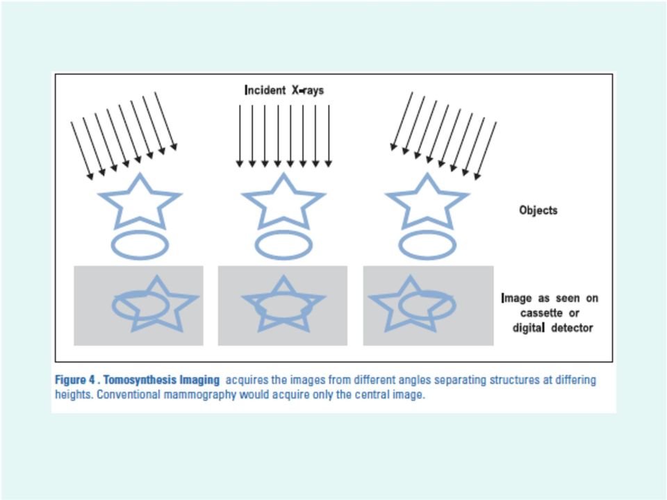
2 Digital Tomosynthesis An imaging technique in which multiple X-rays of one object are take from a discrete number angles. These cross-sectional images are used to reconstruct 3-D images of the object being scanned. Tomosynthesis differs from computed tomography because the range of angles used is less than 360, which is used in CT.

3 The leading Cause of death for women ages Is only behind lung and bronchus cancer in terms of number of deaths in US. Early detection of breast cancer is believed to save thousands of lives Breast Cancer

4 A method for detecting growths in breasts using a dedicated machine. Achieved by compressing the breast tissue to both spread it out and reduce motion blur, followed by X-ray exposure. X-rays will be absorbed to different degrees with different tissue. Bone absorbs the most while soft tissue allows the rays to pass through. Mammography

5 Mammography part 2 X-rays are produced using Bremsstrahlung, a process in which electrons are accelerated against an anode, causing photons to be fired off across a continuous spectrum. The rays that pass through the tissue cause photographic film to expose creating an image. A newer process, called full field digital mammography uses digital receptors.

6 Full Field Digital Mammography The use of a reusable digital flat panel to detect incoming X- rays. The energy from incoming photons is converted to a voltage then run through an ADC and processed. An image is generated in seconds. Digital imaging appeared later in mammography than most of radiology due to the high contrast and resolution requirement for mammograms.

7 Kieran Maher, 2000

8 Problems with Mammograms Mammograms require the breast tissue to be compressed between two plates of glass. Many women dislike the feeling, which reduces the likeliness of getting tested often. Compression causes overlapping in the breast tissue, which can obscure imaging. Mammograms typically only take 2 images at orthogonal axes. Mammograms produce false positives and false negatives. Of all biopsies taken from breasts that tested positive, only 20% came back with cancer.

9 Advantages of DTS Minimal pressure is needed, just enough to hold the breast in place. A lower dose of radiation is required, up to 50% reduction for dense breasts. The cost of DTS is expected to drop below the cost a of traditional mammogram. It is the only procedure that is expected to fully replace mammography.

10 Required Dose vs. Breast Thickness for varying anode materials Anode Material Molybdenum/Rhodium Molybdenum Tungsten/Rhodium

11 Mathematics Behind DTS Filtered back projection is used to reconstruct 3D structures from 2D images. This is a form of an inverse Radon Transformation. A radon transformation is an integral of some function all lines passing though the object of interest. Here, in the two dimensional we could integrate over all lines parameterized in the following way: (x(t),y(t)) = t(sin a, -cos a) + s(cos a, sin a) The resulting Radon transform would be:
,y(t))")
12 Application of Math What s been done here is an integral of all line integrals in the space this object is in. In practice, there is no actual integral, rather, the function is the exponential attenuation caused by the X-rays penetrating the tissue. Now, given the functions, we want to put it back together, using an inverse Radon transformation. However, the inverse Radon transformation is very unstable when dealing with noisy data so an alternative is used: filtered back projection.

13
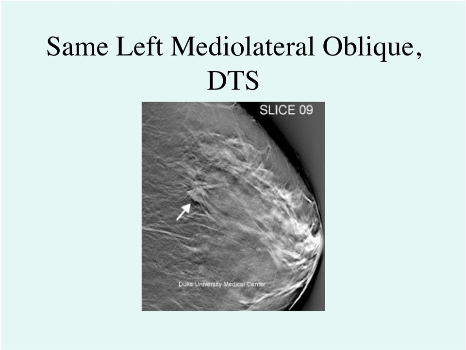
14 Left Mediolateral Oblique, Mammogram

15 Same Left Mediolateral Oblique, DTS
16 Mammomat Inspiration The Mammomat is a prototype developed by Siemens. Comes with complete setup (X-ray machine and computer) Fast, improves workflow. Can be combined with computer aided diagnosis (CAD), Upgradeable User friendly.

17 Technical Specifications: X-Ray Generator Power output - 5kW kv Range - 23kV- 35kV Exposure Time: 10 ms to 4 s (large focus) 60 ms to 6 s (small focus) Source: MAMMOMAT Inspiration - Technical Specifications, Siemens

18 Technical Specifications: Solid-state detector of amorphous selenium. Dimensions: 24 cm x 30 cm (9.5'' x 12'') Pixel Size: 85 µm Image matrix :2816 x 3584 (24 cm x 30 cm) 2016 x 2816 (18 cm x 24 cm) Flat Detector Source: MAMMOMAT Inspiration - Technical Specifications, Siemens
 2016 x 2816 (18 cm x 24 cm) Flat Detector Source: MAMMOMAT")
19 Approval Status The Mammomat Inspiration has been approved in the EU but not in the US by the FDA. Prototype work is being conducted in both the EU and the US.

20 Summary Digital tomosynthesis is a process used to reconstruct 3-D images of from 2-D scans. Advantages of DTS include comfort, speed, and lower radiation dosage. It may eventually replace conventional mammography as it become less expensive. Currently, it is only approved in the EU.

21 Sources Breastcancer.org dig_tomosynth.jsp Joseph Y. Lo, Ph.D. research pages MAMMOMAT Inspiration - Technical Specifications, Siemens Duke Advanced Imaging Laboratory Kieran Maher Flat X-Ray Panel Receptors index.html
22 Questions 1) What are the advantages of Digital Tomosynthesis vs. conventional Mamography A. Comfort B. Lower radiation dose C. Better for dense breast tissue D. All of the above E. None of the above 2) Tomosynthesis involves which of the following: A. Application of electrodes to the skin B. Radio waves C. Images taken at multiple angles D. Consuming chemicals to show up on images E. None of the Above
Mammography. What is Mammography?
 Scan for mobile link. Mammography Mammography is a specific type of breast imaging that uses low-dose x-rays to detect cancer early before women experience symptoms when it is most treatable. Tell your
Scan Time Reduction and X-ray Scatter Rejection in Dual Modality Breast Tomosynthesis. Tushita Patel 4/2/13
 Scan Time Reduction and X-ray Scatter Rejection in Dual Modality Breast Tomosynthesis Tushita Patel 4/2/13 Breast Cancer Statistics Second most common cancer after skin cancer Second leading cause of cancer
R/F. Efforts to Reduce Exposure Dose in Chest Tomosynthesis Targeting Lung Cancer Screening. 3. Utility of Chest Tomosynthesis. 1.
 R/F Efforts to Reduce Exposure Dose in Chest Tomosynthesis Targeting Lung Cancer Screening Department of Radiology, National Cancer Center Hospital East Kaoru Shimizu Ms. Kaoru Shimizu 1. Introduction
Production of X-rays. Radiation Safety Training for Analytical X-Ray Devices Module 9
 Module 9 This module presents information on what X-rays are and how they are produced. Introduction Module 9, Page 2 X-rays are a type of electromagnetic radiation. Other types of electromagnetic radiation
Digital Mammography Update: Design and Characteristics of Current Systems
 : Design and Characteristics of Current Systems Kalpana M. Kanal, Ph.D., DABR Assistant Professor Department of Radiology University of Washington Seattle, Washington AAPM Annual Meeting 2009 Anaheim,
Preparing for your Breast Tomosynthesis
 Preparing for your Breast Tomosynthesis For patients at the Rapid Diagnostic Centre UHN Read this resource to learn: How to prepare What to expect during breast tomosynthesis What questions to ask your
X-ray (Radiography) - Bone
 - Bone") Scan for mobile link. X-ray (Radiography) - Bone Bone x-ray uses a very small dose of ionizing radiation to produce pictures of any bone in the body. It is commonly used to diagnose fractured bones or
Scan for mobile link. X-ray (Radiography) - Bone Bone x-ray uses a very small dose of ionizing radiation to produce pictures of any bone in the body. It is commonly used to diagnose fractured bones or
Patient Prep Information
 Stereotactic Breast Biopsy Patient Prep Information Imaging Services Cannon Memorial Hospital Watauga Medical Center Table Weight Limits for each facility Cannon Memorial Hospital Watauga Medical Center
X-ray (Radiography) - Chest
 - Chest") Scan for mobile link. X-ray (Radiography) - Chest What is a Chest X-ray (Chest Radiography)? The chest x-ray is the most commonly performed diagnostic x-ray examination. A chest x-ray produces images of
Scan for mobile link. X-ray (Radiography) - Chest What is a Chest X-ray (Chest Radiography)? The chest x-ray is the most commonly performed diagnostic x-ray examination. A chest x-ray produces images of
X-ray (Radiography) - Abdomen
 - Abdomen") Scan for mobile link. X-ray (Radiography) - Abdomen Abdominal x-ray uses a very small dose of ionizing radiation to produce pictures of the inside of the abdominal cavity. It is used to evaluate the stomach,
Scan for mobile link. X-ray (Radiography) - Abdomen Abdominal x-ray uses a very small dose of ionizing radiation to produce pictures of the inside of the abdominal cavity. It is used to evaluate the stomach,
Role of the Medical Physicist in Clinical Implementation of Breast Tomosynthesis
 Role of the Medical Physicist in Clinical Implementation of Breast Tomosynthesis Bob Liu, Ph.D. Department of Radiology Massachusetts General Hospital And Harvard Medical School Digital Breast Tomosynthesis
X-ray (Radiography), Chest
, Chest") X-ray (Radiography), Chest What is a Chest X-ray (Chest Radiography)? The chest x-ray is the most commonly performed diagnostic x-ray examination. A chest x-ray makes images of the heart, lungs, airways,
X-ray (Radiography), Chest What is a Chest X-ray (Chest Radiography)? The chest x-ray is the most commonly performed diagnostic x-ray examination. A chest x-ray makes images of the heart, lungs, airways,
Mammography Applications for MAMMOMAT 1000/3000 Nova
 Mammography Applications for MAMMOMAT 1000/3000 Nova Introduction This booklet is intended as an application handbook for use with Siemens MAMMOMAT 1000 and 3000 Nova. The booklet contains advice on positioning,
A Guide to Breast Imaging: The Latest Technology for Screening and Detecting Breast Cancer
 A Guide to Breast Imaging: The Latest Technology for Screening and Detecting Breast Cancer Sally Herschorn, MD Associate Professor of Radiology University of Vermont College of Medicine Medical Director
Quality Control of Full Field Digital Mammography Units
 Quality Control of Full Field Digital Mammography Units Melissa C. Martin, M.S., FACMP, FACR, FAAPM [email protected] 310-612-8127 ACMP Annual Meeting Virginia Beach, VA May 2, 2009 History of
X-Rays Benefits and Risks. Techniques that use x-rays
 X-Rays Benefits and Risks X-rays are a form of electromagnetic radiation, just like light waves and radiowaves. Because X-rays have higher energy than light waves, they can pass through the body. X-rays
Personalized Breast Screening Service
 Frequently Asked Questions WHAT IS BREAST DENSITY? Breasts are made up of a mixture of fibrous, glandular and fatty tissue. Your breasts are considered if you have predominantly fibrous or glandular tissue
SECTION 1: REQUIREMENTS FOR CERTIFICATES OF COMPLIANCE FOR CLASSES OF RADIATION APPARATUS
 Department of Health and Human services Population Health Radiation Protection Act 2005 Section 17 CERTIFICATE OF COMPLIANCE: STANDARD FOR RADIATION APPARATUS - X-RAY MEDICAL DIAGNOSTIC (MAMMOGRAPHY) SECTION
I-Max Touch Range. PAN / CEPH / 3D digital panoramic unit. Evolutive 3 in 1 panoramic unit
 I-Max Touch Range PAN / CEPH / 3D digital panoramic unit Evolutive 3 in 1 panoramic unit 3D A new dimension for a complete diagnosis I-Max Touch 3D Evolutive, simple, fast The panoramic unit realizes complete
Low-dose CT Imaging. Edgar Fearnow, M.D. Section Chief, Computed Tomography, Lancaster General Hospital
 Lung Cancer Screening with Low-dose CT Imaging Edgar Fearnow, M.D. Section Chief, Computed Tomography, Lancaster General Hospital Despite recent declines in the incidence of lung cancer related to the
PHOTOELECTRIC EFFECT AND DUAL NATURE OF MATTER AND RADIATIONS
 PHOTOELECTRIC EFFECT AND DUAL NATURE OF MATTER AND RADIATIONS 1. Photons 2. Photoelectric Effect 3. Experimental Set-up to study Photoelectric Effect 4. Effect of Intensity, Frequency, Potential on P.E.
Radiation therapy involves using many terms you may have never heard before. Below is a list of words you could hear during your treatment.
 Dictionary Radiation therapy involves using many terms you may have never heard before. Below is a list of words you could hear during your treatment. Applicator A device used to hold a radioactive source
Thinking ahead. Focused on life. REALIZED: GROUNDBREAKING RESOLUTION OF 80 µm VOXEL
 Thinking ahead. Focused on life. REALIZED: GROUNDBREAKING RESOLUTION OF 80 µm VOXEL X-ray ZOOM RECONSTRUCTION Flat Panel Detector (FPD) Automatic Positioning Function For ø 40 x H 40 mm, ø 60 x H 60 mm,
Cutting Edge Digital Radiography Technology in Daily NDT Use
 Cutting Edge Digital Radiography Technology in Daily NDT Use Ron PINCU, Eli DAYAN, Ofra KLEINBERGER Vidisco Ltd. Or-Yehuda, Israel 60375 Tel: +972 (3) 533-3001 Email: [email protected] ABSTRACT Digital Radiography
Atomic and Nuclear Physics Laboratory (Physics 4780)
") Gamma Ray Spectroscopy Week of September 27, 2010 Atomic and Nuclear Physics Laboratory (Physics 4780) The University of Toledo Instructor: Randy Ellingson Gamma Ray Production: Co 60 60 60 27Co28Ni *
Gamma Ray Spectroscopy Week of September 27, 2010 Atomic and Nuclear Physics Laboratory (Physics 4780) The University of Toledo Instructor: Randy Ellingson Gamma Ray Production: Co 60 60 60 27Co28Ni *
Surveying and QC of Stereotactic Breast Biopsy Units for ACR Accreditation
 Surveying and QC of Stereotactic Breast Biopsy Units for ACR Accreditation LORAD Stereotactic Breast Biopsy System AAPM Spring Clinical Meeting Phoenix, AZ March 17, 2013 Melissa C. Martin, M.S., FACR,
Fundamentals of Cone-Beam CT Imaging
 Fundamentals of Cone-Beam CT Imaging Marc Kachelrieß German Cancer Research Center (DKFZ) Heidelberg, Germany www.dkfz.de Learning Objectives To understand the principles of volumetric image formation
The effects of radiation on the body can be divided into Stochastic (random) effects and deterministic or Non-stochastic effects.
 effects and deterministic or Non-stochastic effects.") RADIATION SAFETY: HOW TO EDUCATE AND PROTECT YOURSELF AND YOUR STAFF John Farrelly, DVM, MS, ACVIM (Oncology), ACVR (Radiation Oncology) Cornell University Veterinary Specialists The Veterinary Cancer
RADIATION SAFETY: HOW TO EDUCATE AND PROTECT YOURSELF AND YOUR STAFF John Farrelly, DVM, MS, ACVIM (Oncology), ACVR (Radiation Oncology) Cornell University Veterinary Specialists The Veterinary Cancer
X-ray Production. Target Interactions. Principles of Imaging Science I (RAD119) X-ray Production & Emission
 X-ray Production & Emission") Principles of Imaging Science I (RAD119) X-ray Production & Emission X-ray Production X-rays are produced inside the x-ray tube when high energy projectile electrons from the filament interact with the
Principles of Imaging Science I (RAD119) X-ray Production & Emission X-ray Production X-rays are produced inside the x-ray tube when high energy projectile electrons from the filament interact with the
Contents. X-ray and Computed Tomography. Characterization of X-rays. Production of X-rays
 J. E. Wilhjelm Ørsted TU Technical University of enmark, Bldg. 348, K-2800 Kongens Lyngby, enmark. X-ray and Computed Tomography Contents History and characterization of X-rays Conventional (projection)
MDCT Technology. Kalpana M. Kanal, Ph.D., DABR Assistant Professor Department of Radiology University of Washington Seattle, Washington
 MDCT Technology Kalpana M. Kanal, Ph.D., DABR Assistant Professor Department of Radiology University of Washington Seattle, Washington ACMP Annual Meeting 2008 - Seattle, WA Educational Objectives Historical
CT scanning. By Mikael Jensen & Jens E. Wilhjelm Risø National laboratory Ørsted DTU. (Ver. 1.2 4/9/07) 2002-2007 by M. Jensen and J. E.
 2002-2007 by M. Jensen and J. E.") 1 Overview CT scanning By Mikael Jensen & Jens E. Wilhjelm Risø National laboratory Ørsted DTU (Ver. 1.2 4/9/07) 2002-2007 by M. Jensen and J. E. Wilhjelm) As it can be imagined, planar X-ray imaging has
1 Overview CT scanning By Mikael Jensen & Jens E. Wilhjelm Risø National laboratory Ørsted DTU (Ver. 1.2 4/9/07) 2002-2007 by M. Jensen and J. E. Wilhjelm) As it can be imagined, planar X-ray imaging has
for breast cancer detection
 Microwave imaging for breast cancer detection Sara Salvador Summary Epidemiology of breast cancer The early diagnosis: State of the art Microwave imaging: The signal processing algorithm 2D/3D models and
Mammomat Inspiration Mammomat Inspiration Prime
 s Data Sheet Mammomat Inspiration Mammomat Inspiration Prime Digital Mammography Platform for Screening, Diagnostics, Biopsy and Tomosynthesis www.siemens.com/inspiration Mammomat Inspiration Digital Mammography
The Field. Radiologic technologists take x-rays and administer nonradioactive materials into patients' bloodstreams for diagnostic purposes.
 Radiologic Technologist Overview The Field - Specialty Areas - Preparation - Day in the Life - Earnings - Employment - Career Path Forecast - Professional Organizations The Field Radiologic technologists
Digital radiography conquers the veterinary world
 Digital radiography conquers the veterinary world Author: Dirk De Langhe Increasingly, veterinarians are using medical imaging to diagnose their patients. There is a corresponding tendency towards replacing
Development of High Speed Oblique X-ray CT System for Printed Circuit Board
 計 測 自 動 制 御 学 会 産 業 論 文 集 Vol. 6, No. 9, 72/77 (2007) Development of High Speed Oblique X-ray CT System for Printed Circuit Board Atsushi TERAMOTO*, Takayuki MURAKOSHI*, Masatoshi TSUZAKA**, and Hiroshi
Patient Exposure Doses During Diagnostic Radiography
 Patient Exposure Doses During Diagnostic Radiography JMAJ 44(11): 473 479, 2001 Shoichi SUZUKI Associated Professor, Faculty of Radiological Technology, School of Health Sciences, Fujita Health University
Reduce Your Risk of Breast Cancer
 Reduce Your Risk of Breast Cancer Reduce Your Risk of Breast Cancer There was no history in my family. But the test was positive and it was breast cancer. I was so shocked, I couldn t believe it. ~ Colette
CHAPTER 3: DIGITAL IMAGING IN DIAGNOSTIC RADIOLOGY. 3.1 Basic Concepts of Digital Imaging
 Physics of Medical X-Ray Imaging (1) Chapter 3 CHAPTER 3: DIGITAL IMAGING IN DIAGNOSTIC RADIOLOGY 3.1 Basic Concepts of Digital Imaging Unlike conventional radiography that generates images on film through
Feasibility Study of Neutron Dose for Real Time Image Guided. Proton Therapy: A Monte Carlo Study
 Feasibility Study of Neutron Dose for Real Time Image Guided Proton Therapy: A Monte Carlo Study Jin Sung Kim, Jung Suk Shin, Daehyun Kim, EunHyuk Shin, Kwangzoo Chung, Sungkoo Cho, Sung Hwan Ahn, Sanggyu
PRACTICAL TIPS IN ENSURING RADIATION SAFETY IN THE USE OF MEDICAL DIAGNOSTIC X-RAY EQUIPMENT
 PRACTICAL TIPS IN ENSURING RADIATION SAFETY IN THE USE OF MEDICAL DIAGNOSTIC X-RAY EQUIPMENT Although the medical uses of X-rays to examine a patient without surgery became an amazing medical breakthrough,
Dental Radiography Core Subject. Digital Radiography
 Dental Radiography Core Subject Digital Radiography Aims: To develop an understanding of the history of digital radiography, the different types of digital x-rays and the advantages and disadvantages of
im3d S.p.A. La Ricerca fa Impresa
 im3d S.p.A. La Ricerca fa 26 gennaio 2011 Alberto Bert Outline Introduction CAD COLON CAD BREAST DTS Introduction La Ricerca fa Who we are and what we do The company im3d S.p.A. is a company developing
Dose Measurement in Mammography; What are we measuring? David E. Hintenlang, Ph.D. DABR University of Florida
 Dose Measurement in Mammography; What are we measuring? David E. Hintenlang, Ph.D. DABR University of Florida Average Glandular Dose Required measurement performed by medical physicist as part of Mammography
Radiographic Image Production. Radiographic Image Production. Principles of Imaging Science I (RAD 119) Film, Screens, and Cassettes
 Film, Screens, and Cassettes") Principles of Imaging Science I (RAD 119) Film, Screens, and Cassettes Radiographic Image Production X-ray photons emitted from the x-ray tube interact with the body, exit the patient (exit beam) and interact
Principles of Imaging Science I (RAD 119) Film, Screens, and Cassettes Radiographic Image Production X-ray photons emitted from the x-ray tube interact with the body, exit the patient (exit beam) and interact
CT Scan Thorax and Upper Abdomen. Respiratory Unit Patient Information Leaflet
 CT Scan Thorax and Upper Abdomen Respiratory Unit Patient Information Leaflet Introduction This leaflet gives you general information about your CT (computerised tomography) scan. It does not replace the
DENTAL Cone beam 3D X-RAY SYSTEM with
 VERSATILE INTUITIVE efficient DENTAL Cone beam 3D X-RAY SYSTEM with dedicated panoramic imaging With thirty years of experience in designing and manufacturing state-of-the-art dental panoramic and tomographic
5.2 ASSESSMENT OF X-RAY TUBE LEAKAGE RADIATION AND X-RAY TUBE OUTPUT TOTAL FILTRATION
 5.2 ASSESSMENT OF X-RAY TUBE LEAKAGE RADIATION AND X-RAY TUBE OUTPUT TOTAL FILTRATION 5.2.1 Task The bremsstrahlung produced by the X-ray tube has a continuous spectrum, limited by the set and spreads
X-ray Imaging Systems
 Principles of Imaging Science I (RAD 119) X-ray Tube & Equipment X-ray Imaging Systems Medical X-ray Equipment Classified by purpose or energy/current levels kvp, ma Radiographic Non-dynamic procedures
Digital Mammogram National Database
 Digital Mammogram National Database Professor Michael Brady FRS FREng Medical Vision Laboratory Oxford University Chairman: Mirada Solutions Ltd PharmaGrid 2/7/03 ediamond aims construct a federated database
Veraview IC5 HD High definition, digital imaging excellence. Thinking ahead. Focused on life.
 Veraview IC5 HD High definition, digital imaging excellence Thinking ahead. Focused on life. Thinking ahead. Focused on life. 2 3 IC5 HD Super High-Speed with High Definition Clarity For dental radiology
Official reprint from UpToDate www.uptodate.com 2013 UpToDate
 Official reprint from UpToDate www.uptodate.com 2013 UpToDate Patient information: Breast cancer screening (The Basics) Written by the doctors and editors at UpToDate What is breast cancer screening? Breast
Radiation Exposure in X-ray and CT Examinations
 Patient Safety-Xray: Radiation Exposure in X-ray and CT Examinations What are x-rays and what do they do? X-rays are forms of radiant energy, like light or radio waves. Unlike light, x-rays can penetrate
CALCULATION METHODS OF X-RAY SPECTRA: A COMPARATIVE STUDY
 243 CALCULATION METHODS OF X-RAY SPECTRA: A COMPARATIVE STUDY B. Chyba, M. Mantler, H. Ebel, R. Svagera Technische Universit Vienna, Austria ABSTRACT The accurate characterization of the spectral distribution
NORMAL CHEST RADIOGRAPHY. Front and lateral view
 NORMAL CHEST RADIOGRAPHY Front and lateral view Dr Etienne Leroy-Terquem Centre hospitalier de Meulan les Mureaux. France French-cambodian association for pneumology (OFCP) OFCP How to obtain a good quality
BREAST CHARACTERISTICS AND DOSIMETRIC DATA IN X RAY MAMMOGRAPHY - A LARGE SAMPLE WORLDWIDE SURVEY
 BREAST CHARACTERISTICS AND DOSIMETRIC DATA IN X RAY MAMMOGRAPHY - A LARGE SAMPLE WORLDWIDE SURVEY N. GEERAERT a,b,c, R. KLAUSZ a, S. MULLER a, I. BLOCH c, H. BOSMANS b a GE Healthcare, Buc, France b Department
Breast Screening Explained. We can supply this information in other languages, in large print, on audio or in Braille.
 Breast Screening Explained We can supply this information in other languages, in large print, on audio or in Braille. Breast Screening Explained This leaflet tells you about free breast screening. Breast
Concepts for High-Resolution Low-Dose CT of the Breast
 RSNA 2012 Refresher Course 721B, Chicago, Nov. 30, 2012 Concepts for High-Resolution Low-Dose CT of the Breast Disclosures WAK is founder, shareholder and CEO of CT Imaging GmbH, Erlangen, Germany. Willi
Automation of a CT Acquisition: A System-based User-Support. 1. Introduction. Bärbel KRATZ 1, Frank HEROLD 1, Malte KURFISS 1
 11th European Conference on Non-Destructive Testing (ECNDT 2014), October 6-10, 2014, Prague, Czech Republic Automation of a CT Acquisition: A System-based User-Support More Info at Open Access Database
PHYSICAL METHODS, INSTRUMENTS AND MEASUREMENTS Vol. III - Medical and Industrial Tomography - W.B.Gilboy
 MEDICAL AND INDUSTRIAL TOMOGRAPHY Department of Physics, University of Surrey, Guildford, Surrey, U.K. Keywords: Radiography, transmission tomography, emission tomography, microtomography, SPECT (single
An Overview of Digital Imaging Systems for Radiography and Fluoroscopy
 An Overview of Digital Imaging Systems for Radiography and Fluoroscopy Michael Yester, Ph.D. University of Alabama at Birmingham Outline Introduction Imaging Considerations Receptor Properties General
Breast Density Legislation: Implications for primary care providers
 Breast Density Legislation: Implications for primary care providers Deborah J. Rhodes MD Associate Professor of Medicine 2012 MFMER slide-1 Disclosure Relevant financial relationship(s) None Off-label
CONTENT SPECIFICATIONS FOR THE FLUOROSCOPY EXAMINATION
 CONTENT SPECIFICATIONS FOR THE FLUOROSCOPY EXAMINATION Publication Date: November 2010 Implementation Date: March 2011 The purpose of the American Registry of Radiologic Technologists Fluoroscopy Examination
Hologic Selenia Dimensions C-View Software Module. October 24, 2012
 Hologic Selenia Dimensions C-View Software Module October 24, 2012 Introduction and Agenda Peter Soltani, Ph.D. Senior VP & GM, Breast Health Hologic, Inc. Agenda Technology Overview Clinical Overview
Data. microcat +SPECT
 Data microcat +SPECT microcat at a Glance Designed to meet the throughput, resolution and image quality requirements of academic and pharmaceutical research, the Siemens microcat sets the standard for
Digital Breast Tomosynthesis QC Requirements
 Digital Breast Tomosynthesis QC Requirements AAPM Spring Clinical Meeting March 8, 2015 Michael S Glaser, MS, DABR Alliance Medical Physics, LLC Learning Objectives 1. GE SenoClaire - Physicist & Technologist
Lectures about XRF (X-Ray Fluorescence)
") 1 / 38 Lectures about XRF (X-Ray Fluorescence) Advanced Physics Laboratory Laurea Magistrale in Fisica year 2013 - Camerino 2 / 38 X-ray Fluorescence XRF is an acronym for X-Ray Fluorescence. The XRF technique
1 / 38 Lectures about XRF (X-Ray Fluorescence) Advanced Physics Laboratory Laurea Magistrale in Fisica year 2013 - Camerino 2 / 38 X-ray Fluorescence XRF is an acronym for X-Ray Fluorescence. The XRF technique
Development of on line monitor detectors used for clinical routine in proton and ion therapy
 Development of on line monitor detectors used for clinical routine in proton and ion therapy A. Ansarinejad Torino, february 8 th, 2010 Overview Hadrontherapy CNAO Project Monitor system: Part1:preliminary
Q.A. Collectible. Sponsored by CRCPD s Committee on Quality Assurance in Diagnostic X-Ray (H-7)
") Q.A. Collectible Sponsored by CRCPD s Committee on Quality Assurance in Diagnostic X-Ray (H-7) Mammography Phantom Image Quality Evaluation (from the American College of Radiology 1999 Mammography Quality
Q.A. Collectible Sponsored by CRCPD s Committee on Quality Assurance in Diagnostic X-Ray (H-7) Mammography Phantom Image Quality Evaluation (from the American College of Radiology 1999 Mammography Quality
Study the Quality Assurance of Conventional X-ray Machines Using Non Invasive KV meter
 Study the Quality Assurance of Conventional X-ray Machines Using Non Invasive KV meter T.M.Taha Radiation Protection Department, Nuclear Research Center, Atomic Energy Authority, Cairo.P.O.13759 Egypt.
Mammography. For 35+ years, screenfilm has been the gold standard for breast cancer detection
 Digital Mammography Upright Stereo Mammography For 35+ years, screenfilm has been the gold standard for breast cancer detection IN THE BEGINNING Mammography technology has come a long way since the first
Cardiac CT for Calcium Scoring
 Scan for mobile link. Cardiac CT for Calcium Scoring Cardiac computed tomography (CT) for Calcium Scoring uses special x-ray equipment to produce pictures of the coronary arteries to determine if they
College on Medical Physics. Digital Imaging Science and Technology to Enhance Healthcare in the Developing Countries
 2166-Handout College on Medical Physics. Digital Imaging Science and Technology to Enhance Healthcare in the Developing Countries 13 September - 1 October, 2010 Digital Radiography Image Parameters SNR,
Industrial X-ray for Nondestructive Testing Unrestricted Siemens AG 2014. All rights reserved
 Overview of X-ray Technology and Competence offered by Corporate Technology Industrial X-ray for Nondestructive Testing Nondestructive Testing (NDT) with X-rays: Our offer at a glance High-tech X-ray lab
ACR AAPM SIIM PRACTICE PARAMETER FOR DETERMINANTS OF IMAGE QUALITY IN DIGITAL MAMMOGRAPHY
 The American College of Radiology, with more than 30,000 members, is the principal organization of radiologists, radiation oncologists, and clinical medical physicists in the United States. The College
Production of X-rays and Interactions of X-rays with Matter
 Production of X-rays and Interactions of X-rays with Matter Goaz and Pharoah. Pages 11-20. Neill Serman Electrons traveling from the filament ( cathode) to the target (anode) convert a small percentage
HIGH PERFORMANCE MOBILE SURGICAL C-ARM KMC-950
 HIGH PERFORMANCE MOBILE SURGICAL C-ARM 1K x 1k CCD Digital Camera System H.F. GENERATOR & ROTATING ANODE TUBE WITH DIGITAL WORKSTATION DESCRIPTION: Mobile Surgical C-arm systems are integrated with a triple
Solar Photovoltaic (PV) Cells
 Cells") Solar Photovoltaic (PV) Cells A supplement topic to: Mi ti l S Micro-optical Sensors - A MEMS for electric power generation Science of Silicon PV Cells Scientific base for solar PV electric power generation
Solar Photovoltaic (PV) Cells A supplement topic to: Mi ti l S Micro-optical Sensors - A MEMS for electric power generation Science of Silicon PV Cells Scientific base for solar PV electric power generation
Physics testing of image detectors
 Physics testing of image detectors Parameters to test Spatial resolution Contrast resolution Uniformity/geometric distortion Features and Weaknesses of Phantoms for CR/DR System Testing Dose response/signal
Training needs for professionals in conventional radiology (radiology technicians, physicists, radiologists) joining digital radiology
 joining digital radiology") Training needs for professionals in conventional radiology (radiology technicians, physicists, radiologists) joining digital radiology Guidelines on education and training for digital radiology Author:
Training needs for professionals in conventional radiology (radiology technicians, physicists, radiologists) joining digital radiology Guidelines on education and training for digital radiology Author:
Strahlenschutzbelehrung Allgemeiner Teil. Radiation Protection
 1 Radiation Protection 2 Why radiation protection? - Ionizing radiation (>5eV -> UV; X-rays;α,β,γ-radiation)has physical, chemical and biological effects -> human tissue (70% water!) and genetic material
.org. Osteochondroma. Solitary Osteochondroma
 Osteochondroma Page ( 1 ) An osteochondroma is a benign (noncancerous) tumor that develops during childhood or adolescence. It is an abnormal growth that forms on the surface of a bone near the growth
Mammograms & Breast Health. An Information Guide for Women U.S. DEPARTMENT OF HEALTH AND HUMAN SERVICES. Centers for Disease Control and Prevention
 U.S. DEPARTMENT OF HEALTH AND HUMAN SERVICES Centers for Disease Control and Prevention Mammograms & Breast Health An Information Guide for Women This booklet was developed by the Centers for Disease Control
Breast Cancer. CSC Cancer Experience Registry Member, breast cancer
 ESSENTIALS Breast Cancer Take things one step at a time. Try not to be overwhelmed by the tidal wave of technical information coming your way. Finally you know your body best; you have to be your own advocate.
DETERMINING WHICH COLOR UV BEAD CHANGES COLORS THE FASTEST
 DETERMINING WHICH COLOR UV BEAD CHANGES COLORS THE FASTEST Helen C Cary Academy ABSTRACT The purpose of this experiment was to determine which color UV bead changes colors the fastest. The bead colors
CT: Size Specific Dose Estimate (SSDE): Why We Need Another CT Dose Index. Acknowledgements
: Why We Need Another CT Dose Index. Acknowledgements") CT: Size Specific Dose Estimate (SSDE): Why We Need Another CT Dose Index Keith J. Strauss, MSc, FAAPM, FACR Clinical Imaging Physicist Cincinnati Children s Hospital University of Cincinnati College of
CT: Size Specific Dose Estimate (SSDE): Why We Need Another CT Dose Index Keith J. Strauss, MSc, FAAPM, FACR Clinical Imaging Physicist Cincinnati Children s Hospital University of Cincinnati College of
CT RADIATION DOSE REPORT FROM DICOM. Frank Dong, PhD, DABR Diagnostic Physicist Imaging Institute Cleveland Clinic Foundation Cleveland, OH
 CT RADIATION DOSE REPORT FROM DICOM Frank Dong, PhD, DABR Diagnostic Physicist Imaging Institute Cleveland Clinic Foundation Cleveland, OH CT Patient comes out... Patient goes in... Big Black Box Radiology
Medicare Part B. Mammograms - Updated Billing Guide for Screening and Diagnostic Tests
 Mammograms - Updated Billing Guide for Screening and Diagnostic Tests This article from Medicare B News Issue 223 dated October 21, 2005 is being updated and reprinted to ensure that the Noridian Administrative
www.icrcompany.com I SEE THE FUTURE
 www.icrcompany.com I SEE THE FUTURE Stephen Neushul saw the future when he first designed a film scanner back in 1990 called the OmniMedia scanner. He built the scanner with a fixed light source, an innovation
X-RAY IMAGING Emerging Digital Technology - CMOS Detectors
 Application Note Case Study Technology Primer White Paper X-RAY IMAGING Emerging Digital Technology - CMOS Detectors Image Sensors X-Ray DETECTORS Scanners Image Processing Custom Solutions In all domains
First Warning Systems, Inc.
 First Warning Systems, Inc. Breast Cancer Early Detection System US EU/UK & Russia Executive Summary September 2012 i 1. EXECUTIVE SUMMARY 1.1 FIRST WARNING SYSTEM INTRODUCTION First Warning System (FWS)
Quality Assurance. The selection of the equipment. Equipment Specifications. Medical Exposure Directive 97/43 Euratom. Quality Assurance Programme
 Medical Exposure Directive 97/43 Euratom Quality Assurance Ministry of Health, Radiation Protection Department, Luxembourg Alexandra Schreiner Medical Physicist Quality Assurance (QA): All those planned
Proper Implementation of Industrial CT Scanning to Reduce Inspection Costs & Get to Production Faster. Jesse Garant, JG&A Metrology Center
 Proper Implementation of Industrial CT Scanning to Reduce Inspection Costs & Get to Production Faster Jesse Garant, JG&A Metrology Center Traditional Metrology and Inspection Tactile Devices (Touch Probe)
Focused Learning Lesson Physical Science Grade Levels 9 12 PS-H-G4
 Focused Learning Lesson Physical Science Grade Levels 9 12 PS-H-G4 Overview: This lesson provides students the opportunity to study positive and negative aspects of various types of energy through critical
A PACS-Aware DICOM Image Object
 Designing and Implementing A PACS-Aware DICOM Image Object For Digital X-ray, Mammography and Intraoral Applications David A. Clunie Quintiles Intelligent Imaging Clear Vision for the Healthcare Industry