Digital Breast Tomosynthesis QC Requirements
|
|
|
- Blake Freeman
- 9 years ago
- Views:
Transcription
1 Digital Breast Tomosynthesis QC Requirements AAPM Spring Clinical Meeting March 8, 2015 Michael S Glaser, MS, DABR Alliance Medical Physics, LLC

2 Learning Objectives 1. GE SenoClaire - Physicist & Technologist QC 2. Hologic Selenia Dimensions 3D- Physicist & Technologist QC

3 Disclosures Funding Support- None Conflicts of Interest- None Disclosures- Yes, vendor QC slides Special Thanks for vendor QC slides, courtesy of: Razvan Iordache, PhD of GE Healthcare Nikos Gkanatsios,PhD of Hologic

4 Quality Control for SenoClaire (GE Breast Tomosynthesis)

5 Comment: *Exposure time depends on breast thickness, ; 10 sec for average breast of 4.5cm PMMA. SenoClaire (GE Breast Tomosynthesis) SenoClaire Key Features 9 Projections Stop-and-shoot Sweep angle 25 ( +/- 12.5) Sweep time <10 sec* Detector pixel size 100 um in 2D & 3D 2D/3D-grid for scatter reduction ASIR DBT Iterative Reconstruction No dose increase (3D vs. 2D) BTO DICOM format (Breast Tomosynthesis Object)
 Sweep time <10 sec* Detector pixel size 100 um in 2D & 3D 2D/3D-grid for scatter reduction ASIR DBT")


6 SenoClaire is an Upgrade for Senographe Essential Motorized Tomosynthesis Device (MTD) Senographe Essential Senographe Essential Upgraded with SenoClaire.....

7 Imaging with the MTD Traditional antiscatter grid - Essential View of system positioned for a left MLO view 2D/3D antiscatter grid SenoClaire s MTD View of system positioned for a left MLO view Both 2D & 3D imaging is performed with the MTD 5

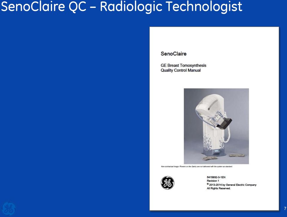
8 SenoClaire QC Manual This quality control (QC) manual adds tests specific to SenoClaire to those listed in the Senographe Essential QC manual 71 pages 6

9 SenoClaire QC Radiologic Technologist 7
10 QC Tests for the Radiologic Technologist Minimum frequency Test Essential SenoClaire (w/ MTD) No 2D No 2D 3D Daily Monitor cleaning Weekly Flat field Weekly Phantom IQ Weekly CNR & MTF Weekly Viewbox & Viewing Monthly AOP Mode & SNR Monthly Visual Checklist Quaterly Repeat Analysis Semianually Compression Force x x Monthly Grid Texture 8

11 QC Tests for the Radiologic Technologist Minimum frequency Test Essential SenoClaire (w/ MTD) No 2D No 2D 3D Daily Monitor cleaning Weekly Flat field Weekly Phantom IQ Weekly CNR & MTF Weekly Viewbox & Viewing Monthly AOP Mode & SNR Monthly Visual Checklist Quaterly Repeat Analysis Semianually Compression Force x x Monthly Grid Texture 9

12 SenoClaire Tests same as Essential Tests Minimum frequency Test Essential SenoClaire (w/ MTD) No 2D No 2D 3D Daily Monitor cleaning Weekly Flat field Weekly Phantom IQ Weekly CNR & MTF Weekly Viewbox & Viewing Monthly AOP Mode & SNR Monthly Visual Checklist Quaterly Repeat Analysis Semianually Compression Force x x Monthly Grid Texture 10

13 SenoClaire Tests specific Visual Checklist Minimum frequency Test Essential SenoClaire (w/ MTD) No 2D No 2D 3D Daily Monitor cleaning Weekly Flat field Weekly Phantom IQ Weekly CNR & MTF Weekly Viewbox & Viewing Monthly AOP Mode & SNR Monthly Visual Checklist Quaterly Repeat Analysis Semianually Compression Force x x Monthly Grid Texture 11

14 Visual Checklist (monthly) Objective To assure that GE Breast Tomosynthesis indicator lights, displays, and mechanical locks and detents are working properly and that the system is mechanically stable. Equipment required Visual checklist Chart 5. Grid texture test, Visual Checklist and Compression Record of Checks (page 36). Procedure Review each item on the visual checklist and indicate its status Action Limit Each of the items listed in the Visual Checklist must pass (ie, receive a check mark) 12

15 SenoClaire Tests New Grid Texture Test Minimum frequency Test Essential SenoClaire (w/ MTD) No 2D No 2D 3D Daily Monitor cleaning Weekly Flat field Weekly Phantom IQ Weekly CNR & MTF Weekly Viewbox & Viewing Monthly AOP Mode & SNR Monthly Visual Checklist Quaterly Repeat Analysis Semianually Compression Force x x Monthly Grid Texture 13

16 Grid Texture Test (monthly) Objective Measures the amount of grid texture in 2D images Equipment required Flat field test object Procedure Automatic acquisition of 10 2D images with increasing mas Record the dispayed test results Action Limit The texture level must not exceed

![Noise [counts] Grid Texture Test (monthly)](/docs-images/40/20839711/images/17-0.jpg "14000 12000 Noise vs dose 10000 8000 y = 0.")

17 Noise [counts] Grid Texture Test (monthly) Noise vs dose y = x x Detector dose [ngy] 15

18 SenoClaire Tests 3D Extensions Minimum frequency Test Essential SenoClaire (w/ MTD) No 2D No 2D 3D Daily Monitor cleaning Weekly Flat field Weekly Phantom IQ Weekly CNR & MTF Weekly Viewbox & Viewing Monthly AOP Mode & SNR Monthly Visual Checklist Quaterly Repeat Analysis Semianually Compression Force x x Monthly Grid Texture 16

19 Flat field 3D Test (Weekly) Objective Ensure flatness and homogeneity when reconstructing planes through a flat field phantom Equipment required Flat field test object Procedure Automatic 3D acquisitiion Record the displayed test results Action Limit Both Brightness non-uniformity* and SNR non-uniformity* tests must pass * Calculated in the plane at 10 mm height 17

20 Phantom IQ 3D Test (Weekly) Objective Ensure adequate and consistent IQ of 3D images Equipment required ACR mammography accreditation phantom Procedure 3D acquisitiion with Rh/Rh track/filter, 29 kv, 56 mas Review the volumes; score the phantom Scroll through the volume to find the best in-focus plane for each structure! Action Limit The score must be: Fibers > 4, Speck groups > 3, Masses > 3 Same technique & action limit as for the 2D test 18

21 AOP 3D Check (Monthly) Objective Check that the correct parameters are selected in AOP 3D mode Equipment required Set of acrylic plates (same as for the AOP 2D Check) Procedure 3D AOP acquisition on 25mm, 50mm, and 60 mm of acyrlic Record the exposure parameters Action Limit Acrylic Thickness (mm) Exposure parameters Track/Filter mas kv 25 Mo/Mo or Mo/Rh Rh/Rh Rh/Rh or 31 19


22 AOP 3D Check (Monthly) Displayed results 20
23 SenoClaire QC Tests for the Technologist 2D Tests: - Section 3: Phantom IQ Test with MTD - Checks for consistency of image quality. - Section 4: CNR and MTF Measurement with MTD - Checks for consistent production of good contrast images. - Section 8: AOP 2D and SNR Check with MTD - Checks for correct operation of AOP mode with MTD. MTD Test: Section 7: Grid Texture Test - Checks for consistency of image quality regarding grid texture. 3D Tests: - Section 5: Flat-field 3D Test - Checks for consistency of image quality. - Section 6: Phantom IQ 3D Test - Checks for consistency of image quality. - Section 9: AOP 3D Test - Checks for correct operation of AOP mode in 3D. Section 10: Visual Checklist (page 27). Section 11: Compression Force Test - Checks for the correct level of compression force. Section 12: Test Results Forms - Provides charts for use in recording test results. It is recommended that you copy these chart pages to record test results. For record keeping and further analysis, data generated on the Acquisition Workstation (AWS) for Flat-field, CNR, MTF, AOP and SNR tests can be exported as text files to a CD-R. 21

24 SenoClaire QC Medical Physicist 22
25 QC Tests for the Medical Physicist Test Essential SenoClaire (w/ MTD) no 2D no 2D 3D Flat field Phantom IQ CNR & MTF AOP Mode & SNR Artifact Eval & Flat field Unif Collimation (2 alternatives) Sub-system MTF or Focal Spot Perf Breast Entrance Expose, AGD, Reproducibility Flexible paddle deflection kvp Accuracy and Reproducibility HVL Mammo Unit Assembly Eval Grid Texture Compression paddle border to chestwall alignment Volume Coverage 23
26 QC Tests for the Medical Physicist Test Essential SenoClaire (w/ MTD) no 2D no 2D 3D Flat field Phantom IQ CNR & MTF AOP Mode & SNR Artifact Eval & Flat field Unif Collimation (2 alternatives) Sub-system MTF or Focal Spot Perf Breast Entrance Expose, AGD, Reproducibility Flexible paddle deflection kvp Accuracy and Reproducibility HVL Mammo Unit Assembly Eval Grid Texture Compression paddle border to chestwall alignment Volume Coverage 24

27 Test Intervals all at acceptance and at least annually 7 tests from radiologic technologist s section 5 additional only for medical physicists
28 SenoClaire Tests same as Essential Tests Test Essential SenoClaire (w/ MTD) no 2D no 2D 3D Flat field Phantom IQ CNR & MTF AOP Mode & SNR Artifact Eval & Flat field Unif Collimation (2 alternatives) Sub-system MTF or Focal Spot Perf Breast Entrance Expose, AGD, Reproducibility Flexible paddle deflection kvp Accuracy and Reproducibility HVL Mammo Unit Assembly Eval Grid Texture Compression paddle border to chestwall alignment Volume Coverage 26
29 Compression paddle to MTD chest wall alignment test Objective Assure that the paddle chest wall side border aligns with the chest wall side of the MTD Same test as Compression Paddle Chest Wall Test from the Collimation Assessment tests for Essential ( EN) - Job Card VF-P01A - Collimation Assessment with -Ray Cassette, OR - Job Card VF-P01B - Collimation Assessment with Radiation Sensitive Strips 27
30 SenoClaire Tests New Grid Texture Test Test Essential SenoClaire (w/ MTD) no 2D no 2D 3D Flat field Phantom IQ CNR & MTF AOP Mode & SNR Artifact Eval & Flat field Unif Collimation (2 alternatives) Sub-system MTF or Focal Spot Perf Breast Entrance Expose, AGD, Reproducibility Flexible paddle deflection kvp Accuracy and Reproducibility HVL Mammo Unit Assembly Eval Grid Texture Compression paddle border to chestwall alignment Volume Coverage 28
31 SenoClaire Tests 3D Extensions Test Essential SenoClaire (w/ MTD) no 2D no 2D 3D Flat field Phantom IQ CNR & MTF AOP Mode & SNR Artifact Eval & Flat field Unif Collimation (2 alternatives) Sub-system MTF or Focal Spot Perf Breast Entrance Expose, AGD, Reproducibility Flexible paddle deflection kvp Accuracy and Reproducibility HVL Mammo Unit Assembly Eval Grid Texture Compression paddle border to chestwall alignment Volume Coverage 29
32 3D Breast Entrance Exposure and AGD Objective Measure the typical entrance exposure in 3D mode on a standard breast (42-mm 50% fibroglandular); calculate the delivered AGD Equipment required Dosimeter & ACR Mammography accreditation phantom Procedure 3D stationary* acquisition in manual mode acquisition technique should be as close as possible to technique clinically used on a standard breast From the measured Entrance exposure compute the AGD Action Limit The AGD for a standard breast must not exceed 3 mgy per 3D acquisition Same procedure as for the 2D test but entrance dose measured over a sequence of 9 low-dose acquisitions * 3D acquisitions with the tube non-moving (at zero degree) 30
33 SenoClaire Tests New 3D Specific Test Test Essential SenoClaire (w/ MTD) no 2D no 2D 3D Flat field Phantom IQ CNR & MTF AOP Mode & SNR Artifact Eval & Flat field Unif Collimation (2 alternatives) Sub-system MTF or Focal Spot Perf Breast Entrance Expose, AGD, Reproducibility Flexible paddle deflection kvp Accuracy and Reproducibility HVL Mammo Unit Assembly Eval Grid Texture Compression paddle border to chestwall alignment Volume Coverage 31
34 Volume Coverage Objective Ensure that the entire imaged object is reconstructed on the Z-axis (perpendicular to the detector) Equipment required Set of acrylic plates; 2 1-mm Al sheets Procedure Sandwich 25 mm of acrylic plates in between the 2 Al sheets as showed in the picture Manual 3D exposure, clinically used compression force Search for the focal planes for the 2 Al sheets Repeat with 60 mm acrylic Action Limit The focal planes for the 2 Al planes must be in the reconstructed volume 32

35 Volume Coverage 33

36 SenoClaire Quality Control Forms 34
37 Conclusion Additional QC Tests for SenoClaire (with MTD installed) Technologist Tests 1. Phantom IQ 2D Test with MTD 2. CNR and MTF Measurement with MTD 3. Flat-field 3D Test 4. Phantom IQ 3D Test 5. MTD Grid Texture Test 6. AOP 2D and SNR Check with MTD 7. AOP 3D Check 8. Visual Checklist 9. Compression Force Test Weekly Monthly Medical Physicist Tests (in addition to repeating those above) 1. Compression paddle to chest wall alignment with MTD 2. Breast Entrance Exposure and AGD in 2D with MTD 3. Breast Entrance Exposure and AGD in 3D Mode 4. Artifact Evaluation and Flat-field Uniformity with MTD 5. Volume Coverage of DBT Semi-annually Annually 35
38 DBT QUALITY CONTROL PROCEDURES Quality Control Procedures: Digital Breast Tomosynthesis Selenia Dimensions Digital Breast Tomosynthesis Systems
39 S Y S T EDBT M DQUALITY E S C R I P TCONTROL I O A N D PROCEDURES U S E R I N T E R F A C E System Description
40 DBT QUALITY CONTROL PROCEDURES Image Acquisition Modes Conventional Acquires 2D images, only Tomo Acquires tomosynthesis images, only TomoHD Acquires tomosynthesis images, only Produces C-View images Combo Acquires 2D images Acquires tomosynthesis images ComboHD Acquires 2D images Acquires tomosynthesis images Produces C-View images

41 DBT QUALITY CONTROL PROCEDURES QC Modes Quality control procedures test Conventional Tomo Combo iview The following modes do not require separate QC testing TomoHD ComboHD

 With")
42 DBT QUALITY CONTROL PROCEDURES AWS Configurations With AWS-8000 (premium version) With AWS-5000 (standard version)
43 DBT QUALITY CONTROL PROCEDURES -Ray Generation -Ray Tube Tungsten (W) Anode LFS: 0.3 mm; SFS: 0.1 mm -Ray Filtration Conv: 50 μm Rh; 50 μm Ag Tomo: 0.7 mm Al -Ray Generator 200 ma max for LFS 50 ma max for SFS Max ma varies with kvp ma adjusts to target time range
44 DBT QUALITY CONTROL PROCEDURES Selenia Dimensions: Techniques Conventional 2D Imaging a-se detector, cm area 70 μm pixel size Rh and Ag filters HTC grid in contact mode; HTC (High Trans. Cellular) kvp Tomosynthesis Imaging a-se detector, cm area 140 μm pixel size Al filter No anti-scatter grid Moving tube, 15 sweep Moving detector 15 projections ~4 seconds acquisition Reconstruction ~100 μm pixel size 1 mm slice spacing kvp

45 S Y S T EDBT M DQUALITY E S C R I P TCONTROL I O A N D PROCEDURES U S E R I N T E R F A C E DBT Quality Control

46 DBT QUALITY CONTROL PROCEDURES Selenia Dimensions QC Manual 3d-breast-tomosynthesis-dimensions-2d-fullfield-digital-mammography The QC manual covers: - Selenia Dimensions 2D FFDM system - Selenia Dimensions DBT system - Current revisions: - MAN R008, Jul MAN R002, Aug 2014
47 DBT QUALITY CONTROL PROCEDURES Medical Physics QC Tests 12 tests to be performed by the medical physicist 7 of them have DBT components/requirements

48 DBT QUALITY CONTROL PROCEDURES Tomosynthesis Option Tomosynthesis specific tests are marked with an icon Icon indicates that a special action is required under tomosynthesis NOTE: When testing FFDM only, these instructions are ignored

49 S Y S T EDBT M DQUALITY E S C R I P TCONTROL I O A N D PROCEDURES U S E R I N T E R F A C E Quality Control Tests
50 MP Tests for Dimensions 2D 10
51 MP Tests with Tomosynthesis Option 11
52 Technologist Tests Dimensions 2D 12
53 Technologist Tests Tomosynthesis Option 13

54 DBT QUALITY CONTROL PROCEDURES Follow the 1999 ACR Mammography Quality Control Manual 1. MAMMOGRAPHIC UNIT ASSEMBLY EVALUATION
55 DBT QUALITY CONTROL PROCEDURES Follow the Hologic Selenia Dimensions Quality Control Manual 2. COLLIMATOR ASSESSMENT
56 DBT QUALITY CONTROL PROCEDURES 2a. -Ray Field to Light Field ONLY use the 24x29 cm compression paddle Cover the image receptor if repeated, high exposures are required (i.e. self-developing film)
57 DBT QUALITY CONTROL PROCEDURES 2b. -Ray Field to Image Receptor Test with the 24x29 cm compression paddle Test left, center and right x-ray fields with the 18x24 cm compression paddle Use the Zero-Degree Tomo view to test under tomosynthesis Follow the directions in the QC manual
58 DBT QUALITY CONTROL PROCEDURES 2c. Compression Paddle to Image Receptor Compression paddles Manufactured as single pieces Do not have adjusting parts Designed to comply with the regulations Design assumes mild compression (~10lb) to remove play
59 DBT QUALITY CONTROL PROCEDURES Follow the Hologic Selenia Dimensions Quality Control Manual 3. ARTIFACT EVALUATION
60 DBT QUALITY CONTROL PROCEDURES Procedure Highlights DICOM printer Send an artificial flat field image to the printer FFDM testing Test all focus/filter combinations (LFS/Rh; LFS/Ag; SFS/Rh; SFS/Ag) Preview image in full resolution DBT testing Test using middle projection Preview image in full resolution

61 DBT QUALITY CONTROL PROCEDURES Follow the 1999 ACR Mammography Quality Control Manual 4. KVP ACCURACY AND REPRODUCIBILITY
62 DBT QUALITY CONTROL PROCEDURES Procedure Highlights Cover the image receptor to protect it from radiation exposure FFDM extends to 39 kvp; DBT extends to 49 kvp Use the Zero-Degree Tomo mode to test beyond 39 kvp, if needed Non-invasive meters must be calibrated to the specific filters and energy range used Hologic Service can assist with equipment
63 DBT QUALITY CONTROL PROCEDURES Follow the 1999 ACR Mammography Quality Control Manual 5. BEAM QUALITY ASSESSMENT HVL MEASUREMENT
64 DBT QUALITY CONTROL PROCEDURES Procedure Highlights Cover the image receptor to protect it from radiation exposure Use the Zero-Degree Tomo mode to measure HVL under DBT (Al filter) NOTE: compression thickness should be <24cm for the system to allow exposure Non-invasive meters must be calibrated to the specific filters and energy range used [HVL > (kvp/100) ] in mm Al
65 DBT QUALITY CONTROL PROCEDURES Follow the Hologic Selenia Dimensions Quality Control Manual 6. EVALUATION OF SYSTEM RESOLUTION
66 DBT QUALITY CONTROL PROCEDURES Procedure Highlights Place the line pair phantom on top of the 4 cm acrylic c Rotate the line pair phantom 45 Apply lb of compression to avoid vibration during DBT Use the Flat Field view (no image processing) Resolution guidelines: FFDM: > 7 45 DBT: > 3 45
67 DBT QUALITY CONTROL PROCEDURES Procedure Highlights
68 DBT QUALITY CONTROL PROCEDURES Follow the Hologic Selenia Dimensions Quality Control Manual 7. AEC FUNCTION PERFORMANCE
69 DBT QUALITY CONTROL PROCEDURES AEC Function Description AEC modes Auto-Filter Auto-kV Auto-Time AEC positions Auto AEC: 2, 1cm 2 floating sensors at 5x14cm 2 area One of seven manual positions (marked on compression paddle) AEC function kvp and filter parameters are determined by compression thickness and AEC technique tables Starting mas is determined from short pre-exposure targeting a specific exposure index (EI) Final mas is adjusted by CNR correction factor
70 DBT QUALITY CONTROL PROCEDURES Procedure Highlights Compression thickness must be set using the compression display FFDM testing Range of phantom thickness Different operating modes (i.e. mag) Exposure compensation steps DBT testing Range of phantom thickness

 ROI mean DC Offset CNR Correction")
71 DBT QUALITY CONTROL PROCEDURES Exposure Index (EI) EI is defined as the digital value of a detector element Raw EI values need to be corrected by Subtracting the DC offset (value of 50) Pixel Value = Normalizing by the CNR correction factor (given in Appendix D of the Hologic QC Manual) ROI mean DC Offset CNR Correction Factor

72 DBT QUALITY CONTROL PROCEDURES CNR Correction Factors, FFDM LFS

73 DBT QUALITY CONTROL PROCEDURES CNR Correction Factors, FFDM SFS
74 DBT QUALITY CONTROL PROCEDURES CNR Correction Factors, FFDM LFS

75 DBT QUALITY CONTROL PROCEDURES Calculation Example
76 DBT QUALITY CONTROL PROCEDURES Follow the Hologic Selenia Dimensions Quality Control Manual 8. BREAST ENTRANCE EPOSURE, AEC REPRODUCIBILITY AND AGD
77 DBT QUALITY CONTROL PROCEDURES Procedure Highlights Wait until the image receptor goes from Warming to Ready status Use ACR Phantom view to overwrite compression thickness to 4.2 cm

78 DBT QUALITY CONTROL PROCEDURES Procedure Highlights Test AGD in all three modes FFDM DBT Combo Hologic AGD recommended dose for ACR phantom FFDM: 1.2 mgy DBT: 1.45 mgy Performance criteria AGD < 3 mgy
79 DBT QUALITY CONTROL PROCEDURES Follow the Hologic Selenia Dimensions Quality Control Manual 10. PHANTOM IMAGE QUALITY EVALUATION

80 DBT QUALITY CONTROL PROCEDURES Procedure Highlights Wait until the image receptor goes from Warming to Ready status Use ACR Phantom view to overwrite compression thickness to 4.2 cm
81 DBT QUALITY CONTROL PROCEDURES Phantom Scoring Score phantom on AWS display Review image in full resolution FFDM scoring 5 fibers, 4 specs, 4 masses Due to phantom variations a score of 4.5/4.0/3.5 is acceptable providing SNR and high contrast resolution tests pass DBT scoring Scroll to the slice that puts the different elements in focus 4 fibers, 3 specs, 3 masses
82 DBT QUALITY CONTROL PROCEDURES Follow the Hologic Selenia Dimensions Quality Control Manual 11. SNR AND CNR MEASUREMENTS


83 DBT QUALITY CONTROL PROCEDURES Procedure Highlights Wait until the image receptor goes from Warming to Ready status Use ACR Phantom view to overwrite compression thickness to 4.2 cm ACR Phantom view allows automatic SNR/CNR calculations Test is performed under FFDM mode only


84 DBT QUALITY CONTROL PROCEDURES Automatic Computation
85 Geometry Calibration
Role of the Medical Physicist in Clinical Implementation of Breast Tomosynthesis
 Role of the Medical Physicist in Clinical Implementation of Breast Tomosynthesis Bob Liu, Ph.D. Department of Radiology Massachusetts General Hospital And Harvard Medical School Digital Breast Tomosynthesis
Surveying and QC of Stereotactic Breast Biopsy Units for ACR Accreditation
 Surveying and QC of Stereotactic Breast Biopsy Units for ACR Accreditation LORAD Stereotactic Breast Biopsy System AAPM Spring Clinical Meeting Phoenix, AZ March 17, 2013 Melissa C. Martin, M.S., FACR,
Introduction. Digital Mammography Detector QC FFDM. What is FFDM QC and why is it. important? What to know before you start. Eric A. Berns, Ph.D.
 Slide 1 Digital Mammography Detector QC Slide 2 Introduction What is FFDM QC and why is it important? Eric A. Berns, Ph.D. What to know before you start [email protected] Overview and compare QC tests
SECTION 1: REQUIREMENTS FOR CERTIFICATES OF COMPLIANCE FOR CLASSES OF RADIATION APPARATUS
 Department of Health and Human services Population Health Radiation Protection Act 2005 Section 17 CERTIFICATE OF COMPLIANCE: STANDARD FOR RADIATION APPARATUS - X-RAY MEDICAL DIAGNOSTIC (MAMMOGRAPHY) SECTION
Quality Control of Full Field Digital Mammography Units
 Quality Control of Full Field Digital Mammography Units Melissa C. Martin, M.S., FACMP, FACR, FAAPM [email protected] 310-612-8127 ACMP Annual Meeting Virginia Beach, VA May 2, 2009 History of
Update on ACR Digital Mammography QC Manual
 Update on ACR Digital Mammography QC Manual Priscilla F. Butler, M.S. Medical Physicist and Senior Director, ACR, Reston, VA (with thanks to Eric Berns, Ph.D.) Overview Phantom Specifications QC Manual
Q.A. Collectible. Sponsored by CRCPD s Committee on Quality Assurance in Diagnostic X-Ray (H-7)
") Q.A. Collectible Sponsored by CRCPD s Committee on Quality Assurance in Diagnostic X-Ray (H-7) Mammography Phantom Image Quality Evaluation (from the American College of Radiology 1999 Mammography Quality
Q.A. Collectible Sponsored by CRCPD s Committee on Quality Assurance in Diagnostic X-Ray (H-7) Mammography Phantom Image Quality Evaluation (from the American College of Radiology 1999 Mammography Quality
Dose Measurement in Mammography; What are we measuring? David E. Hintenlang, Ph.D. DABR University of Florida
 Dose Measurement in Mammography; What are we measuring? David E. Hintenlang, Ph.D. DABR University of Florida Average Glandular Dose Required measurement performed by medical physicist as part of Mammography
Performance testing for Precision 500D Classical R/F System
 Performance testing for Precision 500D Classical R/F System John M. Boudry, Ph.D. Image Quality Systems Engineer GE Healthcare Technologies Outline System background Image Quality Signature Test (IQST)
IAC Ch 41, p.1. Procedure means a stereotactically guided breast biopsy performed on a patient for diagnostic purposes.
 IAC Ch 41, p.1 641 41.7 (136C) X-ray machines used for stereotactically guided breast biopsy. 41.7(1) Definitions. In addition to the definitions provided in rules 641 38.2(136C), 641 40.2(136C), and 641
Mammography. For 35+ years, screenfilm has been the gold standard for breast cancer detection
 Digital Mammography Upright Stereo Mammography For 35+ years, screenfilm has been the gold standard for breast cancer detection IN THE BEGINNING Mammography technology has come a long way since the first
Annual MQSA Inspection Questions NMQAAC April 19, 2004
 1 1 1 1 1 1 1 1 0 1 0 1 0 1 0 1 Annual MQSA Inspection Questions NMQAAC April 1, 00 1.0 Inspection Information 1.1 Name and Address 1. Equipment Registration.0 Facility Inspection Download.0 Facility Inspections.0
Quality Control and Maintenance Programs
 Quality Control and Maintenance Programs Cari Borrás, D.Sc., FACR, FAAPM Visiting Professor DOIN-DEN / UFPE Recife, Pernambuco, Brazil Co-Chair, IUPESM Health Technology Task Group 1 Medical Imaging Equipment
Digital Mammography Update: Design and Characteristics of Current Systems
 : Design and Characteristics of Current Systems Kalpana M. Kanal, Ph.D., DABR Assistant Professor Department of Radiology University of Washington Seattle, Washington AAPM Annual Meeting 2009 Anaheim,
Scan Time Reduction and X-ray Scatter Rejection in Dual Modality Breast Tomosynthesis. Tushita Patel 4/2/13
 Scan Time Reduction and X-ray Scatter Rejection in Dual Modality Breast Tomosynthesis Tushita Patel 4/2/13 Breast Cancer Statistics Second most common cancer after skin cancer Second leading cause of cancer
Study the Quality Assurance of Conventional X-ray Machines Using Non Invasive KV meter
 Study the Quality Assurance of Conventional X-ray Machines Using Non Invasive KV meter T.M.Taha Radiation Protection Department, Nuclear Research Center, Atomic Energy Authority, Cairo.P.O.13759 Egypt.
SUBCHAPTER 22 QUALITY ASSURANCE PROGRAMS FOR MEDICAL DIAGNOSTIC X-RAY INSTALLATIONS
 Note: This is a courtesy copy and is not the official version of this rule. The official, legally effective version of this rule is available through www.lexisnexic.com/bookstore (Phone: (800) 223-1940).
American College of Radiology Mammography Accreditation Program
 American College of Radiology Mammography Accreditation Program Objectives Overview of the ACR Mammography Accreditation Program Interrelationship between mammography accreditation, FDA certification and
CHAPTER 5 QC Test For Radiographic Equipment. Prepared by:- Kamarul Amin bin Abdullah @ Abu Bakar School of Medical Imaging KLMUC
 CHAPTER 5 QC Test For Radiographic Equipment Prepared by:- Kamarul Amin bin Abdullah @ Abu Bakar School of Medical Imaging KLMUC Lesson Outcomes Describe the objectives of each QC test done.(importance)
The American College of Radiology Stereotactic Breast Biopsy Accreditation Program: Frequently Asked Questions (Updated: August 24, 2015)
") The American College of Radiology Stereotactic Breast Biopsy Accreditation Program: Frequently Asked Questions (Updated: August 24, 2015) Table of Contents Stereotactic Breast Biopsy Accreditation Program
The American College of Radiology Stereotactic Breast Biopsy Accreditation Program: Frequently Asked Questions (Updated: August 24, 2015) Table of Contents Stereotactic Breast Biopsy Accreditation Program
An Overview of Digital Imaging Systems for Radiography and Fluoroscopy
 An Overview of Digital Imaging Systems for Radiography and Fluoroscopy Michael Yester, Ph.D. University of Alabama at Birmingham Outline Introduction Imaging Considerations Receptor Properties General
Name: Date: Team: Lab Experiment # 3. Focused Grid Positioning Errors. Computed Radiography and Direct Digital Radiography
 Name: Date: Team: Lab Experiment # 3 Focused Grid Positioning Errors Computed Radiography and Direct Digital Radiography Purpose This experiment is designed to demonstrate the effect of off-level error,
Aspire FCRm/CRm Fujifilm CR for Mammography
 : May 2013 Fujifilm Medical Systems U.S.A., Inc. 2 CONTENTS FOREWORD... 5 Introduction to Measurements... 6 S value... 7 Overview of Testing... 8 Section A Getting Started... 9 0 Set-up & Baseline Images...10
REGULATION: QUALITY ASSURANCE PROGRAMS FOR MEDICAL DIAGNOSTIC X-RAY INSTALLATIONS N.J.A.C. 7:28-22
 REGULATION: QUALITY ASSURANCE PROGRAMS FOR MEDICAL DIAGNOSTIC X-RAY INSTALLATIONS N.J.A.C. 7:28-22 New Jersey Department of Environmental Protection Bureau of Radiological Health PO Box 415 Trenton NJ
IAEA-TECDOC-1447 Optimization of the radiological protection of patients: Image quality and dose in mammography (coordinated research in Europe)
") IAEA-TECDOC-1447 Optimization of the radiological protection of patients: Image quality and dose in mammography (coordinated research in Europe) Results of the Coordinated Research Project on Optimization
IAEA-TECDOC-1447 Optimization of the radiological protection of patients: Image quality and dose in mammography (coordinated research in Europe) Results of the Coordinated Research Project on Optimization
Image Quality and Radiation Dose for Intraoral Radiography: Hand-Held Held (Nomad), Battery Powered
, Battery Powered") Image Quality and Radiation Dose for Intraoral Radiography: Hand-Held Held (Nomad), Battery Powered vs. Wall-Mount X-Ray X Systems Edgar Bailey*, MSEHE, CHP Consultant Joel Gray*, PhD, FAAPM DIQUAD, LLC
Image Quality and Radiation Dose for Intraoral Radiography: Hand-Held Held (Nomad), Battery Powered vs. Wall-Mount X-Ray X Systems Edgar Bailey*, MSEHE, CHP Consultant Joel Gray*, PhD, FAAPM DIQUAD, LLC
PERFORM-X DIGITAL X-RAY SYSTEM PERFORM-X. with ceiling mounted tube support PERFORM-X
 DIGITAL X-RAY SYSTEM with ceiling mounted tube support Universal Digital Radiographic System Conventional system, full DR components Best-in-class image quality Flexible connectivity to PACS systems General
INTRODUCTION. A. Purpose
 New York State Department of Health Bureau of Environmental Radiation Protection Guide for Radiation Safety/Quality Assurance Programs Computed Radiography INTRODUCTION A. Purpose This guide describes
CT: Size Specific Dose Estimate (SSDE): Why We Need Another CT Dose Index. Acknowledgements
: Why We Need Another CT Dose Index. Acknowledgements") CT: Size Specific Dose Estimate (SSDE): Why We Need Another CT Dose Index Keith J. Strauss, MSc, FAAPM, FACR Clinical Imaging Physicist Cincinnati Children s Hospital University of Cincinnati College of
CT: Size Specific Dose Estimate (SSDE): Why We Need Another CT Dose Index Keith J. Strauss, MSc, FAAPM, FACR Clinical Imaging Physicist Cincinnati Children s Hospital University of Cincinnati College of
MDCT Technology. Kalpana M. Kanal, Ph.D., DABR Assistant Professor Department of Radiology University of Washington Seattle, Washington
 MDCT Technology Kalpana M. Kanal, Ph.D., DABR Assistant Professor Department of Radiology University of Washington Seattle, Washington ACMP Annual Meeting 2008 - Seattle, WA Educational Objectives Historical
Mammography: Dosimetry ACMP Annual Meeting Virginia Beach Saturday, May 2, 2009 Lawrence N. Rothenberg, Ph.D. Department of Medical Physics Memorial
 Mammography: Dosimetry ACMP Annual Meeting Virginia Beach Saturday, May 2, 2009 Lawrence N. Rothenberg, Ph.D. Department of Medical Physics Memorial Sloan-Kettering Cancer Center New York, NY [email protected]
5.2 ASSESSMENT OF X-RAY TUBE LEAKAGE RADIATION AND X-RAY TUBE OUTPUT TOTAL FILTRATION
 5.2 ASSESSMENT OF X-RAY TUBE LEAKAGE RADIATION AND X-RAY TUBE OUTPUT TOTAL FILTRATION 5.2.1 Task The bremsstrahlung produced by the X-ray tube has a continuous spectrum, limited by the set and spreads
Comparing dose and image quality of a conventional 400 speed class film/screen system with a CR system by assessing pediatric chest examinations
 Comparing dose and image quality of a conventional 400 speed class film/screen system with a CR system by assessing pediatric chest examinations Alzen,, G., Beiderwellen,, K., Volz,, M., Berthold,, L.D.,
HIGH PERFORMANCE MOBILE SURGICAL C-ARM KMC-950
 HIGH PERFORMANCE MOBILE SURGICAL C-ARM 1K x 1k CCD Digital Camera System H.F. GENERATOR & ROTATING ANODE TUBE WITH DIGITAL WORKSTATION DESCRIPTION: Mobile Surgical C-arm systems are integrated with a triple
MQSA Quality Control Manual for Monochrome Displays for Mammography
 MQSA Quality Control Manual for Monochrome Displays for Mammography Ver1.0 Applicable Models LMD-DM50, LMD-DM30 Sony Corporation - 1 - MQSA Quality Control Manual for Monochrome Displays for Mammography
Easy Quality Control with - PTW Equipment. Code of Practice - Quality control of X-ray equipment in diagnostic radiology
 Easy Quality Control with - PTW Equipment Code of Practice - Quality control of X-ray equipment in diagnostic radiology Revised edition October 2010 Code of Practice- Quality control of X-ray equipment
BREAST CHARACTERISTICS AND DOSIMETRIC DATA IN X RAY MAMMOGRAPHY - A LARGE SAMPLE WORLDWIDE SURVEY
 BREAST CHARACTERISTICS AND DOSIMETRIC DATA IN X RAY MAMMOGRAPHY - A LARGE SAMPLE WORLDWIDE SURVEY N. GEERAERT a,b,c, R. KLAUSZ a, S. MULLER a, I. BLOCH c, H. BOSMANS b a GE Healthcare, Buc, France b Department
SECTION 1: REQUIREMENTS FOR CERTIFICATES OF COMPLIANCE FOR CLASSES OF RADIATION APPARATUS
 Department of Health and Human Services Population Health Radiation Protection Act 2005 Section 17 CERTIFICATE OF COMPLIANCE: STANDARD FOR RADIATION APPARATUS - X-RAY MEDICAL DIAGNOSTIC (BIOPSY) SECTION
R/F. Efforts to Reduce Exposure Dose in Chest Tomosynthesis Targeting Lung Cancer Screening. 3. Utility of Chest Tomosynthesis. 1.
 R/F Efforts to Reduce Exposure Dose in Chest Tomosynthesis Targeting Lung Cancer Screening Department of Radiology, National Cancer Center Hospital East Kaoru Shimizu Ms. Kaoru Shimizu 1. Introduction
SECTION 1: REQUIREMENTS FOR CERTIFICATES OF COMPLIANCE FOR CLASSES OF RADIATION APPARATUS
 Department of Health and Human Services Population Health Radiation Protection Act 2005 Section 17 CERTIFICATE OF COMPLIANCE: STANDARD FOR RADIATION APPARATUS - X-RAY MEDICAL DIAGNOSTIC (MOBILE RADIOGRAPHY)
THE ROYAL AUSTRALIAN AND NEW ZEALAND COLLEGE OF RADIOLOGISTS
 guidelines for quality control testing for digital (cr DR) mammography THE ROYAL AUSTRALIAN AND NEW ZEALAND COLLEGE OF RADIOLOGISTS The Royal Australian and New Zealand College of Radiologists Eligibility
Physics testing of image detectors
 Physics testing of image detectors Parameters to test Spatial resolution Contrast resolution Uniformity/geometric distortion Features and Weaknesses of Phantoms for CR/DR System Testing Dose response/signal
CT RADIATION DOSE REPORT FROM DICOM. Frank Dong, PhD, DABR Diagnostic Physicist Imaging Institute Cleveland Clinic Foundation Cleveland, OH
 CT RADIATION DOSE REPORT FROM DICOM Frank Dong, PhD, DABR Diagnostic Physicist Imaging Institute Cleveland Clinic Foundation Cleveland, OH CT Patient comes out... Patient goes in... Big Black Box Radiology
Update on MQSA and ACR Mammography Accreditation
 MQSA - Who s Who Update on MQSA and ACR Mammography Accreditation The Law: Mammography Quality Standards Act (MQSA) The Regulator: US Food and Drug Administration (FDA) Pamela L. Platt, BSRT(R)(M)(CV)
QUALITY ASSURANCE PROGRAMS FOR DIAGNOSTIC RADIOLOGY FACILITIES. (a) Applicability. (b) Definitions
 Applicability. (b) Definitions") QUALITY ASSURANCE PROGRAMS FOR DIAGNOSTIC RADIOLOGY FACILITIES (a) Applicability Quality assurance programs as described in paragraph (c) of this section are recommended for all diagnostic radiology facilities.
QUALITY ASSURANCE PROGRAMS FOR DIAGNOSTIC RADIOLOGY FACILITIES (a) Applicability Quality assurance programs as described in paragraph (c) of this section are recommended for all diagnostic radiology facilities.
The Exposure Index and Its Standardization
 The Exposure Index and Its Standardization Ulrich Neitzel Philips Medical Systems Clinical Science Hamburg, Germany [email protected] Content The Exposure Index (EI) What is it? What is it good
Purchasing a cardiac CT scanner: What the radiologist needs to know
 Purchasing a cardiac CT scanner: What the radiologist needs to know Maria Lewis ImPACT St George s Hospital, London [email protected] CT scanner development Slice wars 1998 Increased z-coverage
SECTION 1: REQUIREMENTS FOR CERTIFICATES OF COMPLIANCE FOR CLASSES OF RADIATION SOURCES
 Department of Health and Human Services Population Health Radiation Protection Act 2005 Section 17 CERTIFICATE OF COMPLIANCE: STANDARD FOR RADIATION APPARATUS - X-RAY DIAGNOSTIC (VETERINARY) SECTION 1:
Applicable Models ME511L, ME551i2, MS31i2, MS33i2, MS51i2, MS53i2
 Version 3.1 Applicable Models ME511L, ME551i2, MS31i2, MS33i2, MS51i2, MS53i2 MQSA Quality Control Manual for Monochrome Monitors for Mammography Foreword Purpose of this Document This document is for
Digital Mammography Quality Control
 Digital Mammography Quality Control Advanced Health Education Center Quality Assurance All-encompassing management program used to ensure excellence in healthcare through the systematic collection and
CONTENT SPECIFICATIONS FOR THE FLUOROSCOPY EXAMINATION
 CONTENT SPECIFICATIONS FOR THE FLUOROSCOPY EXAMINATION Publication Date: November 2010 Implementation Date: March 2011 The purpose of the American Registry of Radiologic Technologists Fluoroscopy Examination
Spiral CT: Single and Multiple Detector Systems. AAPM Refresher Course Nashville, TN July 28,1999
 Spiral CT: Single and Multiple Detector Systems AAPM Refresher Course Nashville, TN July 28,1999 Mike McNitt-Gray, PhD, DABR Assistant Professor UCLA Radiological Sciences [email protected] X-Ray
Radiographic Grid. Principles of Imaging Science II (RAD 120) Image-Forming X-Rays. Radiographic Grids
 Image-Forming X-Rays. Radiographic Grids") Principles of Imaging Science II (RAD 120) Radiographic Grids 1 Image-Forming X-Rays Four X-ray paths a. X-rays interact with patient and scatter away from the receptor b. X-rays interact and are absorbed
Principles of Imaging Science II (RAD 120) Radiographic Grids 1 Image-Forming X-Rays Four X-ray paths a. X-rays interact with patient and scatter away from the receptor b. X-rays interact and are absorbed
X-ray Imaging Systems
 Principles of Imaging Science I (RAD 119) X-ray Tube & Equipment X-ray Imaging Systems Medical X-ray Equipment Classified by purpose or energy/current levels kvp, ma Radiographic Non-dynamic procedures
MQSA and ACR Digital Breast Tomosynthesis Mammography Accreditation
 US FFDM Mammography Facilities and Units MQSA and ACR Digital Breast Tomosynthesis Mammography Accreditation Pamela L. Platt, BSRT(R)(M)(CV) FDA Liaison, ACR Breast Imaging Accreditation Program In 2000
X-ray Imaging Systems
 Principles of Imaging Science I (RAD 119) X-ray Tube & Equipment X-ray Imaging Systems Medical X-ray Equipment Classified by purpose or energy/current levels kvp, ma Radiographic Non-dynamic procedures
ACR AAPM SIIM PRACTICE PARAMETER FOR DETERMINANTS OF IMAGE QUALITY IN DIGITAL MAMMOGRAPHY
 The American College of Radiology, with more than 30,000 members, is the principal organization of radiologists, radiation oncologists, and clinical medical physicists in the United States. The College
Chest CT protocols. Mannudeep K. Kalra, MD, DNB. Dianna D. Cody, PhD. Massachusetts General Hospital Harvard Medical School
 Chest CT protocols Mannudeep K. Kalra, MD, DNB Dianna D. Cody, PhD Massachusetts General Hospital Harvard Medical School M.D. Anderson Cancer Center Specific principles Routine chest CT Lung nodule follow
Fundamentals of Cone-Beam CT Imaging
 Fundamentals of Cone-Beam CT Imaging Marc Kachelrieß German Cancer Research Center (DKFZ) Heidelberg, Germany www.dkfz.de Learning Objectives To understand the principles of volumetric image formation
How To Improve Your Ct Image Quality
 Translating Protocols Between Scanner Manufacturer and Model Cynthia H. McCollough, PhD, FACR, FAAPM Professor of Radiologic Physics Director, CT Clinical Innovation Center Department of Radiology Mayo
QUALITY CONTROL IN DIAGNOSTIC RADIOLOGY
 AAPM REPORT NO. 74 QUALITY CONTROL IN DIAGNOSTIC RADIOLOGY Report of Task Group #12 Diagnostic X-ray Imaging Committee Members S. Jeff Shepard, Chairman Pei-Jan Paul Lin, Co-Chairman John M. Boone Dianna
Concepts for High-Resolution Low-Dose CT of the Breast
 RSNA 2012 Refresher Course 721B, Chicago, Nov. 30, 2012 Concepts for High-Resolution Low-Dose CT of the Breast Disclosures WAK is founder, shareholder and CEO of CT Imaging GmbH, Erlangen, Germany. Willi
Quality Assurance. The selection of the equipment. Equipment Specifications. Medical Exposure Directive 97/43 Euratom. Quality Assurance Programme
 Medical Exposure Directive 97/43 Euratom Quality Assurance Ministry of Health, Radiation Protection Department, Luxembourg Alexandra Schreiner Medical Physicist Quality Assurance (QA): All those planned
COST EFFECTIVE FLAT PANEL DIGITAL RADIOGRAPHY UPGRADE SOLUTIONS
 COST EFFECTIVE FLAT PANEL DIGITAL RADIOGRAPHY UPGRADE SOLUTIONS DRive is a digital imaging DR hardware & Software solution designed for General Radiography of anatomy. It is intended to replace film/screen
Moving Forward What does this mean for the Medical Physicist and the Imaging Community?
 Moving Forward What does this mean for the Medical Physicist and the Imaging Community? John M. Boone, Ph.D., FAAPM, FACR Professor and Vice Chairman of Radiology University of California Davis Medical
DICOM Correction Item
 Correction Number DICOM Correction Item CP-626 Log Summary: Type of Modification Clarification Rationale for Correction Name of Standard PS 3.3 2004 + Sup 83 The description of pixel spacing related attributes
Quality control tests of diagnostic radiology equipment in Hungary, and its radiation protection aspects
 Quality control tests of diagnostic radiology equipment in Hungary, and its radiation protection aspects Tamás Porubszky *, Csaba Váradi, László Ballay, Olivér Turák, Géza Gáspárdy, István Turai Frédéric
I-Max Touch Range. PAN / CEPH / 3D digital panoramic unit. Evolutive 3 in 1 panoramic unit
 I-Max Touch Range PAN / CEPH / 3D digital panoramic unit Evolutive 3 in 1 panoramic unit 3D A new dimension for a complete diagnosis I-Max Touch 3D Evolutive, simple, fast The panoramic unit realizes complete
Thinking ahead. Focused on life. REALIZED: GROUNDBREAKING RESOLUTION OF 80 µm VOXEL
 Thinking ahead. Focused on life. REALIZED: GROUNDBREAKING RESOLUTION OF 80 µm VOXEL X-ray ZOOM RECONSTRUCTION Flat Panel Detector (FPD) Automatic Positioning Function For ø 40 x H 40 mm, ø 60 x H 60 mm,
PET/CT QC/QA. Quality Control in PET. Magnus Dahlbom, Ph.D. Verify the operational integrity of the system. PET Detectors
 Quality Control in PET PET/CT QC/QA Magnus Dahlbom, Ph.D. Division of Nuclear Medicine Ahmanson Biochemical Imaging Clinic David Geffen School of Medicine at UCLA Los Angeles Verify the operational integrity
Radiography: 2D and 3D Metrics of Performance Towards Quality Index
 AAPM COMP 011 Radiography: D and 3D Metrics of Performance Towards Quality Index Ehsan Samei, Sam Richard Duke University Learning objectives Outlook 1. To understand methods for D and 3D resolution measurements..
Supports screening, diagnostic, and multimodality workflows
 GE Healthcare Universal Viewer Breast Imaging Product Data Sheet Supports screening, diagnostic, and multimodality workflows Introduction Centricity PACS with Universal Viewer 1 puts clinical insight within
Compliance Guidance for FLUOROSCOPIC QUALITY CONTROL
 Compliance Guidance for FLUOROSCOPIC QUALITY CONTROL New Jersey Department of Environmental Protection Bureau of Radiological Health PO Box 415 Trenton NJ 08625 FAX 609-984-5811 Website: http://www.state.nj.us/dep/rpp
Chapter 15. Creating the Digital Image. Objectives. Key Terms
 Chapter 15 Creating the Digital Image Objectives Describe how a digital image is created and processed. Sequence the steps of image production and evaluation of the digital image. Identify the components
Correcting the Lateral Response Artifact in Radiochromic Film Images from Flatbed Scanners
 Correcting the Lateral Response Artifact in Radiochromic Film Images from Flatbed Scanners Background The lateral response artifact (LRA) in radiochromic film images from flatbed scanners was first pointed
CT Protocol Optimization over the Range of CT Scanner Types: Recommendations & Misconceptions
 CT Protocol Optimization over the Range of CT Scanner Types: Recommendations & Misconceptions Frank N. Ranallo, Ph.D. Associate Professor of Medical Physics & Radiology University of Wisconsin School of
Mammomat Inspiration Mammomat Inspiration Prime
 s Data Sheet Mammomat Inspiration Mammomat Inspiration Prime Digital Mammography Platform for Screening, Diagnostics, Biopsy and Tomosynthesis www.siemens.com/inspiration Mammomat Inspiration Digital Mammography
Mammography. What is Mammography?
 Scan for mobile link. Mammography Mammography is a specific type of breast imaging that uses low-dose x-rays to detect cancer early before women experience symptoms when it is most treatable. Tell your
Acknowledgement. Diagnostic X-Ray Shielding. Nomenclature for Radiation Design Criteria. Shielding Design Goal (Air Kerma):
:") Diagnostic X-Ray Shielding Multi-Slice CT Scanners Using NCRP 47 Methodology Melissa C. Martin, M.S., FAAPM, FACR Therapy Physics Inc., Bellflower, CA AAPM Annual Meeting, Orlando, FL Refresher Course
Diagnostic X-Ray Shielding Multi-Slice CT Scanners Using NCRP 47 Methodology Melissa C. Martin, M.S., FAAPM, FACR Therapy Physics Inc., Bellflower, CA AAPM Annual Meeting, Orlando, FL Refresher Course
Understanding Digital Modalities: System Integration and Use
 Understanding Digital Modalities: System Integration and Use Donald J. Peck, PhD Henry Ford Health System Detroit, MI [email protected] Dicom Digital Imaging and Communications in Medicine Part 3: Information
SECTION 1: REQUIREMENTS FOR CERTIFICATES OF COMPLIANCE FOR CLASSES OF RADIATION APPARATUS
 Department of Health and Human Services Population Health Radiation Protection Act 2005 Section 17 CERTIFICATE OF COMPLIANCE: STANDARD FOR RADIATION APPARATUS - X-RAY MEDICAL DIAGNOSTIC (MOBILE RADIOSCOPY)
How To Compare Mammography To A 3D System
 Radiology Advisory Panel Meeting Hologic Selenia Dimensions 3D System with C-View Software Module FDA Review Robert Ochs, PhD Branch Chief Mammography, Ultrasound, and Imaging Software Branch Division
Breast MRI Quality Control
 Donna M. Reeve, MS, DABR, DABMP Department of Imaging Physics Educational Objectives Discuss the importance of breast MRI quality control (QC). Provide an overview of the new ACR Breast MRI Accreditation
Hologic Selenia Dimensions C-View Software Module. October 24, 2012
 Hologic Selenia Dimensions C-View Software Module October 24, 2012 Introduction and Agenda Peter Soltani, Ph.D. Senior VP & GM, Breast Health Hologic, Inc. Agenda Technology Overview Clinical Overview
Application of Digital Radiography for the Detection and Classification of Pneumoconiosis
 Application of Digital Radiography for the Detection and Classification of Pneumoconiosis DEPARTMENT OF HEALTH AND HUMAN SERVICES Centers for Disease Control and Prevention National Institute for Occupational
QUANTITATIVE IMAGING IN MULTICENTER CLINICAL TRIALS: PET
 Centers for Quantitative Imaging Excellence (CQIE) LEARNING MODULE QUANTITATIVE IMAGING IN MULTICENTER CLINICAL TRIALS: PET American College of Radiology Clinical Research Center v.1 Centers for Quantitative
Motion Activated Camera User Manual
 Brinno MAC200 User Manual Last Modified on 12/23/2015 7:51 pm EST Motion Activated Camera User Manual www.brinno.com Register@online http://www.brinno.com/support/register.html contact us: [email protected]
X-Mind. Instinct for perfection
 X-Mind Instinct for perfection X-Mind tubes are located at the back of the head which gives the patient better protection because the distance between the focal spot and the skin is 50% greater than in
PLANMED SOPHIE MAMMOGRAPHY X-RAY UNIT TECHNICAL MANUAL 788405/11 LAT LAT SIN DEX. kv kv. mas. mas CTL
 LAT mas 8 3 2 1 LAT mas PLANMED SOPHIE MAMMOGRAPHY X-RAY UNIT OBL CC OBL SIN DEX kv kv TECHNICAL MANUAL En 788405/11 TABLE OF CONTENTS Chapter A Chapter B Chapter C GENERAL & TECHNICAL DATA 1 WARNINGS...............................................
Assessing Radiation Dose: How to Do It Right
 Assessing Radiation Dose: How to Do It Right Michael McNitt-Gray, PhD, DABR, FAAPM Professor, Department of Radiology Director, UCLA Biomedical Physics Graduate Program David Geffen School of Medicine
A software tool for Quality Assurance of Computed / Digital Radiography (CR/DR) systems
 systems") A software tool for Quality Assurance of Computed / Digital Radiography (CR/DR) systems Nikunj Desai a and Daniel J Valentino a,b a icr Company Inc, 2580 West 237th Street, Torrance, CA 90505, USA b Department
A software tool for Quality Assurance of Computed / Digital Radiography (CR/DR) systems Nikunj Desai a and Daniel J Valentino a,b a icr Company Inc, 2580 West 237th Street, Torrance, CA 90505, USA b Department
Checklist for the Qualification of Digital Detector Array Systems
 Paper No. 010-12 Checklist for the Qualification of Digital Detector Array Systems 1 February 2012 This document was created by the Federal Working Group on Industrial Digital Radiography. Reproduction
Digital Dental X-ray. Midmark Animal Health Products
 Digital Dental X-ray Midmark Animal Health Products Disease you can't see... 60% of periodontal disease lies below the gum line, and many issues aren't detectable via probing or visual inspection.* Don't
Low-dose CT for Pulmonary Embolism
 Low-dose CT for Pulmonary Embolism Gautham Gautham P. P. Reddy, Reddy, MD, MD, MPH MPH University University of of Washington Washington Introduction Introduction CT CT accounts accounts for for > 50%
CT QC Under the ACR QC Manual. What? There s a Manual?? Learning Objectives 8/6/12! ! YES!!! Almost. ! Doesn t have a pretty cover yet
 CT QC Under the ACR QC Manual Douglas Pfeiffer, MS, DABR Boulder Community Hospital What? There s a Manual??! YES!!! Almost! Doesn t have a pretty cover yet! Should be out by RSNA! Really seriously I mean
TISSUE MIMICKING GEL QUALITY LE PHANTOM SERIES DESIGN. performance the ultrasound labs ofand. icking material has the same attenuation mim-
 QUALITY Tissue Benefits Mimicking of s RMI recognized RMI as the ultrasound standard phantoms for quality are performance the ultrasound labs ofand hospitals, manufacturers. clinics Sophisticated and ultrasound
The disclaimer on page 1 is an integral part of this document. Copyright February 23, 2016 by AAPM. All rights reserved.
 DISCLAIMER: TO THE EXTENT ALLOWED BY LOCAL LAW, THIS INFORMATION IS PROVIDED TO YOU BY THE AMERICAN ASSOCIATION OF PHYSICISTS IN MEDICINE, A NON-PROFIT ORGANIZATION ORGANIZED TO PROMOTE THE APPLICATION