Projection fibres- Internal capsule
|
|
|
- Cecily Butler
- 9 years ago
- Views:
Transcription
1 INTERNAL CAPSULE

2 Projection fibres- Internal capsule
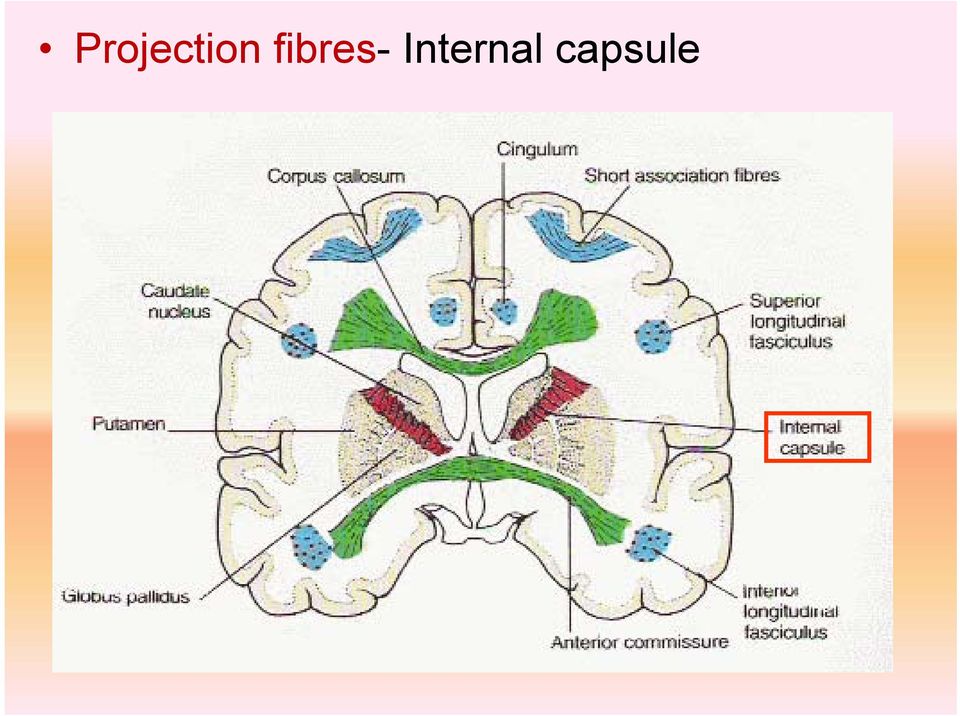
3 DEFINITION Projection fibres (white matter) between caudate nucleus and thalamus medially lentiform nucleus laterally

4 Internal Capsule- A compact bundle of fibres through which the large collections of fibres pass, including- Thalamocortical fibres Corticothalamic fibres Corticopontine fibres Corticobulbar fibres Corticospinal fibres

5 The fibres project from the cerebral cortex to the various nuclei of the extrapyramidal system (e.g., the putamen and caudate nucleus). It is a continuous sheet of fibres that forms the medial boundary of the lenticular nucleus. It continues around posteriorly and inferiorly to partially envelop this nucleus. Inferiorly, many of the fibres of the internal capsule funnel into the cerebral peduncles.

6 Superiorly, the fibres fan out into the corona radiata. Here, they travel in the cerebral white matter to reach their cortical origins or destinations. The internal capsule is divided into 5 regions: The anterior limb is the portion between the lenticular nucleus and the head of the caudate nucleus; The posterior limb is the portion between the lenticular nucleus and the thalamus; The genu is the portion at the junction of the above 2 parts and is adjacent to the interventricular foramen; The retrolenticular part is the portion posterior to the lenticular nucleus; The sublenticular part is the portion inferior to the lenticular nucleus.

7 Portion Description Origin Destination Anterior Limb Anterior thalamic Anterior nucleus Cingulate gyrus radiation DM Prefrontal cortex Genu Posterior Limb Retrolenticular Relays to motor areas 1. Motor pathways: o o Corticospinal tract Corticobulbar tract 2. Somatosensory relays Association relay VA VL 1. Motor cortex 2. VPL/VPM Pulvinar Premotor cortex Primary motor cortex 1. Motor pathways: o o Spinal cord Brainstem 2. Somatosensory relays: o Primary somatosentory cortex Association cortex Optic radiation LGN Visual cortex Sublenticular Optic radiation LGN Visual cortex Auditory radiation MGN Auditory cortex

8 Portion Descending fibres Ascending fibres Radiations Anterior Limb Frontopontine Frontothalamic Thalamofrontal Anterior thalamic radiation Genu Posterior Limb Frontopontine Corticonuclear Fibres carrying somaesthetic sensations from thalamus (VP nu.) to postcentral gyrus Frontopontine Corticospinal Corticorubral Corticoreticular Parietothalamic Thalamoparietal fibres Subthalamic fasciculus Superior or Dorsal thalamic radiation Retrolenticular Parietopontine Occipitopontine Corticorubral Occipitothalamic Optic radiation Thalamo-occipital Thalamo-parietal Posterior or Caudal thalamic radiation Sublenticular Parietopontine Temporopontine Temporothalamic Acoustic radiation Thalamotemporal Inferior thalamic radiation

9 CAUDATE NUCLEUS Internal capsule External capsule Globus palidus LENTIFORM NUCLEUS Extreme capsule Putamen THALAMUS CLAUSTRUM

10

11 Parts of internal capsule Caudate nucleus Anterior limb Genu Thalamus Lentiform nucleus Sublentiform Thalamocortical fibres Corticopontine fibres Corticonuclear & corticospinal fibres Posterior limb Retrolentiform

12 Types of fibres Thalamic radiation Superior thalamic radiation Anterior thalamic radiation Posterior thalamic radiation- Inferior thalamic radiation- Corticospinal Corticonuclear Corticopontine Fronto pontine Parieto pontine Temporo pontine Extrapyramidal Anterior Superior Inferior Posterior

13 Thalamic radiation Thalamocortical fibres Thalamic nuclei -project to ipsilateral cerebral cortex (except for reticular nucleus) Reach neocortex Located entirely within internal capsule Superior-from ventral nucleus Becomes corona radiata Anterior-from anterior & medial nuclei Posterior-from optic radiation Inferior-from auditory radiation

14 ANTERIOR LIMB Anterior thalamic radiation Frontopontine GENU Part of superior thalamic radiation Frontopontine Corticonuclear POSTERIOR LIMB Superior thalamic radiation Frontopontine Corticonuclear (corticobulbar) Corticospinal Extrapyrimidal RETROLENTIFORM Post thalamic radiation - Optic radiation Parieto-pontine Temporo-pontine SUBLENTIFORM Inf thalamic radiation - Auditory radiation Thalamocortical fibres Corticopontine fibres Corticonuclear & corticospinal fibres

15 BLOOD SUPPLY Lateral striate fr middle cerebral artery Ant limb Genu Post limb Basal ganglia Medial striate fr anterior cerebral artery Ant limb Genu Basal ganglia Ant choroidal fr internal carotid Post limb Retrolenticular part HORIZONTAL SECTION

16 CORONAL SECTION Lenticulostriate arteries Internal capsule Anterior cerebral artery Medialstriate arteries Middle cerebral artery Internal carotid artery

17 CORONAL SECTION Lateral striate -MCA Medial striate ACA Left middle cerebral artery Right middle cerebral artery

18 ANTERIOR LIMB Ant cerebral artery through medial striate br. Middle cerebral artery through lateral striate and lenticulostriate br. - pass through the lentiform N to supply the striate

19 GENU Ant cerebral artery through medial striate br. Middle cerebral artery through lateral striate and lenticulostriate br. Branches of internal carotid artery

20 CORONAL SECTION Lenticulostriate arteries Internal capsule Anterior cerebral artery Medialstriate arteries Middle cerebral artery Internal carotid artery

21 POSTERIOR LIMB Middle cerebral artery through lateral striate and lenticulostriate br. Charcot s artery of cerebral haemorrhage Anterior choroidal artery, direct branch of internal carotid artery Long and slender, thus has tendency to get thrombosis
22 INTERNAL CAPSULE CAUDATE NUCLEUS Globus palidus LENTIFORM NUCLEUS Putamen THALAMUS CLAUSTRUM
23 EXTERNAL CAPSULE CAUDATE NUCLEUS Globus palidus LENTIFORM NUCLEUS Putamen Projection fibres from cerebral cortex to basal ganglia and midbrain THALAMUS CLAUSTRUM
24 APPLIED ANATOMY Microaneurysm to lenticulostriate arteries - contralateral side of the body Hemiplegia Impaired sensation Paralysis of lower half of face Thrombosis recurrent br of ACA contralateral side of the body Upper limb Paralysis of lower half of face Anterior choroidal artery may be symptomless collateral circulation
NEUROANATOMY 6 Limbic System
 NEUROANATOMY 6 Limbic System The Limbic System The part of the brain involved with learning, memory and emotion. It is affected in many neuropsychiatric diseases including schizophrenia, Alzheimer s disease
1 PYRAMIDS - CORTICOSPINAL FIBERS
 151 Brain stem Pyramids/Corticospinal Tract 1 PYRAMIDS - CORTICOSPINAL FIBERS The pyramids are two elongated swellings on the ventral aspect of the medulla. Each pyramid contains approximately 1,000,000
Mini-atlas of the Marmoset Brain
 Mini-atlas of the Marmoset Brain http://marmoset-brain.org Aya Senoo Tokyo University of Agriculture and Technology Hironobu Tokuno Tokyo Metropolitan Institute of Medical Science Charles Watson Curtin
Sheep Brain Dissection
 Sheep Brain Dissection http://www.carolina.com/product/preserved+organisms/preserved+animals+%28mammal s%29/sheep+organs/preserved+sheep+dissection.do Michigan State University Neuroscience Program Brain
BIO130 Chapter 14 The Brain and Cranial Nerves Lecture Outline
 BIO130 Chapter 14 The Brain and Cranial Nerves Lecture Outline Brain structure 1. Cerebrum Hemispheres: left & right Cerebral cortex Gyri Sulci Fissures Longitudinal fissure Corpus callosum Lobes Central
CSE511 Brain & Memory Modeling. Lect04: Brain & Spine Neuroanatomy
 CSE511 Brain & Memory Modeling CSE511 Brain & Memory Modeling Lect02: BOSS Discrete Event Simulator Lect04: Brain & Spine Neuroanatomy Appendix of Purves et al., 4e Larry Wittie Computer Science, StonyBrook
Basal Ganglia. Motor systems
 409 systems Basal Ganglia You have just read about the different motorrelated cortical areas. Premotor areas are involved in planning, while MI is involved in execution. What you don t know is that the
Brain Matters: Brain Anatomy
 1 : Brain Anatomy Lesson Overview Students share what they already know about brain structure and function, and then, guided by descriptions of brain regions explore the G2C Online 3-D Brain to learn more
CENTRAL NERVOUS SYSTEM. Sensory Pathway (PNS) OVERVIEW OF SPINAL CORD ANATOMY OF THE SPINAL CORD FUNCTIONS OF THE SPINAL CORD
 OVERVIEW OF SPINAL CORD ANATOMY OF THE SPINAL CORD FUNCTIONS OF THE SPINAL CORD") CENTRAL NERVOUS SYSTEM Central nervous system (CNS) brain and spinal cord enclosed in bony coverings Functions of the spinal cord spinal cord reflexes integration ti (summation of inhibitory and excitatory)
CENTRAL NERVOUS SYSTEM Central nervous system (CNS) brain and spinal cord enclosed in bony coverings Functions of the spinal cord spinal cord reflexes integration ti (summation of inhibitory and excitatory)
OLFACTORY PATHWAYS AND LIMBIC SYSTEM
 903 Olfactory and Limbic OLFACTORY PATHWAYS AND LIMBIC SYSTEM I. OLFACTORY PATHWAYS The sense of smell is much less essential than vision, audition or the somatic senses, and will therefore receive less
DISSECTION OF THE SHEEP'S BRAIN
 DISSECTION OF THE SHEEP'S BRAIN Introduction The purpose of the sheep brain dissection is to familiarize you with the threedimensional structure of the brain and teach you one of the great methods of studying
MEDULLA OBLONGATA AND PONS form lower part of brainstem (oblongata, pons, midbrain)
") MEDULLA OBLONGATA AND PONS form lower part of brainstem (oblongata, pons, midbrain) Medulla oblongata - is uper continuation of the spinal cord Its caudal part is alike the spinal cord, while - its cranial
MEDULLA OBLONGATA AND PONS form lower part of brainstem (oblongata, pons, midbrain) Medulla oblongata - is uper continuation of the spinal cord Its caudal part is alike the spinal cord, while - its cranial
The Normal Neurological Examination
 Normal Neurological Examination 1 of 10 The Normal Neurological Examination Introduction One of the most daunting aspects of the medical examination for the Emergency Physician and the primary care provider
Nervous System Organization. PNS and CNS. Nerves. Peripheral Nervous System. Peripheral Nervous System. Motor Component.
 Nervous System Organization PNS and CNS Chapters 8 and 9 Peripheral Nervous System (PNS) connects CNS to sensory receptors, muscles and glands Central Nervous System (CNS) control/integrating center brain
Adapted from Human Anatomy & Physiology by Marieb and Hoehn (9 th ed.)
") BRAIN ANATOMY Adapted from Human Anatomy & Physiology by Marieb and Hoehn (9 th ed.) The anatomy of the brain is often discussed in terms of either the embryonic scheme or the medical scheme. The embryonic
BRAIN ANATOMY Adapted from Human Anatomy & Physiology by Marieb and Hoehn (9 th ed.) The anatomy of the brain is often discussed in terms of either the embryonic scheme or the medical scheme. The embryonic
Unit 2 - Subcortical systems, neurochemistry and brain function
 Unit 2 - Subcortical systems, neurochemistry and brain function Subcortical anatomy: Most of the five major subdivisions of the brain are subcortical. I. Telencephalon (cortical - part of forebrain) -
Introduction to the Structure and Function of the Central Nervous System
 CHAPTER 3 Introduction to the Structure and Function of the Central Nervous System GENERAL TERMINOLOGY AN OVERVIEW OF THE CENTRAL NERVOUS SYSTEM The Central and Peripheral Nervous Systems Major Divisions
Cerebellum and Basal Ganglia
 Cerebellum and Basal Ganglia 1 Contents Cerebellum and Basal Ganglia... 1 Introduction... 3 A brief review of cerebellar anatomy... 4 Basic Circuit... 4 Parallel and climbing fiber input has a very different
1. Which of the following is NOT part of the diencephalon? a. Pineal gland b. Tectum c. Interthalamic adhesion d. Hypothalamus e.
 1. Which of the following is NOT part of the diencephalon? a. Pineal gland b. Tectum c. Interthalamic adhesion d. Hypothalamus e. Thalamus 2. The is the primary relay station for sensory information coming
The Hypothalamus. Functions of the Hypothalamus. The Hypothalamus. Medical Neuroscience Dr. Wiegand
 The Medical Neuroscience Dr. Wiegand Neural Influences Neural Influences Hormonal Influences Hormonal Influences The Autonomic Nervous System Limbic System Endocrine System system Endocrine 1 The Diencephalon
Transverse Sections of the Spinal Cord
 Transverse Sections of the Spinal Cord The spinal cord is perhaps the most simply arranged part of the CNS. Its basic structure, indicated in a schematic drawing of the eighth cervical segment (Figure
Chapter 15: Neural Integration I: Sensory Pathways and the Somatic Nervous System
 Chapter 15: Neural Integration I: Sensory Pathways and the Somatic Nervous System I. An Overview of Sensory Pathways and the Somatic Nervous System, p. 496 Figure 15-1 Specialized cells called sensory
CHAPTER 11: NERVOUS SYSTEM II: DIVISIONS OF THE NERVOUS SYSTEM OBJECTIVES: 1. Outline the major divisions of the nervous system.
 CHAPTER 11: NERVOUS II: DIVISIONS OF THE NERVOUS OBJECTIVES: 1. Outline the major divisions of the nervous system. NERVOUS CENTRAL NERVOUS (BRAIN & SPINAL CORD) (INTERNEURONS) PERIPHERAL NERVOUS (CRANIAL
Function (& other notes)
") LAB 8. ANATOMY OF THE HUMAN BRAIN In this exercise you each will map the human brain both anatomy and function so that you can develop a more accurate picture of what s going on in your head :-) EXTERNAL
LAB 8. ANATOMY OF THE HUMAN BRAIN In this exercise you each will map the human brain both anatomy and function so that you can develop a more accurate picture of what s going on in your head :-) EXTERNAL
1 Cornea 6 Macula 2 Lens 7 Vitreous humor 3 Iris 8 Optic disc 4 Conjunctiva 9 Ciliary muscles 5 Sclera 10 Choroid
 Anatomy and Physiology Quiz 1 Sample Question Answers Use the following table to answer Questions 1 2. 1 Cornea 6 Macula 2 Lens 7 Vitreous humor 3 Iris 8 Optic disc 4 Conjunctiva 9 Ciliary muscles 5 Sclera
2401 : Anatomy/Physiology
 Dr. Chris Doumen Week 7 2401 : Anatomy/Physiology The Brain Central Nervous System TextBook Readings Pages 431 through 435 and 463-467 Make use of the figures in your textbook ; a picture is worth a thousand
Ascending Sensory Pathways
 CHAPTER 10 Ascending Sensory Pathways CLINICAL CASE SENSORY RECEPTORS ANTEROLATERAL SYSTEM TACTILE SENSATION AND PROPRIOCEPTION SENSORY PATHWAYS TO THE CEREBELLUM CLINICAL CONSIDERATIONS MODULATION OF
NEUROLOCALIZATION MADE EASY
 NEUROLOCALIZATION MADE EASY Jared B. Galle, DVM, Diplomate ACVIM (Neurology) Dogwood Veterinary Referral Center 4920 Ann Arbor-Saline Road Ann Arbor, MI 48103 Localizing a neurologic problem to an anatomical
About Brain Injury: A Guide to Brain Anatomy Information from http://www.waiting.com, 1997-2002, Becca, Ltd.
 About Brain Injury: A Guide to Brain Anatomy Information from http://www.waiting.com, 1997-2002, Becca, Ltd. Brain Anatomy Definitions Brainstem: The lower extension of the brain where it connects to the
Human Anatomy & Physiology Spinal Cord, Spinal Nerves and Somatic Reflexes 13-1
 Human Anatomy & Physiology Spinal Cord, Spinal Nerves and Somatic Reflexes 13-1 Spinal Cord, Spinal Nerves and Somatic Reflexes Spinal cord Spinal nerves Somatic reflexes 13-2 Overview of Spinal Cord Information
3) Cerebral Cortex & Functions of the 4 LOBES. 5) Cranial Nerves (Nerves In the Cranium, i.e., Head)
 Cerebral Cortex & Functions of the 4 LOBES. 5) Cranial Nerves (Nerves In the Cranium, i.e., Head)") Lecture 5 (Oct 8 th ): ANATOMY and FUNCTION OF THE NERVOUS SYSTEM Lecture Outline 1) Basic Divisions (CNS vs. PNS, Somatic vs. Autonomic) and Directional Terms 2) The Brain (Hindbrain/ Midbrain/ Forebrain)
Lecture 5 (Oct 8 th ): ANATOMY and FUNCTION OF THE NERVOUS SYSTEM Lecture Outline 1) Basic Divisions (CNS vs. PNS, Somatic vs. Autonomic) and Directional Terms 2) The Brain (Hindbrain/ Midbrain/ Forebrain)
Movement Disorders and Extrapyramidal System. Doç.. Dr. Sibel Ertan
 Movement Disorders and Extrapyramidal System Doç.. Dr. Sibel Ertan İ.Ü.. CTF. Nöroloji N ABD. Motor System I. neuron (corticospinal pathway) Extrapyramidal system (basal ganglia) Cerebellum Loops for praxis
Myelinization. THOMAS P. NAIDICH, MD FACR Mt. Sinai Medical Center New York, NY USA
 Myelinization THOMAS P. NAIDICH, MD FACR Mt. Sinai Medical Center New York, NY USA ALTERS BRAIN WATER LOCALLY MYELIN CONTAINS: GLYCOLIPIDS PHOSPHOLIPIDS & CHOLESTEROL Maturation of the White Matter Maturation
THE BRAIN, SPINAL CORD, AND CRANIAL NERVES
 THE BRAIN, SPINAL CORD, AND CRANIAL NERVES I. BRAIN ANATOMY A. Meninges (coverings) of the brain and spinal cord (Fig. [13.120 p. 452 [457]) Use text illustrations to study these. Note that the singular
3D Slicer. John Muschelli and Vadim Zipunnikov. Department of Biostatistics. November 18, 2011
 3D Slicer John Muschelli and Vadim Zipunnikov Department of Biostatistics November 18, 2011 John Muschelli and Vadim Zipunnikov (JHU) 3D-Slicer November 18, 2011 1 / 39 What is Slicer http://www.slicer.org;
Anatomy of the Brain > 1. Figure 1. Eight bones form the skull and fourteen bones form the face.
 Anatomy of the Brain Overview The human brain is an amazing three-pound organ that controls all functions of the body, interprets information from the outside world, and embodies the essence of the mind
Nervous System sensor input integration motor output sensory organs central nervous system
 Nervous System Nervous system performs three overlapping functions of sensor input, integration, and motor output. This process is generally the same even at a very primitive level of nervous system, but
Chapter 3 The Anatomy of the Nervous System
 Chapter 3 The Anatomy of the Nervous System Systems, Structures, and Cells That Make Up Your Nervous System 1 General Layout of the Nervous System Central Nervous System (CNS) Brain (in the skull) Spinal
Autonomic Nervous System of the Neck. Adam Koleśnik, MD Department of Descriptive and Clinical Anatomy Center of Biostructure Research, MUW
 Autonomic Nervous System of the Neck Adam Koleśnik, MD Department of Descriptive and Clinical Anatomy Center of Biostructure Research, MUW Autonomic nervous system sympathetic parasympathetic enteric Autonomic
Neuroanatomy and Cortical Landmarks
 Neuroanatomy and Cortical Landmarks 2 Stephan Ulmer 2.1 Neuroanatomy and Cortical Landmarks of Functional Areas Prior to any type of functional mapping, a profound knowledge of neuroanatomy is mandatory.
Vocabulary & General Concepts of Brain Organization
 Vocabulary & General Concepts of Brain Organization Jeanette J. Norden, Ph.D. Professor Emerita Vanderbilt University School of Medicine Course Outline Lecture 1: Vocabulary & General Concepts of Brain
Lab Exercise 9. Nervous Tissue. Brain. Cranial Nerves. Spinal Cord. Spinal Nerves
 Lab Exercise 9 Nervous Tissue Brain Cranial Nerves Spinal Cord Spinal Nerves Textbook Reference: See Chapter 11 for histology of nerve tissue and spinal cord See Chapter 12 for brain and spinal cord anatomy
MEDIAL TEMPORAL LOBE (THE LIMBIC SYSTEM)
") MEDIAL TEMPORAL LOBE (THE LIMBIC SYSTEM) On the medial surface of the temporal lobe are three structures critical for normal human functioning. From rostral to caudal, they are the olfactory cortex, the
MEDIAL TEMPORAL LOBE (THE LIMBIC SYSTEM) On the medial surface of the temporal lobe are three structures critical for normal human functioning. From rostral to caudal, they are the olfactory cortex, the
Effective SLP Interventions for Children with Cerebral Palsy
 Effective SLP Interventions for Children with Cerebral Palsy NDT/Traditional/Eclectic, PhD, CCC-SLP, C/NDT Contents Preface Acknowledgments Contributors ix x xi Chapter 1. The Development in Neurodevelopmental
THE BRAIN AND CRANIAL NERVES
 THE BRAIN AND CRANIAL NERVES The Brain - made up of a trillion neurons - weighs about 3 lbs - has four principle parts 1. Brain stem - medulla oblongata, pons, midbrain (mesencephalon) 2. Diencephalon
Wessex Neurological Centre. Neuro-anatomy & Physiology Workbook
 Wessex Neurological Centre Neuro-anatomy & Physiology Workbook Sarah Irwin, Senior Sister NICU, SUHT & Rachel Palmer, Lecturer Practitioner Neurosciences (SUHT & School of Health Professionals, University
Human Neuroanatomy. Grades 9-12. Driving Question: How did the evolution of the human brain impact the structure and function it has today?
 Human Neuroanatomy Grades 9-12 Driving Question: How did the evolution of the human brain impact the structure and function it has today? Objectives: Students will be able to Describe the basic parts and
Chapter 9 - Nervous System
 Chapter 9 - Nervous System 9.1 Introduction (p. 215; Fig. 9.1) A. The nervous system is composed of neurons and neuroglia. 1. Neurons transmit nerve impulses along nerve fibers to other neurons. 2. Nerves
Structure of the Kidney Laboratory Exercise 56
 Structure of the Kidney Laboratory Exercise 56 Background The two kidneys are the primary organs of the urinary system. They are located in the upper quadrants of the abdominal cavity, against the posterior
Theme 39. Anatomy of the brain stem. Medulla oblongata, its external and internal structure. The construction of the bridge. Reticulated formation.
 Theme 39. Anatomy of the brain stem. Medulla oblongata, its external and internal structure. The construction of the bridge. Reticulated formation. BRAINSTEM. MEDULLA OBLONGATA (EXTERNAL AND INTERNAL STRUCTURE).
Cranial Nerves CHAPTER 15 CLINICAL CASE
 CHAPTER 15 Cranial Nerves CLINICAL CASE OLFACTORY NERVE (CN I) OPTIC NERVE (CN II) OCULOMOTOR NERVE (CN III) TROCHLEAR NERVE (CN IV) TRIGEMINAL NERVE (CN V) ABDUCENT NERVE (CN VI) FACIAL NERVE (CN VII)
Motor dysfunction 2: Spinal cord injury and subcortical motor disorders ANATOMY REVIEW: Basal Ganglia
 Motor dysfunction 2: Spinal cord injury and subcortical motor disorders ANATOMY REVIEW: Basal Ganglia A group of subcortical nuclei caudate, putamen, globus pallidus Caudate & Putamen = Neostriatum caudate
Peripheral Nervous System
 Nervous system consists of: Peripheral Nervous System CNS = brain and spinal cord ~90% (90 Bil) of all neurons in body are in CNS PNS = Cranial nerves and spinal nerves, nerve plexuses & ganglia ~10% (10
NEURO M203 & BIOMED M263 WINTER 2014
 NEURO M203 & BIOMED M263 WINTER 2014 MRI Lab 1: Structural and Functional Anatomy During today s lab, you will work with and view the structural and functional imaging data collected from the scanning
Page 1 of 10 MBBS FIRST PROFESSIONAL (PART- I EXAMINATION) Anatomy (MCQs) Model Paper. Marks 45 Time 45 minutes
 Anatomy (MCQs) Model Paper. Marks 45 Time 45 minutes") Page 1 of 10 Marks 45 Time 45 minutes 01. The plane of reference that divides the body into anterior and posterior portions is: A. Sagittal. B. Transverse. C. Coronal. D. Cross-sectional. E. Oblique. Sub-
Page 1 of 10 Marks 45 Time 45 minutes 01. The plane of reference that divides the body into anterior and posterior portions is: A. Sagittal. B. Transverse. C. Coronal. D. Cross-sectional. E. Oblique. Sub-
Figure 2 Figure 3 Figure 1
 The brain is organized into three tiers; a lower tier made up by the brainstem and cerebellum, a middle tier containing the thalamus, basal ganglia and many components of the limbic system and an upper
MCQ : Central Nervous System. General Functional Organization of the Nervous System
 Section 1 MCQ : Central Nervous System General Functional Organization of the Nervous System 1 ) The central nervous system includes all the following components, except :- a- spinal cord b- medulla oblongata
Primary Motor Pathway
 Understanding Eye Movements Abdullah Moh. El-Menaisy, MD, FRCS Chief, Neuro-ophthalmology ophthalmology & Investigation Units, Dhahran Eye Specialist Hospital, Dhahran, Saudi Arabia Primary Motor Pathway
Weakness More diffuse More focal Atrophy Mild, general Severe, focal Atrophy versus weakness
 Spinal Cord Disorders (Dr. Merchut) Clinical signs and symptoms in spinal cord lesions 1. Motor signs and symptoms Lower motor neuron (LMN) signs (Table 1) are found in a limb if some of its muscles are
Nervous System: Spinal Cord and Spinal Nerves (Chapter 13) Lecture Materials for Amy Warenda Czura, Ph.D. Suffolk County Community College
 Lecture Materials for Amy Warenda Czura, Ph.D. Suffolk County Community College") Nervous System: Spinal Cord and Spinal Nerves (Chapter 13) Lecture Materials for Amy Warenda Czura, Ph.D. Suffolk County Community College Primary Sources for figures and content: Eastern Campus Marieb,
Nervous System: Spinal Cord and Spinal Nerves (Chapter 13) Lecture Materials for Amy Warenda Czura, Ph.D. Suffolk County Community College Primary Sources for figures and content: Eastern Campus Marieb,
Chapter 13: The Spinal Cord, Spinal Nerves, and Spinal Reflexes
 Chapter 13: The Spinal Cord, Spinal Nerves, and Spinal Reflexes I. General Organization of the Nervous System, p. 422 Objectives 1. Describe the basic structural and organizational characteristics of the
The intermedius nucleus of the medulla: A potential site for the integration of cervical information and the generation of autonomic responses
 The intermedius nucleus of the medulla: A potential site for the integration of cervical information and the generation of autonomic responses 1 Journal of Chemical Neuroanatomy November 2009, 38, pp.
Course Syllabus [NEUROSCI 380L] Course description. Course instructors. NEUROSCI 380L Functional Neuroanatomy
![Course Syllabus [NEUROSCI 380L] Course description. Course instructors. NEUROSCI 380L Functional Neuroanatomy](/thumbs/24/4244909.jpg "Course Syllabus [NEUROSCI 380L] Course description. Course instructors. NEUROSCI 380L Functional Neuroanatomy") [NEUROSCI 380L] Course Syllabus Course description The focus of this course is the structure of the human brain and spinal cord, and the functional organization of the central nervous system. This course
[NEUROSCI 380L] Course Syllabus Course description The focus of this course is the structure of the human brain and spinal cord, and the functional organization of the central nervous system. This course
The Visual Cortex 0 http://www.tutis.ca/neuromd/index.htm 20 February 2013
 T he Visual Cortex 0 Chapter contents Contents Chapter 2... 0 T he Visual Cortex... 0 Chapter Contents... 1 Introduction... 2 Optic Chiasm... 2 Where do the eye's ganglion cells project to?... 3 To where
1. Clinical Consequences of Stroke
 1. Clinical Consequences of Stroke Robert Teasell MD, Norhayati Hussein MBBS MRehabMed, Ricardo Viana MD, Sarah Donaldson BHSc, Mona Madady MSc Cerebrovascular disorders represent the third leading cause
BIOL 1108 Vertebrate Anatomy Lab
 BIOL 1108 Vertebrate Anatomy Lab This lab explores major organs associated with the circulatory, excretory, and nervous systems of mammals. Circulatory System Vertebrates are among the organisms that have
of computational neuroscience. The chapter is organized as follows. Section 1 describes the
 Chapter 1 This chapter presents elements of neurobiology that form the necessary preparation for a student of computational neuroscience. The chapter is organized as follows. Section 1 describes the biology
DIAGNOSIS AND MANAGEMENT OF HEMIPLEGIA
 P a g e 1 DIAGNOSIS AND MANAGEMENT OF HEMIPLEGIA INTRODUCTION Hemiplegia is paralysis of one half of the body-which includes arm, leg and often face on the affected side. Terms used to describe weakness
Introduction. I. Objectives. II. Introduction. A. To become familiar with the terms of direction and location.
 E X E R C I S E Introduction I. Objectives A. To become familiar with the terms of direction and location. B. To become familiar with different types of planes and sections. C. To learn the names and locations
Vision: Receptors. Modes of Perception. Vision: Summary 9/28/2012. How do we perceive our environment? Sensation and Perception Terminology
 How do we perceive our environment? Complex stimuli are broken into individual features, relayed to the CNS, then reassembled as our perception Sensation and Perception Terminology Stimulus: physical agent
Sonographic Demonstration of Couinaud s Liver Segments
 PICTORIL ESSY Sonographic Demonstration of Couinaud s Liver Segments Dean Smith, MD, FRCPC, Donal Downey, M, Ch, FRCPC, lison Spouge, MD, FRCPC, Sue Soney, RT, RDMS, RCMS The segmental localization of
The Brain of a Normal Human
 The Brain of a Normal Human Your Brain Evolved Over Time Human Brain Logic and reasoning Mammalian Brain More complex feelings and reactions Lizard Brain Basic functions The Brain Stem or Hindbrain (The
Structure and Function of Neurons
 CHPTER 1 Structure and Function of Neurons Varieties of neurons General structure Structure of unique neurons Internal operations and the functioning of a neuron Subcellular organelles Protein synthesis
GAZE STABILIZATION SYSTEMS Vestibular Ocular Reflex (VOR) Purpose of VOR Chief function is to stabilize gaze during locomotion. Acuity declines if
 Purpose of VOR Chief function is to stabilize gaze during locomotion. Acuity declines if") GAZE STABILIZATION SYSTEMS Vestibular Ocular Reflex (VOR) Purpose of VOR Chief function is to stabilize gaze during locomotion. Acuity declines if slip exceeds 3-5 deg/sec. Ex: Head bobbing and heel strike
GAZE STABILIZATION SYSTEMS Vestibular Ocular Reflex (VOR) Purpose of VOR Chief function is to stabilize gaze during locomotion. Acuity declines if slip exceeds 3-5 deg/sec. Ex: Head bobbing and heel strike
III./8.4.2: Spinal trauma. III./8.4.2.1 Injury of the spinal cord
 III./8.4.2: Spinal trauma Introduction Causes: motor vehicle accidents, falls, sport injuries, industrial accidents The prevalence of spinal column trauma is 64/100,000, associated with neurological dysfunction
How To Understand The Function Of The Basal Ganglia
 122 NEUROCIRCUITRY OF PARKINSON S DISEASE THOMAS WICHMANN MAHLON R. DELONG Recent progress in neuroscience research has led to major insights into the structure and function of the basal ganglia and into
The Anatomy of Spinal Cord Injury (SCI)
") The Anatomy of Spinal Cord Injury (SCI) What is the Spinal Cord? The spinal cord is that part of your central nervous system that transmits messages between your brain and your body. The spinal cord has
The Anatomy of Spinal Cord Injury (SCI) What is the Spinal Cord? The spinal cord is that part of your central nervous system that transmits messages between your brain and your body. The spinal cord has
Doctor, I See Double : Managing Cranial Nerve Palsies
 1 Doctor, I See Double : Managing Cranial Nerve Palsies Joseph W. Sowka, OD, FAAO, Diplomate Professor of Optometry Nova Southeastern University, College of Optometry 3200 South University Drive Fort Lauderdale,
Nervous System: PNS and CNS
 Nervous System: PNS and CNS Biology 105 Lecture 10 Chapter 8 Outline I. Central Nervous System vs Peripheral Nervous System II. Peripheral Nervous System A. Somatic Nervous System B. Autonomic Nervous
2 Neurons. 4 The Brain: Cortex
 1 Neuroscience 2 Neurons output integration axon cell body, membrane potential Frontal planning control auditory episodes soma motor Temporal Parietal action language objects space vision Occipital inputs
Parts of the Brain. Chapter 1
 Chapter 1 Parts of the Brain Living creatures are made up of cells. Groups of cells, similar in appearance and with the same function, form tissue. The brain is a soft mass of supportive tissues and nerve
SPINAL CORD CIRCUITS AND MOTOR CONTROL
 OVERVEW The proximate control of movement is provided by neurons in the spinal cord and brainstem. The primary motor neurons located in the ventral horn of the spinal cord gray matter (and the corresponding
3. The neuron has many branch-like extensions called that receive input from other neurons. a. glia b. dendrites c. axons d.
 Chapter Test 1. A cell that receives information and transmits it to other cells via an electrochemical process is called a(n) a. neuron b. hormone c. glia d. endorphin Answer: A difficulty: 1 factual
An Introduction To Human Neuroanatomy
 An Introduction To Human Neuroanatomy Harvard Brain Tissue Resource Center, McLean Hospital, Belmont, MA 02478, 1-800-BRAIN BANK. Created by Tim Wheelock, Assistant Director of Neuropathology/Instructor
IV. DEFINITION OF LYMPH NODE GROUPS (FIGURE 1) Level IA: Submental Group
 Level IA: Submental Group") IV. DEFINITION OF LYMPH NODE GROUPS (FIGURE 1) Fig. 1 The level system is used for describing the location of lymph nodes in the neck: Level I, submental and submandibular group; Level II, upper jugular
IV. DEFINITION OF LYMPH NODE GROUPS (FIGURE 1) Fig. 1 The level system is used for describing the location of lymph nodes in the neck: Level I, submental and submandibular group; Level II, upper jugular
Circuit Disorders of the Basal Ganglia: Parkinson s Disease Pathophysiology and Surgical Treatments
 Circuit Disorders of the Basal Ganglia: Parkinson s Disease Pathophysiology and Surgical Treatments Mahlon R. DeLong M.D. W. P. Timmie Professor of Neurology Emory University School of Medicine The Basal
Vertebral anatomy study guide. Human Structure Summer 2015. Prepared by Daniel Schmitt, Angel Zeininger, and Karyne Rabey.
 Vertebral anatomy study guide. Human Structure Summer 2015 Prepared by Daniel Schmitt, Angel Zeininger, and Karyne Rabey. 1. Plan of Action: In this guide you will learn to identify these structures: Cervical
Sheep Brain Dissection Picture Guide
 Sheep Brain Dissection Picture Guide Figure 1: Right Hemisphere of Sheep s Brain Figure 2: Underside of Sheep s Brain Figure 3: Saggital cut of Sheep s Brain to reveal subcortical structures Figure 4:
Nervous System Pathology
 Nervous System Pathology Nervous System Central Nervous System CNS Brain & Spinal cord Nervous System Peripheral Nervous System PNS Spinal and cranial nerves CNS Close relationship with endocrine system
Chapter 9 Nervous System
 Chapter 9 Nervous System Nervous System function: The nervous system is composed of neurons and neuroglia. at the ends of peripheral nerves gather information and convert it into nerve impulses. When sensory
Parkinson s Disease: A Review of Motor and Nonmotor Symptoms
 Diana Hong Parkinson s Disease: A Review of Motor and Nonmotor Symptoms Abstract: Although Parkinson s disease (PD) is considered a motor system disorder caused by the degeneration of dopaminergic neurons
Welcome to Anatomy & Physiology
 Welcome to Anatomy & Physiology Chapter 1 -Human Organization What do you need to do to pass this class? MEMORIZE! The Scope of Human Anatomy Human anatomy is the study of the structure of the human body.
The Human Nervous System
 The Human Nervous System S tructure and Function S ixth Ed ition The Human Nervous System Structure and Function S ixth Edition Charles R. Noback, PhD Professor Emeritus Department of Anatomy and Cell