ENG4BF3 Medical Image Processing. Image Visualization
|
|
|
- Belinda Preston
- 10 years ago
- Views:
Transcription
1 ENG4BF3 Medical Image Processing Image Visualization

2 Visualization Methods Visualization of medical images is for the determination of the quantitative information about the properties of anatomic tissues and their functions that relate to and are affected by disease For 2D visualization, three types of multiplanar sectioning and display, including orthogonal, oblique, and curved planes For 3D visualization, two types of display, including surface renderings and volume renderings (both projection and surface types) 2

3 Two-dimensional Image Generation and Visualization The utility of 2D images depends on the physical orientation of the image plane with respect to the structure of interest Most biomedical imaging systems have limited capability to create optimal 2D image directly, as structure positioning and scanner orientation are generally restricted We need techniques to generate and display optimal 2D images from 3D images, and to allow the orientation of 2D image plane to ultimately result in clear, unrestricted view of important features 3

4 Multiplanar Reformating 3D isotropic volume images allow for simple and efficient computation of images that lie along the nonacquired orthogonal orientations of the volume This is achieved by readdressing the order of voxels in the volume images With anatomic reference, the orthogonal planes can be depicted by the terms for orthogonal orientation: transaxial, coronal, and sagittal Display of multiplanar images usually consists of multipanel displays 4

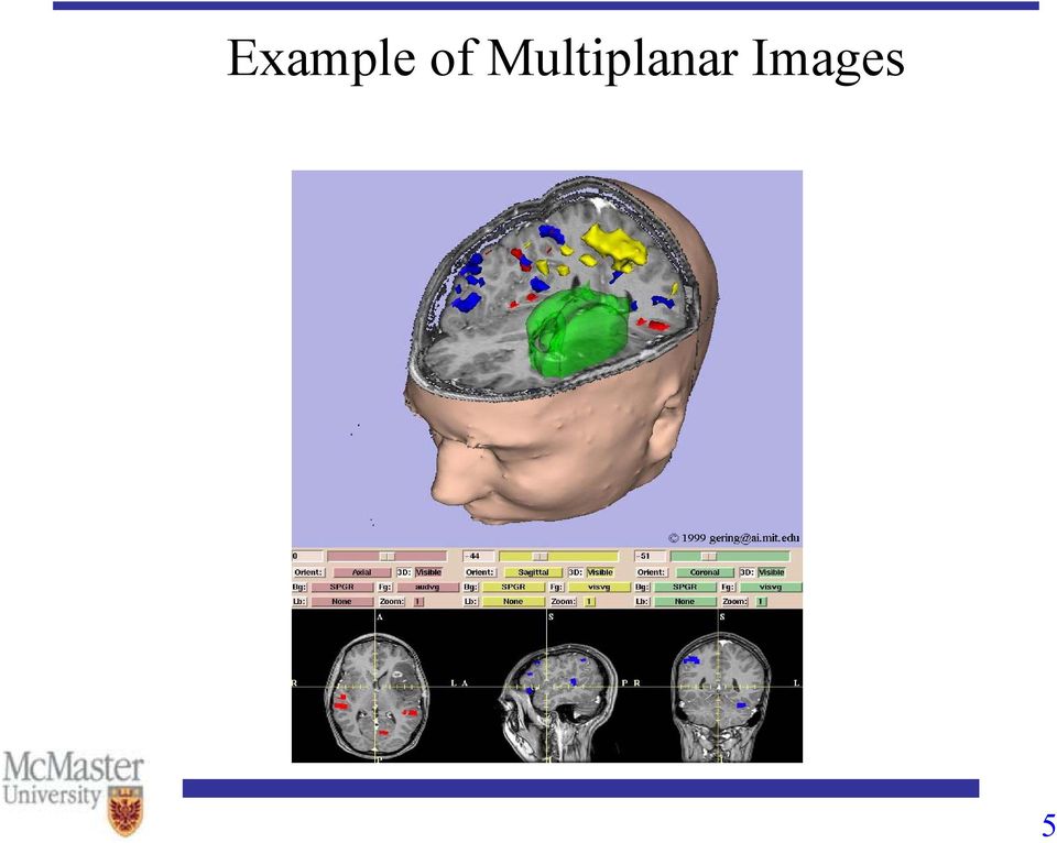
5 Example of Multiplanar Images 5
6 Multiplanar Image (Lung) Axial Coronal Sagittal-right Sagittal-left 6

7 Multiplanar Sectioning 7

8 Oblique Sectioning (I) The desired 2D image may not be parallel to the orthogonal orientation of the 3D image Oblique images are less intuitive and harder to compute, as simple readdressing of the voxels in a given order will not generate the proper oblique image Specification of the orientation and efficient generation of the oblique image require additional visualization and compuation techniques 8

9 Oblique Sectioning (II) Specification and identification of oblique image can be done using structural landmarks in the orthogonal image data Several images need to be presented to allow unambiguous selection of landmarks, through multiplanar orthogonal images Selection of any three points or landmarks will uniquely define the orientation of the plane Two selected points define an axis along which oblique planes perpendicular to the axis can be generated (not uniquely) 9

10 Oblique Sectioning (III) Interactive oblique sectioning involves some method of visualization for plane placement and orientation verification One method is to superimpose a line on orthogonal images indicating the intersection of the oblique images with the orthogonal images Another depicts the intersection of oblique plane with a rendered surface image providing direct 3D visual feedback with familiar structural features 10

11 Example of Oblique Sectioning (II) 11

12 Curved Sectioning A trace along an arbitrary path on any orthogonal image defines a set of pixels in the orthogonal image that have a corresponding row of voxels through the volume image Each row of voxels for each pixel on the trace can be displayed as a line of a new image This is useful for curved structures that remain constant in shape through one orthogonal dimension, like the spinal canal, orbits of the eyes 12

13 Example of Curved Sectioning 13

14 Three-dimensional Image Generation and Display Visualization of 3D volume images can be divided into two techniques characteristically: surface rendering and volume rendering Both produce a visualization of selected structures in the 3D volume image, but the methods involved are quite different Selection of techniques is predicted on the particular nature of the biomedical image data, the application to which the visualization is being applied, and the desired result of visualization 14

15 Surface Rendering Surface rendering techniques require the extraction of contours (edges) that define the surface of the structure to be visualized An algorithm is applied to place surface patches or tiles at each contour point, and with hidden surface removal and shading, the surface is rendered visible They use a relatively small amount of contour data, resulting in fast rendering speeds 15

16 Surface Rendering (II) Standard computer graphics techniques can be applied, including shading models Particular graphics hardware of the computer can be utilized to speed up the geometric transformation and rendering processes Contour descriptions are transformed into analytical descriptions Other analytically defined structures can be easily superposed with the surface-rendered structures 16

17 Surface Rendering - Drawbacks The need to discretely extract the contours defining the structures to be visualized Other volume image information may be lost in the extraction process, which may be important for slice generation or value measurement Difficult to interactive and dynamic determination of the surface to be rendered Prone to sampling and aliasing artifacts on the rendered surface due to the discrete nature of the surface patch placement 17

18 4.2e: Surface Rendering Examples 18

19 A Volume Date Set of Voxels 19

20 Transverse (Axial) Slices in the XY Plane 20

21 Sagittal Slices are in the ZY Plane 21
22 Coronal Slices are in the ZX Plane 22
23 What is the View of this Slice? 23
24 What is the View of this Slice? 24
25 Interpolation and Transformation Most 3D biomedical volume images are sampled anisotropically, with the slice thickness often significantly greater than the in-plane pixel size Visualization and measurement usually require volume data to be isotropic, and the data must be post-processed before it can be used properly Possible problems for anisotropic data: incorrect aspect ratio along each dimension for visualization, and aliasing artifacts due to difference in sampling frequency 25
26 Interpolation First-order linear interpolation of the gray level values is commonly used for 3D volume image Values of new pixels between existing pixels are simply interpolated (averaged) values of the existing pixels. It is a tri-linear interpolation in 3D. The interpolation in each of the dimensions is completely separable Tri-linear interpolation works well for most biomedical images 26
27 High Order Interpolation When the ratio in size between any of the voxel dimensions in 3D becomes greater than approximately 5 to 1, tri-linear interpolation does not work well, which will provide poor approximations High order interpolation, such as cubic interpolation, may be used, with increased computational cost Cubic interpolation uses more than the immediate adjacent voxels and uses a cubic polynomial to estimate the intermediate values Shape-based interpolation methods may be used for certain particular (known) structure of interest 27
28 Volume Rendering (I) The most popular volume rendering methods in 3D biomedical image visualization are ray-casting algorithms They provide direct visualization of the volume images without the need for prior surface or object segmentation, preserving the values and context of the original image data The rendered surface can be dynamically determined by changing the ray-casting and surface recognition conditions during the rendering process 28
29 Volume Rendering (II) Volume rendering can display surfaces with shading and other parts of the volume simultaneously 3D biomedical volume image data sets are characteristically large, taxing the computation abilities of volume rendering algorithms and the computer systems Given the discrete voxel-based nature of the volume image, there is no direct connection to other geometric objects, which may be desired for inclusion in the rendering or for output of the rendered structure to other devices 29
30 Example Volume Rendering (I) 30
31 Example of Volume Rendering (II) Rendered from CT scans 256X256X226 31
32 A Volume Rendering Model A source point (the eye) A focal point (where the eye is looking) A matrix of pixels (the screen) The visible object to display (scene) is in the projection path within a truncated volume (viewing pyramid) The purpose of a ray-tracing model is to define geometry of the rays cast through the scene A ray is defined as a straight line from the source point passing through the pixel 32
33 Diagram of Ray Casting Rendering 33
34 High Quality Volume Rending 34
35 CT Angiography 35
36 Surgery Planning and Rehearsal Patients with brain tumors, arterial venous malformations, or other complicated internal brain pathology undergo multimodality image scanning preoperatively to help neurosurgeons understand the anatomy of interest Direct scans can be co-registered in order to produce single visualizations of complementary information Specific anatomical objects may be identified and segmented 36
37 Neurosurgery Visualization procedures enable more precise and expedient navigation to the target site and provide more accurate delineation of margins for resection in brain surgery than traditional procedures It can provide online, updated information to accommodate any shifts in brain position during the operational procedure This results in significantly increased physician performance, reduction in time in the operating room, and an associated increase in patient throughput and decrease in healthcare costs 37
38 Brain Internal Tissues 38
39 Isolating Brain Tumor 39
40 Understanding Environment 40
41 Storage of Biomedical Images Biomedical images can usually be stored in slow storage media as they are not viewed frequently Design decisions for storage are based on the needs of the practitioners who use images, and balanced against the cost of data storage and software systems needed to make them accessible, reliable, and durable New standards, such as DICOM (Digital Imaging and Communications in Medicine), are evolving that will combine the universal access of film with the flexibility of digital imagery 41
42 DICOM DICOM (Digital Image Communications in Medicine) provides a protocol for transmission of image based on their modality, and incorporates metadata for each image within the message Each DICOM image message provides the basic information required to attach it to a patient or an imaging procedure, encoded information may be redundant DICOM requires the sending and receiving computers to agree on a common basic method of communication, and a set of well-defined services (such as image storage) specified before the message is sent 42
43 Metadata: The Image Header The first part of many image files provides a description of the image file header The information in the header is called the metadata (the information about the image) Some file formats use formatted header fields to describe the image, e.g., information can be image type, height and width of the image Flexible fields such as TIFF (tagged image file format) defines a set of tags, or field definitions, that may be present or absent in an image file 43
44 DICOM File A saved DICOM transmission message file has a type of tagged file format. Everything has a tag, a size, and a value Image pixels are described as the value of a pixel tag There are a minimal set of standard tags required, followed by a minimal set of tags for each modality Many optional tagged values may be included, and the specification includes tags for proprietary data, allowing vendors or developers to encode data specific to their machines or process 44
45 Pixel/Voxel Packing Most pixel values are stored as binary numeric values with a fixed number of bits Monochrome data needs a single bit; gray scale data is stored as 8 bits, 12 bits, or 16 bits For images with multiple channels in each pixel, such as color encoded as RGB, image file formats may incorporate packed or planar schemes In packed scheme, pixels are grouped by pixel, such as RGB, RGB, RGB, etc In planar scheme, all of the pixels for one color are placed together as a 2D image, such as RRR, followed by GGG followed by BBB 45
46 Image Compression Image compression means to express in compact form the information contained in an image The resulting image should retain the salient (or exact) features necessary for the purpose for which it was captured For legal purpose, it is often necessary to insure that decompression restores the image to the same values by which any diagnosis was based Two main types of image compression algorithms: lossless and lossy 46
47 Lossless Image Compression Lossless compression perfectly recovers the original image after decompression, and works by simply taking advantage of any redundancy in the image, e.g., Huffman encoding, run-length encoding, etc. Most image data has pixel values or groups of pixels that repeat in a predictable pattern It typically achieves ratios of 2:1 or 3:1 on medical images 47
48 Run-length Encoding The image pixels are scanned by rows, and a count of successive pixel values along with one copy of the pixel are sent to the output Very effective for large area of constant color Not good for images containing many smooth gradations of gray or color values An example: Original (16 characters) Compressed (12 characters) 48
49 Huffman and Limpel-Ziv Coding These schemes search for patterns in the data that can be represented by a smaller number of bits These patterns are mapped to a number of representative bytes and stored by probabilities Original (17 characters) Map = A, 32 = B Compressed AABA (13 characters) 49
50 Lossy Image Compression Lossy compression changes the image values, but attempts to do so in a way that is difficult to detect or has negligible effect on the purpose for which the image will be used It can accomplish 10:1 to 80:1 compression ratios before change in the image is detectable or deleterious Typical techniques are JPEG and wavelets Most often used to reduce bandwidth required to transmit image data over internet 50
51 JPEG The JPEG standard was developed for still images, and performs best on photographs of real-world scenes The first pass transforms the color space of the image to a luminance-dominated color space, and then downsamples the image and partitions it into selected blocks of pixels A discrete cosine transform is applied to the blocks The resulting DCT values for each block are divided by a quantization coefficients and rounded to integers The reduced data is encoded using Huffman etc 51
52 JPEG Lossy Compression Original: 43K Medium compress: 13K Too much: 3.5k 256 colors in Netscape 52
53 Wavelets Wavelet transform coefficients are partially localized in both space and frequency, and form a multiscale representation of the image with a constant scale factor, leading to localized frequency subbands with equal widths on a logarithmic scale Wavelet compression exploits the fact that real-world images tend to have internal morphological consistency, locally luminance values, oriented edge continuations, and higher order correlations, like textures For CT images, the compression ratio can be 80:1 For chest X-rays, the ratio may be around 10:1 53
54 Example of Wavelet Compression Original image bytes JPEG image bytes Wavelet image bytes 54
55 Image Database Basic requirements for an image database are a means to efficiently store and retrieve the images using an indexing method Hierarchical file systems, combine hard disk drives with optical or magnetic tape media, and through a file system database provide what appears to be a monolithic set of files to the users Frequent requests for a large number of files may swamp such a system 55
56 Where to Store the Images The images can be stored inside or outside of the databases A much smaller metadata about the image can be stored in the database A small-scale reference image, called thumbnail, extracted from the image can be used in a metadata. It is several orders of magnitude smaller than the original image An index is needed to link the metadata with the original image 56
57 DICOM Database DICOM database provides a hierarchical tree structure wherein the patient is at the top of the tree For each patient, there can be several studies, including an image examination by a given modality Within each study, there can be a series of images, where each series can represent different viewpoints of the patient within the study Each series may be a single or a set of images DICOM inherently organizes images in a most suitable way for use in a treatment setting 57
58 DICOM Data Hierarchy 58
59 DICOM Database (I) 59
60 DICOM Database (II) 60
Comparison of different image compression formats. ECE 533 Project Report Paula Aguilera
 Comparison of different image compression formats ECE 533 Project Report Paula Aguilera Introduction: Images are very important documents nowadays; to work with them in some applications they need to be
A Short Introduction to Computer Graphics
 A Short Introduction to Computer Graphics Frédo Durand MIT Laboratory for Computer Science 1 Introduction Chapter I: Basics Although computer graphics is a vast field that encompasses almost any graphical
Image Compression through DCT and Huffman Coding Technique
 International Journal of Current Engineering and Technology E-ISSN 2277 4106, P-ISSN 2347 5161 2015 INPRESSCO, All Rights Reserved Available at http://inpressco.com/category/ijcet Research Article Rahul
Video-Conferencing System
 Video-Conferencing System Evan Broder and C. Christoher Post Introductory Digital Systems Laboratory November 2, 2007 Abstract The goal of this project is to create a video/audio conferencing system. Video
CHAPTER 2 LITERATURE REVIEW
 11 CHAPTER 2 LITERATURE REVIEW 2.1 INTRODUCTION Image compression is mainly used to reduce storage space, transmission time and bandwidth requirements. In the subsequent sections of this chapter, general
CHAPTER 3: DIGITAL IMAGING IN DIAGNOSTIC RADIOLOGY. 3.1 Basic Concepts of Digital Imaging
 Physics of Medical X-Ray Imaging (1) Chapter 3 CHAPTER 3: DIGITAL IMAGING IN DIAGNOSTIC RADIOLOGY 3.1 Basic Concepts of Digital Imaging Unlike conventional radiography that generates images on film through
balesio Native Format Optimization Technology (NFO)
") balesio AG balesio Native Format Optimization Technology (NFO) White Paper Abstract balesio provides the industry s most advanced technology for unstructured data optimization, providing a fully system-independent
balesio AG balesio Native Format Optimization Technology (NFO) White Paper Abstract balesio provides the industry s most advanced technology for unstructured data optimization, providing a fully system-independent
resource Osirix as a Introduction
 as a osirix-viewer.com/snapshots.html by Tonya Limberg 2008, MS, Biomedical Visualization, University of Illinois at Chicago Introduction is a freeware program available to the public on the Apple Inc.
Efficient Storage, Compression and Transmission
 Efficient Storage, Compression and Transmission of Complex 3D Models context & problem definition general framework & classification our new algorithm applications for digital documents Mesh Decimation
Hybrid Lossless Compression Method For Binary Images
 M.F. TALU AND İ. TÜRKOĞLU/ IU-JEEE Vol. 11(2), (2011), 1399-1405 Hybrid Lossless Compression Method For Binary Images M. Fatih TALU, İbrahim TÜRKOĞLU Inonu University, Dept. of Computer Engineering, Engineering
Twelve. Figure 12.1: 3D Curved MPR Viewer Window
 Twelve The 3D Curved MPR Viewer This Chapter describes how to visualize and reformat a 3D dataset in a Curved MPR plane: Curved Planar Reformation (CPR). The 3D Curved MPR Viewer is a window opened from
ANALYSIS OF THE COMPRESSION RATIO AND QUALITY IN MEDICAL IMAGES
 ISSN 392 24X INFORMATION TECHNOLOGY AND CONTROL, 2006, Vol.35, No.4 ANALYSIS OF THE COMPRESSION RATIO AND QUALITY IN MEDICAL IMAGES Darius Mateika, Romanas Martavičius Department of Electronic Systems,
http://www.springer.com/0-387-23402-0
 http://www.springer.com/0-387-23402-0 Chapter 2 VISUAL DATA FORMATS 1. Image and Video Data Digital visual data is usually organised in rectangular arrays denoted as frames, the elements of these arrays
Introduction to image coding
 Introduction to image coding Image coding aims at reducing amount of data required for image representation, storage or transmission. This is achieved by removing redundant data from an image, i.e. by
Introduction to Medical Image Compression Using Wavelet Transform
 National Taiwan University Graduate Institute of Communication Engineering Time Frequency Analysis and Wavelet Transform Term Paper Introduction to Medical Image Compression Using Wavelet Transform 李 自
A Proposal for OpenEXR Color Management
 A Proposal for OpenEXR Color Management Florian Kainz, Industrial Light & Magic Revision 5, 08/05/2004 Abstract We propose a practical color management scheme for the OpenEXR image file format as used
Introduction to Computer Graphics
 Introduction to Computer Graphics Torsten Möller TASC 8021 778-782-2215 [email protected] www.cs.sfu.ca/~torsten Today What is computer graphics? Contents of this course Syllabus Overview of course topics
MEDICAL IMAGE COMPRESSION USING HYBRID CODER WITH FUZZY EDGE DETECTION
 MEDICAL IMAGE COMPRESSION USING HYBRID CODER WITH FUZZY EDGE DETECTION K. Vidhya 1 and S. Shenbagadevi Department of Electrical & Communication Engineering, College of Engineering, Anna University, Chennai,
MMGD0203 Multimedia Design MMGD0203 MULTIMEDIA DESIGN. Chapter 3 Graphics and Animations
 MMGD0203 MULTIMEDIA DESIGN Chapter 3 Graphics and Animations 1 Topics: Definition of Graphics Why use Graphics? Graphics Categories Graphics Qualities File Formats Types of Graphics Graphic File Size Introduction
ANALYSIS OF THE EFFECTIVENESS IN IMAGE COMPRESSION FOR CLOUD STORAGE FOR VARIOUS IMAGE FORMATS
 ANALYSIS OF THE EFFECTIVENESS IN IMAGE COMPRESSION FOR CLOUD STORAGE FOR VARIOUS IMAGE FORMATS Dasaradha Ramaiah K. 1 and T. Venugopal 2 1 IT Department, BVRIT, Hyderabad, India 2 CSE Department, JNTUH,
Michael W. Marcellin and Ala Bilgin
 JPEG2000: HIGHLY SCALABLE IMAGE COMPRESSION Michael W. Marcellin and Ala Bilgin Department of Electrical and Computer Engineering, The University of Arizona, Tucson, AZ 85721. {mwm,bilgin}@ece.arizona.edu
Digital Image Fundamentals. Selim Aksoy Department of Computer Engineering Bilkent University [email protected]
 Digital Image Fundamentals Selim Aksoy Department of Computer Engineering Bilkent University [email protected] Imaging process Light reaches surfaces in 3D. Surfaces reflect. Sensor element receives
Digitisation Disposal Policy Toolkit
 Digitisation Disposal Policy Toolkit Glossary of Digitisation Terms August 2014 Department of Science, Information Technology, Innovation and the Arts Document details Security Classification Date of review
Structures for Data Compression Responsible persons: Claudia Dolci, Dante Salvini, Michael Schrattner, Robert Weibel
 Geographic Information Technology Training Alliance (GITTA) presents: Responsible persons: Claudia Dolci, Dante Salvini, Michael Schrattner, Robert Weibel Content 1.... 2 1.1. General Compression Concepts...3
Big Data: Rethinking Text Visualization
 Big Data: Rethinking Text Visualization Dr. Anton Heijs [email protected] Treparel April 8, 2013 Abstract In this white paper we discuss text visualization approaches and how these are important
Raster Data Structures
 Raster Data Structures Tessellation of Geographical Space Geographical space can be tessellated into sets of connected discrete units, which completely cover a flat surface. The units can be in any reasonable
Essential Mathematics for Computer Graphics fast
 John Vince Essential Mathematics for Computer Graphics fast Springer Contents 1. MATHEMATICS 1 Is mathematics difficult? 3 Who should read this book? 4 Aims and objectives of this book 4 Assumptions made
Extracting, Storing And Viewing The Data From Dicom Files
 Extracting, Storing And Viewing The Data From Dicom Files L. Stanescu, D.D Burdescu, A. Ion, A. Caldare, E. Georgescu University of Kraiova, Romania Faculty of Control Computers and Electronics www.software.ucv.ro/en.
ELECTRONIC DOCUMENT IMAGING
 AIIM: Association for Information and Image Management. Trade association and professional society for the micrographics, optical disk and electronic image management markets. Algorithm: Prescribed set
MODERN VOXEL BASED DATA AND GEOMETRY ANALYSIS SOFTWARE TOOLS FOR INDUSTRIAL CT
 MODERN VOXEL BASED DATA AND GEOMETRY ANALYSIS SOFTWARE TOOLS FOR INDUSTRIAL CT C. Reinhart, C. Poliwoda, T. Guenther, W. Roemer, S. Maass, C. Gosch all Volume Graphics GmbH, Heidelberg, Germany Abstract:
Volume visualization I Elvins
 Volume visualization I Elvins 1 surface fitting algorithms marching cubes dividing cubes direct volume rendering algorithms ray casting, integration methods voxel projection, projected tetrahedra, splatting
Monash University Clayton s School of Information Technology CSE3313 Computer Graphics Sample Exam Questions 2007
 Monash University Clayton s School of Information Technology CSE3313 Computer Graphics Questions 2007 INSTRUCTIONS: Answer all questions. Spend approximately 1 minute per mark. Question 1 30 Marks Total
FCE: A Fast Content Expression for Server-based Computing
 FCE: A Fast Content Expression for Server-based Computing Qiao Li Mentor Graphics Corporation 11 Ridder Park Drive San Jose, CA 95131, U.S.A. Email: qiao [email protected] Fei Li Department of Computer Science
CHAPTER 7 CONCLUSION AND FUTURE WORK
 158 CHAPTER 7 CONCLUSION AND FUTURE WORK The aim of this thesis was to present robust watermarking techniques for medical image. Section 7.1, consolidates the contributions made by the researcher and Section
Direct Volume Rendering Elvins
 Direct Volume Rendering Elvins 1 Principle: rendering of scalar volume data with cloud-like, semi-transparent effects forward mapping object-order backward mapping image-order data volume screen voxel
AW Server 3 for Universal Viewer
 GE Healthcare AW Server 3 for Universal Viewer Powering Advanced Applications in GE s Universal Viewer for Centricity PACS and Centricity PACS-IW. In today s productivity-focused Radiology environment,
Computer Graphics in Medicine
 Computer Graphics in Medicine Loren Block For my presentation, I will discuss several different papers that are related to graphics and imaging in the medical field. The main areas that the papers cover
ParaVision 6. Innovation with Integrity. The Next Generation of MR Acquisition and Processing for Preclinical and Material Research.
 ParaVision 6 The Next Generation of MR Acquisition and Processing for Preclinical and Material Research Innovation with Integrity Preclinical MRI A new standard in Preclinical Imaging ParaVision sets a
Secured Lossless Medical Image Compression Based On Adaptive Binary Optimization
 IOSR Journal of Computer Engineering (IOSR-JCE) e-issn: 2278-0661, p- ISSN: 2278-8727Volume 16, Issue 2, Ver. IV (Mar-Apr. 2014), PP 43-47 Secured Lossless Medical Image Compression Based On Adaptive Binary
Integration and Visualization of Multimodality Brain Data for Language Mapping
 Integration and Visualization of Multimodality Brain Data for Language Mapping Andrew V. Poliakov, PhD, Kevin P. Hinshaw, MS, Cornelius Rosse, MD, DSc and James F. Brinkley, MD, PhD Structural Informatics
SECURE AND EFFICIENT TRANSMISSION OF MEDICAL IMAGES OVER WIRELESS NETWORK
 SECURE AND EFFICIENT TRANSMISSION OF MEDICAL IMAGES OVER WIRELESS NETWORK Deepika.K 1, Varshini Karthik 2 1 Post Graduate Student, Department of BME, SRM University, Tamilnadu, India 2 Assistant Professor,
2695 P a g e. IV Semester M.Tech (DCN) SJCIT Chickballapur Karnataka India
 SJCIT Chickballapur Karnataka India") Integrity Preservation and Privacy Protection for Digital Medical Images M.Krishna Rani Dr.S.Bhargavi IV Semester M.Tech (DCN) SJCIT Chickballapur Karnataka India Abstract- In medical treatments, the integrity
Integrity Preservation and Privacy Protection for Digital Medical Images M.Krishna Rani Dr.S.Bhargavi IV Semester M.Tech (DCN) SJCIT Chickballapur Karnataka India Abstract- In medical treatments, the integrity
Visualization methods for patent data
 Visualization methods for patent data Treparel 2013 Dr. Anton Heijs (CTO & Founder) Delft, The Netherlands Introduction Treparel can provide advanced visualizations for patent data. This document describes
Correcting the Lateral Response Artifact in Radiochromic Film Images from Flatbed Scanners
 Correcting the Lateral Response Artifact in Radiochromic Film Images from Flatbed Scanners Background The lateral response artifact (LRA) in radiochromic film images from flatbed scanners was first pointed
REGION OF INTEREST CODING IN MEDICAL IMAGES USING DIAGNOSTICALLY SIGNIFICANT BITPLANES
 REGION OF INTEREST CODING IN MEDICAL IMAGES USING DIAGNOSTICALLY SIGNIFICANT BITPLANES Sharath T. ChandraShekar Sarayu Technologies, India Gomata L. Varanasi Samskruti, India ABSTRACT Accelerated expansion
What is Visualization? Information Visualization An Overview. Information Visualization. Definitions
 What is Visualization? Information Visualization An Overview Jonathan I. Maletic, Ph.D. Computer Science Kent State University Visualize/Visualization: To form a mental image or vision of [some
Reading.. IMAGE COMPRESSION- I IMAGE COMPRESSION. Image compression. Data Redundancy. Lossy vs Lossless Compression. Chapter 8.
 Reading.. IMAGE COMPRESSION- I Week VIII Feb 25 Chapter 8 Sections 8.1, 8.2 8.3 (selected topics) 8.4 (Huffman, run-length, loss-less predictive) 8.5 (lossy predictive, transform coding basics) 8.6 Image
Digital Imaging and Communications in Medicine (DICOM) Supplement 145: Whole Slide Microscopic Image IOD and SOP Classes
 Supplement 145: Whole Slide Microscopic Image IOD and SOP Classes") 5 Digital Imaging and Communications in Medicine (DICOM) Supplement 145: Whole Slide Microscopic Image IOD and SOP Classes 10 15 20 Prepared by: DICOM Standards Committee, Working Group 26, Pathology 1300
5 Digital Imaging and Communications in Medicine (DICOM) Supplement 145: Whole Slide Microscopic Image IOD and SOP Classes 10 15 20 Prepared by: DICOM Standards Committee, Working Group 26, Pathology 1300
We can display an object on a monitor screen in three different computer-model forms: Wireframe model Surface Model Solid model
 CHAPTER 4 CURVES 4.1 Introduction In order to understand the significance of curves, we should look into the types of model representations that are used in geometric modeling. Curves play a very significant
INTRODUCTION TO RENDERING TECHNIQUES
 INTRODUCTION TO RENDERING TECHNIQUES 22 Mar. 212 Yanir Kleiman What is 3D Graphics? Why 3D? Draw one frame at a time Model only once X 24 frames per second Color / texture only once 15, frames for a feature
A unified representation for interactive 3D modeling
 A unified representation for interactive 3D modeling Dragan Tubić, Patrick Hébert, Jean-Daniel Deschênes and Denis Laurendeau Computer Vision and Systems Laboratory, University Laval, Québec, Canada [tdragan,hebert,laurendeau]@gel.ulaval.ca
From 3D to 2D: Orthographic and Perspective Projection Part 1
 From 3D to 2D: Orthographic and Perspective Projection Part 1 History Geometrical Constructions Types of Projection Projection in Computer Graphics Andries van Dam September 13, 2001 3D Viewing I 1/34
3. Interpolation. Closing the Gaps of Discretization... Beyond Polynomials
 3. Interpolation Closing the Gaps of Discretization... Beyond Polynomials Closing the Gaps of Discretization... Beyond Polynomials, December 19, 2012 1 3.3. Polynomial Splines Idea of Polynomial Splines
CAR Standards for Teleradiology
 CAR Standards for Teleradiology The standards of the Canadian Association of Radiologists (CAR) are not rules, but are guidelines that attempt to define principles of practice that should generally produce
Interactive 3D Medical Visualization: A Parallel Approach to Surface Rendering 3D Medical Data
 Interactive 3D Medical Visualization: A Parallel Approach to Surface Rendering 3D Medical Data Terry S. Yoo and David T. Chen Department of Computer Science University of North Carolina Chapel Hill, NC
Image Compression and Decompression using Adaptive Interpolation
 Image Compression and Decompression using Adaptive Interpolation SUNILBHOOSHAN 1,SHIPRASHARMA 2 Jaypee University of Information Technology 1 Electronicsand Communication EngineeringDepartment 2 ComputerScience
Constrained Tetrahedral Mesh Generation of Human Organs on Segmented Volume *
 Constrained Tetrahedral Mesh Generation of Human Organs on Segmented Volume * Xiaosong Yang 1, Pheng Ann Heng 2, Zesheng Tang 3 1 Department of Computer Science and Technology, Tsinghua University, Beijing
Finite Element Formulation for Plates - Handout 3 -
 Finite Element Formulation for Plates - Handout 3 - Dr Fehmi Cirak (fc286@) Completed Version Definitions A plate is a three dimensional solid body with one of the plate dimensions much smaller than the
encoding compression encryption
 encoding compression encryption ASCII utf-8 utf-16 zip mpeg jpeg AES RSA diffie-hellman Expressing characters... ASCII and Unicode, conventions of how characters are expressed in bits. ASCII (7 bits) -
Adding Animation With Cinema 4D XL
 Step-by-Step Adding Animation With Cinema 4D XL This Step-by-Step Card covers the basics of using the animation features of Cinema 4D XL. Note: Before you start this Step-by-Step Card, you need to have
SkillsUSA 2014 Contest Projects 3-D Visualization and Animation
 SkillsUSA Contest Projects 3-D Visualization and Animation Click the Print this Section button above to automatically print the specifications for this contest. Make sure your printer is turned on before
Solving Simultaneous Equations and Matrices
 Solving Simultaneous Equations and Matrices The following represents a systematic investigation for the steps used to solve two simultaneous linear equations in two unknowns. The motivation for considering
RUN-LENGTH ENCODING FOR VOLUMETRIC TEXTURE
 RUN-LENGTH ENCODING FOR VOLUMETRIC TEXTURE Dong-Hui Xu, Arati S. Kurani, Jacob D. Furst, Daniela S. Raicu Intelligent Multimedia Processing Laboratory, School of Computer Science, Telecommunications, and
MassArt Studio Foundation: Visual Language Digital Media Cookbook, Fall 2013
 INPUT OUTPUT 08 / IMAGE QUALITY & VIEWING In this section we will cover common image file formats you are likely to come across and examine image quality in terms of resolution and bit depth. We will cover
How To Scan A Document
 Guidelines For Scanning University Records Scanning, or digital imaging, is an increasingly popular strategy for dealing with records. Scanning can be a useful tool for managing your records and enhancing
Lecture 14. Point Spread Function (PSF)
") Lecture 14 Point Spread Function (PSF), Modulation Transfer Function (MTF), Signal-to-noise Ratio (SNR), Contrast-to-noise Ratio (CNR), and Receiver Operating Curves (ROC) Point Spread Function (PSF) Recollect
Lecture 14 Point Spread Function (PSF), Modulation Transfer Function (MTF), Signal-to-noise Ratio (SNR), Contrast-to-noise Ratio (CNR), and Receiver Operating Curves (ROC) Point Spread Function (PSF) Recollect
Sachin Patel HOD I.T Department PCST, Indore, India. Parth Bhatt I.T Department, PCST, Indore, India. Ankit Shah CSE Department, KITE, Jaipur, India
 Image Enhancement Using Various Interpolation Methods Parth Bhatt I.T Department, PCST, Indore, India Ankit Shah CSE Department, KITE, Jaipur, India Sachin Patel HOD I.T Department PCST, Indore, India
A NEW LOSSLESS METHOD OF IMAGE COMPRESSION AND DECOMPRESSION USING HUFFMAN CODING TECHNIQUES
 A NEW LOSSLESS METHOD OF IMAGE COMPRESSION AND DECOMPRESSION USING HUFFMAN CODING TECHNIQUES 1 JAGADISH H. PUJAR, 2 LOHIT M. KADLASKAR 1 Faculty, Department of EEE, B V B College of Engg. & Tech., Hubli,
DATA RATE AND DYNAMIC RANGE COMPRESSION OF MEDICAL IMAGES: WHICH ONE GOES FIRST? Shahrukh Athar, Hojatollah Yeganeh and Zhou Wang
 DATA RATE AND DYNAMIC RANGE COMPRESSION OF MEDICAL IMAGES: WHICH ONE GOES FIRST? Shahrukh Athar, Hojatollah Yeganeh and Zhou Wang Dept. of Electrical & Computer Engineering, University of Waterloo, Waterloo,
CHAPTER 6 TEXTURE ANIMATION
 CHAPTER 6 TEXTURE ANIMATION 6.1. INTRODUCTION Animation is the creating of a timed sequence or series of graphic images or frames together to give the appearance of continuous movement. A collection of
COMPRESSION OF 3D MEDICAL IMAGE USING EDGE PRESERVATION TECHNIQUE
 International Journal of Electronics and Computer Science Engineering 802 Available Online at www.ijecse.org ISSN: 2277-1956 COMPRESSION OF 3D MEDICAL IMAGE USING EDGE PRESERVATION TECHNIQUE Alagendran.B
COMP175: Computer Graphics. Lecture 1 Introduction and Display Technologies
 COMP175: Computer Graphics Lecture 1 Introduction and Display Technologies Course mechanics Number: COMP 175-01, Fall 2009 Meetings: TR 1:30-2:45pm Instructor: Sara Su ([email protected]) TA: Matt Menke
dr hab. Paweł Strumiłło ([email protected])
") IE PŁ dr hab. Paweł Strumiłło ([email protected]) One picture is worth more than ten thousand words Anonymous Literature: 1. Lecture notes (*.pdf files) www.eletel.p.lodz.pl 2. R.C. Gonzales, R. E. Woods,
IE PŁ dr hab. Paweł Strumiłło ([email protected]) One picture is worth more than ten thousand words Anonymous Literature: 1. Lecture notes (*.pdf files) www.eletel.p.lodz.pl 2. R.C. Gonzales, R. E. Woods,
A. Scan to PDF Instructions
 Revised 08/17/11 Scan to PDF Instructions (Epson scanner example) Scan to PDF Adobe Acrobat 9.0 A. Scan to PDF Instructions Refer to the user manual for your scanner. These instructions are for an Epson
A Three-Dimensional Correlation Method for Registration of Medical Images in Radiology
 A Three-Dimensional Correlation Method for Registration of Medical Images in Radiology Michalakis F. Georgiou 1, Joachim H. Nagel 2, George N. Sfakianakis 3 1,3 Department of Radiology, University of Miami
SITE IMAGING MANUAL ACRIN 6698
 SITE IMAGING MANUAL ACRIN 6698 Diffusion Weighted MR Imaging Biomarkers for Assessment of Breast Cancer Response to Neoadjuvant Treatment: A sub-study of the I-SPY 2 TRIAL Version: 1.0 Date: May 28, 2012
Understanding Compression Technologies for HD and Megapixel Surveillance
 When the security industry began the transition from using VHS tapes to hard disks for video surveillance storage, the question of how to compress and store video became a top consideration for video surveillance
Version 8 DICOM Conformance Statement. Version 3.04, September 2014
 Version 8 DICOM Conformance Statement Version 3.04, September 2014 1 Conformance Statement Overview The application described in this Conformance Statement VEPRO EMR Manager PACS is a collection of processes
Visualization and Feature Extraction, FLOW Spring School 2016 Prof. Dr. Tino Weinkauf. Flow Visualization. Image-Based Methods (integration-based)
") Visualization and Feature Extraction, FLOW Spring School 2016 Prof. Dr. Tino Weinkauf Flow Visualization Image-Based Methods (integration-based) Spot Noise (Jarke van Wijk, Siggraph 1991) Flow Visualization:
Visualization and Feature Extraction, FLOW Spring School 2016 Prof. Dr. Tino Weinkauf Flow Visualization Image-Based Methods (integration-based) Spot Noise (Jarke van Wijk, Siggraph 1991) Flow Visualization:
What Resolution Should Your Images Be?
 What Resolution Should Your Images Be? The best way to determine the optimum resolution is to think about the final use of your images. For publication you ll need the highest resolution, for desktop printing
Medical Image Processing on the GPU. Past, Present and Future. Anders Eklund, PhD Virginia Tech Carilion Research Institute [email protected].
 Medical Image Processing on the GPU Past, Present and Future Anders Eklund, PhD Virginia Tech Carilion Research Institute [email protected] Outline Motivation why do we need GPUs? Past - how was GPU programming
Morphological analysis on structural MRI for the early diagnosis of neurodegenerative diseases. Marco Aiello On behalf of MAGIC-5 collaboration
 Morphological analysis on structural MRI for the early diagnosis of neurodegenerative diseases Marco Aiello On behalf of MAGIC-5 collaboration Index Motivations of morphological analysis Segmentation of
Thinking ahead. Focused on life. REALIZED: GROUNDBREAKING RESOLUTION OF 80 µm VOXEL
 Thinking ahead. Focused on life. REALIZED: GROUNDBREAKING RESOLUTION OF 80 µm VOXEL X-ray ZOOM RECONSTRUCTION Flat Panel Detector (FPD) Automatic Positioning Function For ø 40 x H 40 mm, ø 60 x H 60 mm,
B2.53-R3: COMPUTER GRAPHICS. NOTE: 1. There are TWO PARTS in this Module/Paper. PART ONE contains FOUR questions and PART TWO contains FIVE questions.
 B2.53-R3: COMPUTER GRAPHICS NOTE: 1. There are TWO PARTS in this Module/Paper. PART ONE contains FOUR questions and PART TWO contains FIVE questions. 2. PART ONE is to be answered in the TEAR-OFF ANSWER
The Scientific Data Mining Process
 Chapter 4 The Scientific Data Mining Process When I use a word, Humpty Dumpty said, in rather a scornful tone, it means just what I choose it to mean neither more nor less. Lewis Carroll [87, p. 214] In
Published International Standards Developed by ISO/IEC JTC 1/SC 37 - Biometrics
 Published International Standards Developed by ISO/IEC JTC 1/SC 37 - Biometrics Revised October 25, 2007 These standards can be obtained (for a fee) at ANSI s estandards Store: http://webstore.ansi.org/
Tibiscus University, Timişoara
 PDF/A standard for long term archiving Ramona Vasilescu Tibiscus University, Timişoara ABSTRACT. PDF/A is defined by ISO 19005-1 as a file format based on PDF format. The standard provides a mechanism
Development and Evaluation of Point Cloud Compression for the Point Cloud Library
 Development and Evaluation of Point Cloud Compression for the Institute for Media Technology, TUM, Germany May 12, 2011 Motivation Point Cloud Stream Compression Network Point Cloud Stream Decompression