Radiology. Floron C. Faries, Jr. DVM, MS
|
|
|
- Emerald Jackson
- 7 years ago
- Views:
Transcription


1 Radiology Floron C. Faries, Jr. DVM, MS

2 Objectives Determine the appropriate machine settings for making a radiograph Describe essential radiograph accessories Describe the positions used to perform radiographs from different views Describe safety precautions and discuss their importance

3 Terminology X-ray tube of radiograph machine Vacuum tube Cathode emits electrons Anode collects electrons Metal target on anode Electron beam (electrical current) perpendicularly directed X-rays (photons, radiation) emitted Patient s tissues absorb Strike patient and redirect (scatter radiation) High voltage power source kilovolts (kv)
 High voltage power source 30-150")
4 Milliamperage (ma) Unit of measure Controls amount of emitted x-rays ma x exposure time (S) = mas Kilovoltage peak (kvp) Value of voltage Controls radiographic contrast of image High mas and low kvp = black and white image Too high mas = black image (over-exposed) Too low kvp = white image (under-exposed) Low mas and high kvp = gray image
 Too low kvp = white image (under-exposed) Low mas and high kvp")
5 Source to image distance (SID) Distance from anode to receptor (film, plate) Increase distance = decreases beam intensity Lightens images Requires increasing mas settings Decrease distance = increases beam intensity Darkens images Requires reducing mas settings Reduces radiation exposure to patient

6 X-ray scatter control (reduces x-ray dose exposure to patient and x-ray scatter) Filtration device on x-ray tube Filtration device of lead strips (x-ray grid) Between anode and receptor (film, plate) Requires increasing mas settings Requires increasing kvp setting for large animal thorax

7 Determination of Machine Settings Measure thickness of patient s part Caliper instrument in centimeters Use technique chart for thickness measurement Position machine settings kvp ma Exposure time Set distance Set needle of voltage line meter 1/16 inch above center line Insures constant voltage coming into machine

8 Radiographic Positions Head, neck, body, tail Dorsal top view Ventral bottom view Lateral side view Leg Anterior front view Posterior back view Lateral outer side view Medial inner side view Two views One plane 90º angle to first view

9 Imaging Modalities Film/Screen Combinations Cassette holds film inside Image produced on film x-ray image (not visible) Film development image visible Chemical, water, and drying times Computer Radiology (CR) or Digital Radiography (DR) Cassette holds phosphor plate Image produced on plate x-ray image (not visible) Plate development image visible Plate in reader laser reads and digitizes into digital image Digital image sent to computer for viewing and storing Plate erased and refreshed for reuse by exposure to light

10 Computerized Axial Tomography (CAT) or Computerized Tomography (CT) Scan Utilizes x-rays Produces cross-sectional images of soft tissue and bone Ultrasonography (Ultrasound) Utilizes sound waves Produces images (sonograms) of soft tissues Motion images Still images Magnetic Resonance Imaging (MRI) Scan Utilizes radio waves Produces images in any plane of body
 Scan Utilizes radio waves Produces images in any")
11 Images and Tissue Density Black image air Gray image soft tissue White image bone, minerals, sand, metal, dental enamel

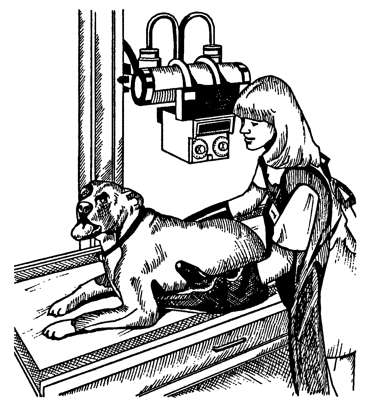
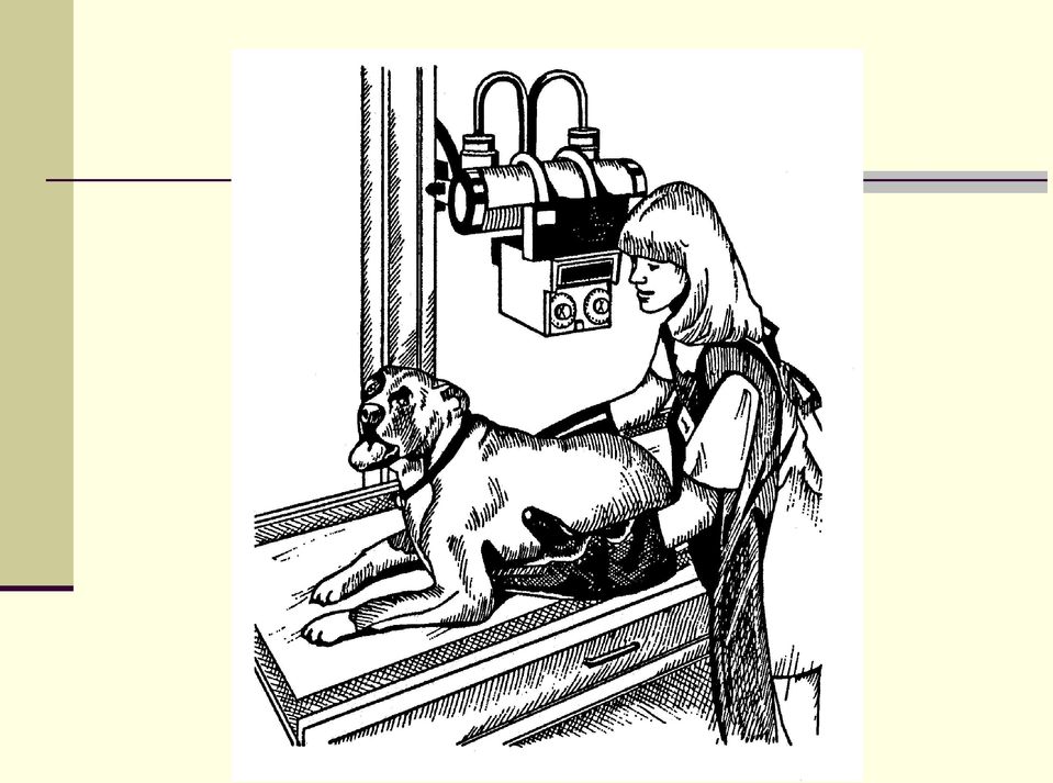
12 Patient Motion Voluntary motion (controlled motion) restraint necessary Looking around Moving away Moving extremeties Paddling Pushing away Involuntary motion (uncontrolled motion) reduce exposure time, increase ma Heart beating Breathing Panting Discomfort moving

13 Heart, lungs, and thorax images Animal inhales make exposure Abdomen, pelvis, and spine images Animal exhales make exposure

14 Safety Devices and Precautions Remove anyone from the x-ray room that is not needed. Every person should wear a lead apron and lead gloves when holding the x-rayed animal. Check the condition of gloves and aprons periodically by using radiography to determine if they allow x-rays to pass through. Limit the beam to the size of the film with a cone or lead diaphragm. Do not direct the x-ray beam into another room or work area. Install an aluminum filter (1 to 2 mm thick) at the tube housing opening to eliminate radiation from useless wave lengths. Cover the bottom side of the x-ray table with lead to protect the feet. The hands should not be placed in the path of the direct beam. Do not use fluoroscopy when radiography will do the job. Fluoroscopy is more hazardous and requires additional safety precautions.

15
16 Lead Gloves and Aprons To help prevent deterioration of lead gloves and lead aprons: Roll or drape apron over curved surface rather than folding it. Store gloves by hanging or by placing cans with both ends open inside gloves to keep the gloves open and to allow moisture to evaporate.

Production of X-rays. Radiation Safety Training for Analytical X-Ray Devices Module 9
 Module 9 This module presents information on what X-rays are and how they are produced. Introduction Module 9, Page 2 X-rays are a type of electromagnetic radiation. Other types of electromagnetic radiation
PRACTICAL TIPS IN ENSURING RADIATION SAFETY IN THE USE OF MEDICAL DIAGNOSTIC X-RAY EQUIPMENT
 PRACTICAL TIPS IN ENSURING RADIATION SAFETY IN THE USE OF MEDICAL DIAGNOSTIC X-RAY EQUIPMENT Although the medical uses of X-rays to examine a patient without surgery became an amazing medical breakthrough,
Radiographic Grid. Principles of Imaging Science II (RAD 120) Image-Forming X-Rays. Radiographic Grids
 Image-Forming X-Rays. Radiographic Grids") Principles of Imaging Science II (RAD 120) Radiographic Grids 1 Image-Forming X-Rays Four X-ray paths a. X-rays interact with patient and scatter away from the receptor b. X-rays interact and are absorbed
Principles of Imaging Science II (RAD 120) Radiographic Grids 1 Image-Forming X-Rays Four X-ray paths a. X-rays interact with patient and scatter away from the receptor b. X-rays interact and are absorbed
Radiation safety in dental radiography
 Radiation safety in dental radiography Dental Radiography Series The goal of dental radiography is to obtain diagnostic information while keeping the exposure to the patient and dental staff at minimum
X-ray (Radiography) - Bone
 - Bone") Scan for mobile link. X-ray (Radiography) - Bone Bone x-ray uses a very small dose of ionizing radiation to produce pictures of any bone in the body. It is commonly used to diagnose fractured bones or
Scan for mobile link. X-ray (Radiography) - Bone Bone x-ray uses a very small dose of ionizing radiation to produce pictures of any bone in the body. It is commonly used to diagnose fractured bones or
The Field. Radiologic technologists take x-rays and administer nonradioactive materials into patients' bloodstreams for diagnostic purposes.
 Radiologic Technologist Overview The Field - Specialty Areas - Preparation - Day in the Life - Earnings - Employment - Career Path Forecast - Professional Organizations The Field Radiologic technologists
Production of X-rays and Interactions of X-rays with Matter
 Production of X-rays and Interactions of X-rays with Matter Goaz and Pharoah. Pages 11-20. Neill Serman Electrons traveling from the filament ( cathode) to the target (anode) convert a small percentage
Digital radiography conquers the veterinary world
 Digital radiography conquers the veterinary world Author: Dirk De Langhe Increasingly, veterinarians are using medical imaging to diagnose their patients. There is a corresponding tendency towards replacing
Radiation safety in dental radiography
 Radiation safety in dental radiography Kodak s dental radiograph series The goal of dental radiography is to obtain diagnostic information while keeping the exposure to the patient and dental staff at
X-ray Production. Target Interactions. Principles of Imaging Science I (RAD119) X-ray Production & Emission
 X-ray Production & Emission") Principles of Imaging Science I (RAD119) X-ray Production & Emission X-ray Production X-rays are produced inside the x-ray tube when high energy projectile electrons from the filament interact with the
Principles of Imaging Science I (RAD119) X-ray Production & Emission X-ray Production X-rays are produced inside the x-ray tube when high energy projectile electrons from the filament interact with the
NHS Imaging and Radiodiagnostic activity in England. 2012/13 Release. August 2013
 NHS Imaging and Radiodiagnostic activity in England 2012/13 Release August 2013 Commentary This National Statistics release covers Imaging and Radiodiagnostic examinations or tests carried out in the NHS
Radiation therapy involves using many terms you may have never heard before. Below is a list of words you could hear during your treatment.
 Dictionary Radiation therapy involves using many terms you may have never heard before. Below is a list of words you could hear during your treatment. Applicator A device used to hold a radioactive source
The effects of radiation on the body can be divided into Stochastic (random) effects and deterministic or Non-stochastic effects.
 effects and deterministic or Non-stochastic effects.") RADIATION SAFETY: HOW TO EDUCATE AND PROTECT YOURSELF AND YOUR STAFF John Farrelly, DVM, MS, ACVIM (Oncology), ACVR (Radiation Oncology) Cornell University Veterinary Specialists The Veterinary Cancer
RADIATION SAFETY: HOW TO EDUCATE AND PROTECT YOURSELF AND YOUR STAFF John Farrelly, DVM, MS, ACVIM (Oncology), ACVR (Radiation Oncology) Cornell University Veterinary Specialists The Veterinary Cancer
Patient Prep Information
 Stereotactic Breast Biopsy Patient Prep Information Imaging Services Cannon Memorial Hospital Watauga Medical Center Table Weight Limits for each facility Cannon Memorial Hospital Watauga Medical Center
X-ray (Radiography) - Chest
 - Chest") Scan for mobile link. X-ray (Radiography) - Chest What is a Chest X-ray (Chest Radiography)? The chest x-ray is the most commonly performed diagnostic x-ray examination. A chest x-ray produces images of
Scan for mobile link. X-ray (Radiography) - Chest What is a Chest X-ray (Chest Radiography)? The chest x-ray is the most commonly performed diagnostic x-ray examination. A chest x-ray produces images of
X-ray (Radiography), Chest
, Chest") X-ray (Radiography), Chest What is a Chest X-ray (Chest Radiography)? The chest x-ray is the most commonly performed diagnostic x-ray examination. A chest x-ray makes images of the heart, lungs, airways,
X-ray (Radiography), Chest What is a Chest X-ray (Chest Radiography)? The chest x-ray is the most commonly performed diagnostic x-ray examination. A chest x-ray makes images of the heart, lungs, airways,
X-ray (Radiography) - Abdomen
 - Abdomen") Scan for mobile link. X-ray (Radiography) - Abdomen Abdominal x-ray uses a very small dose of ionizing radiation to produce pictures of the inside of the abdominal cavity. It is used to evaluate the stomach,
Scan for mobile link. X-ray (Radiography) - Abdomen Abdominal x-ray uses a very small dose of ionizing radiation to produce pictures of the inside of the abdominal cavity. It is used to evaluate the stomach,
X-ray Imaging Systems
 Principles of Imaging Science I (RAD 119) X-ray Tube & Equipment X-ray Imaging Systems Medical X-ray Equipment Classified by purpose or energy/current levels kvp, ma Radiographic Non-dynamic procedures
X-Rays Benefits and Risks. Techniques that use x-rays
 X-Rays Benefits and Risks X-rays are a form of electromagnetic radiation, just like light waves and radiowaves. Because X-rays have higher energy than light waves, they can pass through the body. X-rays
Page: 1 of 6 Page: 1 of 6
 Page: 1 of 6 Page: 1 of 6 CR Basics and FAQ Overview Computed Radiography is a term used to describe a system that electronically records a radiographic image. Computed Radiographic systems use unique
X-ray Radiation Safety Course. James Kane & Rob Deters Office of Radiological Control 545-7581
 X-ray Radiation Safety Course James Kane & Rob Deters Office of Radiological Control 545-7581 About the Course X-ray Radiation Safety X-ray radiation safety training is mandatory for radiation workers
CHAPTER 5 QC Test For Radiographic Equipment. Prepared by:- Kamarul Amin bin Abdullah @ Abu Bakar School of Medical Imaging KLMUC
 CHAPTER 5 QC Test For Radiographic Equipment Prepared by:- Kamarul Amin bin Abdullah @ Abu Bakar School of Medical Imaging KLMUC Lesson Outcomes Describe the objectives of each QC test done.(importance)
3701-72-03 Standards for accreditation of educational programs and approval of continuing education courses.
 1 3701-72-03 Standards for accreditation of educational programs and approval of continuing education courses. (A) Any person may apply to the director for approval to conduct an educational program for
5.2 ASSESSMENT OF X-RAY TUBE LEAKAGE RADIATION AND X-RAY TUBE OUTPUT TOTAL FILTRATION
 5.2 ASSESSMENT OF X-RAY TUBE LEAKAGE RADIATION AND X-RAY TUBE OUTPUT TOTAL FILTRATION 5.2.1 Task The bremsstrahlung produced by the X-ray tube has a continuous spectrum, limited by the set and spreads
REGULATION: QUALITY ASSURANCE PROGRAMS FOR MEDICAL DIAGNOSTIC X-RAY INSTALLATIONS N.J.A.C. 7:28-22
 REGULATION: QUALITY ASSURANCE PROGRAMS FOR MEDICAL DIAGNOSTIC X-RAY INSTALLATIONS N.J.A.C. 7:28-22 New Jersey Department of Environmental Protection Bureau of Radiological Health PO Box 415 Trenton NJ
Image Quality and Radiation Dose for Intraoral Radiography: Hand-Held Held (Nomad), Battery Powered
, Battery Powered") Image Quality and Radiation Dose for Intraoral Radiography: Hand-Held Held (Nomad), Battery Powered vs. Wall-Mount X-Ray X Systems Edgar Bailey*, MSEHE, CHP Consultant Joel Gray*, PhD, FAAPM DIQUAD, LLC
Image Quality and Radiation Dose for Intraoral Radiography: Hand-Held Held (Nomad), Battery Powered vs. Wall-Mount X-Ray X Systems Edgar Bailey*, MSEHE, CHP Consultant Joel Gray*, PhD, FAAPM DIQUAD, LLC
SECTION 1: REQUIREMENTS FOR CERTIFICATES OF COMPLIANCE FOR CLASSES OF RADIATION APPARATUS
 Department of Health and Human services Population Health Radiation Protection Act 2005 Section 17 CERTIFICATE OF COMPLIANCE: STANDARD FOR RADIATION APPARATUS - X-RAY MEDICAL DIAGNOSTIC (MAMMOGRAPHY) SECTION
Running head: OBESITY AND MEDICAL IMAGING 1. Obesity and Medical Imaging. November 13, 2012
 Running head: OBESITY AND MEDICAL IMAGING 1 Obesity and Medical Imaging November 13, 2012 OBESITY AND MEDICAL IMAGING 2 Abstract Obesity is a growing problem in the United States, and it affects the health
Dental Radiography collimator Ionising radiation image radiolucent area radiopaque area controlled zone scatter radiation intraoral
 Dental Radiography X-rays for dental radiography are produced by high voltages of electricity within an x-ray head and come out through a metal tube called a collimator. This ensures the x-rays only come
U.S. Bureau of Labor Statistics. Radiology Tech
 From the: U.S. Bureau of Labor Statistics Radiology Tech What They Do Radiologic technologists (RTs) perform diagnostic imaging examinations, such as x rays, on patients. Duties RTs typically do the following:
Required RS Training Info
 C-arm Radiation Safety at Tufts Required RS Training Info What are annual rad. dose limits? Who is our regulator? What should you do in an emergency? Are there health effects of radiation? C-arm beam awareness
Radiologic Technology Training Program
 Radiologic Technology Training Program 438 Radiologic Technology Training Program Radiologic Technology Training Program Program Coordinator: Mansour, Zepure General Information The Radiologic Technology
Radiographic Image Production. Radiographic Image Production. Principles of Imaging Science I (RAD 119) Film, Screens, and Cassettes
 Film, Screens, and Cassettes") Principles of Imaging Science I (RAD 119) Film, Screens, and Cassettes Radiographic Image Production X-ray photons emitted from the x-ray tube interact with the body, exit the patient (exit beam) and interact
Principles of Imaging Science I (RAD 119) Film, Screens, and Cassettes Radiographic Image Production X-ray photons emitted from the x-ray tube interact with the body, exit the patient (exit beam) and interact
INTENSIFYING SCREENS, CASSETTES AND SCREEN FILMS N. Serman & S. Singer
 INTENSIFYING SCREENS, CASSETTES AND SCREEN FILMS N. Serman & S. Singer X-rays were discovered by W.C. Roentgen because of their ability to cause fluorescence. X-ray photons cannot be seen. The image produced
SUBCHAPTER 22 QUALITY ASSURANCE PROGRAMS FOR MEDICAL DIAGNOSTIC X-RAY INSTALLATIONS
 Note: This is a courtesy copy and is not the official version of this rule. The official, legally effective version of this rule is available through www.lexisnexic.com/bookstore (Phone: (800) 223-1940).
Patient Exposure Doses During Diagnostic Radiography
 Patient Exposure Doses During Diagnostic Radiography JMAJ 44(11): 473 479, 2001 Shoichi SUZUKI Associated Professor, Faculty of Radiological Technology, School of Health Sciences, Fujita Health University
Handbook of radiographic positions and projections in the dog
 THE VETERINARY PUBLISHING COMPANY COMPANION ANIMALS Handbook of radiographic positions and projections in the dog Aimed at veterinary surgeons, students, teachers and other professionals in the veterinary
R12-2-602. Recognized Certificate-granting Bodies R12-2-104. Approval of Radiologic Technology Schools
 Arizona Administrative Code Title 12, Ch. 2 TITLE 12. NATURAL RESOURCES CHAPTER 2. RADIATION REGULATORY AGENCY MEDICAL RADIOLOGIC TECHNOLOGY BOARD OF EXAMINERS Authority: A.R.S. 32-2803 et seq. ARTICLE
CLINICAL PERFORMANCE OBJECTIVES
 CLINICAL PERFORMANCE OBJECTIVES CLINICAL COMPETENCY CLINICAL PERFORMANCE CRITERIA 1. Evaluation of Requisition: Student was able to: a. Identify procedures to be performed b. Recall the patient s age,
portable x-ray survey report
 department of HealtH and Human services centers for medicare & medicaid services form approved omb no. 0938-0027 portable x-ray survey report provider number H1 date surveyed (1) initial (2) survey W H2
Standard Test Method for Classification of Film Systems for Industrial Radiography 1
 Designation: E 1815 96 (Reapproved 2001) Standard Test Method for Classification of Film Systems for Industrial Radiography 1 This standard is issued under the fixed designation E 1815; the number immediately
RADIATION SAFETY STANDARD
 Radiation Safety Act 1999 RADIATION SAFETY STANDARD NM004:2010 Standard for radiation apparatus used to carry out industrial radiography Preface Under section 17 of the Radiation Safety Act 1999, a possession
Compliance Guidance for FLUOROSCOPIC QUALITY CONTROL
 Compliance Guidance for FLUOROSCOPIC QUALITY CONTROL New Jersey Department of Environmental Protection Bureau of Radiological Health PO Box 415 Trenton NJ 08625 FAX 609-984-5811 Website: http://www.state.nj.us/dep/rpp
CONTENT SPECIFICATIONS FOR THE FLUOROSCOPY EXAMINATION
 CONTENT SPECIFICATIONS FOR THE FLUOROSCOPY EXAMINATION Publication Date: November 2010 Implementation Date: March 2011 The purpose of the American Registry of Radiologic Technologists Fluoroscopy Examination
CT Scan Thorax and Upper Abdomen. Respiratory Unit Patient Information Leaflet
 CT Scan Thorax and Upper Abdomen Respiratory Unit Patient Information Leaflet Introduction This leaflet gives you general information about your CT (computerised tomography) scan. It does not replace the
SUMMARY OF CURRENT UK LEGISLATION AND GUIDELINES
 SUMMARY OF CURRENT UK LEGISLATION AND GUIDELINES Legislation There are two sets of regulations in the UK governing the use of ionizing radiation. They both form part of The Health and Safety at Work Act
Contents. X-ray and Computed Tomography. Characterization of X-rays. Production of X-rays
 J. E. Wilhjelm Ørsted TU Technical University of enmark, Bldg. 348, K-2800 Kongens Lyngby, enmark. X-ray and Computed Tomography Contents History and characterization of X-rays Conventional (projection)
Bon Secours St. Mary s School of Medical Imaging Course Descriptions by Semester Class of 2009. CRS 101 Clinical Radiation Science 3
 Bon Secours St. Mary s School of Medical Imaging Course Descriptions by Semester Class of 2009 First Year Fall REV.5/07 Credits CRS 101 Clinical Radiation Science 3 A clinical education course designed
SOP #: Revision #: Current Version Implementation Date: Page #: Page 1 of 10 Last Reviewed/Update Date: Expiration
 Implementation Page #: Page 1 of 10 Last Reviewed/Update 1. Purpose and Scope The purpose of this document is to describe the Medical Physics and Radiation Safety program at Boston University (BU) and
Name: Date: Team: Lab Experiment # 3. Focused Grid Positioning Errors. Computed Radiography and Direct Digital Radiography
 Name: Date: Team: Lab Experiment # 3 Focused Grid Positioning Errors Computed Radiography and Direct Digital Radiography Purpose This experiment is designed to demonstrate the effect of off-level error,
Computed Tomography (CT) - Sinuses
 - Sinuses") Scan for mobile link. Computed Tomography (CT) - Sinuses Computed tomography (CT) of the sinuses uses special x-ray equipment to evaluate the paranasal sinus cavities hollow, air-filled spaces within the
Scan for mobile link. Computed Tomography (CT) - Sinuses Computed tomography (CT) of the sinuses uses special x-ray equipment to evaluate the paranasal sinus cavities hollow, air-filled spaces within the
 P R E S E N T S Dr. Mufa T. Ghadiali is skilled in all aspects of General Surgery. His General Surgery Services include: General Surgery Advanced Laparoscopic Surgery Surgical Oncology Gastrointestinal
X-RAY REGULATORY GUIDE
 Minnesota Department of Health Radiation Control, X-ray Unit Protecting, maintaining and improving the health of all Minnesotans by promoting radiation safety through guidance and collaboration with the
SECTION 1: REQUIREMENTS FOR CERTIFICATES OF COMPLIANCE FOR CLASSES OF RADIATION SOURCES
 Department of Health and Human Services Population Health Radiation Protection Act 2005 Section 17 CERTIFICATE OF COMPLIANCE: STANDARD FOR RADIATION APPARATUS - X-RAY DIAGNOSTIC (VETERINARY) SECTION 1:
IONISING RADIATION. X-rays: benefit and risk
 IONISING RADIATION X-rays: benefit and risk Impress Federal Office for Radiation Protection Postfach 10 01 49 D - 38201 Salzgitter Telephone: + 49 (0) 30 18333-0 Fax: + 49 (0) 30 18333-1885 Website: www.bfs.de
Cardiac CT for Calcium Scoring
 Scan for mobile link. Cardiac CT for Calcium Scoring Cardiac computed tomography (CT) for Calcium Scoring uses special x-ray equipment to produce pictures of the coronary arteries to determine if they
Agency for Health Care Administration
 Page 1 of 20 FED - H0000 - INITIAL COMMENTS Title INITIAL COMMENTS CFR Type Memo Tag FED - H0005 - COMPLIANCE WITH FED/STATE/LOCAL LAWS Title COMPLIANCE WITH FED/STATE/LOCAL LAWS CFR 486.100 Type Condition
I.7. SOFT TECHNIQUE STILL IN USE IN CHEST RADIOGRAPHY PROS AND CONS
 I.7. SOFT TECHNIQUE STILL IN USE IN CHEST RADIOGRAPHY PROS AND CONS A. Slavtchev 1, I. Manolov 2 1 National Centre of Radiobiology and Radiation Protection 2 Medrom Ltd. Abstract In recent years the number
Compliance Guidance for QUALITY ASSURANCE MANUAL (2 nd Edition)
") Compliance Guidance for QUALITY ASSURANCE MANUAL (2 nd Edition) New Jersey Department of Environmental Protection Bureau of Radiological Health PO Box 415 Trenton NJ 08625 FAX 609-984-5811 Website: http://www.state.nj.us/dep/rpp
Compliance Guidance for QUALITY ASSURANCE MANUAL (2 nd Edition) New Jersey Department of Environmental Protection Bureau of Radiological Health PO Box 415 Trenton NJ 08625 FAX 609-984-5811 Website: http://www.state.nj.us/dep/rpp
RADIOLOGY SERVICES. By Dr Lim Eng Kok 1
 INTRODUCTION RADIOLOGY SERVICES By Dr Lim Eng Kok 1 Radiology is the branch of medicine that deals with the use of ionising (e.g. x- rays and radio-isotopes) and non-ionising radiation (e.g. ultrasound
Preadmission Information
 Preadmission Information British Columbia Institute of Technology 3700 Willingdon Avenue Burnaby, BC V5G 3H2 Program Head: 604 451 6918 Administrative Coordinator: 604 456 1257 medrad/web/medrad_medrad_preadmission_info
Cabrillo College Catalog 2015-2016
 RADIOLOGIC TECHNOLOGY Health, Athletics, Wellness, and Kinesiology Division Ian Haslam, Division Dean Division Office, Room 1102 Ann Smeltzer, Department Chair, (831) 479-5056 Aptos Counselor: (831) 479-6274
Bon Secours St. Mary s Hospital School of Medical Imaging Course Descriptions by Semester 18 Month Program
 Bon Secours St. Mary s Hospital School of Medical Imaging Course Descriptions by Semester 18 Month Program First Semester RAD 1101 Patient Care, Ethics, Law and Diversity Credits This 16 week course prepares
Understanding Digital Modalities: System Integration and Use
 Understanding Digital Modalities: System Integration and Use Donald J. Peck, PhD Henry Ford Health System Detroit, MI donaldp@rad.hfh.edu Dicom Digital Imaging and Communications in Medicine Part 3: Information
Bitewing Radiography B.E. DIXON. B.D.S., M.Sc., D.P.D.S.
 Bitewing Radiography B.E. DIXON B.D.S., M.Sc., D.P.D.S. Main Indications Detection of Dental Caries Monitoring progression of caries Assessment of existing restorations Assessment of Periodontal status
CPT Radiology Codes Requiring Review by AIM Effective 01/01/2016
 CPT Radiology Codes Requiring Review by AIM Effective 01/01/2016 When a service is authorized only one test per group is payable. *Secondary codes or add-on codes do not require preauthorization or separate
33-10-06-03. General requirements. 1. Administrative Controls.
 33-10-06-03. General requirements. 1. Administrative Controls. a. Registrant. The registrant shall be responsible for directing the operation of the x-ray systems which have been registered with the department.
X-ray Imaging Systems
 Principles of Imaging Science I (RAD 119) X-ray Tube & Equipment X-ray Imaging Systems Medical X-ray Equipment Classified by purpose or energy/current levels kvp, ma Radiographic Non-dynamic procedures
An Overview of Digital Imaging Systems for Radiography and Fluoroscopy
 An Overview of Digital Imaging Systems for Radiography and Fluoroscopy Michael Yester, Ph.D. University of Alabama at Birmingham Outline Introduction Imaging Considerations Receptor Properties General
Radiation Safety Issues for Radiologic Technologists
 Radiation Safety Issues for Radiologic Technologists Greg Sackett, M.S., CHP Medical Physicist Radiation Worker Risks? 1 Patient Risks? Acute Effects? Delayed Effects? Patient Questions? Radiation Dose
Mammography. What is Mammography?
 Scan for mobile link. Mammography Mammography is a specific type of breast imaging that uses low-dose x-rays to detect cancer early before women experience symptoms when it is most treatable. Tell your
.org. Fractures of the Thoracic and Lumbar Spine. Cause. Description
 Fractures of the Thoracic and Lumbar Spine Page ( 1 ) Spinal fractures can vary widely in severity. While some fractures are very serious injuries that require emergency treatment, other fractures can
R/F. Efforts to Reduce Exposure Dose in Chest Tomosynthesis Targeting Lung Cancer Screening. 3. Utility of Chest Tomosynthesis. 1.
 R/F Efforts to Reduce Exposure Dose in Chest Tomosynthesis Targeting Lung Cancer Screening Department of Radiology, National Cancer Center Hospital East Kaoru Shimizu Ms. Kaoru Shimizu 1. Introduction
Scan Time Reduction and X-ray Scatter Rejection in Dual Modality Breast Tomosynthesis. Tushita Patel 4/2/13
 Scan Time Reduction and X-ray Scatter Rejection in Dual Modality Breast Tomosynthesis Tushita Patel 4/2/13 Breast Cancer Statistics Second most common cancer after skin cancer Second leading cause of cancer
Procedures/risks: Radiology (CT, DXA, MRI, ultrasound, X-ray)
") Procedures/risks: Radiology (CT, DXA, MRI, ultrasound, X-ray) Computerized Axial Tomography (CT): Procedure: A Computerized Axial Tomography (CT) scan [of your heart] involves holding your breath for a
Procedures/risks: Radiology (CT, DXA, MRI, ultrasound, X-ray) Computerized Axial Tomography (CT): Procedure: A Computerized Axial Tomography (CT) scan [of your heart] involves holding your breath for a
420-3-26-.06 RADIATION SAFETY REQUIREMENTS FOR USERS OF X-RAY IN HEALING ARTS OR SERVICERS OF X-RAY EQUIPMENT
 420-3-26-.06 RADIATION SAFETY REQUIREMENTS FOR USERS OF X-RAY IN HEALING ARTS OR SERVICERS OF X-RAY EQUIPMENT (1) Scope. Rule 420-3-26-.03 establishes standards for use of x-rays in the healing arts including
Radiologic Technology (RAD, CLE) courses are open only to Radiologic Technology majors.
 courses are open only to Radiologic Technology majors.") RADIOLOGIC TECHNOLOGY (A.A.S Degree) Director: Prof. Virginia Mishkin, M.S., R.T. (R) (M) (QM) A radiologic technologist is a skilled professional who provides a specialized health care service. This rewarding
RADIOLOGIC TECHNOLOGY (A.A.S Degree) Director: Prof. Virginia Mishkin, M.S., R.T. (R) (M) (QM) A radiologic technologist is a skilled professional who provides a specialized health care service. This rewarding
European Academy of DentoMaxilloFacial Radiology
 European Academy of DentoMaxilloFacial Radiology Framework for Specialist Training in Dental and Maxillofacial Radiology Background The scope of DentoMaxilloFacial Radiology DMFR (Dental and Maxillofacial
ARTICLE 1. GENERAL PROVISIONS ARTICLE 2. SCHOOLS AND TRAINING APROVALS AND REQUIREMENTS OF RADIOLOGIC TECHNOLOGY
 GRRC Approved April 7, 2015 Filed with Arizona Secretary of State April 7, 2015 NOTICE OF FINAL RULEMAKING TITLE 12. NATURAL RESOURCES CHAPTER 2. RADIATION REGULATORY AGENCY MEDICAL RADIOLOGIC TECHNOLOGY
Staff Doses & Practical Radiation Protection in DEXA
 Patient Xray X Doses Staff Doses & Practical Radiation Protection in DEXA Una O ConnorO Dept. of Medical Physics & Bioengineering, St. James s s Hospital. Examination Types General XrayX Fluoroscopy /
Elements of required physical infrastructures: space, schielding, and patient flow..
 Elements of required physical infrastructures: space, schielding, and patient flow.. IAEA Following the IAEA guidelines, adapded by Anna Benini for workshop on Health Technology IUPESM Task Group, Porto
THORACIC DIAGNOSTIC ASSESMENT PROGRAM (DAP) PATIENT INFORMATION FOR:
 PATIENT INFORMATION FOR:") central east regional cancer program in partnership with cancer care ontario THORACIC DIAGNOSTIC ASSESMENT PROGRAM (DAP) PATIENT INFORMATION FOR: Thoracic dap booklet March2012.indd 1 SCHEDULED TESTS YOUR
central east regional cancer program in partnership with cancer care ontario THORACIC DIAGNOSTIC ASSESMENT PROGRAM (DAP) PATIENT INFORMATION FOR: Thoracic dap booklet March2012.indd 1 SCHEDULED TESTS YOUR
Equipment. Lin muyan. Presented by: CocticaUwititonze Email: costicauwitonze@yahoo.com Cell:+250 (0)7888 44764
7888 44764") Digital Medical Radiography Diagnostic (DR) system X-ray Equipment Presented by: CocticaUwititonze Email: costicauwitonze@yahoo.com Cell:+250 (0)7888 44764 Lin muyan 1. Goals 2. Introduction 3. X-ray machine
Digital Medical Radiography Diagnostic (DR) system X-ray Equipment Presented by: CocticaUwititonze Email: costicauwitonze@yahoo.com Cell:+250 (0)7888 44764 Lin muyan 1. Goals 2. Introduction 3. X-ray machine
Smoking and misuse of certain pain medicines can affect the risk of developing renal cell cancer.
 Renal cell cancer Renal cell cancer is a disease in which malignant (cancer) cells form in tubules of the kidney. Renal cell cancer (also called kidney cancer or renal adenocarcinoma) is a disease in which
Study the Quality Assurance of Conventional X-ray Machines Using Non Invasive KV meter
 Study the Quality Assurance of Conventional X-ray Machines Using Non Invasive KV meter T.M.Taha Radiation Protection Department, Nuclear Research Center, Atomic Energy Authority, Cairo.P.O.13759 Egypt.
Small cell lung cancer
 Small cell lung cancer Small cell lung cancer is a disease in which malignant (cancer) cells form in the tissues of the lung. The lungs are a pair of cone-shaped breathing organs that are found within
What is Radiology and Radiologic Technology?
 What is Radiology and Radiologic Technology? Note: Prospective CSI Radiologic Technology students are encouraged to print this document out and study it thoroughly before applying for admission to the
QUALITY CONTROL IN DIAGNOSTIC RADIOLOGY
 AAPM REPORT NO. 74 QUALITY CONTROL IN DIAGNOSTIC RADIOLOGY Report of Task Group #12 Diagnostic X-ray Imaging Committee Members S. Jeff Shepard, Chairman Pei-Jan Paul Lin, Co-Chairman John M. Boone Dianna
Thinking ahead. Focused on life. REALIZED: GROUNDBREAKING RESOLUTION OF 80 µm VOXEL
 Thinking ahead. Focused on life. REALIZED: GROUNDBREAKING RESOLUTION OF 80 µm VOXEL X-ray ZOOM RECONSTRUCTION Flat Panel Detector (FPD) Automatic Positioning Function For ø 40 x H 40 mm, ø 60 x H 60 mm,
In the previous presentation, we discussed how x-rays were discovered and how they are generated at the atomic level. Today we will begin the
 In the previous presentation, we discussed how x-rays were discovered and how they are generated at the atomic level. Today we will begin the discussion on the major components of the x-ray machine. Today
Froedtert Hospital School of Radiology Curriculum Analysis Grid. Clinical Practice
 Professional Curriculum Clinical Practice Procedural Performance Clinical Competency Basic Principles of Digital Radiography Image Acquisition Image Acquisition Errors Fundamental Principles of Exposure
XRS-3 X-RAY SOURCE OPERATOR S MANUAL
 XRS-3 X-RAY SOURCE OPERATOR S MANUAL CONTENTS ITEM PAGE 1.0 INTRODUCTION... 2 2.0 WARNINGS... 2 EXCLUSION ZONE DIAGRAM. 4 3.0 PHYSICAL DESCRIPTION... 5 3.1 HIGH VOLTAGE PULSER/TUBEHEAD... 5 3.2 BASE...
PHYS 222 Spring 2012 Final Exam. Closed books, notes, etc. No electronic device except a calculator.
 PHYS 222 Spring 2012 Final Exam Closed books, notes, etc. No electronic device except a calculator. NAME: (all questions with equal weight) 1. If the distance between two point charges is tripled, the
Computed Tomography (CT) - Chest
 - Chest") Scan for mobile link. Computed Tomography (CT) - Chest Computed tomography (CT) of the chest uses special x-ray equipment to examine abnormalities found in other imaging tests and to help diagnose the
Scan for mobile link. Computed Tomography (CT) - Chest Computed tomography (CT) of the chest uses special x-ray equipment to examine abnormalities found in other imaging tests and to help diagnose the
Chapter 7: Clinical Treatment Planning in External Photon Beam Radiotherapy
 Chapter 7: Clinical Treatment Planning in External Photon Beam Radiotherapy Set of 232 slides based on the chapter authored by W. Parker, H. Patrocinio of the IAEA publication (ISBN 92-0-107304-6): Review
A PACS-Aware DICOM Image Object
 Designing and Implementing A PACS-Aware DICOM Image Object For Digital X-ray, Mammography and Intraoral Applications David A. Clunie Quintiles Intelligent Imaging Clear Vision for the Healthcare Industry
Radiation Safety In Dental Practice
 Radiation Safety In Dental Practice A study guide and excerpts from The California Radiaton Control Regulations pertaining to dental practice Radiation Safety Protection Program template December 2013
Inferior Vena Cava filter and removal
 Inferior Vena Cava filter and removal What is Inferior Vena Cava Filter Placement and Removal? An inferior vena cava filter placement procedure involves an interventional radiologist (a specialist doctor)
QUALITY ASSURANCE PROGRAMS FOR DIAGNOSTIC RADIOLOGY FACILITIES. (a) Applicability. (b) Definitions
 Applicability. (b) Definitions") QUALITY ASSURANCE PROGRAMS FOR DIAGNOSTIC RADIOLOGY FACILITIES (a) Applicability Quality assurance programs as described in paragraph (c) of this section are recommended for all diagnostic radiology facilities.
QUALITY ASSURANCE PROGRAMS FOR DIAGNOSTIC RADIOLOGY FACILITIES (a) Applicability Quality assurance programs as described in paragraph (c) of this section are recommended for all diagnostic radiology facilities.
Health Care Careers in the Field of Imaging. Shari Workman, MSM,PHR,CIR MultiCare Health System Senior Recruiter/Employment Specialist
 Health Care Careers in the Field of Imaging Shari Workman, MSM,PHR,CIR MultiCare Health System Senior Recruiter/Employment Specialist What is Health Care Imaging? Technologists working with patients, using