BNFO601 Introduction to Molecular Biology Protein
|
|
|
- Aubrey Lyons
- 9 years ago
- Views:
Transcription
1 BNFO601 Introduction to Molecular Biology Protein Outline: A. What can protein do? B. What are proteins? C. Structure and basis for catalysis D. Targeting protein E. Alteration of protein structure and function by mutation F. Summary A. What can protein do? DNA is often depicted as the blueprint of the cell. A blueprint is something an architect refers to in building a structure. It contains a representation of the final shape of the building, its dimensions, what's connected to what, and so forth. If you examine DNA, you will find none of this. The molecule has no knowledge of the cell's final shape, nor any other of the things that characterize blueprints. DNA is not the blueprint of the cell; it s just the parts list, giving the components that make up the proteins of a cell. Fortunately, that is enough. The weight of action, then, lies squarely on protein. Table 1 gives a synopsis of some functions performed by protein. At the top of the list is the catalysis of the chemical reactions, as emphasized in the last section. The enzyme tyrosine hydroxylase, for example, catalyzes the conversion of tyrosine to the neurotransmitter L-DOPA. Proteins are responsible for other functions besides catalysis. They are required for the transport of a variety of compounds through membranes or, in the case of hemoglobin, the transport of oxygen in solution. Protein also plays a passive, structural role, for example in connective tissue. There are many other roles for protein, and Table 1 could have been many times as big as it is. B. What are proteins? The function of a protein is determined ultimately by its particular shape and structure. At its most basic level, the structure of a protein is simple. It has to be, otherwise DNA could not specify it. Understanding the structure of protein thus answers two profound questions: How do proteins control the activities of a cell? How do genes exert control over those activities? FUNCTION Catalysis Binding: transport Binding: defense Binding: information Mechanical Support Mechanical Work Table 1. Some Biological Functions of Proteins EXAMPLE Tyrosine Hydroxylase (hormone & neurotransmitter production) Hemoglobin (oxygen transport) Immunoglobins (immune system) Insulin (hormone) & Insulin Receptor Collagen (connective tissue) Actin/Myosin (muscle contraction) Protein - 1

2 In brief, a protein is a linear array of amino acids. If you grasp all that sentence has to say, then you've come a long way towards understanding protein. Notice the pattern in Figure 1c. A protein is a polymer of a unit repeated again and again. That unit consists of a carboxylic acid, connected to a carbon. The carbon is called the "alpha-carbon" because it's the closest one to the carboxylic acid group. An amino group is attached to the alphacarbon. The subunits are thus (alphaamino acids. Amino acids differ from one another only in what else is connected to the alpha-carbon, represented in Figure 1a as a variable "R-group". The synthesis of proteins is the process of combining alpha-amino acids in a linear chain, connecting alpha-amino groups to carboxylate groups (Figure 1a and 1b). The backbone of this chain is identical for all proteins. If the R groups were similarly invariable, then all proteins would be alike, and protein would be able to do only one thing, a not very interesting thing at that. Fortunately, the R groups vary from one amino acid to the next, amongst the 20 possibilities shown in Figure 2. This listing of the twenty major amino acids is a very good list to get to know, but not to memorize. If you go into biochemistry, you'll find that they will become etched Figure 1. Protein as a polymer of alpha-amino acids. 1a. Structure of alpha-amino acid. "R" represents side group, as shown in Figure 2. 1b. Formation of dipeptide by joining two amino acids. 1c. Polypeptide chain composed of linked amino acids. The shapes represent the different R-groups, each with its own chemical properties. into your brain without having to memorize them, and if you don't, there's probably no need to know the structures. Some R groups of amino acids are acidic carboxylic acids, giving rise to negative charges at physiological ph. Aspartic acid is an example of an acidic amino acid. Some R-groups are basic, giving rise to positive charges at physiological ph. The charged amino acids interact strongly with water and so are hydrophilic. There are other R groups that interact strongly with water but are uncharged. For example, serine contains a hydroxyl group (an OH group), just like water does, and it's no surprise that serine is hydrophilic. There are also hydrophobic amino acids, like leucine, whose R-groups would tend to segregate away from water, because they interact less strongly with water than water does with itself. Protein - 2

3 Protein - 4
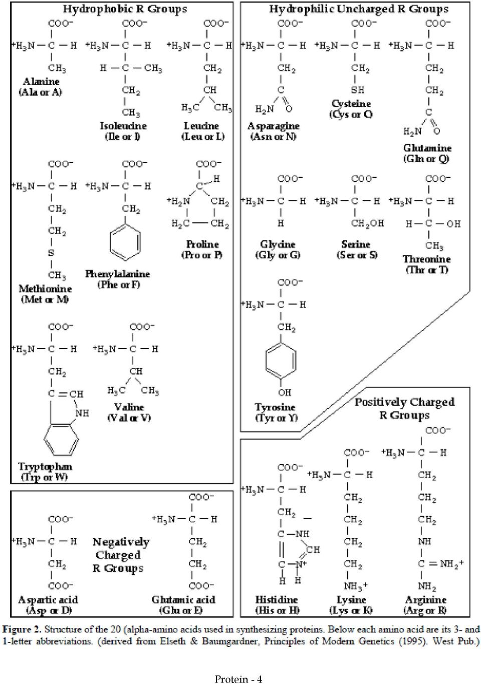
4 There are many other properties in which the twenty amino acids differ from one another: some are bulky, some small; some are capable of donating electrons, others not; some are chemically reactive. And so forth. Each amino acid represents a different flavor, and the structure and properties of a protein are defined by the properties and order of its amino acids: its primary structure. There are only twenty amino acids used to synthesize proteins, which limits what proteins are possible in nature. How constricting is this limitation? Consider the number of possible dipeptides (two amino acids joined together by a peptide bond). There are 20 possible amino acids in the first position and 20 possible amino acids in the second position. That makes 202 = 400 possible dipeptides. Similarly, there are 203 = 8000 possible tripeptides. Proteins range in size from a smallish 100 amino acids to a The number of possible proteins in nature is therefore staggering! SQ7. What is a protein? SQ8. Glycogen is a linear array of glucose. Why isn't glycogen as varied in its properties as protein? SQ9. Find an amino acid with the following properties: a. Small, negatively charged. b. Large, has double bonds (and so can participate in electron transfer reactions), and has a free -OH group (and so can participate in hydrogen bonding). SQ10. How do proteins control the activities of a cell? SQ11. How do genes exert control over these processes? C. Structure and basis for catalysis Unfortunately, knowing merely that proteins are linear arrays of alpha-amino acids doesn't tell us how they can have the varied properties required of proteins in a living cell. In particular, it doesn't explain how proteins can act as catalysts. For this we have to see the protein in three dimensions. The protein hexokinase (Figure 3), is the enzyme that begins the degradation of glucose in the liver. If you were to see this molecule, the first thing you might notice is that the enzyme has a hole just the right size for glucose to fit into. The binding of glucose to the enzyme alters the enzyme in such a way that glucose cannot escape unless the enzyme again changes shape. This normally occurs only after the reaction catalyzed by the enzyme is complete. So glucose goes in and glucose 6-phosphate goes out. The function of hexokinase is clearly tied up in its shape. How did the protein get to this shape? The answer lies ultimately in the primary structure * of the protein, that is the order of its amino acids. The local structure resulting from the interaction of nearby amino acids is called the secondary structure. For example, a secondary structure known as the alpha-helix is formed by the interaction of carboxyl groups of amino acids and the alpha-amino groups on neighbors three amino acids removed. We can with some confidence predict the secondary structure of a protein * Illustrations of the primary, secondary, tertiary, and quaternary structures of protein can be found in any standard genetics text (which I can t reproduce here owing to copyright restrictions, but I can show in class if you like). Protein - 5
.")
5 from its amino acid sequence. Globular, watersoluble proteins (such as hexokinase and most enyzymes) tend to have short alpha-helices. In contrast, proteins with long extended regions of secondary structure are fibrous and generally play a structural role. An example is the protein fibrin, which forms the protein network that makes up blood clots. In some cases structures common to several proteins with similar functions have been identified. One example is the helix-turn-helix motif, a stretch of about 20 amino acids consisting of two alpha-helices separated by a bend. Proteins that have this structure, with specific amino acids in key positions, are able to bind to DNA. One of the two alpha-helices fits nicely into the famous double helix of DNA (Figure 4). There are many such motifs known, and it is sometimes possible to guess the function of a protein simply by knowing its primary structure and deducing its secondary structure. Fig. 3: Three dimensional representation of the enzyme hexokinase. Each ball represents one amino acid out of a total of 457. Blue amino acids are hydrophobic, red negatively charged, orange positively charged, yellow and pink uncharged but hydrophilic. The identities of grey amino acids are unknown. Note the hole in the central part of the protein, where glucose binds. Interactions between distant amino acids, particularly their R-groups, give rise to a protein s tertiary structure, the folding of a polypeptide chain in three dimensions. For example, the hydrophobic amino acids would tend to be Figure 4. Two views of a protein binding DNA. A virus-encoded protein, Cro, is shown attached to a region of viral DNA. The panels show only the portion of the protein that interacts with the DNA. Cro consists of two polypeptides, arranged head-to-head (note the symmetry). The DNA is shown as a stick figure cartoon, with redviolet representing cytosine, blue-violet representing guanine, green representing thymine, and cyano representing adenine. White and yellow sticks represent oxygen and phosphate, respectively. In the left panel, the amino acids of the protein use the same color conventions as in Figure 3. In the right panel, only the backbone of the two polypeptide chains are shown, with a-helices in yellow. For each polypeptide, one helix of the helix-turn-helix motif is inserted in a major groove of the DNA. Protein - 6

6 sequestered in the middle of the protein, away from water, just as the hydrophobic chains of soap aggregate to minimize contact with water. Charged and other hydrophilic amino acids would tend to lie outside the protein. You can see this to some extent with hexokinase (Figure 3). It may be, however, that any way the chain may twist, there is no folding that can avoid patches of hydrophobic amino acids from appearing at the surface of the protein. What then? In some cases, further aggregation may occur between separate protein chains, so that in the end, the completely assembled protein consists of multiple chains formed by the interaction between them. Such proteins are said to have quaternary structure. An example of this is the protein hemoglobin, the oxygen-carrying protein in blood. It consists of four separate polypeptide chains that interact with each other. Separately, each subunit can bind oxygen, due in part to the oxygen-binding molecule, heme, which fits into a hole created by the tertiary structure. But the regulation of oxygen binding, essential to the functioning of hemoglobin in the body, is apparent only when four subunits aggregate together. The positions of specific amino acids determine not only the shape of the protein but also its capacity for catalysis (Figure 5). The folding of chymotrypsin, a digestive enzyme that catalyzes the hydrolysis (breakdown) of ingested protein in the gut, creates a local region of the enzyme called the active site. The folding happens to place the 195th amino acid in the chain, serine, near a hole that has the shape of the amino acid phenylalanine. When a phenylalanine within a protein you eat finds its way into the phenylalanine-shaped hole of chymotrypsin, the amide bond adjacent to phenylalanine is positioned close enough to serine-195 that a chemical reaction takes place, breaking the amide bond. Once that occurs, the broken protein is released. The ability of chymotrypsin to do this depends upon the precise geometry of the active site. is dependent upon a serine occurring precisely at position number 195 and upon folding occurring that places serine in exactly the right position relative to the protein being digested. Figure 5. Active site of the chymotrypsin, an enzyme that breaks specific peptide bonds of protein. A phenylalanine residue (blue ring) of a blue protein (could be any protein) slips into the active site of chymotrypsin (green) at its binding site. This positions the peptide bond (red) next to phenylalanine in such a way that it becomes susceptible to cleavage. It becomes susceptible because of a particular confluence of amino acids at the active site. In brief, the oxygen of a serine at the 195 th position in the amino acid chain of chymotrypsin is close to the carboxylate group of the phenylalanine residue and can attack the carbon. The carbon is poised for attack because hydrogens from both serine-195 and glycine 193 hydrogen bond with an oxygen from the carboxylate group, reducing the electron density around the carbon. At the same time, the hydrogen that is normally on the serine oxygen is drawn off by the electrons on the ring of histidine at position 57. Those electrons are more available owing to the interaction of a hydrogen on the same histidine ring with the carboxylate group of aspartate at position 102. After the peptide bond is cleaved, the two resulting fragments of the substrate protein float away, leaving the active site of chymotrypsin available for a new substrate. Protein - 7

7 SQ12. If the critical part of an enzyme is its active site, consisting typically of several amino acids, what's the use of the rest of the protein? D. Targeting protein Similar considerations govern the placement of protein. Figure 6 shows a cartoon of glycophorin, a protein that spans the membrane of red blood cells. You can see that most of the amino acids in the membrane-spanning region are hydrophobic, while the amino acids inside or outside the cell are generally hydrophilic. This arrangement of amino acids serves to anchor the protein in the membrane, because the hydrophilic amino acids would not be happy in the oily, lipid environment of the membrane, and the hydrophobic amino acids would not be happy outside that environment (or more accurately, the water wouldn't be happy to accommodate the weakly interacting hydrophobic residues). Note that some amino acids in the membrane are hydrophilic and some amino acids in the two aqueous compartments are hydrophobic. Why might that be? Figure 6. Primary structure of glycophorin A. Amino acid sequence of one polypeptide of glycophorin A. Amino acids are colored to show different chemical properties. Note that the charged amino acids all lie not in the membrane but either inside or outside the red blood cell, as do most of the other hydrophilic amino acids. Many of the hydroxylated amino acids on the outside of the cell are also charged, owing to negatively charged sugars (not shown) attached to the amino acids after the protein is made. In contrast, the portion of the protein that spans the membrane consists of amino acids that are predominantly hydrophobic (see also inset at bottom of the figure). These 19 amino acids form an alpha-helix. The regions on both sides of the membrane also have secondary and tertiary structures, not shown in this cartoon. Finally, glycophorin A has a quaternary structure: in nature (but not in this cartoon) it is a dimer consisting of two identical polypeptide chains associated with one another. Protein - 8

8 The cartoon of glycophorin raises more questions than it answers. The protein was surely made inside the cell... then how did those many hydrophilic amino acids pass through the hydrophobic environment of the membrane to get outside? Worse, what about the case of the protein hormone insulin, made within pancreatic cells and secreted into the circulatory system? Insulin must have hydrophilic amino acids on its exterior (since it's soluble in blood), so how did it completely cross the hydrophobic cell membrane? Well, a cell could provide a hole in the membrane for the protein to pass through, but that simply replaces one problem with many: How can you make sure only the protein you want to leave can leave? How can you make sure that protein supposed to leave the cell go through holes in the cell membrane and protein bound to the mitochondria go through holes in the mitochondrial membrane? How come the cell's guts don't spill out the holes? A blueprint would solve these problems, specifying for each protein where it's supposed to go. This is not the answer nature found. There are no blueprints, and the protein must contain within itself information specifying its ultimate location. Since protein are nothing more than sequences of amino acids, something within the sequence must carry the information, and indeed this is the case. Protein that must pass membranes have N-terminal amino acid sequences, called signal peptides, that function as routing slips. Transport proteins on certain membranes recognize the appropriate signal peptide and ferry the attached polypeptide chain through the membrane. The signal peptide binds to the membrane protein and passes through and an aqueous channel formed through the bilayer, dragging the rest of the protein with it. Once the signal peptide has initiated transfer across the membrane, it is cleaved off. What is the nature of the amino acid sequence of a signal peptide that enables it to be recognized by the transport apparatus? Figure 7 shows the N-terminal amino acids of the precursor to bovine growth hormone. The cell export signal peptide consists of a string of hydrophobic amino acids preceded by polar amino acids. The exact amino acids don't seem to be important -- just the types. This signal peptide enables growth hormone made within pituitary cells to be secreted into the circulatory system, and any protein that begins with this pattern of amino acids would also be secreted. SQ13. Describe the process by which glycophorin A presumably gained its proper position in the red blood cell membrane. Figure 7. Signal peptide of bovine growth hormone. The first 31 amino acids of unprocessed growth hormone are shown. The polypeptide is made in pituitary cells, and the N-terminal signal sequence binds to Signal Recognition Proteins on the cell surface, which facilitate the transport of the polypeptide outside the cell. Once outside, the polypeptide is cleaved between the 27th and 28th amino acids, forming mature growth hormone. Protein - 9
, so how did it completely cross the hydrophobic cell membrane?")
9 E. Alteration of Protein Structure and Function by Mutation A protein's primary structure (the linear order of its amino acids) ultimately determines the shape of the protein, its function, and its location within or without the cell. The specific characteristics of a protein result from the interplay of the chemical properties of its component amino acids. These properties, particularly hydrophobicity, enable the protein to assemble itself into a structure that places reactive groups critical to protein function at their proper locations in space. This is the connection between genetics and life. The centrality of the primary structure of protein is so critical to our understanding, that I will restate the point from two directions: What is the nature of mutation? and How can we control protein function? Most simple genetic mutations cause a change in an amino acid within a protein. What effect might that have? Here are some not mutually exclusive possibilities: Changing an amino acid at the active site of an enzyme could alter or destroy the catalytic properties of the enzyme. Mutation in an amino acid distant from the active site might nonetheless alter the three dimensional structure and, for example, make amino acids within the active site too distant from one another to be effective. More specifically, a mutation might alter the secondary structure of a region, perhaps by inserting an amino acid that prevents an alpha-helix from forming. A mutation might prevent proper placement in the membrane by replacing a hydrophobic amino acid with a charged amino acid. The change in three-dimensional structure might be subtle, just making the structure more prone to falling apart at high temperature, for example. Replacing one amino acid with another might alter a motif that enables the protein to bind to DNA, or perform some other function. The mutation might affect a purely informational part of the protein, a signal sequence, so that the protein is improperly targeted. We will see that mutation occurs directly in DNA, not protein, but the ultimate effects of mutation are felt as aberrant protein. The importance of the primary structure of a protein can be restated in the following way: if you can specify a protein's amino acids, i.e. its primary structure, you can determine its properties and its capacity to catalyze biochemical reactions. For example, consider hexokinase once more (Figure 3). If you knew what amino acids to change, you might alter the enzyme so that it could no longer act on glucose but only on the larger sugar, sucrose. As a matter of fact, in principle, you could design a protein to catalyze virtually any energetically feasible reaction you could imagine -- make plastic from starch! Make azat or other expensive drugs at a fraction of the current cost! We can already make proteins to order. The only reason these applications are presently out of reach is that we don't know how to predict the complete folding of a protein or its catalytic properties from the sequence of amino acids. Most proteins assemble themselves, but what is simple in nature is fiendishly difficult to predict. There is considerable research aimed at learning how to predict the three dimensional structures of proteins from their primary structures. When this is achieved, you may expect a societal change comparable to what resulted from the transformation of 19th century organic chemistry to 20th century practice. Protein - 10

10 SQ14. Suppose a gene suffers a mutation and the enzyme encoded by it doesn't work. What kind of change in the amino acid sequence of the protein might account for this outcome? F. Summary Proteins have a wide variety of catalytic and structural roles Proteins consist of a linear sequence of amino acid (its primary structure) The three dimensional structure of a protein determines its activity, for example by placing amino acids in a critical spatial relationship to form a catalytic site A protein s structure is determined by local interactions between amino acids (secondary structure), distant interactions (tertiary structures), and interactions between distinct polypeptide chains (quaternary structure). The protein contains within it the information to direct its proper placement within (or outside) the cell Mutations in a protein s amino acid sequence may have one of a variety of effects on its structure or targeting and thus on its function Protein - 11
, distant interactions (tertiary structures), and interactions between distinct polypeptide chains (quaternary structure).")
Carbohydrates, proteins and lipids
 Carbohydrates, proteins and lipids Chapter 3 MACROMOLECULES Macromolecules: polymers with molecular weights >1,000 Functional groups THE FOUR MACROMOLECULES IN LIFE Molecules in living organisms: proteins,
Chapter 3 Molecules of Cells
 Bio 100 Molecules of cells 1 Chapter 3 Molecules of Cells Compounds containing carbon are called organic compounds Molecules such as methane that are only composed of carbon and hydrogen are called hydrocarbons
The Molecules of Cells
 The Molecules of Cells I. Introduction A. Most of the world s population cannot digest milk-based foods. 1. These people are lactose intolerant because they lack the enzyme lactase. 2. This illustrates
Biological molecules:
 Biological molecules: All are organic (based on carbon). Monomers vs. polymers: Monomers refer to the subunits that, when polymerized, make up a larger polymer. Monomers may function on their own in some
Proteins and Nucleic Acids
 Proteins and Nucleic Acids Chapter 5 Macromolecules: Proteins Proteins Most structurally & functionally diverse group of biomolecules. : o Involved in almost everything o Enzymes o Structure (keratin,
Biochemistry of Cells
 Biochemistry of Cells 1 Carbon-based Molecules Although a cell is mostly water, the rest of the cell consists mostly of carbon-based molecules Organic chemistry is the study of carbon compounds Carbon
Chapter 3: Biological Molecules. 1. Carbohydrates 2. Lipids 3. Proteins 4. Nucleic Acids
 Chapter 3: Biological Molecules 1. Carbohydrates 2. Lipids 3. Proteins 4. Nucleic Acids Elements in Biological Molecules Biological macromolecules are made almost entirely of just 6 elements: Carbon (C)
4. Which carbohydrate would you find as part of a molecule of RNA? a. Galactose b. Deoxyribose c. Ribose d. Glucose
 1. How is a polymer formed from multiple monomers? a. From the growth of the chain of carbon atoms b. By the removal of an OH group and a hydrogen atom c. By the addition of an OH group and a hydrogen
Lecture Overview. Hydrogen Bonds. Special Properties of Water Molecules. Universal Solvent. ph Scale Illustrated. special properties of water
 Lecture Overview special properties of water > water as a solvent > ph molecules of the cell > properties of carbon > carbohydrates > lipids > proteins > nucleic acids Hydrogen Bonds polarity of water
A disaccharide is formed when a dehydration reaction joins two monosaccharides. This covalent bond is called a glycosidic linkage.
 CH 5 Structure & Function of Large Molecules: Macromolecules Molecules of Life All living things are made up of four classes of large biological molecules: carbohydrates, lipids, proteins, and nucleic
Chapter 5. The Structure and Function of Macromolecule s
 Chapter 5 The Structure and Function of Macromolecule s Most Macromolecules are polymers: Polymer: (poly: many; mer: part) Large molecules consisting of many identical or similar subunits connected together.
2007 7.013 Problem Set 1 KEY
 2007 7.013 Problem Set 1 KEY Due before 5 PM on FRIDAY, February 16, 2007. Turn answers in to the box outside of 68-120. PLEASE WRITE YOUR ANSWERS ON THIS PRINTOUT. 1. Where in a eukaryotic cell do you
Elements in Biological Molecules
 Chapter 3: Biological Molecules 1. Carbohydrates 2. Lipids 3. Proteins 4. Nucleic Acids Elements in Biological Molecules Biological macromolecules are made almost entirely of just 6 elements: Carbon (C)
Chapter 5: The Structure and Function of Large Biological Molecules
 Name Period Concept 5.1 Macromolecules are polymers, built from monomers 1. The large molecules of all living things fall into just four main classes. Name them. 2. Circle the three classes that are called
18.2 Protein Structure and Function: An Overview
 18.2 Protein Structure and Function: An Overview Protein: A large biological molecule made of many amino acids linked together through peptide bonds. Alpha-amino acid: Compound with an amino group bonded
Helices From Readily in Biological Structures
 The α Helix and the β Sheet Are Common Folding Patterns Although the overall conformation each protein is unique, there are only two different folding patterns are present in all proteins, which are α
Chemical Basis of Life Module A Anchor 2
 Chemical Basis of Life Module A Anchor 2 Key Concepts: - Water is a polar molecule. Therefore, it is able to form multiple hydrogen bonds, which account for many of its special properties. - Water s polarity
Disaccharides consist of two monosaccharide monomers covalently linked by a glycosidic bond. They function in sugar transport.
 1. The fundamental life processes of plants and animals depend on a variety of chemical reactions that occur in specialized areas of the organism s cells. As a basis for understanding this concept: 1.
Built from 20 kinds of amino acids
 Built from 20 kinds of amino acids Each Protein has a three dimensional structure. Majority of proteins are compact. Highly convoluted molecules. Proteins are folded polypeptides. There are four levels
Biological Molecules
 Biological Molecules I won t lie. This is probably the most boring topic you have ever done in any science. It s pretty much as simple as this: learn the material deal with it. Enjoy don t say I didn t
IV. -Amino Acids: carboxyl and amino groups bonded to -Carbon. V. Polypeptides and Proteins
 IV. -Amino Acids: carboxyl and amino groups bonded to -Carbon A. Acid/Base properties 1. carboxyl group is proton donor! weak acid 2. amino group is proton acceptor! weak base 3. At physiological ph: H
How To Understand The Chemistry Of Organic Molecules
 CHAPTER 3 THE CHEMISTRY OF ORGANIC MOLECULES 3.1 Organic Molecules The chemistry of carbon accounts for the diversity of organic molecules found in living things. Carbon has six electrons, four of which
A. A peptide with 12 amino acids has the following amino acid composition: 2 Met, 1 Tyr, 1 Trp, 2 Glu, 1 Lys, 1 Arg, 1 Thr, 1 Asn, 1 Ile, 1 Cys
 Questions- Proteins & Enzymes A. A peptide with 12 amino acids has the following amino acid composition: 2 Met, 1 Tyr, 1 Trp, 2 Glu, 1 Lys, 1 Arg, 1 Thr, 1 Asn, 1 Ile, 1 Cys Reaction of the intact peptide
Chemistry 20 Chapters 15 Enzymes
 Chemistry 20 Chapters 15 Enzymes Enzymes: as a catalyst, an enzyme increases the rate of a reaction by changing the way a reaction takes place, but is itself not changed at the end of the reaction. An
Structure of proteins
 Structure of proteins Primary structure: is amino acids sequence or the covalent structure (50-2500) amino acids M.Wt. of amino acid=110 Dalton (56 110=5610 Dalton). Single chain or more than one polypeptide
Lab 3 Organic Molecules of Biological Importance
 Name Biology 3 ID Number Lab 3 Organic Molecules of Biological Importance Section 1 - Organic Molecules Section 2 - Functional Groups Section 3 - From Building Blocks to Macromolecules Section 4 - Carbohydrates
Organic Molecules of Life - Exercise 2
 Organic Molecules of Life - Exercise 2 Objectives -Know the difference between a reducing sugar and a non-reducing sugar. -Distinguish Monosaccharides from Disaccharides and Polysaccharides -Understand
Protein Physics. A. V. Finkelstein & O. B. Ptitsyn LECTURE 1
 Protein Physics A. V. Finkelstein & O. B. Ptitsyn LECTURE 1 PROTEINS Functions in a Cell MOLECULAR MACHINES BUILDING BLOCKS of a CELL ARMS of a CELL ENZYMES - enzymatic catalysis of biochemical reactions
http://faculty.sau.edu.sa/h.alshehri
 http://faculty.sau.edu.sa/h.alshehri Definition: Proteins are macromolecules with a backbone formed by polymerization of amino acids. Proteins carry out a number of functions in living organisms: - They
Biochemistry - I. Prof. S. Dasgupta Department of Chemistry Indian Institute of Technology, Kharagpur Lecture-11 Enzyme Mechanisms II
 Biochemistry - I Prof. S. Dasgupta Department of Chemistry Indian Institute of Technology, Kharagpur Lecture-11 Enzyme Mechanisms II In the last class we studied the enzyme mechanisms of ribonuclease A
H H N - C - C 2 R. Three possible forms (not counting R group) depending on ph
 depending on ph") Amino acids - 0 common amino acids there are others found naturally but much less frequently - Common structure for amino acid - C, -N, and functional groups all attached to the alpha carbon N - C - C
Amino acids - 0 common amino acids there are others found naturally but much less frequently - Common structure for amino acid - C, -N, and functional groups all attached to the alpha carbon N - C - C
This class deals with the fundamental structural features of proteins, which one can understand from the structure of amino acids, and how they are
 This class deals with the fundamental structural features of proteins, which one can understand from the structure of amino acids, and how they are put together. 1 A more detailed view of a single protein
AP BIOLOGY 2008 SCORING GUIDELINES
 AP BIOLOGY 2008 SCORING GUIDELINES Question 1 1. The physical structure of a protein often reflects and affects its function. (a) Describe THREE types of chemical bonds/interactions found in proteins.
Pipe Cleaner Proteins. Essential question: How does the structure of proteins relate to their function in the cell?
 Pipe Cleaner Proteins GPS: SB1 Students will analyze the nature of the relationships between structures and functions in living cells. Essential question: How does the structure of proteins relate to their
Chapter 2. The Chemistry of Life Worksheets
 Chapter 2 The Chemistry of Life Worksheets (Opening image courtesy of David Iberri, http://en.wikipedia.org/wiki/file:camkii.png, and under the Creative Commons license CC-BY-SA 3.0.) Lesson 2.1: Matter
Amino Acids, Proteins, and Enzymes. Primary and Secondary Structure Tertiary and Quaternary Structure Protein Hydrolysis and Denaturation
 Amino Acids, Proteins, and Enzymes Primary and Secondary Structure Tertiary and Quaternary Structure Protein Hydrolysis and Denaturation 1 Primary Structure of Proteins H 3 N The particular sequence of
1. The diagram below represents a biological process
 1. The diagram below represents a biological process 5. The chart below indicates the elements contained in four different molecules and the number of atoms of each element in those molecules. Which set
8/20/2012 H C OH H R. Proteins
 Proteins Rubisco monomer = amino acids 20 different amino acids polymer = polypeptide protein can be one or more polypeptide chains folded & bonded together large & complex 3-D shape hemoglobin Amino acids
Ch18_PT MULTIPLE CHOICE. Choose the one alternative that best completes the statement or answers the question.
 Ch18_PT MULTIPLE CHOICE. Choose the one alternative that best completes the statement or answers the question. 1) All of the following can be classified as biomolecules except A) lipids. B) proteins. C)
Keystone Review Practice Test Module A Cells and Cell Processes. 1. Which characteristic is shared by all prokaryotes and eukaryotes?
 Keystone Review Practice Test Module A Cells and Cell Processes 1. Which characteristic is shared by all prokaryotes and eukaryotes? a. Ability to store hereditary information b. Use of organelles to control
Proteins. Proteins. Amino Acids. Most diverse and most important molecule in. Functions: Functions (cont d)
") Proteins Proteins Most diverse and most important molecule in living i organisms Functions: 1. Structural (keratin in hair, collagen in ligaments) 2. Storage (casein in mother s milk) 3. Transport (HAEMOGLOBIN!)
Proteins Proteins Most diverse and most important molecule in living i organisms Functions: 1. Structural (keratin in hair, collagen in ligaments) 2. Storage (casein in mother s milk) 3. Transport (HAEMOGLOBIN!)
Worksheet 13.1. Chapter 13: Human biochemistry glossary
 Worksheet 13.1 Chapter 13: Human biochemistry glossary α-helix Refers to a secondary structure of a protein where the chain is twisted to form a regular helix, held by hydrogen bonds between peptide bonds
Exam 4 Outline CH 105 Spring 2012
 Exam 4 Outline CH 105 Spring 2012 You need to bring a pencil and your ACT card. Chapter 24: Lipids 1. Describe the properties and types of lipids a. All are hydrophobic b. Fatty acid-based typically contain
Enzymes: Practice Questions #1
 Enzymes: Practice Questions #1 1. Compound X increases the rate of the reaction below. Compound X is most likely A. an enzyme B. a lipid molecule C. an indicator D. an ADP molecule 2. The equation below
Ionization of amino acids
 Amino Acids 20 common amino acids there are others found naturally but much less frequently Common structure for amino acid COOH, -NH 2, H and R functional groups all attached to the a carbon Ionization
PRESTWICK ACADEMY NATIONAL 5 BIOLOGY CELL BIOLOGY SUMMARY
 Name PRESTWICK ACADEMY NATIONAL 5 BIOLOGY CELL BIOLOGY SUMMARY Cell Structure Identify animal, plant, fungal and bacterial cell ultrastructure and know the structures functions. Plant cell Animal cell
Name: Hour: Elements & Macromolecules in Organisms
 Name: Hour: Elements & Macromolecules in Organisms Most common elements in living things are carbon, hydrogen, nitrogen, and oxygen. These four elements constitute about 95% of your body weight. All compounds
Advanced Medicinal & Pharmaceutical Chemistry CHEM 5412 Dept. of Chemistry, TAMUK
 Advanced Medicinal & Pharmaceutical Chemistry CHEM 5412 Dept. of Chemistry, TAMUK Dai Lu, Ph.D. [email protected] Tel: 361-221-0745 Office: RCOP, Room 307 Drug Discovery and Development Drug Molecules Medicinal
Chapter 2 Chemical Principles
 Chapter 2 Chemical Principles I. Chemistry. [Students should read this section on their own]. a. Chemistry is the study of the interactions between atoms and molecules. b. The atom is the smallest unit
The Organic Chemistry of Amino Acids, Peptides, and Proteins
 Essential rganic Chemistry Chapter 16 The rganic Chemistry of Amino Acids, Peptides, and Proteins Amino Acids a-amino carboxylic acids. The building blocks from which proteins are made. H 2 N C 2 H Note:
Shu-Ping Lin, Ph.D. E-mail: [email protected]
 Amino Acids & Proteins Shu-Ping Lin, Ph.D. Institute te of Biomedical Engineering ing E-mail: [email protected] Website: http://web.nchu.edu.tw/pweb/users/splin/ edu tw/pweb/users/splin/ Date: 10.13.2010
10.1 The function of Digestion pg. 402
 10.1 The function of Digestion pg. 402 Macromolecules and Living Systems The body is made up of more than 60 % water. The water is found in the cells cytoplasm, the interstitial fluid and the blood (5
Amino Acids and Proteins
 Amino Acids and Proteins Proteins are composed of amino acids. There are 20 amino acids commonly found in proteins. All have: N2 C α R COO Amino acids at neutral p are dipolar ions (zwitterions) because
MCAT Organic Chemistry - Problem Drill 23: Amino Acids, Peptides and Proteins
 MCAT rganic Chemistry - Problem Drill 23: Amino Acids, Peptides and Proteins Question No. 1 of 10 Question 1. Which amino acid does not contain a chiral center? Question #01 (A) Serine (B) Proline (C)
The Lipid Bilayer Is a Two-Dimensional Fluid
 The Lipid Bilayer Is a Two-Dimensional Fluid The aqueous environment inside and outside a cell prevents membrane lipids from escaping from bilayer, but nothing stops these molecules from moving about and
Chapter 3. Protein Structure and Function
 Chapter 3 Protein Structure and Function Broad functional classes So Proteins have structure and function... Fine! -Why do we care to know more???? Understanding functional architechture gives us POWER
NO CALCULATORS OR CELL PHONES ALLOWED
 Biol 205 Exam 1 TEST FORM A Spring 2008 NAME Fill out both sides of the Scantron Sheet. On Side 2 be sure to indicate that you have TEST FORM A The answers to Part I should be placed on the SCANTRON SHEET.
Anatomy and Physiology Placement Exam 2 Practice with Answers at End!
 Anatomy and Physiology Placement Exam 2 Practice with Answers at End! General Chemical Principles 1. bonds are characterized by the sharing of electrons between the participating atoms. a. hydrogen b.
Lab # 12: DNA and RNA
 115 116 Concepts to be explored: Structure of DNA Nucleotides Amino Acids Proteins Genetic Code Mutation RNA Transcription to RNA Translation to a Protein Figure 12. 1: DNA double helix Introduction Long
Recognizing Organic Molecules: Carbohydrates, Lipids and Proteins
 Recognizing Organic Molecules: Carbohydrates, Lipids and Proteins Oct 15 8:05 PM What is an Organic Molecule? An Organic Molecule is a molecule that contains carbon and hydrogen and oxygen Carbon is found
Myoglobin and Hemoglobin
 Myoglobin and Hemoglobin Myoglobin and hemoglobin are hemeproteins whose physiological importance is principally related to their ability to bind molecular oxygen. Myoglobin (Mb) The oxygen storage protein
BIOMOLECULES. reflect
 reflect A child s building blocks are relatively simple structures. When they come together, however, they can form magnifi cent structures. The elaborate city scene to the right is made of small, simple
The Molecules of Life - Overview. The Molecules of Life. The Molecules of Life. The Molecules of Life
 The Molecules of Life - Overview The Molecules of Life The Importance of Carbon Organic Polymers / Monomers Functions of Organic Molecules Origin of Organic Molecules The Molecules of Life Water is the
Peptide Bond Amino acids are linked together by peptide bonds to form polypepetide chain.
 Peptide Bond Peptide Bond Amino acids are linked together by peptide bonds to form polypepetide chain. + H 2 O 2 Peptide bonds are strong and not broken by conditions that denature proteins, such as heating.
Part A: Amino Acids and Peptides (Is the peptide IAG the same as the peptide GAI?)
") ChemActivity 46 Amino Acids, Polypeptides and Proteins 1 ChemActivity 46 Part A: Amino Acids and Peptides (Is the peptide IAG the same as the peptide GAI?) Model 1: The 20 Amino Acids at Biological p See
ChemActivity 46 Amino Acids, Polypeptides and Proteins 1 ChemActivity 46 Part A: Amino Acids and Peptides (Is the peptide IAG the same as the peptide GAI?) Model 1: The 20 Amino Acids at Biological p See
Ms. Campbell Protein Synthesis Practice Questions Regents L.E.
 Name Student # Ms. Campbell Protein Synthesis Practice Questions Regents L.E. 1. A sequence of three nitrogenous bases in a messenger-rna molecule is known as a 1) codon 2) gene 3) polypeptide 4) nucleotide
Carbon-organic Compounds
 Elements in Cells The living substance of cells is made up of cytoplasm and the structures within it. About 96% of cytoplasm and its included structures are composed of the elements carbon, hydrogen, oxygen,
What happens to the food we eat? It gets broken down!
 Enzymes Essential Questions: What is an enzyme? How do enzymes work? What are the properties of enzymes? How do they maintain homeostasis for the body? What happens to the food we eat? It gets broken down!
Structures of Proteins. Primary structure - amino acid sequence
 Structures of Proteins Primary structure - amino acid sequence Secondary structure chain of covalently linked amino acids folds into regularly repeating structures. Secondary structure is the result of
BIOLOGICAL MOLECULES OF LIFE
 BIOLOGICAL MOLECULES OF LIFE C A R B O H Y D R A T E S, L I P I D S, P R O T E I N S, A N D N U C L E I C A C I D S The Academic Support Center @ Daytona State College (Science 115, Page 1 of 29) Carbon
Elements & Macromolecules in Organisms
 Name: Date: Per: Table # Elements & Macromolecules in rganisms Most common elements in living things are carbon, hydrogen, nitrogen, and oxygen. These four elements constitute about 95% of your body weight.
Chapter 12 - Proteins
 Roles of Biomolecules Carbohydrates Lipids Proteins 1) Catalytic 2) Transport 3) Regulatory 4) Structural 5) Contractile 6) Protective 7) Storage Nucleic Acids 12.1 -Amino Acids Chapter 12 - Proteins Amino
Disulfide Bonds at the Hair Salon
 Disulfide Bonds at the Hair Salon Three Alpha Helices Stabilized By Disulfide Bonds! In order for hair to grow 6 inches in one year, 9 1/2 turns of α helix must be produced every second!!! In some proteins,
Preliminary MFM Quiz
 Preliminary MFM Quiz 1. The major carrier of chemical energy in all cells is: A) adenosine monophosphate B) adenosine diphosphate C) adenosine trisphosphate D) guanosine trisphosphate E) carbamoyl phosphate
3120-1 - Page 1. Name:
 Name: 1) Which series is arranged in correct order according to decreasing size of structures? A) DNA, nucleus, chromosome, nucleotide, nitrogenous base B) chromosome, nucleus, nitrogenous base, nucleotide,
Structure and Function of DNA
 Structure and Function of DNA DNA and RNA Structure DNA and RNA are nucleic acids. They consist of chemical units called nucleotides. The nucleotides are joined by a sugar-phosphate backbone. The four
Chapter 16 Amino Acids, Proteins, and Enzymes
 Chapter 16 Amino Acids, Proteins, and Enzymes 1 Functions of Proteins Proteins in the body are polymers made from 20 different amino acids differ in characteristics and functions that depend on the order
PRACTICE TEST QUESTIONS
 PART A: MULTIPLE CHOICE QUESTIONS PRACTICE TEST QUESTIONS DNA & PROTEIN SYNTHESIS B 1. One of the functions of DNA is to A. secrete vacuoles. B. make copies of itself. C. join amino acids to each other.
Human Physiology Lab (Biol 236L) Digestive Physiology: Amylase hydrolysis of starch
 Digestive Physiology: Amylase hydrolysis of starch") Human Physiology Lab (Biol 236L) Digestive Physiology: Amylase hydrolysis of starch Introduction Enzymes are proteins composed of amino acid building blocks. Enzymes catalyze or increase the rate of metabolic
Human Physiology Lab (Biol 236L) Digestive Physiology: Amylase hydrolysis of starch Introduction Enzymes are proteins composed of amino acid building blocks. Enzymes catalyze or increase the rate of metabolic
Organic Compounds. Essential Questions: What is Organic? What are the 4 major Organic Compounds? How are they made? What are they used for?
 Organic Compounds Essential Questions: What is Organic? What are the 4 major Organic Compounds? How are they made? What are they used for? Aristotle: Francesco Redi: What do we already know? Spontaneous
Replication Study Guide
 Replication Study Guide This study guide is a written version of the material you have seen presented in the replication unit. Self-reproduction is a function of life that human-engineered systems have
Teacher Guide: Have Your DNA and Eat It Too ACTIVITY OVERVIEW. http://gslc.genetics.utah.edu
 ACTIVITY OVERVIEW Abstract: Students build an edible model of DNA while learning basic DNA structure and the rules of base pairing. Module: The Basics and Beyond Prior Knowledge Needed: DNA contains heritable
Thymine = orange Adenine = dark green Guanine = purple Cytosine = yellow Uracil = brown
 1 DNA Coloring - Transcription & Translation Transcription RNA, Ribonucleic Acid is very similar to DNA. RNA normally exists as a single strand (and not the double stranded double helix of DNA). It contains
13.2 Ribosomes & Protein Synthesis
 13.2 Ribosomes & Protein Synthesis Introduction: *A specific sequence of bases in DNA carries the directions for forming a polypeptide, a chain of amino acids (there are 20 different types of amino acid).
Molecular Genetics. RNA, Transcription, & Protein Synthesis
 Molecular Genetics RNA, Transcription, & Protein Synthesis Section 1 RNA AND TRANSCRIPTION Objectives Describe the primary functions of RNA Identify how RNA differs from DNA Describe the structure and
Endocrine System: Practice Questions #1
 Endocrine System: Practice Questions #1 1. Removing part of gland D would most likely result in A. a decrease in the secretions of other glands B. a decrease in the blood calcium level C. an increase in
Recap. Lecture 2. Protein conformation. Proteins. 8 types of protein function 10/21/10. Proteins.. > 50% dry weight of a cell
 Lecture 2 Protein conformation ecap Proteins.. > 50% dry weight of a cell ell s building blocks and molecular tools. More important than genes A large variety of functions http://www.tcd.ie/biochemistry/courses/jf_lectures.php
Paper: 6 Chemistry 2.130 University I Chemistry: Models Page: 2 of 7. 4. Which of the following weak acids would make the best buffer at ph = 5.0?
 Paper: 6 Chemistry 2.130 University I Chemistry: Models Page: 2 of 7 4. Which of the following weak acids would make the best buffer at ph = 5.0? A) Acetic acid (Ka = 1.74 x 10⁻⁵ ) B) H 2 PO - 4 (Ka =
PROTEINS THE PEPTIDE BOND. The peptide bond, shown above enclosed in the blue curves, generates the basic structural unit for proteins.
 Ca 2+ The contents of this module were developed under grant award # P116B-001338 from the Fund for the Improvement of Postsecondary Education (FIPSE), United States Department of Education. However, those
CHAPTER 4: Enzyme Structure ENZYMES
 CHAPTER 4: ENZYMES Enzymes are biological catalysts. There are about 40,000 different enzymes in human cells, each controlling a different chemical reaction. They increase the rate of reactions by a factor
DNA Worksheet BIOL 1107L DNA
 Worksheet BIOL 1107L Name Day/Time Refer to Chapter 5 and Chapter 16 (Figs. 16.5, 16.7, 16.8 and figure embedded in text on p. 310) in your textbook, Biology, 9th Ed, for information on and its structure
ENZYME SCIENCE AND ENGINEERING PROF. SUBHASH CHAND DEPARTMENT OF BIOCHEMICAL ENGINEERING AND BIOTECHNOLOGY IIT DELHI LECTURE 4 ENZYMATIC CATALYSIS
 ENZYME SCIENCE AND ENGINEERING PROF. SUBHASH CHAND DEPARTMENT OF BIOCHEMICAL ENGINEERING AND BIOTECHNOLOGY IIT DELHI LECTURE 4 ENZYMATIC CATALYSIS We will continue today our discussion on enzymatic catalysis
Lecture 15: Enzymes & Kinetics Mechanisms
 ROLE OF THE TRANSITION STATE Lecture 15: Enzymes & Kinetics Mechanisms Consider the reaction: H-O-H + Cl - H-O δ- H Cl δ- HO - + H-Cl Reactants Transition state Products Margaret A. Daugherty Fall 2004
1.1.2. thebiotutor. AS Biology OCR. Unit F211: Cells, Exchange & Transport. Module 1.2 Cell Membranes. Notes & Questions.
 thebiotutor AS Biology OCR Unit F211: Cells, Exchange & Transport Module 1.2 Cell Membranes Notes & Questions Andy Todd 1 Outline the roles of membranes within cells and at the surface of cells. The main
green B 1 ) into a single unit to model the substrate in this reaction. enzyme
 into a single unit to model the substrate in this reaction. enzyme") Teacher Key Objectives You will use the model pieces in the kit to: Simulate enzymatic actions. Explain enzymatic specificity. Investigate two types of enzyme inhibitors used in regulating enzymatic activity.
Teacher Key Objectives You will use the model pieces in the kit to: Simulate enzymatic actions. Explain enzymatic specificity. Investigate two types of enzyme inhibitors used in regulating enzymatic activity.
Genetic information (DNA) determines structure of proteins DNA RNA proteins cell structure 3.11 3.15 enzymes control cell chemistry ( metabolism )
 determines structure of proteins DNA RNA proteins cell structure 3.11 3.15 enzymes control cell chemistry ( metabolism )") Biology 1406 Exam 3 Notes Structure of DNA Ch. 10 Genetic information (DNA) determines structure of proteins DNA RNA proteins cell structure 3.11 3.15 enzymes control cell chemistry ( metabolism ) Proteins
Biology 1406 Exam 3 Notes Structure of DNA Ch. 10 Genetic information (DNA) determines structure of proteins DNA RNA proteins cell structure 3.11 3.15 enzymes control cell chemistry ( metabolism ) Proteins
BIOLOGICAL MEMBRANES: FUNCTIONS, STRUCTURES & TRANSPORT
 BIOLOGICAL MEMBRANES: FUNCTIONS, STRUCTURES & TRANSPORT UNIVERSITY OF PNG SCHOOL OF MEDICINE AND HEALTH SCIENCES DISCIPLINE OF BIOCHEMISTRY AND MOLECULAR BIOLOGY BMLS II / B Pharm II / BDS II VJ Temple
Macromolecules 1 Carbohydrates, Lipids & Nucleic Acids
 VEA Bringing Learning to Life Program Support Notes Macromolecules 1 Carbohydrates, Lipids & Nucleic Acids Grades 10 - College 25mins Teacher Notes by Sue Wright, B. Sc., Dip. Ed. Produced by VEA Pty Ltd
Lecture 11 Enzymes: Kinetics
 Lecture 11 Enzymes: Kinetics Reading: Berg, Tymoczko & Stryer, 6th ed., Chapter 8, pp. 216-225 Key Concepts Kinetics is the study of reaction rates (velocities). Study of enzyme kinetics is useful for
Biological cell membranes
 Unit 14: Cell biology. 14 2 Biological cell membranes The cell surface membrane surrounds the cell and acts as a barrier between the cell s contents and the environment. The cell membrane has multiple