An overview of the anatomy of the canine hindlimb
|
|
|
- Rachel Bailey
- 7 years ago
- Views:
Transcription
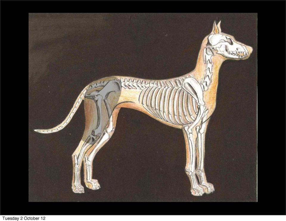
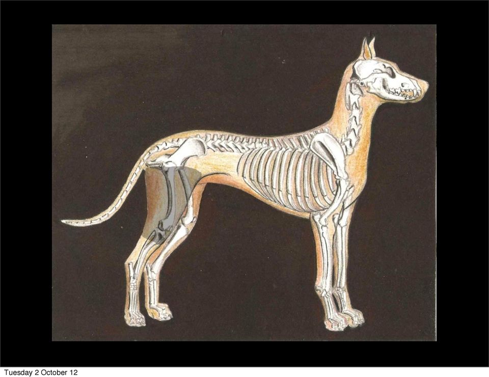
1 An overview of the anatomy of the canine hindlimb Darren Kelly Artwork by Paddy Lennon Original photos courtesy of Mary Ferguson Students at University College Dublin, School of Veterinary Medicine. Video clip by Dr. David Kilroy

2

3

4

5
6 Lateral view of the right hip and stifle regions. 1. Biceps Femoris 2. Semitendonosis 3. Superficial Gluteal 4 and 4. Tensor Fascia Lata 5. Sartorius 6. Vastus Lateralis of Quadriceps 7. Fascia Lata (cut)

7 The Quadriceps is a large muscle which lies on the cranial aspect of the femur. It is made up of four heads; Vastus Lateralis Vastus Medialis Vastus Intermedia Rectus Femoris The tendons of insertion of all four heads join to form the patellar tendon (often called the patellar ligament) which crosses the stifle joint to insert on the tibial tuberosity. The patellar tendon contains the largest sesamoid bone, the patella. All four heads therefor act to extend the stifle joint but only one acts to also flex the hip joint, the rectus femoris. Because this head originates just cranial to the acetabulum of the ilium, it crosses the hip joint and can therefor act to flex it. The other heads originate on the femur and so do not cross the hip joint.

8 In the dog, we see four sesamoid bones in the stifle joint. The largest is the patella which is found in the tendon of insertion of the quadriceps femoris on the cranial aspect of the joint. A patella can be found in all domestic species. However in the dog there are three sesamoid bones found caudal to the stifle joint, the fabellae. Two of these develop in the tendons of origin of the two heads of the gastrocnemius muscle. The other develops in the tendon of origin of the popliteus muscle. These three sesamoid bones are not found in the ox or horse.

9 Radiograph of the left stifle joint of a dog. 1. Patella 2, 3, and 4. Fabellae 5. Patellar Tendon 6. Tibial Tuberosity 2 and 3 develop in the tendons of origin of the two heads of the gastrocnemius while 4 develops in the tendon of origin of the popliteus. These are absent in the ox and horse.

10 There are three gluteal muscles; Superficial gluteal Middle gluteal Deep gluteal They act to extend the hip joint and are the abductors of the hindlimb. Abduction of a limb is to move it further away from the body. Adduction of a limb is to move it closer to the body. The gluteal muscles are therefor essential for the male dog when it comes to lifting the leg during urination! The deep gluteal originates on the body of the Ilium. The middle gluteal originates on the wing of the Ilium. The superficial gluteal originates on the gluteal fascia.

11 Muscle Origin Insertion Innervation Function Biceps Femoris Ischial Tuber Patella, Tibial Crest & Calcaneus Sciatic Nerve Extend Hip Joint, Flex or Extend Stifle Joint, Extend Hock Joint Gluteals (3) Ilium Greater Trochanter of Femur Gluteal Nerves Extend Hip and Abduct the Limb Tensor Fascia Lata Ventral aspect of the Wing of the Ilium Patella Cranial Gluteal Nerve Tenses the Fascia Lata to Extend and Stabilise the Stifle Joint Sartorius (2 parts in Dog) Wing of the Ilium Patella and Disto-medial Femur Femoral Nerve Flex Hip and Adduct Limb Quadriceps See slide number 5 See slide number 5 Femoral Nerve See slide number 5

12
13 Medial view of the hip and stifle regions of the right hindlimb. 1 and 1. Sartorius 2. Vastus Medialis of Quadriceps 3. Patellar Tendon 4. Gracilis (large part removed)

14 Five muscles play a role in adducting the hindlimb. They are; Adductor Gracilis Semimembranosus Sartorius Pectineus

15

16 Caudal view of the hip and stifle region of the right hindlimb 1. Gracilis 2. Semimembranosis 3. Semitendonosis 4. Biceps Femoris Medial Lateral

17 The biceps femoris, semitendonosus and semimembranosis lie of the caudal aspect of the femur and are often together referred to as the hamstring group of muscles. All three originate from the ventral aspect of the ischial tuber and are innervated by the sciatic nerve.

18 Muscle Origin Insertion Innervation Function Adductor Ventral aspect of the Pelvis Medial aspect of the Femur Obturator Nerve Adduct the Limb Gracilis Pelvic Symphysis Medial aspect of the Stifle and Calcaneus Obturator Nerve Adduct the Limb and extend the Hock Pectineus Prepubic tendon Medial aspect of the Femur Obturator Nerve Adduct the Limb Semitendonosis Ventral aspect of the Ischial Tuber Tibial Crest and Calcaneus Sciatic Nerve Extend Hip Joint, Flex Stifle Joint, Extend Hock Joint Semimembranosis Ventral aspect of the Ischial Tuber Medial aspect of the Femur and Tibia Sciatic Nerve Extend Hip Joint and Flex Stifle Joint

19 Notice that three of the adductors of the hindlimb are innervated by the obturator nerve. This nerve can become compressed and damaged during a difficult birthing in cows as the foetus passes through the birth canal and compresses the the nerve against the wall of the pelvic cavity. This can cause an inability of the newly calved cow to stand, and a splits stance can be seen due to the inability to adduct the limbs. The obturator nerve comes off the lumbosacral plexus and leaves the pelvic cavity through the obturator foramen.

20 The nerves of the hindlimb arise from the lumbosacral plexus. Starting cranially they are the femoral nerve, obturator nerve, gluteal nerves and the sciatic nerve. The video on the next slide shows and explains the lumbosacral plexus in a dissected dog.

21 Double click on the video to play it. It may take a few seconds to start. If it does not play it can be downloaded individually from the OVAM website.
22
23 Medial view of the hip and stifle regions. 1 and 1. Sartorius 2. Gracilis (cut) * Here we can see the External Iliac Artery. This is a direct branch off the aorta and is continued as the femoral artery which supplies blood to the hindlimb.
24
25 Lateral view of hip and stifle regions. 1. Biceps Femoris 2. Semitendonosis 3. Adductor 4. Vastus Lateralis 5. Tensor Fascia Lata 6. Superficial Gluteal 7. Sartorius 8. Gastrocnemius
26 In the previous image we can see the large sciatic nerve running between the biceps and the adductor. It is continued distal to the stifle as the tibial nerve and the fibular nerve. The fibular nerve is sometimes called the peroneal nerve. This can be seen in the previous picture crossing the lateral head of the gastrocnemius muscle to innervate the muscles which lie on the cranial aspect of the tibia and fibula. The tibial nerve branches off the sciatic nerve and dives between the two heads of the gastrocnemius to innervate the muscles on the caudal aspect of the tibia and fibula. If the sciatic nerve is severed at a point proximal to the stifle, by a broken femur for example, paralysis of all the muscles distal to the stifle may be seen as both the tibial and fibular nerves are continuations of the sciatic nerve.
Chapter 9 The Hip Joint and Pelvic Girdle
 Copyright The McGraw-Hill Companies, Inc. Reprinted by permission. The Hip Joint and Pelvic Girdle Chapter 9 The Hip Joint and Pelvic Girdle Structural Kinesiology R.T. Floyd, Ed.D, ATC, CSCS Hip joint
Anterior Superior Iliac Spine. Anterior Inferior Iliac Spine. head neck greater trochanter intertrochanteric line lesser trochanter
 Ilium Bones The Skeleton Ischium Pubis Sacro-iliac Joint Iliac Crest Anterior Superior Superior Pubic Ramus Anterior Inferior Acetabulum Obturator Foramen Ischio-pubic ramus Ischial tuberosity Pubic Crest
Structure & Function of the Knee. One of the most complex simple structures in the human body. The middle child of the lower extremity.
 Structure & Function of the Knee One of the most complex simple structures in the human body. The middle child of the lower extremity. Osteology of the Knee Distal femur (ADDuctor tubercle) Right Femur
Buccinator Presses cheek against molar teeth Facial (CNVII) wrinkles forehead
 wrinkles forehead") Muscles to Identify on the Cadaver and/or Models You are required to identify each of the following muscles or associated structures on the cadavers and/or models in lab. If the box is shaded in a particular
Muscles to Identify on the Cadaver and/or Models You are required to identify each of the following muscles or associated structures on the cadavers and/or models in lab. If the box is shaded in a particular
Anatomy and Physiology 121: Muscles of the Human Body
 Epicranius Anatomy and Physiology 121: Muscles of the Human Body Covers upper cranium Raises eyebrows, surprise, headaches Parts Frontalis Occipitalis Epicranial aponeurosis Orbicularis oculi Ring (sphincter)
Anatomy and Pathomechanics of the Sacrum and Pelvis. Charles R. Thompson Head Athletic Trainer Princeton University
 Anatomy and Pathomechanics of the Sacrum and Pelvis Charles R. Thompson Head Athletic Trainer Princeton University Simplify Everything There are actually only three bones: Two innominates, one sacrum.
Chapter 9 The Hip Joint and Pelvic Girdle
 Chapter 9 The Hip Joint and Pelvic Girdle Manual of Structural Kinesiology R.T. Floyd, EdD, ATC, CSCS Hip relatively joint (acetabularfemoral) functions bony strong large stable due to The enhanced architecture
Musculoskeletal Ultrasound Technical Guidelines. IV. Hip
 European Society of MusculoSkeletal Radiology Musculoskeletal Ultrasound Technical Guidelines IV. Ian Beggs, UK Stefano Bianchi, Switzerland Angel Bueno, Spain Michel Cohen, France Michel Court-Payen,
Lower limb nerve blocks
 Lower limb nerve blocks Barry Nicholls is Consultant in Anaesthesia and Pain Management at Musgrove Hospital, Taunton, UK. He qualified from Liverpool University and trained in Newcastle, UK, and Seattle,
Muscle Name Origin Insertion Action Innervation Muscles of Upper Extremity Pectoralis Major Medial half of clavicle, front of sternum, costal
 Muscle Name Origin Insertion Action Innervation Muscles of Upper Extremity Pectoralis Major Medial half of clavicle, front of sternum, costal Crest of greater tubercle (Lateral lip of bicipital groove)
The Knee Internal derangement of the knee (IDK) The Knee. The Knee Anatomy of the anteromedial aspect. The Knee
 The Knee. The Knee Anatomy of the anteromedial aspect. The Knee") Orthopedics and Neurology James J. Lehman, DC, MBA, FACO University of Bridgeport College of Chiropractic Internal derangement of the knee (IDK) This a common provisional diagnosis for any patient with
Orthopedics and Neurology James J. Lehman, DC, MBA, FACO University of Bridgeport College of Chiropractic Internal derangement of the knee (IDK) This a common provisional diagnosis for any patient with
Diagnostic MSK Case Submission Requirements
 Diagnostic MSK Case Submission Requirements Note: MSK Ultrasound-Guided Interventional Procedures (USGIP) is considered a separate specialty. Corresponds with 4/21/16 Accred Newsletter* From the main site:
NETWORK FITNESS FACTS THE HIP
 NETWORK FITNESS FACTS THE HIP The Hip Joint ANATOMY OF THE HIP The hip bones are divided into 5 areas, which are: Image: www.health.com/health/static/hw/media/medical/hw/ hwkb17_042.jpg The hip joint is
SECTION II General Osteopathic Techniques
 SECTION II General Osteopathic Techniques Chapter Four The Lower Extremities 40 Ligamentous Articular Strain The lower extremities are among the most important structures of the body and yet are often
Muscular System. Student Learning Objectives: Identify the major muscles of the body Identify the action of major muscles of the body
 Muscular System Student Learning Objectives: Identify the major muscles of the body Identify the action of major muscles of the body Structures to be identified: Muscle actions: Extension Flexion Abduction
13 Adductor Muscle Group Excision
 Malawer Chapter 13 21/02/2001 15:36 Page 243 13 Adductor Muscle Group Excision Martin Malawer and Paul Sugarbaker OVERVIEW The adductor muscle group is the second most common site for high- and low-grade
Skin of eyebrows galea aponeurotica. Muscle and skin of mouth
 : SEE ALSO THE AP SITE FOR OTHER TABLES GROSS ANATOMY OF THE MUSCULAR SYSTEMM Muscles of the Head and Neck: Occipitofrontalis Frontalis Occipitalis Orbicularis oculi Orbicularis oris Buccinator Masseter
Femoral head and neck ostectomy (FHO) is a commonly
 is a commonly") P ro c e d u re s P ro O R T H O P E D I C S Peer Reviewed Laura E. Peycke, DVM, MS, Diplomate CVS Texas &M University Femoral Head & Neck Ostectomy Femoral head and neck ostectomy (FHO) is a commonly
P ro c e d u re s P ro O R T H O P E D I C S Peer Reviewed Laura E. Peycke, DVM, MS, Diplomate CVS Texas &M University Femoral Head & Neck Ostectomy Femoral head and neck ostectomy (FHO) is a commonly
Structure & Function of the Ankle and Foot. A complicated model of simplicity that you really think little about until you have a problem with one.
 Structure & Function of the Ankle and Foot A complicated model of simplicity that you really think little about until you have a problem with one. The Foot and Ankle Terminology Plantar flexion Dorsi flexion
MET: Posterior (backward) Rotation of the Innominate Bone.
 Rotation of the Innominate Bone.") MET: Posterior (backward) Rotation of the Innominate Bone. Purpose: To reduce an anterior rotation of the innominate bone at the SI joint. To increase posterior (backward) rotation of the SI joint. Precautions:
MET: Posterior (backward) Rotation of the Innominate Bone. Purpose: To reduce an anterior rotation of the innominate bone at the SI joint. To increase posterior (backward) rotation of the SI joint. Precautions:
Musculoskeletal Ultrasound Technical Guidelines. V. Knee
 European Society of MusculoSkeletal Radiology Musculoskeletal Ultrasound Technical Guidelines V. Knee Ian Beggs, UK Stefano Bianchi, Switzerland Angel Bueno, Spain Michel Cohen, France Michel Court-Payen,
Chapter 8. Muscular System: Skeletal Muscles of the Body
 Chapter 8 Muscular System: Skeletal Muscles of the Body INTRODUCTION This chapter continues our study of the muscular system by examining the distribution of muscles throughout the body. We learned in
Muscles of Mastication
 Muscles of Mastication Masseter Zygomatic Arch Mandibular angle Elevates mandible Mandibular ramus Temporalis Temporal fossa of the temporal bone Coronoid process of the mandible Elevates mandible Retracts
Skeletal system. 2012 Pearson Education, Inc.
 NURS1004 Week 6 Part I Prepared by Didy Button Skeletal system An Introduction to the Skeletal System The Skeletal System Includes: Bones of the skeleton Cartilages, ligaments, and connective tissues 6-1
The Pilates Studio of Los Angeles / PilatesCertificationOnline.com
 Anatomy Review Part I Anatomical Terminology and Review Questions (through pg. 80) Define the following: 1. Sagittal Plane 2. Frontal or Coronal Plane 3. Horizontal Plane 4. Superior 5. Inferior 6. Anterior
Surgical Approaches to Total Hip Arthroplasty
 Surgical Approaches to Total Hip Arthroplasty Daniel Kelmanovich, 1 Michael L. Parks, MD, 2 Raj Sinha, MD, PhD, 3 and William Macaulay, MD 4 Surgical exposure of the hip for trauma, infection, or reconstruction
Stretching the Major Muscle Groups of the Lower Limb
 2 Stretching the Major Muscle Groups of the Lower Limb In this chapter, we present appropriate stretching exercises for the major muscle groups of the lower limb. All four methods (3S, yoga, slow/static,
Muscles of the Neck and Vertebral Column Sternocleidomastoid (anterior neck) Origin Insertion Action
 Origin Insertion Action") Muscular movements of the head (at the cervical spine/neck) and of the torso (thoracic and lumbar spine/upper, middle, and lower back): flexion, extension, lateral flexion, rotation. Muscles of the Neck
Muscular movements of the head (at the cervical spine/neck) and of the torso (thoracic and lumbar spine/upper, middle, and lower back): flexion, extension, lateral flexion, rotation. Muscles of the Neck
Avoiding Meniscus Surgery
 Avoiding Meniscus Surgery Robert Tarantino February 9, 2008 2007 NY CTTC-1 1 WARNING THIS MATERIAL DOES NOT CONSTITUTE MEDICAL ADVICE. IT IS INTENDED FOR INFORMATIONAL PURPOSES ONLY. PLEASE CONSULT A PHYSICIAN
Learning IRM. The Knee: lateral ligaments and anatomical quadrants.
 Learning IRM. The Knee: lateral ligaments and anatomical quadrants. Poster No.: C-1733 Congress: ECR 2014 Type: Educational Exhibit Authors: A. Amador Gil, M. D. C. Jurado Gómez, V. de Lara Bendahan ;
Chapter 10: The Muscular System
 Chapter 10: The Muscular System Objectives: 1. Describe the function of prime movers, antagonists, synergists, and fixators. 2. List the criteria used in naming muscles. Provide an example to illustrate
Lower Extremity Orthopedic Surgery in Cerebral Palsy. Hank Chambers, MD Rady Children s Hospital - San Diego
 Lower Extremity Orthopedic Surgery in Cerebral Palsy Hank Chambers, MD Rady Children s Hospital - San Diego Indications Fixed contracture Joint dislocations Shoe wear problems Pain Perineal hygiene problems
Anatomy of Human Muscles
 Anatomy of Human Muscles PURPOSE: To develop skill in identifying muscle names and locations relative to other regional structures. To determine origin, insertion and principle action of muscles through
Screening Examination of the Lower Extremities BUY THIS BOOK! Lower Extremity Screening Exam
 Screening Examination of the Lower Extremities Melvyn Harrington, MD Department of Orthopaedic Surgery & Rehabilitation Loyola University Medical Center BUY THIS BOOK! Essentials of Musculoskeletal Care
www.ghadialisurgery.com
 P R E S E N T S Dr. Mufa T. Ghadiali is skilled in all aspects of General Surgery. His General Surgery Services include: General Surgery Advanced Laparoscopic Surgery Surgical Oncology Gastrointestinal
Rehabilitation with Pilates
 Rehabilitation with Pilates Flat Feet Pes Planus Naiké Durel January 2016 Montreal, Canada Table of contents Title Table of contents Abstract Anatomy of the foot p.1 p.2 p.3 p.4-5- 6-7 Case of study BASI
ULTRASOUND GUIDED FEMORAL NERVE BLOCK ANAESTHESIA TUTORIAL OF THE WEEK 284
 ULTRASOUND GUIDED FEMORAL NERVE BLOCK ANAESTHESIA TUTORIAL OF THE WEEK 284 15 TH APRIL 2013 Dr Suresh Kumar Jeyaraj, Specialty Doctor Dr Tim Pepall, Consultant Anaesthetist Frimley Park Hospital NHS foundation
PRIMARY HUMAN ANATOMY: BIOL20600 SPRING 2014
 PRIMARY HUMAN ANATOMY: BIOL20600 SPRING 2014 Instructors: Kit Muma, Rm. 158 CNS, (607) 274-3610, muma@ithaca.edu Michelle Bamberger, Rm. 118A Williams Hall, mbamberger@ithaca.edu Mark Baustian, Rm. 118A
What is the Hip? Femur pull-back prone stabilize pelvis and pull the femur up at the top
 Spinning the Hip Dr. George Russell 1133 Broadway, #1125 New York, NY 10010 george@georgerusselldc.com georgerusselldc.com Copyright George Russell 2014 What is the Hip? Front bone of the pelvis Anterior
Medical Billing Basics
 Ingenix Learning: Medical Billing Basics 2010 8th edition Contents Introduction... 1 Welcome to the Career of Medical Coding...1 The Nature of Medical Coding...1 Coding and the Financial Picture...2 Supporting
PART B - Human Tissue List February 2015
 PART B - Human Tissue List February 2015 Billing Code CARDIO-THORACIC Conduit - Aortic TBV56 Donor Tissue Bank of Victoria $1,505.00 Conduit - Aortic + Valve QHV09 The Prince Charles Hospital - Queensland
Lower Extremity Special Tests. Hip Special Tests
 Lower Extremity Special Tests Hip Special Tests Trendelenburg Test: a test for weakness of the gluteus medius muscle during unilateral weight bearing. Therapist is positioned behind patient to observe
Equine Bodywork Basics Clinic
 Comprehensive Care. Tailored Solutions. Real Results. Ilene Nessenson, J.D., CEBW Certified Equine Body Worker English and Western Saddle Fitting Saddle Reflocking and Refleecing Tack Repair Certified
Knee Kinematics and Kinetics
 Knee Kinematics and Kinetics Definitions: Kinematics is the study of movement without reference to forces http://www.cogsci.princeton.edu/cgi-bin/webwn2.0?stage=1&word=kinematics Kinetics is the study
LABORATORY EXERCISE 12 BONE STRUCTURE AND CLASSIFICATION
 LABORATORY EXERCISE 12 BONE STRUCTURE AND CLASSIFICATION FIG. 12.1 1. Articular cartilage (hyaline cartilage) 6. Periosteum 2. Spongy bone (red marrow) 7. Proximal epiphysis 3. Medullary cavity 8. Diaphysis
Fascia Iliaca Compartment Block: LANDMARK AND ULTRASOUND APPROACH
 Fascia Iliaca Compartment Block: LANDMARK AND ULTRASOUND APPROACH ANAESTHESIA TUTORIAL OF THE WEEK 193 23 rd AUGUST 2010 Dr Christine Range, Specialist Registrar Anaesthesia Dr Christian Egeler, Consultant
Anatomy & Physiology 120. Lab #7 Muscle Tissue and Skeletal Muscles
 Anatomy & Physiology 120 Lab #7 Muscle Tissue and Skeletal Muscles What you Need to Know Look briefly at the Structure of: 1) Skeletal, 2) Smooth & 3) Cardiac Muscle Naming, Identification, Functions You
Surgical Treatment in Cerebral Palsy
 Surgical Treatment in Cerebral Palsy พ.ญ.ก ต วรรณ ว ป ลากร ภาคว ชาออร โธป ด กส คณะแพทยศาสตร มหาว ทยาล ยขอนแก น General consideration important to speak clearly and frankly with the family about the goals
Chapter 11 The Muscular System. Muscle Attachment Sites: Origin and Insertion
 Chapter 11 The Muscular System Skeletal muscle major groupings How movements occur at specific joints Learn the origin, insertion, function and innervation of all major muscles Important to allied health
Structure and Function of the Hip
 Structure and Function of the Hip Objectives Identify the bones and bony landmarks of the hip and pelvis Identify and describe the supporting structures of the hip joint Describe the kinematics of the
ANATOMY 1 LEARNING TARGETS
 ANATOMY 1 LEARNING TARGETS ORGANIZATION OF THE BODY 1. Define "anatomy" and "physiology." 2. Describe homeostasis. 3. Identify examples of homeostasis 4. Describe the organization of the body according
Patellofemoral Pain Syndrome and the Pilates Client
 Patellofemoral Pain Syndrome and the Pilates Client Aliza Nizet Comprehensive Teacher Training Course Body Arts and Science International Mind & Body Wellness Studio January 26 May 18, 2008 1 Abstract
Critical Reviews in Physical and Rehabilitation Medicine, 12:277-282, 2000
 HAMSTRING INJURIES R.L.Morgan-Jones, T.Cross, M.J.Cross Critical Reviews in Physical and Rehabilitation Medicine, 12:277-282, 2000 ABSTRACT Hamstring injuries are common and have a significant impact on
Functional Anatomy and Lower Extremity Biomechanics
 Functional Anatomy and Lower Extremity Biomechanics Eric Folmar, MPT, OCS Functional Lower Extremity Biomechanics The science of foot, ankle, knee and hip biomechanics and their relationships and interactions
The Nervous System: The Spinal Cord and Spinal Nerves
 14 The Nervous System: The Spinal Cord and Spinal Nerves PowerPoint Lecture Presentations prepared by Steven Bassett Southeast Community College Lincoln, Nebraska Introduction The Central Nervous System
The Muscular System. Appendicular Musculature
 11 The Muscular System Appendicular Musculature CHAPTER OBJECTIVES 1. Describe the functions of the appendicular musculature. 2. Identify and locate the principal appendicular muscles of the body, together
Surgical Art. Formulaic Drawing Method. DRAWING WORKSHOP Learning to sketch for patient notes
 DRAWING WORKSHOP Learning to sketch for patient notes Surgical Art Formulaic Drawing Method Formulaic figure drawing systems involve using abstract rhythms and interlocking shapes to construct the human
Hip Bursitis/Tendinitis
 Hip Bursitis/Tendinitis Anatomy and Biomechanics The hip is a ball and socket joint that occurs between the head of the femur (ball) and the acetabulum of the pelvis (socket). It is protected by several
GROSS ANATOMY. Unit #4: Upper and Lower Limbs. Lecture Syllabus 2008
 GROSS ANATOMY Lecture Syllabus 2008 Unit #4: Upper and Lower Limbs ANAT 6010 - Gross Anatomy Department of Neurobiology and Anatomy University of Utah School of Medicine G24- Upper Limb Overview, Shoulder,
SPORT AND PHYSICAL ACTIVITY
 2016 Suite Cambridge TECHNICALS LEVEL 3 SPORT AND PHYSICAL ACTIVITY Unit 1 Body systems and the effects of physical activity K/507/4452 Guided learning hours: 90 Version 2 - Revised content - March 2016
The Muscular System. PowerPoint Lecture Presentations prepared by Jason LaPres. Lone Star College North Harris. 2012 Pearson Education, Inc.
 11 The Muscular System PowerPoint Lecture Presentations prepared by Jason LaPres Lone Star College North Harris An Introduction to the Muscular System Learning Outcomes 11-1 Describe the arrangement of
TOTAL BODY: POWER/EXPLOSIVE EXERCISES
 Referring to Chapters 12-14 TOTAL BODY: POWER/EXPLOSIVE EXERCISES Power Snatch hip extension Muscle group/ gluteals gluteus maximis hamstrings semimembranosus semitendinosus biceps femoris knee extension
Canine Muscle Origins, Insertions, Actions and Nerve Innervations
 Canine Muscle Origins, Insertions, Actions and Nerve Innervations! The purpose of this document is to provide students of canine anatomy a simple reference for muscular origins, insertions, actions and
EXERCISE MANUAL PERSONALITY GYM
 EXERCISE MANUAL PERSONALITY GYM EXERCISE MANUAL PERSONALITY GYM legs. 1 calves raise Stand with the wide part of one foot on the seated row foot support. Start in a position with your calves stretched.
Understanding back pain 1 The anatomy & physiology of back pain
 Understanding back pain 1 The anatomy & physiology of back pain Dr James Tang, MBA, BDS, LDS RCS General Dental Practitioner, Level 3 Personal Trainer (REP registration no R1045463), Sports Nutritionist
American Osteopathic Academy of Sports Medicine James McCrossin MS ATC, CSCS Philadelphia Flyers April 23 rd, 2015
 American Osteopathic Academy of Sports Medicine James McCrossin MS ATC, CSCS Philadelphia Flyers April 23 rd, 2015 Coming together is a beginning; keeping together is progress; working together is success.
LABS 8 & 9: CAT MUSCULATURE
 ZOO 4377L - VERTEBRATE MORPHOLOGY LAB LABS 8 & 9: CAT MUSCULATURE Name: Name: SSN: SSN: ---------------------------------------------------------------------------------------------------------------------
RUNNING INJURIES: PREVENTION AND REHABILITATION
 RUNNING INJURIES: PREVENTION AND REHABILITATION Topics of Tonight s s Lecture Common Injuries and Treatments Causes of Common Injuries Measures to Avoid Injury Most Common Running Injuries Plantar Fascitis
Hip Anatomy and Biomechanics in the Athlete
 Sports Medicine and Arthroscopy Review 10:103 114 2002 Lippincott Williams & Wilkins, Inc., Philadelphia in the Athlete Paul E. Hughes, M.D., Jim C. Hsu, M.D., and Matthew J. Matava, M.D. SUMMARY The hip
The Muscular System General & Anatomy
 The Muscular System General & Anatomy General Functions: 1. movement voluntary skeletal muscles 2. internal movement of substances through various tubes and passageways eg blood, food, urine heart pumps
Hamstring Apophyseal Injuries in Adolescent Athletes
 Hamstring Apophyseal Injuries in Adolescent Athletes Kyle Nagle, MD MPH University of Colorado Department of Orthopedics Children s Hospital Colorado Orthopedics Institute June 14, 2014 Disclosures I have
MR Imaging of the Anatomy of and Injuries to the Lateral and Posterolateral Aspects of the Knee
 Downloaded from www.ajronline.org by 37.44.192.158 on 07/15/16 from IP address 37.44.192.158. opyright RRS. For personal use only; all rights reserved Pictorial Essay MR Imaging of the natomy of and Injuries
The Correlation between Hamstring Tightness and Low Back Pain in Seated Workers Ahmed Radwan PT, DPT, PhD Thomas A. Crist, PhD
 The Correlation between Hamstring Tightness and Low Back Pain in Seated Workers Ahmed Radwan PT, DPT, PhD Thomas A. Crist, PhD Ahmed Radwan, PT, DPT, Ph.D. Associate Professor Physical Therapy Program
CONTINUING EDUCATION COURSES. for Massage Therapists. Online!
 CONTINUING EDUCATION COURSES for Massage Therapists Online! ccmh Halifax Canadian College of Massage & Hydrotherapy Online Continuing Education Program CCMH Halifax offers a variety of Continuing Education
SPECIAL TESTS ANKLE Anterior Drawer anterior talofibular ligament Positive Sign pain, laxity Talar Tilt calcaneofibular ligament; deltoid ligament
 SPECIAL TESTS ANKLE Anterior Drawer anterior talofibular ligament pain, laxity Talar Tilt calcaneofibular ligament; deltoid ligament pain, laxity Kleiger deltoid ligament medial and lateral pain, displaced
Classification of bones Any bone may be classified into one of the following groups:
 Skeletal system This system is made up of hard tissues like bone and cartilages. This system gives form and shape to animal body The skeleton of a living animal is made up living structures of bones. The
Glossary (continued)
") 1. Maxillary bone 2. Maxillary teeth 3. Mandibular arch 4. Vomerine teeth 5. Lining of buccal (mouth) cavity 6. Inferior aspect of eyeball (floor of eye socket) 7. Tongue (with right half cut away) 8.
1. Maxillary bone 2. Maxillary teeth 3. Mandibular arch 4. Vomerine teeth 5. Lining of buccal (mouth) cavity 6. Inferior aspect of eyeball (floor of eye socket) 7. Tongue (with right half cut away) 8.
The patellofemoral joint and the total knee replacement
 Applied and Computational Mechanics 1 (2007) The patellofemoral joint and the total knee replacement J. Pokorný a,, J. Křen a a Faculty of AppliedSciences, UWB inpilsen, Univerzitní 22, 306 14Plzeň, CzechRepublic
DSM Spine+Sport - Mobility
 To set yourself up for success, practice keeping a neutral spine throughout all of these movements. This will ensure the tissue mobilization is being applied to the correct area, and make the techniques
Vertebrate Skeletal Muscle Structure
 VertebrateSkeletalMuscleStructure HumanAnatomy&PhysiologyLab#1 Todaywewillexaminethemuscularsystemoftherat,withparticularemphasison themusclesofthehindlimb.thegoalofthislabistoconsidertherelationship betweenfineandgrossmusclestructureandfunction.byfocusingonrathindlimb
Integrated Manual Therapy & Orthopedic Massage For Low Back Pain, Hip Pain, and Sciatica
 Integrated Manual Therapy & Orthopedic Massage For Low Back Pain, Hip Pain, and Sciatica Assessment Protocols Treatment Protocols Treatment Protocols Corrective Exercises By Author & International Lecturer
Trigger Point Master Course. Chapter 12. Muscles of the Leg and Foot
 Trigger Point Master Course Chapter 12 Muscles of the Leg and Foot 12 Muscles of the Leg and Foot Regional Trigger Points for Lower Leg, Ankle, and Foot Pain MUSCLE PAGE REFERENCE Abductor digiti minimi...
Lengthening Reconstruction Surgery for Congenital Femoral Deficiency
 Lengthening Reconstruction Surgery for Congenital Femoral Deficiency 13 Dror Paley and Fran Guardo Contents 13.1 Introduction... 000 13.1.1 Classification... 000 13.2 Evaluating the Child with Unilateral
Integrated Manual Therapy & Orthopedic Massage For Complicated Knee Conditions
 Integrated Manual Therapy & Orthopedic Massage For Complicated Knee Conditions Assessment Protocols Treatment Protocols Treatment Protocols Corrective Exercises Artwork and slides taken from the book Clinical
II. Axial Skeleton (Skull, Thoracic Cage, and Vertebral Column)
") THE SKELETAL SYSTEM Lab Objectives Students should be able to: 1. Recognize bones and bone markings for the axial and appendicular skeleton 2. Recognize bones disarticulated and/or articulated 3. Identify
THE SKELETAL SYSTEM Lab Objectives Students should be able to: 1. Recognize bones and bone markings for the axial and appendicular skeleton 2. Recognize bones disarticulated and/or articulated 3. Identify
Muscular System. Principles of Health Science Dr. Wood
 Muscular System Principles of Health Science Dr. Wood Characteristics of muscles Excitability: : irritability or ability to respond to stimulus Contractibility: : ability to contract (become short and
Thank You for Your Support!
 Thank You for Your Support! This PDF document has been placed on the Internet with the goal of providing quality learning material at a low price to cover web operating expenses. This document is shareware,
Is Your Horse Off Behind?? Hindlimb Facts. Common Hindlimb Lameness. Diagnostic Techniques. Gait Analysis 3/21/2012
 Is Your Horse Off Behind?? Nathaniel A. White II DVM MS DACVS Jean Ellen Shehan Professor and Director Common Hindlimb Lameness Sacroiliac joint pain Hip Lameness Stifle Lameness Stress Fractures Hock
EHFA Assessment Strategy (EAD 03) Name of Training Organisation:
 Name of Training Organisation:") EHFA Assessment Strategy (EAD 03) Name of Training Organisation: Introduction European Health and Fitness Association (EHFA) Standards The following EHFA standards are currently available: Fitness Instructor
EHFA Assessment Strategy (EAD 03) Name of Training Organisation: Introduction European Health and Fitness Association (EHFA) Standards The following EHFA standards are currently available: Fitness Instructor
CORRESPONDENCE EDUCATION PROGRAM
 Deep Tissue Massage CORRESPONDENCE EDUCATION PROGRAM Check your receipt for course expiration date. After that date no credit will be awarded for this program. 1 How to Complete this Program Thank you
Flexibility Assessment and Improvement Compiled and Adapted by Josh Thompson
 Flexibility Assessment and Improvement Compiled and Adapted by Josh Thompson Muscles must have a full and normal range of motion in order for joints and skeletal structure to function properly. Flexibility
ACUTE AVULSION FRACTURE OF THE ANTERIOR SUPERIOR ILIAC SPINE IN A HIGH SCHOOL TRACK AND FIELD ATHLETE
 ACUTE AVULSION FRACTURE OF THE ANTERIOR SUPERIOR ILIAC SPINE IN A HIGH SCHOOL TRACK AND FIELD ATHLETE Christopher Mings, LAT, ATC University of Central Florida Alumnus & Florida Gulf Coast University Graduate
Middle California Region USPC Upper Level Horse Management Education. Tendons, Ligaments, Joints & the Skeletal System By Claudia Deffenbaugh
 Tendons - connect Muscle to Bone Middle California Region USPC Upper Level Horse Management Education Tendons, Ligaments, Joints & the Skeletal System By Claudia Deffenbaugh Tendons are fibrous cords of
A Woman s Guide to Muscle & Strength
 A Woman s Guide to Muscle & Strength CORRESPONDENCE EDUCATION PROGRAM # 140. Check your receipt for course expiration date. After that date no credit will be awarded for this program. 2012 by Exercise
This is your Anatomy and Physiology Mid-term.
 This is your Anatomy and Physiology Mid-term. 1 Run for your lives!!! (Just Kidding) Matching - Anatomy or Physiology? A 1. Study of structure A. Anatomy 2. muscle actions B. Physiology 3. Brain Function
CVM 6100 Veterinary Gross Anatomy. Twelve Small Animal Clinical Practice Problems. Alvin F. Weber, DVM, PhD
 CVM 6100 Veterinary Gross Anatomy Twelve Small Animal Clinical Practice Problems by Alvin F. Weber, DVM, PhD CONTENTS Problem One: Tissue masses palpable from the skin surface. Problem Two: Anatomy related
International Standards for the Classification of Spinal Cord Injury Motor Exam Guide
 C5 Elbow Flexors Biceps Brachii, Brachialis Patient Position: The shoulder is in neutral rotation, neutral flexion/extension, and adducted. The elbow is fully extended, with the forearm in full supination.
Dr. O Meara s. Anterior Knee Pain (PatelloFemoral Syndrome) Rehabilitation Protocol www.palomarortho.com
 Rehabilitation Protocol www.palomarortho.com") Dr. O Meara s Anterior Knee Pain (PatelloFemoral Syndrome) Rehabilitation Protocol www.palomarortho.com Anterior Knee Pain (PatelloFemoral Syndrome) Rehabilitation Protocol Hamstring Stretching & Strengthening
Dr. O Meara s Anterior Knee Pain (PatelloFemoral Syndrome) Rehabilitation Protocol www.palomarortho.com Anterior Knee Pain (PatelloFemoral Syndrome) Rehabilitation Protocol Hamstring Stretching & Strengthening
Total hip arthroplasty (THA) through the anterior approach
 through the anterior approach") Primary Total Hip Arthroplasty with a Minimally Invasive Anterior Approach Jonathan G. Yerasimides, MD, and Joel M. Matta, MD Total hip arthroplasty (THA) through the anterior approach is a minimally invasive
Primary Total Hip Arthroplasty with a Minimally Invasive Anterior Approach Jonathan G. Yerasimides, MD, and Joel M. Matta, MD Total hip arthroplasty (THA) through the anterior approach is a minimally invasive