Dental Anatomy: A Review
|
|
|
- Bertina Marsh
- 10 years ago
- Views:
Transcription
1 Dental Anatomy: A Review Antoinette Metivier, CDA; Kimberly Bland, CDA, EFDA, M.Ed. Continuing Education Units: 2 hours Disclaimer: Participants must always be aware of the hazards of using limited knowledge in integrating new techniques or procedures into their practice. Only sound evidence-based dentistry should be used in patient therapy. It is important for the dental team to know the appearance of normal anatomy of the face and oral cavity. This knowledge provides a sound basis for identifying abnormal conditions. The dentist holds sole responsibility for diagnosis and treatment of the patient, however, the entire dental team should always be alert for abnormal conditions in all patients oral cavities. There are, of course, wide variations of what can be considered normal, but with careful attention to detail the dental team will gain confidence and become more adept at identifying conditions that may require further attention. Conflict of Interest Disclosure Statement The authors report no conflicts of interest associated with this work. ADA CERP The Procter & Gamble Company is an ADA CERP Recognized Provider. ADA CERP is a service of the American Dental Association to assist dental professionals in identifying quality providers of continuing dental education. ADA CERP does not approve or endorse individual courses or instructors, nor does it imply acceptance of credit hours by boards of dentistry. Concerns or complaints about a CE provider may be directed to the provider or to ADA CERP at: Approved PACE Program Provider The Procter & Gamble Company is designated as an Approved PACE Program Provider by the Academy of General Dentistry. The formal continuing education programs of this program provider are accepted by AGD for Fellowship, Mastership, and Membership Maintenance Credit. Approval does not imply acceptance by a state or provincial board of dentistry or AGD endorsement. The current term of approval extends from 8/1/2009 to 7/31/

2 Overview It is important for the dental team to know the appearance of normal anatomy of the face and oral cavity. This knowledge provides a sound basis for identifying abnormal conditions. The dentist holds sole responsibility for diagnosis and treatment of the patient, however, the entire dental team should always be alert for abnormal conditions in all patients oral cavities. There are, of course, wide variations of what can be considered normal, but with careful attention to detail the dental team will gain confidence and become more adept at identifying conditions that may require further attention. Learning Objectives Upon completion of this course, the dental professional should be able to: Identify and describe the appearance of the normal anatomic structures of the face and oral cavity. Identify and describe the following: types of teeth and their functions, the divisions of a tooth, the tissues of a tooth, the surfaces of a tooth, and the anatomical landmarks of teeth. Discuss the importance of occlusion and describe the classifications of occlusion. Describe the two dentitions and the types of teeth in each dentition. Discuss the importance of the primary teeth. Identify and describe the components of the periodontium, and discuss the importance of the periodontium. Course Contents Glossary Normal Anatomic Structures of the Face and Oral Cavity Facial Landmarks Landmarks of the Oral Cavity Landmarks of the Palate Landmarks of the Tongue Landmarks of the Floor of the Mouth Types of Teeth and Their Functions Divisions of a Tooth Tissues of a Tooth Surfaces of Teeth Anatomical Landmarks of the Teeth Arrangement of the Teeth in the Oral Cavity Arches and Quadrants Occlusion Classifications of Occlusion Dentitions Primary Dentition Permanent Dentition D-A-Q-T System Periodontium Gingival Unit Attachment Unit Conclusion Course Test References About the Authors Glossary adjacent Next to, or nearby. anterior Toward the front. apex The end of the root of a tooth. apical foramen The opening at the end of the root of a tooth. bifurcated One tooth with two roots. bifurcation The area in a two-rooted tooth where the roots divide. buccal The surface of a posterior tooth facing the cheeks. cementoenamel junction (CEJ)/cervical line The area of a tooth where the cementum of the root and the enamel of the crown meet. cementum The tissue covering the root of a tooth. cingulum A smooth, rounded bump on the cervical third of the lingual surface of anterior teeth. contact area The area on the proximal surface of a tooth where it touches an adjacent tooth. 2

3 deciduous teeth The first set of teeth; the primary teeth. dentin The tissue of a tooth that comprises the main inner portion of the tooth; it is covered by cementum on the root and enamel on the crown. dentition Set of teeth; the natural teeth in position in the dental arches. distal The surface of a tooth that is away from the midline. facial The surface of a tooth that faces the lips or cheeks; includes the labial and buccal surfaces. fissure A groove or natural depression on the surface of a tooth. fossa A shallow, rounded or angular depression on the surface of a tooth. gingiva The mucous membrane tissue that surrounds the teeth. incisal Cutting edge of anterior teeth. incisive foramen A passageway through the bone of the hard palate for vessels and nerves; found just lingual to the central incisor region. interproximal Between the proximal surfaces of adjacent teeth. labial The surface of anterior teeth facing the lips. lingual The surface of a tooth that faces the tongue. mandibular Pertaining to the lower jaw. maxillary Pertaining to the upper arch. mesial The surface of a tooth that faces the midline. midline An imaginary vertical plane that divides the body into equal right and left halves. mucogingival junction The line or area where the alveolar mucosa meets the attached gingiva. occlusal The biting surface of a posterior tooth. periapical The area surrounding the apex of a tooth. periodontium The tissues that surround and support the teeth; includes the gingiva, the alveolar mucosa, the cementum, the periodontal ligament, and the alveolar bone. posterior Toward the back. quadrant One-fourth of the mouth; half of the maxillary or mandibular arch. succedaneous Permanent teeth that replace primary teeth. sulcus, gingival The space between the tooth and the free gingiva. Normal Anatomic Structures of the Face and Oral Cavity Facial Landmarks The following are landmarks that are found on the face (Figures 1-3). 1 Ala: Outer edge (wing) of the nostril. Philtrum: Shallow depression between the bottom of the nose and the top of the upper lip. Vermilion Zone: Extraoral reddish portion of the lips. Vermilion Border: The border of the lips where the skin of the face meets the Vermilion zone. Nasolabial Sulcus: Linear depression of the face that runs from the ala to the corner of the mouth. Labial Commissures: Corners of the mouth where upper lip meets the lower lip. Tubercle of the Lip: Small projection of tissue in the middle of the upper lip. Nasion: Midpoint between the eyes just below the eyebrows. Outer Canthus of the Eye: Fold of tissue at the outer corner of the eyelids. Inner Canthus of the Eye: Fold of tissue at the inner corner of the eyelids. Tragus of the Ear: The triangular cartilage projection anterior to the external opening of the ear. mucosa The tissue lining the oral cavity. 3

4 Figure 1. Regions of the Face. There are nine regions of the face as shown in the photograph. Image courtesy of Modern Dental Assisting, 10th Ed. Figure 3. Frontal View of the Lips. This photograph points out the parts of the lips. Image courtesy of Modern Dental Assisting, 10th Ed. Figure 2. Features of the Face. This photograph highlights different facial features. Image courtesy of Modern Dental Assisting, 10th Ed. Landmarks of the Oral Cavity There are various methods of compensation The following are landmarks found in the oral cavity (Figures 4-7). 1,2 Masticatory Mucosa: Heavily keratinized tissue that lines the hard palate and tongue. Alveolar Mucosa: Lightly keratinized tissue that lines floor of the mouth and covers the alveolar processes. Labial and Buccal Mucosa: Thinly keratinized tissue that lines the inner surface of the lips and cheeks (Figures 5 & 6). Oral Vestibule: A pocket formed by the soft tissue of the lips/cheeks and the gingiva, its deepest point is called the vestibule fornix or the muccobuccal fold. 4

5 Figure 4. Normal Structures of the Oral Cavity. Diagram with structures of the mouth labeled. Image courtesy of Mosby s Comprehensive Dental Assisting: A Clinical Approach. Figure 5. Labial Mucosa. Photograph of facial mucosa. Image courtesy of Modern Dental Assisting, 10th Ed. 5

6 Frenum (pl. Frena): Raised lines of oral mucosa that extend from the alveolar mucosa to the labial and buccal mucosa. Fordyce Granules/Spots: Yellowish ectopic sebaceous glands found on the facial mucosa near the corners of the mouth. Linea Alba: A raised white line of keratinized tissue on the buccal mucosa that runs parallel to the line of the occlusal plane (Figure 7). Parotid Papilla: Flap of tissue found opposite the maxillary 2nd molar on the buccal mucosa and contains the terminal end of the Parotid Duct (Stenson s Duct). Landmarks of the Palate The following are landmarks of the palate. Hard Palate: Bony structure that separates oral cavity from nasal cavity. Incisive Papilla: Thick keratinized tissue that covers the incisive foramen. Palatine Raphe: Midline ridge of tissue that covers the bony suture of the palate. Figure 6. Buccal Mucosa. Photograph of mucosa in the back of the mouth. Image courtesy of Modern Dental Assisting, 10th Ed. Figure 7. Linea Alba. Example of a linea alba in the patient s cheek. Image courtesy of Modern Dental Assisting, 10th Ed. 6
.")
7 Palatine Rugae: Irregular ridges or folds of masticatory mucosa that extend horizontally from either side of the palatine raphe. Soft Palate: Forms the posterior section of the palate and is not supported by underlying bone. It can be lifted to meet the posterior pharyngeal wall to seal the nasopharynx during swallowing and speech. Uvula: Small conical mass of tissue that hangs from the palatine velum (free edge of the soft palate). Fauces: The passageway from the oral cavity to the pharynx (throat). Pillars of Fauces: Two arches of muscle tissue surrounding posterior oral cavity. The Anterior Pillar of Fauces is also called palatoglossal arch, the Posterior Pillar of Fauces is also called the palatopharyngeal arch. They contract and narrow the fauces during deglutition (swallowing). Isthmus of Fauces: The space between the left and right anterior and posterior pillars of fauces. It contains the palatine tonsils. (These landmarks are identified in Figure 4) Landmarks of the Tongue The following are landmarks of the tongue. Dorsum of the Tongue: Superior (top) surface of the tongue (Figure 8). 3 Median Sulcus: A centralized linear indentation on the dorsum of the tongue running anterior to posterior. Ventral Surface of the Tongue: Inferior (underneath) surface of the tongue. The ventral surface of the tongue is very vascular and covered with thin, alveolar mucosa. Apex of the Tongue: Anterior tip of the tongue. Filiform Papillae: Small cone shaped papillae found in the anterior 2/3 of the dorsum that are responsible for the sense of touch. Fungiform Papillae: Mushroom-shaped papillae spread evenly over the entire dorsum of the tongue. They are deep red in color and each contains a taste bud. Circumvallate Papillae (Vallate Papillae): Cup-shaped papillae that are approximately 1-2 mm wide and found on the posterior dorsum of the tongue. They are usually arranged in 2 rows that form a V-shape. Each papilla contains a taste bud. Landmarks of the Floor of the Mouth The following are landmarks of the floor of the mouth located under the tongue (Figures 9 & 10). 3 Lingual Frenum: Midline fold of tissue between ventral surface of the tongue and floor of the mouth. Sublingual Caruncles: Two small, raised folds of tissue found on either side of the lingual frenum. They each contain a salivary duct opening for Wharton s Duct (duct leading Figure 8. Dorsal View of the Tongue. Diagram of the top of the tongue. Image courtesy of Illustrated Anatomy of the Head and Neck, 3rd Ed. 7
. Fauces: The passageway from the oral cavity to the pharynx (throat).")
8 from the Submandibular Salivary gland). Sublingual Folds: Folds of tissue that begin at the Sublingual Caruncles on either side of the lingual frenum and run backward toward the base of the tongue. They contain the many ducts from the sublingual salivary gland. Types of Teeth and Their Functions The four front teeth in each arch are called incisors, and their function is to cut food with their sharp thin edges. On each side of the incisors, at the corners of the mouth, are the canines. These teeth have one cusp, or pointed edge, and are used for holding or grasping food, and are very strong, stable teeth. Behind the canines are the premolars, which are designed for holding food like the canines because they have cusps, but they also function to crush food. Sometimes these teeth are referred to as bicuspids, meaning two cusps, but this is not always accurate because some premolars may have three cusps. Therefore the term premolar is preferred. The Figure 9. View of the Floor of the Mouth. Photograph of the area under the tongue. Image courtesy of Illustrated Anatomy of the Head and Neck, 3rd Ed. Figure 10. The Major Salivary Glands and Associated Structures. Diagram of location of major salivary glands. Image courtesy of Illustrated Anatomy of the Head and Neck, 3rd Ed. 8

9 Figure 11. Primary Dentition. Primary dentition diagram of primary teeth in order in the arch. Image source: General Chairside Assisting: A Review for a National General Chairside Exam. Figure 12. Permanent Dentition. Permanent dentition diagram of permanent teeth in order in the arch. Image source: General Chairside Assisting: A Review for a National General Chairside Exam. 9

10 teeth farthest back in the mouth are the molars. These teeth have broad chewing surfaces with four or five cusps, and are designed for grinding food. The incisors and canines are called anterior teeth, because they are located in the front of the mouth, while the premolars and molars are called posterior teeth because they are located in the back of the mouth (Figures 11 & 12). In addition to aiding in acquiring and chewing food, teeth perform several other important functions within the oral cavity. They begin the digestive process by breaking down food; they protect the oral cavity; they aid in proper speech; and they affect physical appearance. There are several types of teeth, and each performs its own special function in the chewing process, depending on its size, shape and location within the jaws. Starting at the midline, the permanent dentition is comprised of incisors, canines, premolars and molars. The primary dentition is the same except it has no premolars. Incisors There are four incisors in each arch. Two central incisors and two lateral incisors. Location the central incisors are side by side at the midline. There is a lateral incisor on each side of the central incisors. Shape single rooted, crowns are arched and angle toward one sharp incisal edge. Function to cut or incise food with their thin edges. Canines There are two canines in each arch. They are sometimes referred to as cuspids. Location next to the lateral incisors, establishes the cornering of the arches. Shape anchored with the longest root, one pointed cusp. Function used for holding, grasping, and tearing food. Referred to as the cornerstone of the mouth. Premolars There are for premolars in each arch. Two first premolars and two second premolars. They are sometimes referred to as bicuspids. There are no premolars in the primary dentition. Location first premolars are next to the canines followed by the second premolars. Shape maxillary first premolars have a bifurcated root, all others have one root, one prominent cusp with one or two lesser lingual cusps. Function holding food, like canines because they have cusps; also to crush food. Molars There are three molars in each arch of the permanent dentition. Two first molars, two second molars and two third molars. Third molars are sometimes called wisdom teeth. There are two molars in each arch of the primary dentition. Two first molars and two second molars. Location first molars are next to the second premolars, second molars next to the first molars and third molars next to the second molars. The third molars are the farthest teeth in the mouth. Shape bifurcated or trifurcated roots, broad chewing surfaces with four to five cusps. Function grinding food. Divisions of a Tooth A tooth is divided into two main sections, the crown and the root. The crown is the portion of the tooth that is most visible in the mouth, and the root is the portion that is normally not visible because it is embedded within the bone. A tooth either will have a single root, or will have multiple roots. If a root is divided into two segments, it has a bifurcation; if a root is divided into three segments it has a trifurcation. Each root has an apex, which is the end of the root. The crown of a tooth is covered with enamel and the root is covered with cementum. Therefore, the area where the crown and the root meet is known as the cementoenamel junction (CEJ). This area is also known as the cervix or cervical line of the tooth (Figure 13). 4 Tissues of a Tooth There are four tissues that make up a tooth. Enamel, dentin, and cementum are the hard tissues of a tooth. The pulp is the soft tissue. Enamel, which forms the outer surface of the crown of the tooth, is the hardest tissue in the body, thus making the tooth able to withstand a great amount of stress, chewing pressure and temperature change. Enamel is formed by ameloblasts. Once enamel is completely formed, 10

11 Figure 13. Divisions and Tissues of a Tooth. Parts of the teeth and surrounding tissues. Image courtesy of The Anatomy and Physiology Learning System, 2nd Ed. it does not have the ability for further growth or repair, but it does have the ability to remineralize. This means that areas experiencing early demineralization (loss of minerals) are able to regain minerals and stop the caries process. This process of demineralization and remineralization can occur without loss of tooth structure when adhering to proper nutrition and oral care. Enamel is somewhat translucent and due to the fact that is covers the dentin, the tooth receives it hue and tint from the underlying dentin. Dentin comprises the main portion of the tooth; it is softer than enamel but harder than bone. Dentin is infiltrated in its entirely by microscopic canals called dentinal tubules. These tubules contain dentinal fibers that transmit pain stimuli and nutrition throughout the tissues. The dentin is formed by odontoblasts. There are three types of dentin referred to as primary, secondary and tertiary. The dentin that forms when a tooth erupts is called primary dentin. Unlike enamel, dentin does have the ability for further growth, and the dentin that forms inside the primary dentin is called secondary dentin. This secondary dentin will continue to grow throughout the life of the tooth and can result in a narrowing of the pulp canal. The third type of dentin, also known as reparative dentin, forms as a response to irritation and trauma such as erosion and dental caries. Cementum is the tissue that covers the root of the tooth in a very thin layer. It is not as hard as enamel or dentin, but it is harder than bone. It contains attachment fibers that help to anchor the tooth within the bone. There are two types of cementum. Primary cementum (or acellular cementum) covers the entire length of the root, and does not have growth capability. Once a tooth has erupted and has reached functional occlusion (when it is used for chewing) secondary, or cellular, cementum continues to form on the apical half of the root. The pulp is located in the center of the tooth, and is surrounded by dentin. The pulp cavity is divided into two areas: the pulp chamber, located in the crown of the tooth; and the pulp canal(s), located in the root(s) of the tooth. When a tooth first erupts, the pulp chamber and canal are large, but as secondary dentin forms they decrease in size. The pulp is composed of blood vessels, lymph vessels, connective tissue, nerve tissue, and cells which are able to produce dentin; therefore the pulp nourishes the tooth and repairs dentin. The nerve supply in the pulp transmits the signals of sensitivity and pain through a small foramen (hole) in the apex of the root. If the pulp tissues become necrotic (die) then a root canal procedure is recommended to save the tooth (see Figure 13). 11

12 Surfaces of Teeth In order to identify specific locations on a tooth, it is divided into surfaces and each surface has a specific name. The surfaces are named according to the direction in which they face (Figure 14). 2 The surfaces of teeth are as follows: Lingual the surface of a tooth facing the tongue. Facial the surface of a tooth facing the cheeks or lips. This surface can also be known as: labial the surface of an anterior tooth facing the lips. buccal the surface of a posterior tooth facing the cheeks. Proximal the surface of a tooth that faces a neighboring tooth s surface; each tooth has two proximal surfaces. mesial the surface of a tooth that is closest to the midline (middle) of the face. distal the surface of a tooth that faces away from the midline of the face. Occlusal the chewing surface of posterior teeth. Incisal Ridge (or edge) the biting edge of anterior teeth. Anatomical Landmarks of the Teeth Depending on the type of tooth and where it is located in the mouth, it is important to be able to recognize the various anatomical structures of a tooth. Each tooth has certain features that set it apart. The contact point where a tooth touches another tooth is often at the height of contour, or the widest bulging point. An embrasure is the triangular space formed between the contouring angles of adjacent teeth and gingiva. Figure 15 identifies additional landmarks found on certain anterior or posterior teeth. 1 Arrangement of the Teeth in the Oral Cavity Arches and Quadrants Arches The teeth are arranged in the oral cavity in two separate arches. The upper teeth are located in the maxillary arch; the lower teeth are located in the mandibular arch (Figure 16). The maxillary arch is immovable, while the mandibular arch is capable of movement. The teeth are normally arranged in the maxillary and mandibular arches in such a way that they will function properly and the position of each tooth is maintained. Quadrants Each arch can be divided in half by an imaginary vertical line drawn through the center of the face (the midline). Each half of the arch is called a quadrant. Thus there are four quadrants: maxillary right, maxillary left, mandibular right, and mandibular left (Figure 16). 2 Figure 14. Surfaces of the Teeth. Diagram of surfaces of the teeth. Images courtesy of Mosby s Comprehensive Dental Assisting: A Clinical Approach. 12

13 Figure 15. Anatomical Landmarks of Teeth. Diagram of landmarks of the teeth. Image courtesy of Modern Dental Assisting, 6th Ed. 13

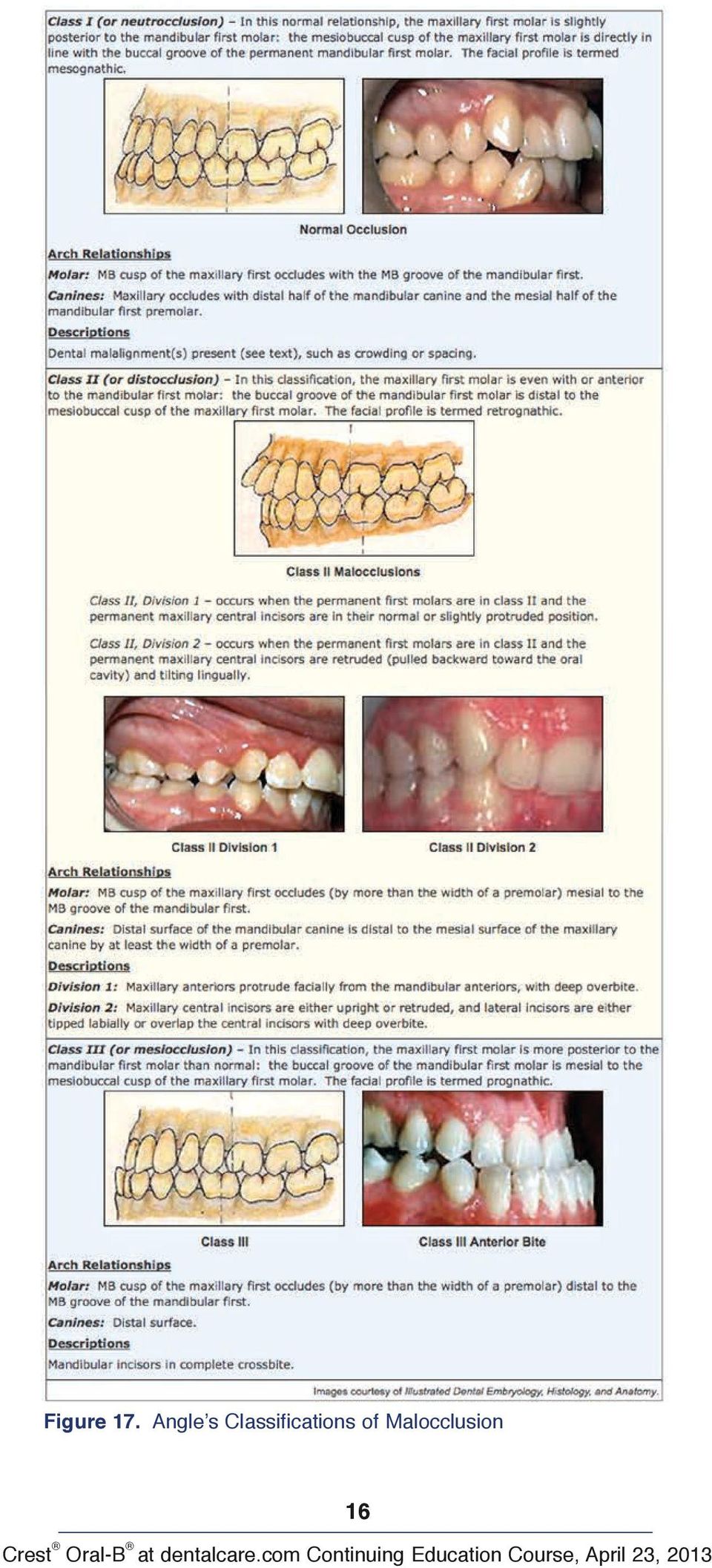
14 Figure 15. Anatomical Landmarks of Teeth. Diagram of landmarks of the teeth. (continued) Image courtesy of Modern Dental Assisting, 6th Ed. Figure 16a. Primary Teeth Arches and Quadrants Image courtesy of Mosby s Comprehensive Dental Assisting: A Clinical Approach. Occlusion Occlusion is the contact between the maxillary and mandibular teeth in any functional relationship. Normal occlusion is important for optimal oral functions, for prevention of dental diseases, and for esthetics. Any deviation from normal occlusion is considered as malocclusion. Malocclusion may involve a single tooth, groups of teeth, or entire arches. With malocclusion, oral functions may be affected, such as difficulty in chewing, swallowing, and speech; it may also cause pain in the temporomandibular joint (TMJ). Malocclusion may be caused by several factors including heredity, diseases that disturb dental development, injuries, and habits such as thumb sucking or tongue thrusting. Classifications of Occlusion Angle s classification system is a common method used to classify various occlusal relationships. This system is based upon the relationship between the permanent maxillary and mandibular first molars. Figure 17 shows the classifications of occlusion as well as the facial profiles of each. 5 14

15 Figure 16b. Permanent Teeth Arches and Quadrants. Examples of the arches and quadrants of both primary and permanent dentition. Image courtesy of Mosby s Comprehensive Dental Assisting: A Clinical Approach. Dentitions The term dentition refers to the natural teeth in the dental arches. There are two major dentitions: primary and permanent. In children between the ages of approximately 5 and 12, each arch will contain a mixture of primary and permanent teeth. This is referred to as mixed dentition. Primary Dentition The primary dentition refers to the first twenty teeth to erupt in the oral cavity. These teeth are also called deciduous teeth, and will be exfoliated (shed) to make way for the permanent teeth. There are 20 teeth in the primary dentition, there are 2 central incisors, 2 lateral incisors, 2 canines, 2 first molars, and 2 second molars in each arch. Table 1 shows the average eruption and exfoliation (shedding) dates of the primary dentition. Permanent Dentition The permanent dentition contains 32 teeth, with each arch having 2 central incisors, 2 lateral incisors, 2 canines, 2 first premolars, 2 second premolars, 2 first molars, 2 second molars, and 2 third molars. This period of dentition begins when the last primary tooth is shed. The permanent teeth that replace primary teeth are called succedaneous teeth. The permanent molars are not succedaneous teeth because they do not replace any primary teeth. The primary molars are replaced with the permanent premolars. Table 2 shows the eruption dates of the permanent dentition. D-A-Q-T System The correct sequence of words when describing a tooth is based on the D-A-Q-T system. D stands for dentition. 15

16 Figure 17. Angle s Classifications of Malocclusion 16
17 Figure 17a. Discrepancies 17

18 Table 1. Eruption and Exfoliation Dates of the Primary Dentition Table 2. Eruption Dates of the Permanent Dentition A stands for arch. Q stands for quadrant. T stands for the tooth type. The dentition is named first, followed by the arch, then the quadrant, and finally the tooth name. For example, a primary first molar would be identified as the primary maxillary right first molar. A permanent central incisor would be identified as the permanent mandibular left central incisor. Teeth can also be referred to by number/letter. Periodontium The Periodontium consists of the hard and soft tissues that help to anchor, support, and protect the teeth. It is made up of the gingival unit and the attachment unit. Figure 18 illustrates the components of the Periodontium. 5 18

19 Figure 18. Periodontium. Diagram of periodontium which surround the teeth. Image courtesy of Illustrated Dental Embryology, Histology, and Anatomy. Gingival Unit The gingival unit consists of the gingiva (soft tissues that surround the teeth) and the alveolar mucosa (soft tissues that line the oral cavity). Gingiva The gingiva, also known as gum tissue, surrounds the teeth and can be attached to the underlying bone (attached gingiva) or unattached (free gingiva). When healthy, the gingiva should be firm and well adapted to the teeth, and have a stippled appearance. This means that its texture appears similar to an orange peel. The color of healthy gingiva depends on the pigmentation of each person, but in general it should appear light pink. Alveolar Mucosa The alveolar mucosa consists of the tissue inside the cheeks, vestibule (the space between the lips or cheeks and the teeth), lips, soft palate, and under the tongue. This tissue is more movable and is lightly attached to the underlying bone and muscles. Its texture is smooth and its color is red to bright red. Attachment Unit The attachment unit consists of the cementum, the alveolar bone (bone surrounding the teeth), and the periodontal ligaments (fibers or ligaments that anchor and support the teeth in their sockets). Cementum The cementum, a tooth tissue covering the root of the tooth; periodontal ligament fibers are imbedded in the cementum which serve the function of anchoring the teeth in their sockets. Alveolar Bone The alveolar bone is also called the alveolar process. It is the bone that forms the sockets for the teeth. Periodontal Ligament The periodontal ligament is connective tissue arranged into groups of fibers; these fibers are attached to the cementum, to the alveolar bone and to the cervical gingivae. Conclusion A working knowledge of normal anatomy of the face and oral cavity is critical for the entire dental team. This is one of many facets necessary in providing oral healthcare. Communication between team members and other healthcare providers is an important component to the overall health and well being of the dental patient. 19

20 Course Test Preview To receive Continuing Education credit for this course, you must complete the online test. Please go to and find this course in the Continuing Education section. 1. is the portion of the tooth that comprises the main inner portion of the tooth. a. Enamel b. Cementum c. Dentin d. Apex 2. The consists of hard and soft tissues and helps anchor, support and protect the teeth. a. gingiva b. periodontium c. sulcus d. hard palate 3. The area where two roots divide on a two rooted tooth is called the. a. cingulum b. bifurcation c. midline d. uvula 4. The incisors are responsible for food. a. cutting b. holding c. crushing d. chewing 5. If a patient s maxillary first molar is slightly posterior to the mandibular first molar, that is considered occlusion. a. Class III b. Class I c. Class II, Division 2 d. Class II 6. The primary dentition is the same as the permanent dentition, except it has no. a. canines b. lateral incisors c. premolars d. first molars 7. The inner portion of the tooth that is comprised of blood vessels, lymph vessels, connective tissue, nerve tissue and cells is the. a. cementum b. dentin c. pulp d. enamel 20

21 8. During an exam, the dentist states the patient has decay on the cervical buccal of the maxillary right first molar. The decay is located and. a. facing the cheek / on the biting surface b. facing the tongue / near the gingiva c. facing the tongue / the midline d. near the gingiva / facing the cheek 9. The consists of the tissues inside the cheeks, vestibule, lips, soft palate and under the tongue and covers bone. a. periodontium b. alveolar mucosa c. periodontal ligament d. attachment unit 10. The permanent dentition has a total of teeth, while the primary dentition has a total of teeth. a. 20 / 32 b. 30 / 22 c. 30 / 20 d. 32 / The surface of a tooth facing the cheek or lip would be known as labial or buccal as well as. a. occlusal b. facial c. incisal d. lingual 12. There are four hard tissues that make up a tooth, the hardest tissue in the body is the. a. cementum b. dentin c. pulp d. enamel 13. The triangular space formed between the contouring angles of adjacent teeth and the gingiva is a(n). a. contact b. incisal ridge c. embrasure d. fossa 14. The correct sequence of words used when describing a tooth is. a. dentition, arch, quadrant and tooth b. arch, quadrant, dentition and tooth c. tooth, dentition, arch and quadrant d. dentition, quadrant, tooth and arch 15. A parent should expect their child to get his/her first tooth at approximately months. a. 36 b c d
22 16. The crown of a tooth is covered with enamel and the root is covered with. a. dentin b. cementum c. enamel d. apex 17. Gingiva surrounds the teeth. Healthy gingiva should be and. a. soft / stippled b. red / soft c. firm / stippled d. smooth / loose 18. The Attachment Unit consists of cementum, alveolar bone and. a. periodontal ligaments b. gingiva c. alveolar mucosa d. cementum 19. The line angle of a tooth is away from the midline and faces the cheek. a. mesiobuccal b. distobuccal c. distolingual d. mesiolingual 20. A child would be about years old if the first permanent molars have erupted but the canines have not. a. 7 b. 4 c. 12 d
23 References 1. Bird DL, Robinson DS. Modern Dental Assisting, 10th Edition. Philadelphia: Saunders. 2. Finkbeiner BL, Johnson CS. Mosby s Comprehensive Review of Dental Assisting: A Clinical Approach, Normal Structures of the Oral Cavity. St. Louis: Mosby, Fehrenbach MJ, Kerring SW, Thomas P. Illustrated Anatomy of the Head and Neck, 3rd Edition. Philadelphia: Saunders. 4. Applegate EJ. The Anatomy and Physiology Learning System, 2nd Edition. Philadelphia: Saunders. p Bath-Balogh M, Fehrenbach MJ. Illustrated Dental Embryology, Histology, and Anatomy. Philadelphia: Saunders. p About the Authors Antoinette Metivier, CDA Antoinette Metivier has experience as a chairside assistant and as an assistant professor of dental assisting at the New Hampshire Technical Institute in Concord, NH. She has developed and presented a radiology review course for the New Hampshire Dental Assistants Association, and has also authored or co-authored several other workbook courses for the American Dental Assistants Association. When Antoinette authored this course she was a Certified Dental Assistant with a Bachelor of Science in Education from the University of Maine (She is now retired and no longer certified by the Dental Assisting National Board.). She has served on the Council on Education of The American Dental Assistants Association. Kimberly Bland, CDA, EFDA, M.Ed. Kimberly Bland has served as ADAA s President as well as held all national offices and the position of ADAA Fifth District Trustee. She has held several offices in both the local and state ADAA organizations, having been President of the Florida Dental Assistants Association for three terms. Ms. Bland is a member of the Florida Board of Dentistry Dental Assisting Council and has held offices in the Florida Allied Dental Educators Association. She is currently Florida Region V Postsecondary Advisor of the Florida Health Occupation Students of America (HOSA). Kimberly Bland is a graduate of the University of South Florida and holds an undergraduate degree in Industrial Technical Education and a Masters Degree in Educational Leadership She earned her Dental Assisting Certificate at Manatee Technical Institute in 1983 where she is now the dental assisting program director. 23
Introduction to Dental Anatomy
 Introduction to Dental Anatomy Vickie P. Overman, RDH, MEd Continuing Education Units: N/A This continuing education course is intended for dental students and dental hygiene students. Maintaining the
LESSON ASSIGNMENT. Topography of the Mouth and Tooth Structure. After completing this lesson, you should be able to:
 LESSON ASSIGNMENT LESSON 3 Topography of the Mouth and Tooth Structure. LESSON ASSIGNMENT Paragraphs 3-1 through 3-9. LESSON OBJECTIVES After completing this lesson, you should be able to: 3-1. Identify
A. DEVELOPMENT OF THE DENTAL ORGAN (ENAMEL ORGAN):
:") A. DEVELOPMENT OF THE DENTAL ORGAN (ENAMEL ORGAN): AS EARLY AS THE SECOND MONTH OF FETAL LIFE, THE DEVELOPMENT OF THE DECIDUOUS TEETH MAY FIRST BECOME EVIDENT. 1. Dental lamina and Bud stage At about six
A. DEVELOPMENT OF THE DENTAL ORGAN (ENAMEL ORGAN): AS EARLY AS THE SECOND MONTH OF FETAL LIFE, THE DEVELOPMENT OF THE DECIDUOUS TEETH MAY FIRST BECOME EVIDENT. 1. Dental lamina and Bud stage At about six
CLASSIFICATION OF CARIOUS LESIONS AND TOOTH PREPARATION.
 CLASSIFICATION OF CARIOUS LESIONS AND TOOTH PREPARATION. ١ G.V. BLACK who is known as the father of operative dentistry,he classified carious lesions into groups according to their locations in permanent
Calibrated Periodontal Probes and Basic Probing Technique
 Module 11 Calibrated Periodontal Probes and Basic Probing Technique MODULE OVERVIEW This module presents the (1) design characteristics of calibrated periodontal probes and (2) step-by-step instructions
Classification of Malocclusion
 Classification of Malocclusion What s going on here? How would you describe this? Dr. Robert Gallois REFERENCE: Where Do We Begin? ESSENTIALS FOR ORTHODONTIC PRACTICE By Riolo and Avery Chapter 6 pages
The Digestive System. Chapter 15
 The Digestive System Chapter 15 Introduction Digestion refers to the mechanical and chemical breakdown of food so the nutrients can be absorbed by cells Carried out by the digestive system Consists of
Molar Uprighting Dr. Margherita Santoro Division of Orthodontics School of Dental and Oral surgery. Consequences of tooth loss.
 Molar Uprighting Dr. Margherita Santoro Division of Orthodontics School of Dental and Oral surgery Molars The wide occlusal surface is designed for food grinding. The surface needs to be aligned with the
CHAPTER 10 RESTS AND PREPARATIONS. 4. Serve as a reference point for evaluating the fit of the framework to the teeth.
 CHAPTER 10 RESTS AND DEFINITIONS A REST is any rigid part of an RPD framework which contacts a properly prepared surface of a tooth. A REST PREPARATION or REST SEAT is any portion of a tooth or restoration
The Gastrointestinal System It consists of: The digestive tract Mouth Pharynx Oesophagus Stomach Small intestine Large intestine
 The Gastrointestinal System It consists of: The digestive tract Mouth Pharynx Oesophagus Stomach Small intestine Large intestine The digestive organs Teeth Tongue Salivary glands Liver Gall bladder Pancreas
Development of Teeth
 Development of Teeth Dr. Khaldoun Darwich Specialist in Oral and Maxillo-Facial Surgery Hamburg University PhD Hamburg University Academic Teacher - Department of OMF Surgery in Damascus University Instructor
The Business of Dentistry: Patient Records and Records Management
 The Business of Dentistry: Patient Records and Records Management Natalie Kaweckyj, LDARF, CDA, CDPMA, COMSA, COA, CRFDA, CPFDA, MADAA, BA; Wendy Frye, CDA, RDA, EFDA, FADAA, MADAA; Lynda Hilling, CDA,
DEVELOPMENT AND GROWTH OF THE MANDIBLE
 2012-2013 ORAL BIOLOGY DEVELOPMENT AND GROWTH OF THE MANDIBLE Ass. Prof. Dr. Heba M. Elsabaa Development and Growth of the Mandible DEVELOPMENT OF THE MANDIBLE The Mandible Is the largest and strongest
Veterinary Dentistry Basics
 Veterinary Dentistry Basics Introduction This program will guide you, step by step, through the most important features of veterinary dentistry in current best practice. This chapter covers the basics
porcelain fused to metal crown
 Lectur.5 Dr.Adel F.Ibraheem porcelain fused to metal crown the most widely used fixed restoration,it is full metal crown having facial surface (or all surfaces) covered by ceramic material. It consist
A collection of pus. Usually forms because of infection. A tooth or tooth structure which is responsible for the anchorage of a bridge or a denture.
 Abscess A collection of pus. Usually forms because of infection. Abutment A tooth or tooth structure which is responsible for the anchorage of a bridge or a denture. Amalgam A silver filling material.
NEW YORK CITY COLLEGE OF TECHNOLOGY
 NEW YORK CITY COLLEGE OF TECHNOLOGY THE CITY UNIVERSITY OF NEW YORK DEPARTMENT OF RESTORATIVE DENTISTRY DEPARTMENT: COURSE CODE: COURSE TITLE: COURSE DESCRIPTION: CLASS HOURS & CREDITS: NUMBER OF WEEKS:
GRADE 6 DENTAL HEALTH
 GRADE 6 DENTAL HEALTH DENTAL HEALTH GRADE: 6 LESSON: 1 THEME: STRUCTURE AND FUNCTION CONCEPT: THE STRUCTURE OF A TOOTH IS RELATED TO ITS FUNCTION PREPARATION: 1. Prepare an overhead transparency of Parts
Secondary dentition permanent teeth - 32. Primary dentition deciduous teeth - 20
 Department of Histology and Embryology, P. J. Šafárik University, Medical Faculty, Košice DEVELOPMENT OF TEETH: Sylabus for foreign students Dental medicine Author: doc. MVDr. Iveta Domoráková, PhD. Primary
Denture Trouble Shooting Guide
 Denture Trouble Shooting Guide Comfort Sore spot in vestibuleupper or lower denture 1. Overextended borders 2. Rough spot in base 1. Shorten borders and polish. 2. Refinish borders. Sore spot in upper
Full Crown Module: Learner Level 1
 Full Crown Module Restoration / Tooth # Full Gold Crown (FGC) / 30 Extensions: Porcelain Fused to Metal (PFM) / 12 All Ceramic / 8 Learner Level 1 Mastery of Tooth Preparation Estimated Set Up Time: 30
ORTHODONTIC SCREENING GUIDE FOR NORTH DAKOTA HEALTH TRACKS NURSES
 ORTHODONTIC SCREENING GUIDE FOR NORTH DAKOTA HEALTH TRACKS NURSES The North Dakota Department of Human Services Medical Services Division and the North Dakota Department of Health s Oral Health Program
Periapical radiography
 8 Periapical radiography Periapical radiography describes intraoral techniques designed to show individual teeth and the tissues around the apices. Each film usually shows two to four teeth and provides
Clinical Practice Guideline For Orthodontics
 Clinical Practice Guideline For Orthodontics MOH- Oral Health CSN -Orthodontics -2010 Page 1 of 15 Orthodontic Management Guidelines 1. Definitions: Orthodontics is the branch of dentistry concerned with
Page 1 of 10 BDS FINAL PROFESSIONAL EXAMINATION 2007 Prosthodontics (MCQs) Model Paper SECTION I
 Model Paper SECTION I") Page 1 of 10 COMPLETE DENTURES ANATOMICAL LANDMARKS SECTION I 1. There are many landmarks in the oral cavity which helps in designing complete dentures. One of the important landmarks is fovea palatini.
Page 1 of 10 COMPLETE DENTURES ANATOMICAL LANDMARKS SECTION I 1. There are many landmarks in the oral cavity which helps in designing complete dentures. One of the important landmarks is fovea palatini.
Anatomic Anomalies. Anomalies. Anomalies. Anomalies. Supernumerary Teeth. Supernumerary Teeth. Steven R. Singer, DDS 212.305.5674 srs2@columbia.
 Anatomic Anomalies Steven R. Singer, DDS 212.305.5674 [email protected] Anomalies! Anomalies are variations in the:! Size! Morphology! Number! Eruption of the teeth Anomalies Anomalies There are two categories:!
IMPLANT DENTISTRY EXAM BANK
 IMPLANT DENTISTRY EXAM BANK 1. Define osseointegration. (4 points, 1/4 2. What are the critical components of an acceptable clinical trial? (10 points) 3. Compare the masticatory performance of individuals
Alimentary canal (gastrointestinal or GI tract) continuous coiled hollow tube
 continuous coiled hollow tube") The Digestive System and Body Metabolism Gross Anatomy Function The Digestive System Functions Ingestion taking in food Digestion breaking food down both physically and chemically Absorption movement of
The Digestive System and Body Metabolism Gross Anatomy Function The Digestive System Functions Ingestion taking in food Digestion breaking food down both physically and chemically Absorption movement of
MEDICAID DENTAL PROGRAMS CODING, POLICY AND RELATED FEE REVISION INFORMATION
 MEDICAID DENTAL PROGRAMS CODING, POLICY AND RELATED FEE REVISION INFORMATION Effective for dates of service on and after November 1, 2005, the following dental coding, policy and related fee revisions
Simplified Positioning for Dental Radiology
 Simplified Positioning for Dental Radiology Prepared by: Animal Dental Care Tony M. Woodward DVM, Dipl. AVDC 5520 N. Nevada Ave. Suite 150 Colorado Springs, CO 80918 (719) 536-9949 [email protected] www.wellpets.com
Implants in your Laboratory: Abutment Design
 1/2 point CDT documented scientific credit. See Page 41. Implants in your Laboratory: Abutment Design By Leon Hermanides, CDT A patient s anatomical limitations have the greatest predictive value for successful
Preventive Pediatric Dental Care. Lawrence A. Kotlow DDS Practice Limited to Pediatric Dental Care 340 Fuller Road Albany, New York 12203
 Preventive Pediatric Dental Care Lawrence A. Kotlow DDS Practice Limited to Pediatric Dental Care 340 Fuller Road Albany, New York 12203 Patient comfort and safety 1. All children are treated using the
PREPARATION OF MOUTH FOR REMOVABLE PARTIAL DENTURES Dr. Mazen kanout
 PREPARATION OF MOUTH FOR REMOVABLE PARTIAL DENTURES Dr. Mazen kanout Mouth preparation includes procedures in four categories: 1. Oral Surgical Preparation. 2. Conditioning of Abused and Irritated Tissue.
ABSTRACT INTRODUCTION. Facial Esthetics. Dental Esthetics
 ABSTRACT The FACE philosophy is characterized by clearly defined treatment goals. This increases diagnostic ability and improves the quality and stability of the end result. The objective is to establish
Case Report(s): Uncomplicated Crown Fractures
: Uncomplicated Crown Fractures") Case Report(s): Uncomplicated Crown Fractures Tooth fractures can be classified as follows: Uncomplicated crown fracture = fracture limited to the crown of the tooth with dentin exposure but no pulp exposure.
Case Report(s): Uncomplicated Crown Fractures Tooth fractures can be classified as follows: Uncomplicated crown fracture = fracture limited to the crown of the tooth with dentin exposure but no pulp exposure.
CLASSIFICATION OF REMOVABLE PARTIAL DENTURES
 Unless otherwise noted, the content of this course material is licensed under a Creative Commons Attribution - Non-Commercial - Share Alike 3.0 License. Copyright 2008, Dr. Jeff Shotwell. The following
Bitewing Radiography B.E. DIXON. B.D.S., M.Sc., D.P.D.S.
 Bitewing Radiography B.E. DIXON B.D.S., M.Sc., D.P.D.S. Main Indications Detection of Dental Caries Monitoring progression of caries Assessment of existing restorations Assessment of Periodontal status
Dent Clin N Am 51 (2007) 299 318. Smile Design. Nicholas C. Davis, DDS, MAGD*
 299 318. Smile Design. Nicholas C. Davis, DDS, MAGD*") Dent Clin N Am 51 (2007) 299 318 Smile Design Nicholas C. Davis, DDS, MAGD* Loma Linda University, School of Dentistry, 11092 Anderson Street, Loma Linda, CA 92354, USA Smile design refers to the many
Dent Clin N Am 51 (2007) 299 318 Smile Design Nicholas C. Davis, DDS, MAGD* Loma Linda University, School of Dentistry, 11092 Anderson Street, Loma Linda, CA 92354, USA Smile design refers to the many
The Digestive System. Chapter 16. Introduction. Histological Organization. Overview of Digestive System. Movement and Mixing of Digestive Materials
 The Digestive System Chapter 16 Introduction Structure of the digestive system A tube that extends from mouth to anus Accessory organs are attached Functions include Ingestion Movement Digestion Absorption
4-1-2005. Dental Clinical Criteria and Documentation Requirements
 4-1-2005 Dental Clinical Criteria and Documentation Requirements Table of Contents Dental Clinical Criteria Cast Restorations and Veneer Procedures... Pages 1-3 Crown Repair... Page 3 Endodontic Procedures...
Phonetics Related to Prosthodontics
 Middle-East Journal of Scientific Research 12 (1): 31-35, 2012 ISSN 1990-9233 IDOSI Publications, 2012 DOI: 10.5829/idosi.mejsr.2012.12.1.988 Phonetics Related to Prosthodontics 1 2 Abdul-Aziz Abdullah
Resorptive Changes of Maxillary and Mandibular Bone Structures in Removable Denture Wearers
 Resorptive Changes of Maxillary and Mandibular Bone Structures in Removable Denture Wearers Dubravka KnezoviÊ-ZlatariÊ Asja»elebiÊ Biserka LaziÊ Department of Prosthodontics School of Dental Medicine University
Digestive System. Gross Anatomy and Physiology
 Digestive System Gross Anatomy and Physiology I. Introduction A. Base Function: Working with the circulatory system the digestive system provides the body with fuel. B. Main players: 1. Digestive tract:
Headgear Appliances. Dentofacial Orthopedics and Orthodontics. A Common Misconception. What is Headgear? Ideal Orthodontic Treatment Sequence
 Ideal Orthodontic Treatment Sequence Headgear Appliances Natalie A. Capan, D.M.D. 580 Sylvan Avenue, Suite 1M Englewood Cliffs, New Jersey 07632 (201)569-9055 www.capanorthodontics.com [email protected]
Welcome back. Today, we embark on Lesson 6 where we ll study the human digestive system.
 Basic Human Anatomy Lesson 6: The Human Digestive System Welcome back. Today, we embark on Lesson 6 where we ll study the human digestive system. After completing this lesson, you should be able to define
Pediatric Dental Trauma. Acute Care Topics Mary Fox Braithwaite June 2008
 Pediatric Dental Trauma Acute Care Topics Mary Fox Braithwaite June 2008 Dental Injuries in Children Nearly 50% of children experience some type of dental injury during childhood, many of which are are
Impression Technique for complete dentures
 Impression Technique for complete dentures Dr. M A Abdullah M.D.S. Associate Professor of Prosthodontics, College of Dentistry, King Saud University. Saudi Arabia. References. Heartwell C M, Rahn A O,Text
Principles of Partial Denture Design
 Principles of Partial Denture Design 1. Keep the RPD design as simple as possible Simple those design elements which promote function, esthetics, comfort, ease of fabrication, and ease of maintenance,
Introduction to Charting. Tooth Surfaces: M = mesial D = distal O = Occlusal B = buccal F = facial I = incisal L = lingual
 Tooth Surfaces: M = mesial D = distal O = Occlusal B = buccal F = facial I = incisal L = lingual When combining tooth surfaces, as in defining cavity preparations or restorations, there are some spelling
A. function: supplies body with oxygen and removes carbon dioxide. a. O2 diffuses from air into pulmonary capillary blood
 A. function: supplies body with oxygen and removes carbon dioxide 1. ventilation = movement of air into and out of lungs 2. diffusion: B. organization a. O2 diffuses from air into pulmonary capillary blood
Best Practices for Oral Health Assessments for School Nurses. Jill Fernandez RDH, MPH. National Association of School Nurses June 22, 2012
 Best Practices for Oral Health Assessments for School Nurses Jill Fernandez RDH, MPH National Association of School Nurses June 22, 2012 Jill Fernandez RDH, MPH Clinical Associate Professor Department
SURGICAL EXTRACTIONS: TECHNIQUE AND CAUTIONS By Tony M. Woodward, DVM, AVDC
 SURGICAL EXTRACTIONS: TECHNIQUE AND CAUTIONS By Tony M. Woodward, DVM, AVDC We continue describing the five basic dental services that all general practitioners should be able to provide for their patients.
Supported by. A seven part series exploring the fantastic world of science.
 Supported by A seven part series exploring the fantastic world of science. Find out about the different types of teeth in your mouth. Milk Teeth As a child you have 20 milk teeth. Your first tooth appears
The Obvious and the Obscure:Diagnostic Steps for Crack Confirmation
 Cracking the Cracked Tooth Code In response to your requests... At the end of each issue of ENDODONTICS: Colleagues for Excellence, the American Association of Endodontists (AAE) asks readers to send in
Universal Crown and Bridge Preparation
 Universal Crown and Bridge Preparation The All-Ceramic Crown Preparation Technique for Predictable Success According to Dr. Ronald E. Goldstein Expect the Best. Buy Direct. The Universal * Crown and Bridge
In Class IV arch: Fulcrum line passes through two abutments adjacent to single edentulous space.
 It is that part of removable partial denture which assists the direct retainers in preventing displacement of distal extension denture bases by resisting lever action from the opposite side of the fulcrum
The etiology of orthodontic problems Fifth session
 بنام خداوند جان و خرد The etiology of orthodontic problems Fifth session دکتر مھتاب نوری دانشيار گروه ارتدنسی Course Outline( 5 sessions) Specific causes of malocclusion Genetic Influences Environmental
Intraoral Radiographic Techniques
 Intraoral Radiographic Techniques Allan G. Farman, BDS, EdS., MBA, PhD; Sandra A. Kolsom, CDA-Emeritus, RDA; ADAA 2014 Council on Education Continuing Education Units: 4 hours Online Course: www.dentalcare.com/en-us/dental-education/continuing-education/ce119/ce119.aspx
Classifications of animals: ruminant vs non-ruminant carnivore: meat-eating herbivore: plant-eating omnivore: both meat and plant-eating
 Digestion and Metabolism Digestive tract one long, continuous tube starting at the mouth and ending at the anus Functions Ingestion Grinding Digestion/absorption of food Elimination of solid wastes Classifications
Treatment planning for the class 0, 1A, 1B dental arches
 Treatment planning for the class 0, 1A, 1B dental arches Dr.. Peter Hermann Dr Reminder: Torquing movement on tooth supported denture : no movement Class 1 movement in one direction (depression) Class
Activity: Can You Identify the Age?
 Activity: Can You Identify the Age? Skeletons are good age markers because teeth and bones mature at fairly predictable rates. How Teeth Reveal Age For toddler to age 21, teeth are the most accurate age
Removable appliances II. Functional jaw orthopedics
 Removable appliances II. Functional jaw orthopedics Melinda Madléna DMD, PhD Associate professor Department of Pedodontics and Orthodontics Faculty of Dentistry Semmelweis University Budapest Classification
Wired for Learning - Orthodontic Basics
 Wired for Learning - Orthodontic Basics Lori Garland Parker, BS, MAOM, RDAEF, CDA, COA Continuing Education Units: 3 hours Online Course: www.dentalcare.com/en-us/dental-education/continuing-education/ce365/ce365.aspx
[PAGE HEADLINE] Improve your Health and Change Your Smile with Complete Dental Services in One [CITYNAME] Location
![[PAGE HEADLINE] Improve your Health and Change Your Smile with Complete Dental Services in One [CITYNAME] Location](/thumbs/31/14971568.jpg "[PAGE HEADLINE] Improve your Health and Change Your Smile with Complete Dental Services in One [CITYNAME] Location") Eddie Stephens//Copywriter Sample: Website copy/internal Dental Services Pages [PAGE HEADLINE] Improve your Health and Change Your Smile with Complete Dental Services in One [CITYNAME] Location [LEAD SENTENCE/PARAGRAPH]
Eddie Stephens//Copywriter Sample: Website copy/internal Dental Services Pages [PAGE HEADLINE] Improve your Health and Change Your Smile with Complete Dental Services in One [CITYNAME] Location [LEAD SENTENCE/PARAGRAPH]
Local Anesthesia in Oral Surgery. Animal Dental Care
 Local Anesthesia in Oral Surgery Presented by: Animal Dental Care Tony M. Woodward DVM, Dipl. AVDC 5520 N. Nevada Ave. Suite 150 Colorado Springs, CO 80918 (719) 536-9949 [email protected] www.wellpets.com
!Financial agreement COST / RISK / BENEFIT
 COMPLETE DENTURES 1 2 Oral Examination, Diagnosis and Treatment Planning Initial Appointment! Get to know your patient! Personally, experiences, expectations! Past medical history! Past dental history!
Ear, Nose, Throat, Teeth and the Jaw
 Many people suffer from ear related problems, such as ear pain, a feeling of pressure or fullness of the ear, or ringing of the ear. If you suffer from any of these symptoms, it may surprise you to learn
Bone augmentation procedure without wound closure
 THE CREATION OF ATTACHED GINGIVA IMMEDIATELY AFTER EXTRACTION Bone augmentation procedure without wound closure One of the characteristics of wound healing after an extraction is that the alveolar process
CDHA NATIONAL LIST OF SERVICE CODES
 CDHA NATIONAL LIST OF SERVICE CODES Prepared and published by The Canadian Dental Hygienists Association First edition 1998 Revised 2012 Intended for the use of provincial dental hygiene associations and
SYSTEMATIC APPROACH TO ORTHODONTIC DIAGNOSIS DENT 656
 SYSTEMATIC APPROACH TO ORTHODONTIC DIAGNOSIS DENT 656 ORTHODONTIC CLASSIFICATION / DIAGNOSIS Goal of diagnosis: An orderly reduction of the data base to a useful list of the patient s problems Useful??
Dental Updates. Excerpted Article e-mail: [email protected]. Why Implant Screws Loosen Part 1. Richard Erickson, MS, DDS
 ¼ ½ ¾ µ mw/cm 2 Volume 17; 2007 Dental Updates "CUTTING EDGE INFORMATION FOR THE DENTAL PROFESSIONAL " 200 SEMINARS AND 30 JOURNALS REVIEWED YEARLY FOR THE LATEST, CUTTING EDGE INFORMATION Excerpted Article
Healing Abutment Selection. Perio Implant Part I. Implant Surface Characteristics. Single Tooth Restorations. Credit and Thanks for Lecture Material
 Healing Abutment Selection Perio Implant Part I Credit and Thanks for Lecture Material Implant Surface Characteristics!CAPT Robert Taft!CAPT Greg Waskewicz!Periodontal Residents NPDS and UMN!Machined Titanium!Tiunite!Osseotite
Periodontal Screening and Recording: Early Detection of Periodontal Diseases
 Periodontal Screening and Recording: Early Detection of Periodontal Diseases Tanya Villalpando Mitchell, RDH, MS Continuing Education Units: 1 hour Online Course: www.dentalcare.com/en-us/dental-education/continuing-education/ce53/ce53.aspx
ORTHODONTIC TREATMENT
 ORTHODONTIC TREATMENT Informed Consent for the Orthodontic Patient As a general rule, positive orthodontic results can be achieved by informed and cooperative patients. Thus, the following information
Chapter 12 The Gingiva
 Page 1 of 31 Chapter 12 The Gingiva The true test of successful treatment, the real evaluation of the effects of scaling and related instrumentation, is the health of the periodontal tissues. The objective
Dental Radiography collimator Ionising radiation image radiolucent area radiopaque area controlled zone scatter radiation intraoral
 Dental Radiography X-rays for dental radiography are produced by high voltages of electricity within an x-ray head and come out through a metal tube called a collimator. This ensures the x-rays only come
OCCLUSION IN COMPLETE DENTURES
 1 OCCLUSION IN COMPLETE DENTURES C P Owen Introduction Occlusion has been described as the most important subject in all the disciplines of dentistry, and for good reason, because the way the teeth come
Replacement of the upper left central incisor with a Straumann Bone Level Implant and a Straumann Customized Ceramic Abutment
 Replacement of the upper left central incisor with a Straumann Bone Level Implant and a Straumann Customized Ceramic Abutment by Dr. Ronald Jung and Master Dental Technician Xavier Zahno Initial situation
Prosthodontist s Perspective
 Unless otherwise noted, the content of this course material is licensed under a Creative Commons Attribution - Non-Commercial - Share Alike 3.0 License. Copyright 2008, Dr. Jeff Shotwell. The following
The Treatment of Traumatic Dental Injuries
 The Recommended Guidelines of the American Association of Endodontists for The Treatment of Traumatic Dental Injuries 2013 American Association of Endodontists Revised 9/13 The Recommended Guidelines of
Tooth preparation J. C. Davenport, 1 R. M. Basker, 2 J. R. Heath, 3 J. P. Ralph, 4 P-O. Glantz, 5 and P. Hammond, 6
 12 5 Tooth preparation J. C. Davenport, 1 R. M. Basker, 2 J. R. Heath, 3 J. P. Ralph, 4 P-O. Glantz, 5 and P. Hammond, 6 This final article in the series describes the modification of teeth to improve
dental nerve strong sensory stimuli anesthesia requirements
 Ban0108_022-033.qxd 2/1/08 2:45 PM Page 22 Dental nerve block techniques Proper training and practice can enhance the level of care you offer to Pets and clients. { Crown Gingival margin By Gary S. Goldstein,
Position Classification Standard for Dental Officer Series, GS-0680
 Position Classification Standard for Dental Officer Series, GS-0680 Table of Contents SERIES DEFINITION... 2 BACKGROUND... 2 TITLES... 3 GRADE-LEVEL EVALUATION CRITERIA... 3 NOTES ON THE USE OF THE STANDARDS...
Local Anesthesia Techniques in Oral and Maxillofacial Surgery
 Local Anesthesia Techniques in Oral and Maxillofacial Surgery Sean M. Healy, D.D.S. Oral and Maxillofacial Surgery Francis B. Quinn, M.D., FACS Otolaryngology Head and Neck Surgery The University of Texas
Humana Health Plans of Florida. Important:
 Humana Health Plans of Florida Important: Dental discount membership in Florida is determined by viewing the member s ID card and verifying that the Humana Logo and Medicare name is listed with an effective
Maxillary Sinus. (Antrum of Higmore)
") Maxillary Sinus (Antrum of Higmore) The maxillary sinus is a pneumatic space. It is the largest bilateral air sinus located in the body of the maxilla and opens in the middle nasal meatus of the nasal
Maxillary Sinus (Antrum of Higmore) The maxillary sinus is a pneumatic space. It is the largest bilateral air sinus located in the body of the maxilla and opens in the middle nasal meatus of the nasal
Lesson 2: Save your Smile from Tooth Decay
 Lesson 2: Save your Smile from Tooth Decay OVERVIEW Objectives: By the end of the lesson, the Lay Health Worker will be able to: 1. Describe what tooth decay is and how it happens. 2. State the causes
Topics for the Orthodontics Board Exam
 Topics for the Orthodontics Board Exam I. Diagnostics, relations to paediatric dentistry, prevention 1. Etiology of dental anomalies. 2. Orthodontic anomalies, relationship between orthodontic treatment
ATLANTIS abutments design guide CAD/CAM patient-specific abutments
 ATLANTIS abutments design guide CAD/CAM patient-specific abutments Contents Introduction 4 This manual helps you to explore all the benefits of ATLANTIS CAD/CAM patient-specific abutments. It gives you
Periodontal surgery report for crown lengthening of tooth number 24,25
 411 PDS Periodontal surgery report for crown lengthening of tooth number -Course director : Dr. Nahid Ashri - instructor: Dr.Fatin Awaratani - - Student Name: Hanadi Alyami Computer Number: K S U - D E
The gingival flap technique,
 Ban0108_034-041.qxd 2/1/08 4:47 PM Page 34 Creating effective dental flaps Practitioners can use this surgery to visualize and treat dental problems. By Beatriz Woodall, DVM Contributing Author The gingival
In the past decade, there has been a remarkable
 TECHNO BYTES Principles of cosmetic dentistry in orthodontics: Part 1. Shape and proportionality of anterior teeth David M. Sarver, DMD, MS Vestavia Hills, Ala In the past decade, there has been a remarkable
Endodontics. Colleagues for Excellence. Access Opening and Canal Location
 Endodontics Colleagues for Excellence Spring 2010 Access Opening and Canal Location Published for the Dental Professional Community by the American Association of Endodontists Cover artwork: Rusty Jones,
Role of dentistry in the health scciences, the dental team. Dr. Dézsi Anna Júlia
 Role of dentistry in the health scciences, the dental team Dr. Dézsi Anna Júlia The art of medicine Herodatus, a historian, described the medical art in Egypt: "The art of medicineis distributed thus:
Dental Services. Dental Centre. HKSH Healthcare Medical Centre Dental Centre. For enquiries and appointments, please contact us
 Dental Services For enquiries and appointments, please contact us HKSH Healthcare Medical Centre Dental Centre Level 22, One Pacific Place 88 Queensway, Hong Kong (852) 2855 6666 (852) 2892 7589 [email protected]
CRACKED TOOTH SYNDROME
 CRACKED TOOTH SYNDROME Dr Vijay Salvi We all come across apparently healthy teeth eliciting complex and often bizarre symptoms. The patient will give a long history of undiagnosed but severe pain, and
Tooth Formation. Chapter 1
 Chapter Tooth Formation Teeth develop from epithelial cells from the mucosal lining of the oral cavity and cranial neural crest-derived ectomesenchymal cells. These latter cells originate at the ectodermal/neuroectodermal