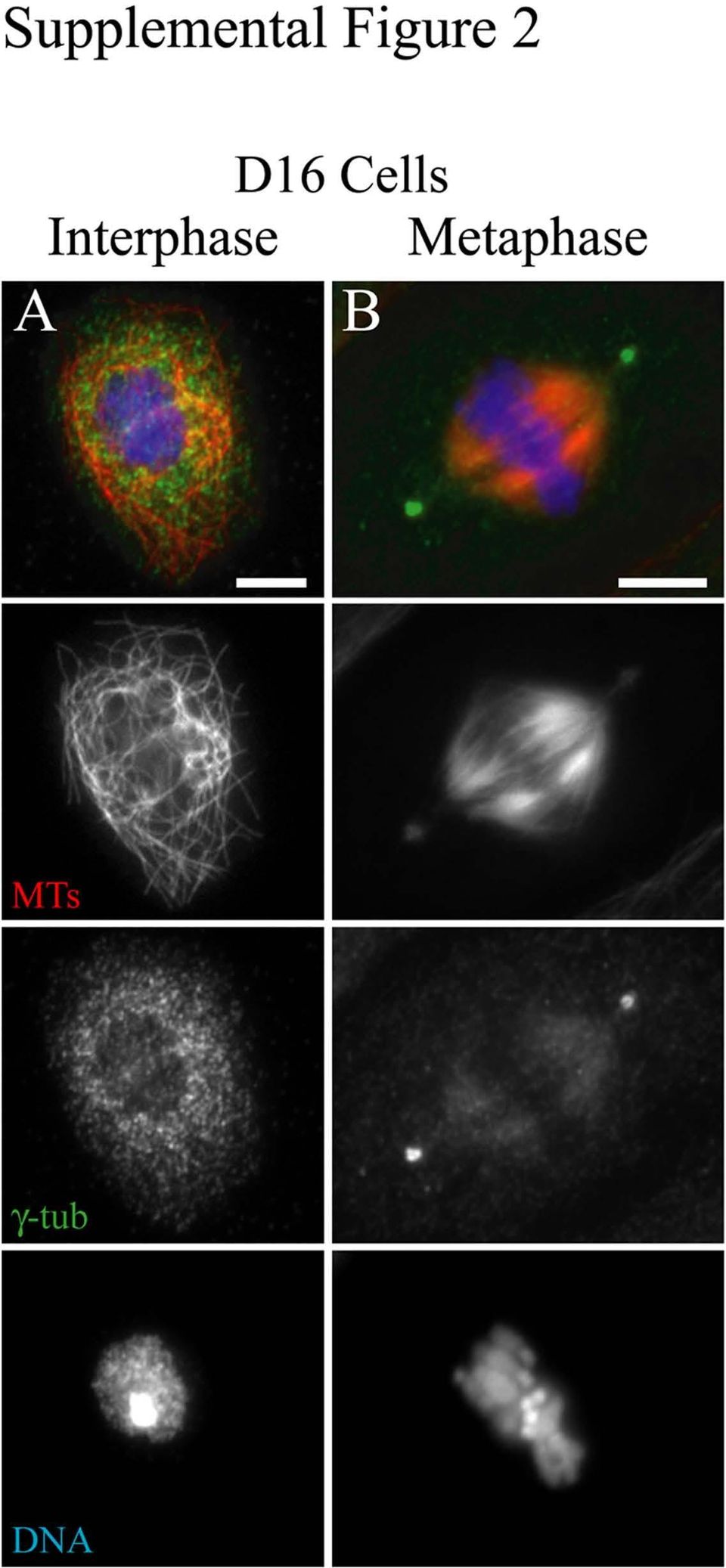
Interphase (A) and mitotic (B) D16 cells were plated on concanavalin A and processed for
|
|
|
- Magnus Daniels
- 10 years ago
- Views:
Transcription
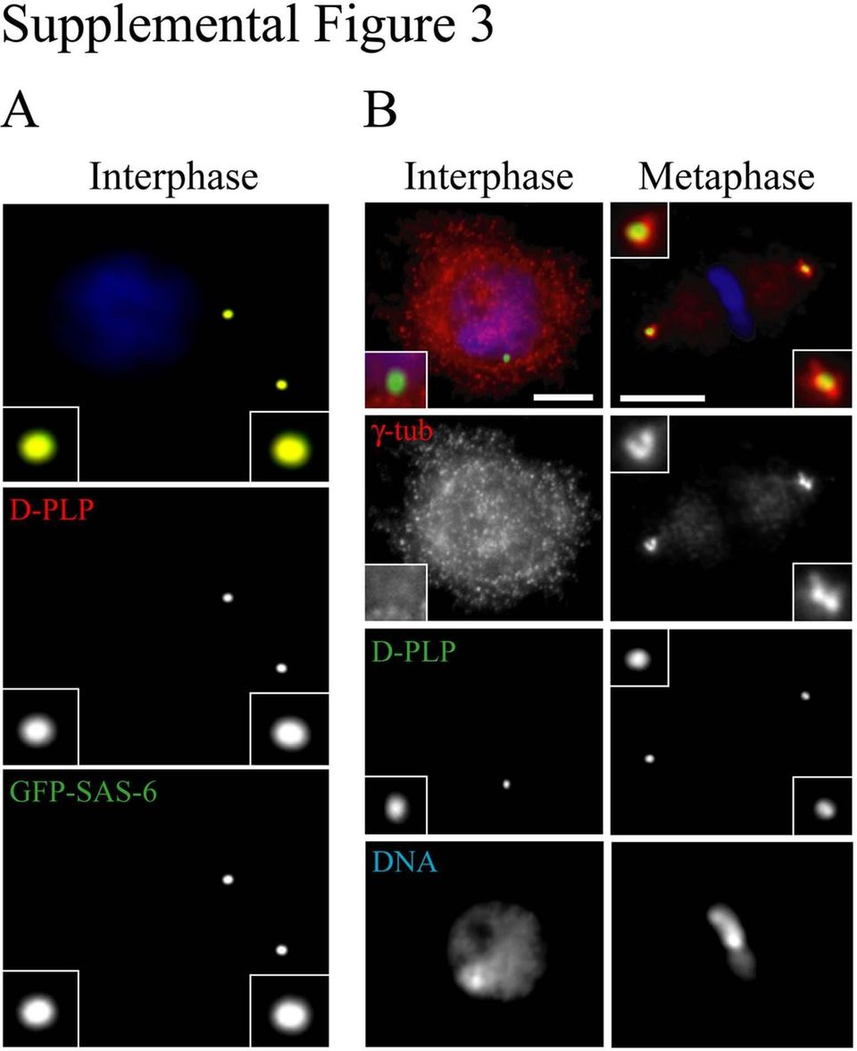
1 E Rogers SUPPLEMENTARY DATA Supplemental Figure 1. Interphase day-7 control and γ-tubulin23c RNAi-treated S2 cells stained for MTs (green), γ-tubulin (red), and DNA (blue). Scale, 5μm. Supplemental Figure 2. Drosophila D16 cells lack a γ-tubulin-containing MTOC during interphase. Interphase (A) and mitotic (B) D16 cells were plated on concanavalin A and processed for immunofluorescence to reveal MTs (red), γ-tubulin (green) and DNA (blue). Scale, 5μm. Supplemental Figure 3. (A) Drosophila SAS-6 co-localizes with the centriolar protein D-PLP. An interphase GFP-SAS-6 (green) expressing stable S2 cell stained for D-PLP (red) and DNA (blue). Magnified views of the centrioles are shown in insets. (B) Centrioles in Drosophila D16 cells co-localize with γ-tubulin in mitosis but not during interphase. D16 cells were plated on concanavalin A and processed for immunofluorescence with antibodies against γ-tubulin (red), D-PLP (green) and stained for DNA (blue). The panels illustrate typical cells in interphase and mitosis. Magnified views of the centrioles are shown in insets. Scale 5μm. Supplemental Figure 4. The centrosomal proteins Centrosomin (Cnn), CP190, and CP60 localize to the PCM during mitosis but do not localize to centrioles during interphase. Interphase and mitotic stable S2 cells expressing GFP-SAS-6 (green) were fixed and stained for DNA (blue) and Cnn (red, top row), CP190 (red, middle row), or CP60 (red, bottom row). Top panel, the mitotic cell shown is in prophase. Magnified views of the centrioles and Cnn (white arrowheads) are shown below the micrographs. Middle and bottom panels, both CP190 and CP69 are restricted to the nucleus during interphase (DNA not shown), but are released into the cytoplasm during mitosis and concentrate on centrosomes at spindle poles (white arrowheads) (GFP-SAS-6 centrioles not shown for CP60). Magnified views of CP190-labeled mitotic centrosomes are shown in insets. Scale 5μm. 1
 Drosophila SAS-6 co-localizes with the centriolar protein D-PLP. An interphase GFP-SAS-6 (green) expressing stable S2 cell stained for D-PLP (red) and DNA (blue).")
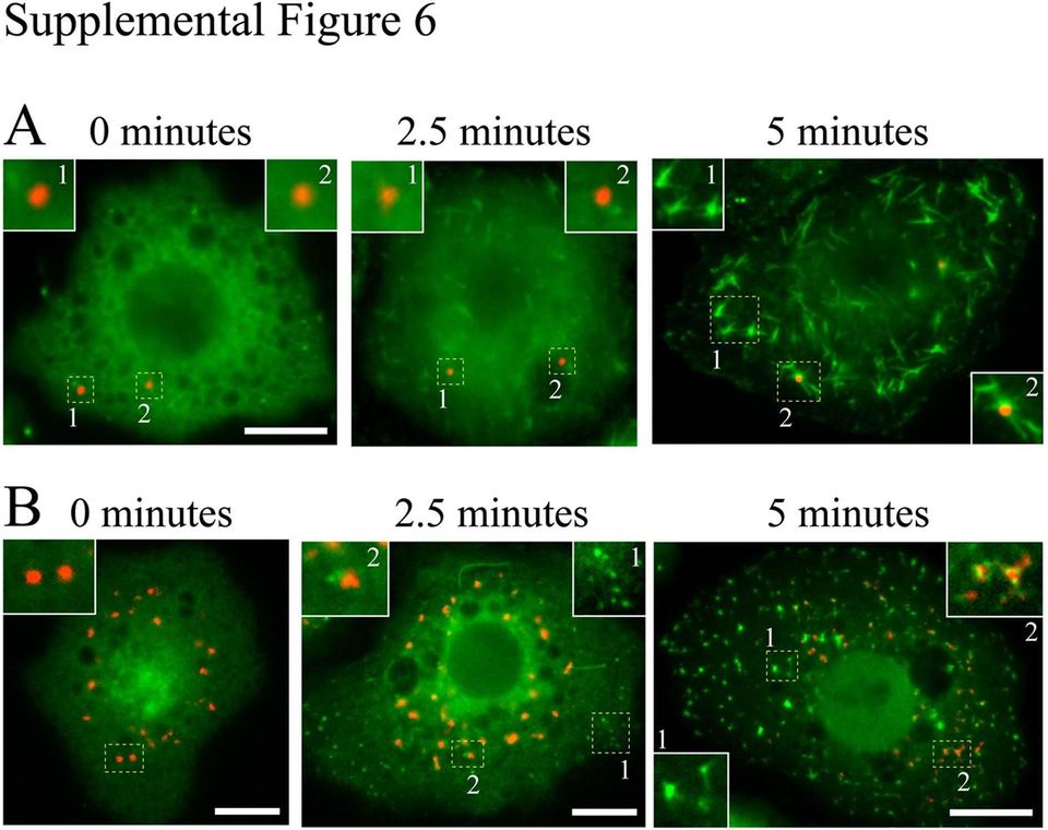
2 Supplemental Figure 5. Drosophila SAS-6 RNAi eliminates centrioles without altering interphase cell cycle progression. (A) Cells were fixed and stained for SAS-6 (green), γ-tubulin (red), and DNA (blue). Anti-SAS-6 antibody labels spots within γ-tubulin-stained rings at the spindle pole. Note a cluster of centrosomes at the right spindle pole (left pole is in a different focal plane). (B) S2 cell lysates were prepared prior to and 7 days after SAS-6 RNAi-treatment and probed by Western blot using anti-sas-6 antibody (α-tubulin was used as a loading control). (C) Day 7 control and SAS-6 RNAi-treated S2 cells stained for D-PLP (red) and DNA (blue). White traces outline the cells. (D) Histogram of centrioles per interphase cell counted in control or SAS-6 RNAi-treated S2 cells. (E) SAS-6 RNAi reduces mitotic centrosome number. Control and SAS-6 RNAi cells were stained for γ-tubulin (red), α-tubulin (green) and DNA (blue). Percentages of spindles types are indicated. Scale, 2.5μm. (F) Cell cycle progression is unaffected by SAS-6 RNAi. Histograms of DNA fluorescence intensity (x-axis) and cell number (yaxis) of 7,000 cells with 2C, 4C and 8C populations denoted. Supplemental Figure 6. Early periods of MT re-growth occur independently of centrioles and Golgi but both organelles can associate with MTs. S2 cells were stained for MTs (green) and either (A) D- PLP centrioles (red) or (B) Golgi (red) at specific early time-points in a MT re-growth assay. MTs were depolymerized by cold-treatment (0 min.) and brought to room temperature to allow polymerization. Cells were fixed at 0, 2.5 and 5 minutes. (A) Centrioles are numbered and shown at higher magnifications (insets). At 2.5 minutes, MTs assemble but most are not associated with centrioles. At 5 minutes, MT foci form, most of which do not contain centrioles (1), although some MT foci do colocalize with centrioles (2). (B) Golgi are numbered and shown at higher magnifications (insets). At 2.5 and 5 minutes, MTs assemble as small (2.5 min.) and larger (5 min.) foci, most of which are not associated with Golgi (1). However, some MT foci do associate with Golgi at these time-points (2). Scale, 5μm. 2
 S2 cell lysates were prepared prior to and 7 days after SAS-6 RNAi-treatment and probed by Western blot using anti-sas-6 antibody (α-tubulin was used as a loading control).")
3 Supplemental Figure 7. Interphase day-7 control, MAST, and Mini-spindles (Msps) RNAi-treated S2 cells stained for MTs (the cell margin for the MAST depleted cell is traced in white). Scale, 5μm. Supplemental Figure 8. The kinetics of MT re-growth is not altered in either Ncd, Klp10A, Shortstop (Shot), or Kinesin heavy chain (KHC)-depleted S2 cells. Time-points show representative day 6 RNAitreated S2 cells stained for MTs during MT re-growth. Scale, 5μm. MOVIE LEGENDS Movie 01. Interphase microtubule nucleation is independent of centrioles. Expression of EB1-mRFP (green) allows the visualization of microtubule nucleation and growth relative to a GFP-SAS-6-labeled centriole (red) in a live interphase S2 cell. Movie 02. Mitotic microtubule nucleation is associated with centrioles. Expression of EB1-mRFP (green) allows the visualization of microtubule nucleation and growth relative to GFP-SAS-6-labeled centrioles (red) in a live prophase S2 cell. Movie 03. Interphase microtubule nucleation is independent of centrioles in live embryonic amnioserosal cells. Expression of EB1-GFP (green) allows the visualization of microtubule nucleation and growth relative to mcherry-sas-6-labeled centrioles (red) in live embryonic amnioserosal cells. These differentiated cells are terminally-arrested in interphase (G2 phase). The central cell in this movie has an elongateddiamond shape. Centrioles display wide oscillations in their movements throughout the cell. Movie 04. Interphase microtubule nucleation is independent of centrioles in live embryonic leading edge cells. Expression of EB1-GFP (green) allows the visualization of microtubule nucleation and growth relative to mcherry-sas-6-labeled centrioles (red) in live embryonic leading edge cells. These differentiated cells are terminally-arrested in interphase (G1 phase), and were observed during dorsal closure. The 3
 allows the visualization of microtubule nucleation and growth relative to a GFP-SAS-6-labeled centriole (red) in a live interphase S2 cell. Movie 02.")
4 direction of leading edge cell migration occurs toward the top of the image. Several leading edge cells are seen in this field of view and each has an elongated morphology. Movie 05. Mitotic microtubule nucleation is associated with centrioles in live embryonic dividing epithelial cells. Expression of EB1-GFP (green) allows the visualization of microtubule nucleation and growth relative to mcherry-sas-6-labeled centrioles (red) in live embryonic dividing epithelial cells. Several metaphase-stage spindles are seen within this region of a stage 9 embryo with centrioles at the spindle poles associated with the brightest EB1 signal. Movie 06. Interphase microtubule nucleation is independent of both centrioles and γtub23c. Expression of EB1-mRFP (green) allows the visualization of microtubule nucleation and growth relative to GFP-SAS-6-labeled centrioles (red) in a live 7-day RNAi-treated interphase S2 cell depleted of the major γtub23c isotype. Two centrioles move throughout this cell and are not associated with the microtubule nucleation that appears indistinguishable relative to control cells (see Movie 01). Centrioles are sometimes seen moving in fast linear trajectories which presumably occurs along MT tracks. 4

5

6

7
8
9

10

11
12

13

J. Cell Sci. 128: doi:10.1242/jcs.164566: Supplementary Material
 Figure S1. Microtubule and microfilament drug treatments severely interfere with mitotic spindle morphology, length and positioning. (A) GFP-fused lamin-a/c, LAP2 or BAF1 localizes to the spindle and all
Chapter 12: The Cell Cycle
 Name Period Chapter 12: The Cell Cycle Overview: 1. What are the three key roles of cell division? State each role, and give an example. Key Role Reproduction Growth and development Tissue removal Example
The Huntington Library, Art Collections, and Botanical Gardens
 The Huntington Library, Art Collections, and Botanical Gardens Rooting for Mitosis Overview Students will fix, stain, and make slides of onion root tips. These slides will be examined for the presence
Cell Division Mitosis and the Cell Cycle
 Cell Division Mitosis and the Cell Cycle A Chromosome and Sister Chromatids Key Points About Chromosome Structure A chromosome consists of DNA that is wrapped around proteins (histones) and condensed Each
CHAPTER 9 CELLULAR REPRODUCTION P. 243-257
 CHAPTER 9 CELLULAR REPRODUCTION P. 243-257 SECTION 9-1 CELLULAR GROWTH Page 244 ESSENTIAL QUESTION Why is it beneficial for cells to remain small? MAIN IDEA Cells grow until they reach their size limit,
Chapter 12: The Cell Cycle
 Name Period Chapter 12: The Cell Cycle Overview: 1. What are the three key roles of cell division? State each role, and give an example. Key Role Example 2. What is meant by the cell cycle? Concept 12.1
LAB 8 EUKARYOTIC CELL DIVISION: MITOSIS AND MEIOSIS
 LAB 8 EUKARYOTIC CELL DIVISION: MITOSIS AND MEIOSIS Los Angeles Mission College Biology 3 Name: Date: INTRODUCTION BINARY FISSION: Prokaryotic cells (bacteria) reproduce asexually by binary fission. Bacterial
Cell Division CELL DIVISION. Mitosis. Designation of Number of Chromosomes. Homologous Chromosomes. Meiosis
 Cell Division CELL DIVISION Anatomy and Physiology Text and Laboratory Workbook, Stephen G. Davenport, Copyright 2006, All Rights Reserved, no part of this publication can be used for any commercial purpose.
MITOSIS IN ONION ROOT TIP CELLS: AN INTRODUCTION TO LIGHT MICROSCOPY
 MITOSIS IN ONION ROOT TIP CELLS: AN INTRODUCTION TO LIGHT MICROSCOPY Adapted from Foundations of Biology I; Lab 6 Introduction to Microscopy Dr. John Robertson, Westminster College Biology Department,
If and when cancer cells stop dividing, they do so at random points, not at the normal checkpoints in the cell cycle.
 Cancer cells have escaped from cell cycle controls Cancer cells divide excessively and invade other tissues because they are free of the body s control mechanisms. Cancer cells do not stop dividing when
Biology 3A Laboratory MITOSIS Asexual Reproduction
 Biology 3A Laboratory MITOSIS Asexual Reproduction OBJECTIVE To study the cell cycle and understand how, when and why cells divide. To study and identify the major stages of cell division. To relate the
The cell cycle, mitosis and meiosis
 The cell cycle, mitosis and meiosis Learning objective This learning material is about the life cycle of a cell and the series of stages by which genetic materials are duplicated and partitioned to produce
SUPPLEMENTARY DATA 1
 SUPPLEMENTARY DATA 1 Supplementary Figure S1. Overexpression of untagged Ago2 inhibits the nuclear transport of the dotted foci of GFP-signal of myc-gfp-tnrc6a-nes-mut. (A C) HeLa cells expressing myc-gfp-tnrc6a-nes-mut
Mitosis in Onion Root Tip Cells
 Mitosis in Onion Root Tip Cells A quick overview of cell division The genetic information of plants, animals and other eukaryotic organisms resides in several (or many) individual DNA molecules, or chromosomes.
Cellular Reproduction
 9 Cellular Reproduction section 1 Cellular Growth Before You Read Think about the life cycle of a human. On the lines below, write some of the stages that occur in the life cycle of a human. In this section,
Cell Viability Assays: Microtitration (MTT) Viability Test Live/Dead Fluorescence Assay. Proliferation Assay: Anti-PCNA Staining
 Viability Test Live/Dead Fluorescence Assay. Proliferation Assay: Anti-PCNA Staining") Cell Viability Assays: Microtitration (MTT) Viability Test Live/Dead Fluorescence Assay Proliferation Assay: Anti-PCNA Staining Spring 2008 1 Objectives To determine the viability of cells under different
Cell Viability Assays: Microtitration (MTT) Viability Test Live/Dead Fluorescence Assay Proliferation Assay: Anti-PCNA Staining Spring 2008 1 Objectives To determine the viability of cells under different
EDF Extended Depth of Field
 EDF Extended Depth of Field An upgrade for the ImageStream system Think outside the dot. Break the classic depth of field barrier Improve precision Enhance discrimination Simplify analysis Increase resolution
From DNA to Protein
 Nucleus Control center of the cell contains the genetic library encoded in the sequences of nucleotides in molecules of DNA code for the amino acid sequences of all proteins determines which specific proteins
Cell Cycle in Onion Root Tip Cells (IB)
") Cell Cycle in Onion Root Tip Cells (IB) A quick overview of cell division The genetic information of plants, animals and other eukaryotic organisms resides in several (or many) individual DNA molecules,
Cell Cycle in Onion Root Tip Cells (IB) A quick overview of cell division The genetic information of plants, animals and other eukaryotic organisms resides in several (or many) individual DNA molecules,
Lecture 11 The Cell Cycle and Mitosis
 Lecture 11 The Cell Cycle and Mitosis In this lecture Cell division Chromosomes The cell cycle Mitosis PPMAT Apoptosis What is cell division? Cells divide in order to reproduce themselves The cell cycle
LABORATORY 2 THE CELL CYCLE AND THE STAGES OF MITOSIS LEARNING OBJECTIVES AFTER COMPLETING THIS LABORATORY, YOU SHOULD BE ABLE TO:
 LABORATORY 2 THE CELL CYCLE AND THE STAGES OF MITOSIS LEARNING OBJECTIVES AFTER COMPLETING THIS LABORATORY, YOU SHOULD BE ABLE TO: 1. Describe the cell cycle. 2. Identify stages of mitosis from prepared
Mitotic Membrane Turnover Coordinates Differential Induction of the Heart Progenitor Lineage
 Developmental Cell Supplemental Information Mitotic Membrane Turnover Coordinates Differential Induction of the Heart Progenitor Lineage Christina D. Cota and Brad Davidson Supplemental Figures S1-S7 S1.
PROTOCOL. Immunostaining for Flow Cytometry. Background. Materials and equipment required.
 PROTOCOL Immunostaining for Flow Cytometry 1850 Millrace Drive, Suite 3A Eugene, Oregon 97403 Rev.0 Background The combination of single cell analysis using flow cytometry and the specificity of antibody-based
Comparing Plant And Animal Cells
 Comparing Plant And Animal Cells http://khanacademy.org/video?v=hmwvj9x4gny Plant Cells shape - most plant cells are squarish or rectangular in shape. amyloplast (starch storage organelle)- an organelle
EXPRESSION ARREST shrna mir GENOME- WIDE LIBRARIES
 C GUGAAG EXPRESSION ARREST shrna mir GENOME- WIDE LIBRARIES MicroRNA-adapted shrna (shrna mir ) for increased, specific and consistent knockdown. MicroRNA PROCESSING PATHWAY UTILIZED FOR shrna mir Developed
Guided Notes: Chapter 9 Cellular Reproduction
 Guided Notes: Cellular Reproduction When do cells divide? Cells grow and function normally until they become too. Cell size is because increases faster than This means that there is not enough area on
Appendix C DNA Replication & Mitosis
 K.Muma Bio 6 Appendix C DNA Replication & Mitosis Study Objectives: Appendix C: DNA replication and Mitosis 1. Describe the structure of DNA and where it is found. 2. Explain complimentary base pairing:
Biology Behind the Crime Scene Week 4: Lab #4 Genetics Exercise (Meiosis) and RFLP Analysis of DNA
 and RFLP Analysis of DNA") Page 1 of 5 Biology Behind the Crime Scene Week 4: Lab #4 Genetics Exercise (Meiosis) and RFLP Analysis of DNA Genetics Exercise: Understanding how meiosis affects genetic inheritance and DNA patterns
Page 1 of 5 Biology Behind the Crime Scene Week 4: Lab #4 Genetics Exercise (Meiosis) and RFLP Analysis of DNA Genetics Exercise: Understanding how meiosis affects genetic inheritance and DNA patterns
Chapter 3. Cell Division. Laboratory Activities Activity 3.1: Mock Mitosis Activity 3.2: Mitosis in Onion Cells Activity 3.
 Chapter 3 Cell Division Laboratory Activities Activity 3.1: Mock Mitosis Activity 3.2: Mitosis in Onion Cells Activity 3.3: Mock Meiosis Goals Following this exercise students should be able to Recognize
Plasma Membrane hydrophilic polar heads
 The Parts of the Cell 3 main parts in ALL cells: plasma membrane, cytoplasm, genetic material this is about the parts of a generic eukaryotic cell Plasma Membrane -is a fluid mosaic model membrane is fluid
No-wash, no-lyse detection of leukocytes in human whole blood on the Attune NxT Flow Cytometer
 APPLICATION NOTE Attune NxT Flow Cytometer No-wash, no-lyse detection of leukocytes in human whole blood on the Attune NxT Flow Cytometer Introduction Standard methods for isolating and detecting leukocytes
LAB 09 Cell Division
 LAB 09 Cell Division Introduction: One of the characteristics of living things is the ability to replicate and pass on genetic information to the next generation. Cell division in individual bacteria and
CHROMOSOME STRUCTURE CHROMOSOME NUMBERS
 CHROMOSOME STRUCTURE 1. During nuclear division, the DNA (as chromatin) in a Eukaryotic cell's nucleus is coiled into very tight compact structures called chromosomes. These are rod-shaped structures made
The illustrations below reflect other scientists results in identifying and counting the stages of the onion root tip and the whitefish blastula.
 Abstract: The purpose of this laboratory experiment was to identify in what stage of mitosis viewed cells were in. The stages of mitosis include prophase, metaphase, anaphase and telophase. Although the
Lecture 7 Mitosis & Meiosis
 Lecture 7 Mitosis & Meiosis Cell Division Essential for body growth and tissue repair Interphase G 1 phase Primary cell growth phase S phase DNA replication G 2 phase Microtubule synthesis Mitosis Nuclear
Use of the Microscope and Cytology
 Use of the Microscope and Cytology Introduction: A true study of anatomy not only considers the large, visible structures of an organism, but also the small structures that provide the organism its form
Recognition of T cell epitopes (Abbas Chapter 6)
") Recognition of T cell epitopes (Abbas Chapter 6) Functions of different APCs (Abbas Chapter 6)!!! Directon Routes of antigen entry (Abbas Chapter 6) Flow of Information Barrier APCs LNs Sequence of Events
Recognition of T cell epitopes (Abbas Chapter 6) Functions of different APCs (Abbas Chapter 6)!!! Directon Routes of antigen entry (Abbas Chapter 6) Flow of Information Barrier APCs LNs Sequence of Events
Biology Chapter 7 Practice Test
 Biology Chapter 7 Practice Test Multiple Choice Write the letter that best answers the question or completes the statement on the line provided. 1. The work of Schleiden and Schwann can be summarized by
Lab 3: Testing Hypotheses about Mitosis
 Lab 3: Testing Hypotheses about Mitosis Why do cells divide? Lab today focuses on cellular division, also known as cellular reproduction. To become more familiar with why cells divide, the types of cell
Supplementary Figure 1 Characterization of 5xFAD derived hippocampal cultures. A-B) Average total numbers of cells per hippocampus as well as average
 Average total numbers of cells per hippocampus as well as average") Supplementary Figure 1 Characterization of 5xFAD derived hippocampal cultures. A-B) Average total numbers of cells per hippocampus as well as average total numbers of cells per field of view were similar
Supplementary Figure 1 Characterization of 5xFAD derived hippocampal cultures. A-B) Average total numbers of cells per hippocampus as well as average total numbers of cells per field of view were similar
Video Links: Differences Between Plant and Animal Cells http://www.youtube.com/watch?v=mwz4ptp_qeu
 Comparing Animal and Plant Cells by Annie Plant and animal cells are known as Eukaryotic cells which contain a nucleus and other genetic material enclosed within membranes. (Science Daily, n.d.) The primary
Investigating the role of a Cryptosporidium parum apyrase in infection
 Investigating the role of a Cryptosporidium parum apyrase in infection David Riccardi and Patricio Manque Abstract This project attempted to characterize the function of a Cryptosporidium parvum apyrase
7.2 Cell Structure. Lesson Objectives. Lesson Summary. Cell Organization Eukaryotic cells contain a nucleus and many specialized structures.
 7.2 Cell Structure Lesson Objectives Describe the structure and function of the cell nucleus. Describe the role of vacuoles, lysosomes, and the cytoskeleton. Identify the role of ribosomes, endoplasmic
The Somatic Cell Cycle
 The Somatic Cell Cycle Maternal chromosome Diploid Zygote Diploid Zygote Paternal chromosome MITOSIS MITOSIS Maternal chromosome Diploid organism Diploid organism Paternal chromosome Int terpha ase The
COMPARING PLANT AND ANIMAL CELLS
 COMPARING PLANT AND ANIMAL CELLS OBJECTIVES: Distinguish between plant and animals cells by their structures Demonstrate the benefit of stains Acquire ability to prepare wet mounts SAFETY: Methylene blue
Cell Growth and Reproduction Module B, Anchor 1
 Cell Growth and Reproduction Module B, Anchor 1 Key Concepts: - The larger a cell becomes, the more demands the cell places on its DNA. In addition, a larger cell is less efficient in moving nutrients
CHAPTER 10 CELL CYCLE AND CELL DIVISION
 CHAPTER 10 CELL CYCLE AND CELL DIVISION Cell division is an inherent property of living organisms. It is a process in which cells reproduce their own kind. The growth, differentiation, reproduction and
List, describe, diagram, and identify the stages of meiosis.
 Meiosis and Sexual Life Cycles In this topic we will examine a second type of cell division used by eukaryotic cells: meiosis. In addition, we will see how the 2 types of eukaryotic cell division, mitosis
The immune response Antibodies Antigens Epitopes (antigenic determinants) the part of a protein antigen recognized by an antibody Haptens small
 the part of a protein antigen recognized by an antibody Haptens small") The immune response Antibodies Antigens Epitopes (antigenic determinants) the part of a protein antigen recognized by an antibody Haptens small molecules that can elicit an immune response when linked
The immune response Antibodies Antigens Epitopes (antigenic determinants) the part of a protein antigen recognized by an antibody Haptens small molecules that can elicit an immune response when linked
Objective: On a team of no more than (2). Build to illustrate a 3D model of a PLANT or ANIMAL cell. 10 pts.
. Build to illustrate a 3D model of a PLANT or ANIMAL cell. 10 pts.") THE CELL model: Activity 4.1 Science / Biology Objective: On a team of no more than (2). Build to illustrate a 3D model of a PLANT or ANIMAL cell. - Your models should clearly demonstrate the following
THE CELL model: Activity 4.1 Science / Biology Objective: On a team of no more than (2). Build to illustrate a 3D model of a PLANT or ANIMAL cell. - Your models should clearly demonstrate the following
Review of the Cell and Its Organelles
 Biology Learning Centre Review of the Cell and Its Organelles Tips for most effective learning of this material: Memorize the names and structures over several days. This will help you retain what you
1. When new cells are formed through the process of mitosis, the number of chromosomes in the new cells
 Cell Growth and Reproduction 1. When new cells are formed through the process of mitosis, the number of chromosomes in the new cells A. is half of that of the parent cell. B. remains the same as in the
MARK SCHEME for the May/June 2012 question paper for the guidance of teachers 9700 BIOLOGY
 www.xtremepapers.com UNIVERSITY OF CAMBRIDGE INTERNATIONAL EXAMINATIONS GCE Advanced Subsidiary Level and GCE Advanced Level MARK SCHEME for the May/June 2012 question paper for the guidance of teachers
The Steps. 1. Transcription. 2. Transferal. 3. Translation
 Protein Synthesis Protein synthesis is simply the "making of proteins." Although the term itself is easy to understand, the multiple steps that a cell in a plant or animal must go through are not. In order
CELL DIVISION. STAGES OF MITOTIC DIVISION (Diag. C1)
") 1 CELL DIVISION Cell division is the process by which cells replicate in order to replace cell loss, repair tissue damage and reproduce the organism. Two types of cell division are encountered in the Eukaryotic
1 CELL DIVISION Cell division is the process by which cells replicate in order to replace cell loss, repair tissue damage and reproduce the organism. Two types of cell division are encountered in the Eukaryotic
www.njctl.org PSI Biology Mitosis & Meiosis
 Mitosis and Meiosis Mitosis Classwork 1. Identify two differences between meiosis and mitosis. 2. Provide an example of a type of cell in the human body that would undergo mitosis. 3. Does cell division
Sample Questions for Exam 3
 Sample Questions for Exam 3 1. All of the following occur during prometaphase of mitosis in animal cells except a. the centrioles move toward opposite poles. b. the nucleolus can no longer be seen. c.
Look for these related items from Learning Resources :
 Look for these related items from Learning Resources : LER 1901 Cross Section Plant Cell LER 1902 Cross Section Heart Model LER 1903 Cross Section Brain Model LER 2437 Cross Section Earth Model For a dealer
Cell Biology Questions and Learning Objectives
 Cell Biology Questions and Learning Objectives (with hypothetical learning materials that might populate the objective) The topics and central questions listed here are typical for an introductory undergraduate
Protein immunoblotting
 Protein immunoblotting (Western blotting) Dr. Serageldeen A. A. Sultan Lecturer of virology Dept. of Microbiology SVU, Qena, Egypt [email protected] Western blotting -It is an analytical technique used to
Myosin II-Dependent Cortical Movement Is Required for Centrosome Separation and Positioning during Mitotic Spindle Assembly
 Cell, Vol. 117, 361 372, April 30, 2004, Copyright 2004 by Cell Press Myosin II-Dependent Cortical Movement Is Required for Centrosome Separation and Positioning during Mitotic Spindle Assembly Jody Rosenblatt,
Modelling cell motion: From microscopic to macroscopic scale. Luigi Preziosi [email protected] calvino.polito.it/~preziosi
 Modelling cell motion: From microscopic to macroscopic scale Luigi Preziosi [email protected] calvino.polito.it/~preziosi Modelling cell-ecm interaction: An opportunity to present different modelling
Microscopy. MICROSCOPY Light Electron Tunnelling Atomic Force RESOLVE: => INCREASE CONTRAST BIODIVERSITY I BIOL1051 MAJOR FUNCTIONS OF MICROSCOPES
 BIODIVERSITY I BIOL1051 Microscopy Professor Marc C. Lavoie [email protected] MAJOR FUNCTIONS OF MICROSCOPES MAGNIFY RESOLVE: => INCREASE CONTRAST Microscopy 1. Eyepieces 2. Diopter adjustment
Lecture 4 Cell Membranes & Organelles
 Lecture 4 Cell Membranes & Organelles Structure of Animal Cells The Phospholipid Structure Phospholipid structure Encases all living cells Its basic structure is represented by the fluidmosaic model Phospholipid
How To Align A Spindle In A Cell
 Research Article 1973 Interphase microtubule bundles use global cell shape to guide spindle alignment in fission yeast Rafael R. Daga 1,2, * and Paul Nurse 1 1 Rockefeller University, 1234 York Avenue,
Cell Structure & Function!
 Cell Structure & Function! Chapter 3! The most exciting phrase to hear in science, the one that heralds new discoveries, is not 'Eureka!' but 'That's funny.! -- Isaac Asimov Animal Cell Plant Cell Cell
How Well Do You Know Your Cells?
 How Well Do You Know Your Cells? Complete each sentence below with words from the box. One word will not be used. cells cell membrane cell walls chloroplasts cytoplasm Hooke Leeuwenhoek mitochondria nucleus
Neuro imaging: looking with lasers in the brain
 Neuro imaging: looking with lasers in the brain Aim: To image life cells, label free, with cellular resolution in deep tissue Marloes Groot Vrije Universiteit Amsterdam Faculteit Exacte Wetenschappen Natuurkunde
An Overview of Cells and Cell Research
 An Overview of Cells and Cell Research 1 An Overview of Cells and Cell Research Chapter Outline Model Species and Cell types Cell components Tools of Cell Biology Model Species E. Coli: simplest organism
Teacher s Guide. Mitosis. Grades 5-9 MTTV
 Teacher s Guide Mitosis Grades 5-9 MTTV CREDITS Program Production Sunburst Visual Media Teacher s Guide Terry Gates Print Material Design Cecile Foshee 2004 Sunburst Visual Media, a division of Global
CELL ANALOGY: AIRPORT. By: Joe Behrmann and Isaac Thompson
 CELL ANALOGY: AIRPORT By: Joe Behrmann and Isaac Thompson MITOCHONDRIA Location: The Mitochondria of a cell is located in both plant and animal cells. They are found floating throughout the cell. Function:
The Cell Cycle: A series of modeling activities
 The Cell Cycle: A series of modeling activities Cancer Education Project University of Rochester Premise: Students learn best when exposed to a variety of activities Overview 1. Information Gathering:
Related topics: Application Note 27 Data Analysis of Tube Formation Assays.
 Tube Formation Assays in µ-slide Angiogenesis Related topics: Application Note 27 Data Analysis of Tube Formation Assays. Contents 1. General Information... 1 2. Material... 2 3. Work Flow Overview...
3.1 Cells and cell function
 BTEC s own resources 3.1 Cells and cell function In this section: P1 How you are made Key terms Tissue a group of similar cells acting together to perform a particular function. Epithelial cells one of
5. The cells of a multicellular organism, other than gametes and the germ cells from which it develops, are known as
 1. True or false? The chi square statistical test is used to determine how well the observed genetic data agree with the expectations derived from a hypothesis. True 2. True or false? Chromosomes in prokaryotic
Western Blot Analysis with Cell Samples Grown in Channel-µ-Slides
 Western Blot Analysis with Cell Samples Grown in Channel-µ-Slides Polyacrylamide gel electrophoresis (PAGE) and subsequent analyses are common tools in biochemistry and molecular biology. This Application
Supplementary Materials for
 www.sciencesignaling.org/cgi/content/full/7/339/ra80/dc1 Supplementary Materials for Manipulation of receptor oligomerization as a strategy to inhibit signaling by TNF superfamily members Julia T. Warren,
Cellular Structure and Function
 Chapter Test A CHAPTER 7 Cellular Structure and Function Part A: Multiple Choice In the space at the left, write the letter of the term or phrase that best answers each question. 1. Which defines a cell?
Predict Reactivity Note Chicken (100%), Sheep (100%), Rhesus Monkey (100%), Chimpanzee (100%), Bovine (100%), Guinea pig (100%)
, Sheep (100%), Rhesus Monkey (100%), Chimpanzee (100%), Bovine (100%), Guinea pig (100%)") Datasheet GeneTex, Inc : Toll Free 1-877-GeneTex (1-877-436-3839) Fax:1-949-309-2888 [email protected] GeneTex International Corporation : Tel:886-3-6208988 Fax:886-3-6208989 [email protected] Date :
Datasheet GeneTex, Inc : Toll Free 1-877-GeneTex (1-877-436-3839) Fax:1-949-309-2888 [email protected] GeneTex International Corporation : Tel:886-3-6208988 Fax:886-3-6208989 [email protected] Date :
Prokaryotic and Eukaryotic Cells
 Lab 2- Bio 201 Prokaryotic and Eukaryotic Cells Name: OBJECTIVES To explore cell structure and morphology in prokaryotes and eukaryotes. To gain more experience using the microscope, and in particular,
Lecture 2: Mitosis and meiosis
 Lecture 2: Mitosis and meiosis 1. Chromosomes 2. Diploid life cycle 3. Cell cycle 4. Mitosis 5. Meiosis 6. Parallel behavior of genes and chromosomes Basic morphology of chromosomes telomere short arm
MetaMorph Software Basic Analysis Guide The use of measurements and journals
 MetaMorph Software Basic Analysis Guide The use of measurements and journals Version 1.0.2 1 Section I: How Measure Functions Operate... 3 1. Selected images... 3 2. Thresholding... 3 3. Regions of interest...
7.2 Cells: A Look Inside
 CHAPTER 7 CELL STRUCTURE AND FUNCTION 7.2 Cells: A Look Inside Imagine a factory that makes thousands of cookies a day. Ingredients come into the factory, get mixed and baked, then the cookies are packaged.
Cell Structure and Function. Eukaryotic Cell: Neuron
 Cell Structure and Function Eukaryotic Cell: Neuron Cell Structure and Function Eukaryotic Cells: Blood Cells Cell Structure and Function Prokaryotic Cells: Bacteria Cell Structure and Function All living
B2 1 Cells, Tissues and Organs
 B2 Cells, Tissues and Organs 5 minutes 5 marks Page of 7 Q. The diagram shows a bacterium. On the drawing, name the structures labelled A, B, C and D. (Total 4 marks) Q2. (a) The diagrams show cells containing
ab139418 Propidium Iodide Flow Cytometry Kit for Cell Cycle Analysis
 ab139418 Propidium Iodide Flow Cytometry Kit for Cell Cycle Analysis Instructions for Use To determine cell cycle status in tissue culture cell lines by measuring DNA content using a flow cytometer. This
Imaris Quick Start Tutorials
 Imaris 1 Introduction Why should you read and practice the Imaris? They provide you with the basic information how-to-use Imaris but may also show yet unrecognized new features of the software to the advanced
DIVISION PLANE ORIENTATION IN PLANT CELLS
 DIVISION PLANE ORIENTATION IN PLANT CELLS Amanda J. Wright and Laurie G. Smith Section of Cell and Developmental Biology, University of California, San Diego, 9500 Gilman Dr., La Jolla, CA 92093-0116 [email protected],
CD3/TCR stimulation and surface detection Determination of specificity of intracellular detection of IL-7Rα by flow cytometry
 CD3/TCR stimulation and surface detection Stimulation of HPB-ALL cells with the anti-cd3 monoclonal antibody OKT3 was performed as described 3. In brief, antibody-coated plates were prepared by incubating