USE OF THE GRAM STAIN FOR DIAGNOSIS OF INFECTIOUS DISEASE
|
|
|
- Florence Austin
- 7 years ago
- Views:
Transcription
1 USE OF THE GRAM STAIN FOR DIAGNOSIS OF INFECTIOUS DISEASE Part One: Introduction and Cell Types Washington C. Winn, Jr., M.D. Clinical Microbiology Laboratory Department of Pathology University of Vermont College of Medicine

2 INTRODUCTION This slide set is an introduction to the use of Gram s stain as a rapid tool in the laboratory diagnosis of infectious disease. The century old Gram stain remains the mainstay of rapid diagnosis. Simple and rapid, requiring only minutes as opposed to hours to even a day for sophisticated immunological techniques. Inexpensive to perform. Requires considerable experience for the correct interpretation.

3 VALUE OF GRAM STAIN When properly interpreted in the light of the clinical history, the Gram stain can provide useful, presumptive information as to the etiology of many infections.

4 INFLAMMATORY EXUDATE This slide demonstrates an inflammatory exudate from the pleural fluid, obtained from a 4-year-old child with pneumonia. There is a mixture of polymorphonuclear leukocytes (PMNL) and mononuclear cells. Many of the cells contain small pleomorphic gramnegative coccobacilli. There are also a few extracellular organisms. Presumptive diagnosis of Hemophilus pneumonia.

5 ADDITIONAL ADVANTAGE OF GRAM STAIN An additional advantage of Gram stain is that it is immunologically non-specific. With all of the immunological tests one is restricted to the organisms for which acceptable antisera are available.

6 Gram Smear from a Draining Abdominal Wound of a 30 yo Man. The specimen was submitted for a culture, but when a gram stain was done these branching filamentous gram-positive bacilli were demonstrated amidst the inflammatory cells.

7 Gram Smear from a Draining Abdominal Wound of a 30 yo Man. Notice that the elongated rods stain rather irregularly. This morphologic appearance is typical of the actinomycetes. On the basis of the Gram stain, the technologist in the microbiology laboratory suggested that an anaerobic culture for Actinomyces sp. and a culture for Nocardia sp. should be considered.

8 ADEQUACY OF THE CULTURE TECHNIQUE The Gram stain also allows assessment of the adequacy of the culture technique. Bacteria may not be recovered in culture for a variety of reasons. They may have been damaged by antimicrobial therapy, so that they are no longer viable or are inhibited from growth. In addition as happened in the next specimen, the culture conditions may not have been appropriate for recovery of the organism.

9 INADEQUATE CULTURE CONDITIONS
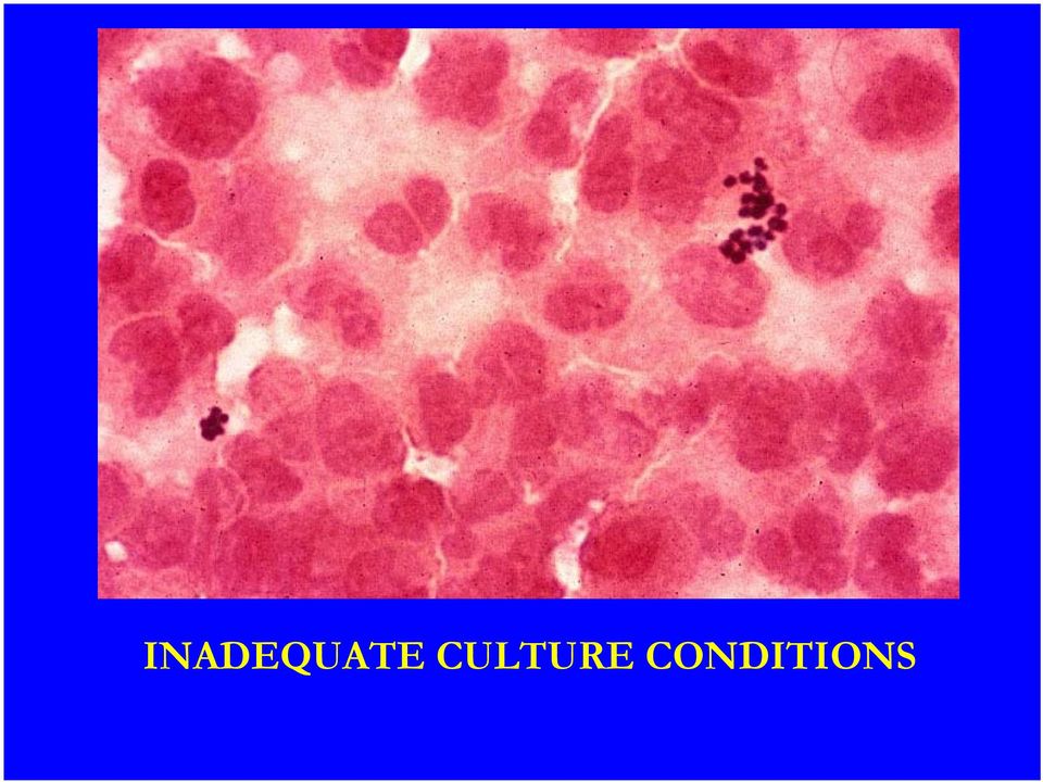
10 INADEQUATE CULTURE CONDITIONS This Gram smear demonstrates many inflammatory cells and several clusters of Gram positive cocci clearly grouped together in clumps. Such a Gram stain would suggest staphylococci, but no bacteria were recovered in the aerobic culture. An anaerobic culture which was subsequently submitted yielded Peptococcus sp., an anaerobic organism that did not grow in the original culture and which resembles Staphylococcus sp. in morphology.

11 SENSITIVTY OF TECHNIQUE The Gram smear is a relatively insensitive technique. It requires organisms per ml of fluid or gram of tissue in order to detect any bacteria. In some instances, this insensitivity is an advantage. For instance, the Gram smear of the undiluted voided urine can be used with reasonable success as a screen for significant bacteriuria (greater than 10 5 bacteria per ml). Similarly, a culture is more likely to detect confusing indigenous oropharyngeal flora that contaminate a sputum specimen than is the corresponding Gram stain. Unfortunately, the indigenous flora is often present in such large amounts that even the Gram stain is able to detect these unwanted elements.

12 OROPHARYNGEAL SQUAMOUS CELLS Oropharyngeal squamous cells are accompanied by large numbers of gram-negative bacilli in this sputum specimen from a patient in one of the special care units.

13 ASSESSMENT OF CELLULAR CONTENT The Gram stain allows assessment of the cellular content of a specimen. The first question in addressing the significance of the culture is whether the specimen came from an inflammatory process. There is no way to evaluate this question by analyzing the results of the culture. For instance, it is not uncommon to receive specimens of bile from which multiple organisms including enteric bacilli are cultured.

14 BILE WITH MULTIPLE PLUMP GRAM NEGATIVE RODS There is clearly a lack of any inflammatory process. The information from working up such a culture is not likely to be helpful and may be misleading. Even if the patient has fever and a post-operative wound infection, there is nothing to suggest that the isolated organisms are the ones that are involved in that infection.

15 FECES FROM A YOUNG MAN WITH GASTROENTERITIS Conversely, the presence of inflammatory cells establishes an inflammatory etiology and may suggest the etiologic agent. In this specimen, clumps of inflammatory cells are present and include both polymorphonuclear neutrophils (PMN) and mononuclear cells.

16 FECES FROM A YOUNG MAN WITH GASTROENTERITIS Although the Gram stain does not establish the etiology of the infection, it: does indicate that the process is not functional (eg., caused by emotional stress) or solely related to disturbances of bowel motility does suggest that an organism that produces gastroenteritis by invading bowel mucosa is responsible. Does suggest that viral gastroenteritis or Giardia enteritis are less likely. In this instance, the infection was caused by Salmonella typhimurium.

17 VALUE OF GRAM STAIN The Gram stain can be most useful for - assessing the adequacy of individual specimens, and - directing attention to specimens most likely to yield the correct answer.

18 VALUE OF GRAM STAIN In addition, one can provide some discrimination in those specimens that contain indigenous flora by trying to assess which morphologic bacteria are predominantly associated with the inflammatory process. Although these guides are clearly not foolproof, they do provide much needed help for interpretation of the corresponding culture. The following set of slides provide examples.

19 TRANSTRACHEAL ASPIRATE OF A PATIENT WITH PULMONARY INFILTRATES

20 Transtracheal Aspirate of a Patient with Pulmonary Infiltrates On the right, is a group of respiratory epithelial cells. They are elongated with basal nuclei and one can clearly see the brush of cilia at the ends of the cell. There were a few scattered polymorphonuclear cells in the specimen. It was basically non-inflammatory, but obviously contained respiratory material. The culture was entirely devoid of bacteria as demonstrated by the chocolate agar plate on the left.

21 SPUTUM CULTURE FROM THE SAME PATIENT
22 Sputum Culture from the Same Patient On the right is the Gram smear, in which one can see many squamous epithelial cells. Notice the gram negative bacilli present in this area of the smear. Once again, there were a few scattered polymorphonuclear cells. The culture, shown on the left, yielded a large number of mixed bacterial flora from this non-inflammatory process. Without the Gram smear, one would have a difficult time evaluating the significance of this culture. With it one can essentially dismiss the significance of these organisms.
23 Gram Smears from a Pair of Sputum Specimens Received on the Same Day from a patient with bacteremic pneumococcal pneumonia
24 Gram Smears from a Pair of Sputum Specimens Received on the Same Day On the left is the first specimen, which contained strands of mucus, proteinaceous debris, a moderate number of squamous epithelial cells, a few PMNs, and a few respiratory epithelial cells. It was a minimally inflammatory specimen and it was impossible to pick an area that was devoid of oral squamous cells. On the right is the Gram smear from the second sputum, which demonstrates clumps of inflammatory material with PMNs, macrophages, and protein exudate.
25 HIGHER POWER VIEW
26 Higher Power View On the left side one can clearly see a large squamous cell with which bacteria of many different morphologies are associated. Several inflammatory cells are also present and there are several pairs of gram-positive cocci in the field. One might wonder about the identity of these organisms as pneumococcus, but with the mixed morphology in the presence of squamous cells, the smear is essentially uninterpretable.
27 Higher Power View On the right is the second, more inflammatory specimen, in which essentially the only bacterial cells present are gram positive cocci in pairs and short chains. Many of the cocci have the pointed ends of a typical pneumococcus. In such a very inflammatory specimen, the morphology of pneumococcus often becomes distorted, as is the case here. The red staining material around the cocci probably represents the abundant polysaccharide capsule that this isolate possessed. It is treacherous, however, to try to assess the presence of a capsule with Gram-stained smear.
28 INTERPRETATION OF BACTERIAL ISOLATES In the sputum and in most other situations, the absence of inflammation makes interpretation of bacterial isolates difficult. There are a few exceptions to this rule, however. One such exception is in the lower urinary tract where bacteriuria without pyuria is a concern in pregnant women, who will have increased risk of pyelonephritis if untreated. Neutropenic patients may have difficulty mobilizing inflammatory cells. Some bacterial infections may typically be associated with minimal inflammation. The two most important are cellulitis produced by group A beta hemolytic streptococci and Clostridium perfringens. These infections are characterized by a watery, edematous, spreading inflammation. An additional, more exotic example is anthrax, caused by Bacillus anthracis, a bacterium that produces an edema toxin. Refusal to culture specimens that lack inflammation is not justifiable, but blind speciation of all isolates from such specimens is not rewarding and a considerable waste of resources.
29 Bacterial Infection with Minimal Inflammation -- Clostridial myonecrosis.
30 Clostridial myonecrosis. In this patient with gas gangrene there is a protein exudate and even at high dry power clearly visible large bacilli. The bacteria are grampositive, but clostridia decolorize easily and may even appear gramnegative, as they do here. Inflammatory cells are characteristically sparse. The diagnosis of clostridial gangrene requires documentation of the clinical syndrome and isolation of the organism.
31 DISADVANTAGES OF GRAM STAIN ARE FOR THE MOST PART THE OBVERSE OF THE ADVANTAGES Lack of immunological specificity means that only a presumptive diagnosis can be rendered Identification is less definitive than that provided by immunological means Insensitivity of the Gram smear means that the lack of demonstration of bacteria in a clinical specimen, particularly a sterile body fluid, does not predict accurately the absence of bacteria from that specimen. Interpretation of the Gram-stained smear requires considerable experience. Many morphologic forms of bacteria and confusing artifacts may be present in clinical specimens.
32 NOTES ON SLIDE PREPARATION In this laboratory the smears are usually prepared by laboratory personnel for your inspection. If you are in a situation where you must prepare your own smears, however, it is important to make the smear so that substantial portions are thin. A thick specimen must be spread very thinly or diluted. Tenacious specimens, such as sputum, may be pulled between two glass slides to give a reasonable separation of material. Use of ringed slides facilitates the observation of relatively noninflammatory material, where it may be hard to identify the location of the smear on the slide. Cytospin preparations provide excellent spreads of cells from body fluids.
33 PROTOCOLS FOR STAINING The length of time that crystal violet and Gram s iodine are left on the smear is not critical. A minimal 10 second staining with these reagents is sufficient. The period of time the decolorizing agent is left on the smear depends on the chemical used. In this laboratory acetone, which is a very rapid decolorizer is employed and the exposure should be brief. In general, the decolorizing solution is rinsed across the smear until the decolorizing fluid is no longer blue. It is very important, no matter what the abbreviations of the earlier steps are, to leave the counterstain in place for at least 30 seconds. Many gram-negative bacilli are stained very faintly by the counterstain and may be missed if this step is abbreviated. In our laboratory 0.05% basic fuchsin is added to the safranin counterstain to enhance contrast.
34 FURTHER NOTES ON SLIDE PREPARATION The smear should be air dried. It should not be heated to speed up drying, because the heat distorts the morphology of bacteria and cells. It should not be placed in front of a fan or waved around the room, because such maneuvers aerosolize material on the slides, including potential pathogens such as Mycobacterium tuberculosis. These strictures will result in a small delay (as much as 5-10 minutes), but a better smear will result in the end.
35 IMPORTANCE OF THIN SMEAR This is a touch preparation from a lung biopsy. Clumps of tissue and much cellular debris are present. If one looks closely, one can perceive in the background darker staining elongated gram negative bacilli, but they are not easy to recognize amidst all the other material in this rather thick area of the smear.
36 THINNER AREA OF SMEAR It is much easier to appreciate that innumerable thin, irregular gram-negative bacilli are present in the exudate. They still are not densely stained, again emphasizing the importance of the counterstain. This smear is from the lung of a patient who had Legionnaires disease during the 1977 epidemic in Burlington. No bacteria were grown from this specimen, because media that were adequate for recovery of Legionella were not available at that time. The Gram stain served as an indicator that the cultural procedures were inadequate.
37 EVALUATING THE SMEAR When evaluating the smear one should scan the slide at low power (10x objective) and roughly quantitate the numbers of cells of different types in the smear. This overall quantitation of cell types will give an appreciation of the inflammatory character of the specimen and potential contamination from mucosal surfaces. For sputum smears our laboratory uses the following scheme for quantitation of cells: 1-10/low power field (LPF) = few 10-25/LPF = moderate >25/LPF = many For other types of specimens a different scheme is used: 1/10 oil immersion fields (OIF) = few 1-10/OIF = moderate >10/OIF = many
38 QUANTITATION OF BACTERIA <10 organisms/smear = rare 1/1-10 oil immersion fields (OIF) = few 2-50/OIF = moderate >50/OIF = many
39 We do not quantitate cells and bacteria from fluid specimens that are centrifuged, because the number of cells and bacteria present will depend on the volume of fluid that has been processed. For evaluation of the bacterial morphotypes a thin area of the smear that contains predominantly PMNs should be selected.
40 There is no difficulty in selecting a field for view in this smear.
41 CONTAMINATING SQUAMOUS CELLS
42 CONTAMINATING SQUAMOUS CELLS Contaminating squamous cells are so mixed in with inflammatory cells that it is impossible to decide which bacteria are associated with the inflammatory component of the specimen. If one or two morphological types predominate it is reasonable to characterize them. If there is a greater mixture of organisms, it is probably best to lump them as mixed flora.
43 CELL TYPES ONE MAY ENCOUNTER IN CLINICAL SPECIMENS
44 SQUAMOUS EPITHELIAL CELL
45 MATURE SQUAMOUS CELLS The mature squamous cells shown here are characteristic and easy to identify. Abundant cytoplasm is present and the nucleus is small and hyperchromatic. Although squamous cells may be present in the lower respiratory tract of a patient with chronic bronchitis and squamous metaplasia, they usually signify the presence of oropharyngeal epithelium in sputum specimens. They may also be present in vaginal smears and may indicate vaginal contamination in urine specimens.
46 POLYMORPHONU CLEAR LEUKOCYTE (Poly)
47 POLYMORPHONUCLEAR NEUTROPHIL The polymorphonuclear neutrophil shown here is identified by the multilobed nucleus. Earlier precursors of the PMN in an inflammatory exudate may be more difficult to separate from other mononuclear cells.
48 ALVEOLAR MACROPHAGE
49 ALVEOLAR MACROPHAGE In respiratory specimens, the alveolar macrophage is a larger cell than the PMN. It contains abundant vaculated cytoplasm and a small nucleus.
50 ALVEOLAR PMNs MACROPHAGE Macrophages Inclusions of various kinds may be present in these cells. They virtually fill the cytoplasm of the cells. These large pigment-containing alveolar macrophages are called dust cells.
51 TUMOR CELLS IN SPUTUM Not all processes in the lung or other tissue are inflammatory and other cell types may be present, but the Gram stain is not a good cytological technique for identification of cells. This slide demonstrates tumor cells from a necrotic squamous carcinoma that were expectorated in sputum and detected by Gram s stain.
52 End of Part One
Gram Staining. The Most Commonly Used Differential Stain. Advantages:
 Gram Staining The Most Commonly Used Differential Stain Advantages: Can observe size and morphology (like other staining) Can find out additional information about the organism- primarily what type of
Medical Microbiology Microscopic slides and media
 Medical Microbiology Microscopic slides and media Head of Microbiology Department and Laboratory Medical Immunology : Janina Grzegorczyk MD, PhD, professor Implementators: Małgorzata Brauncajs MD Zbigniew
GRAM STAIN. Preanalytical Considerations
 GRAM STAIN Preanalytical Considerations I. PRINCIPLE The Gram stain is used to classify bacteria on the basis of their forms, sizes, cellular morphologies, and Gram reactions; in a clinical microbiology
PACKAGE LEAFLET. CLINDAMYCIN capsules Clidamycin. One capsule of 75 mg contains 75 mg Clindamycin (as hydrochloride).
.") PACKAGE LEAFLET CLINDAMYCIN capsules Clidamycin COMPOSITION One capsule of 75 mg contains 75 mg Clindamycin (as hydrochloride). One capsule of 150 mg contains 150 mg Clindamycin (as hydrochloride). PROPERTIES
PACKAGE LEAFLET CLINDAMYCIN capsules Clidamycin COMPOSITION One capsule of 75 mg contains 75 mg Clindamycin (as hydrochloride). One capsule of 150 mg contains 150 mg Clindamycin (as hydrochloride). PROPERTIES
The Gram Stain (2009)
") The Gram Stain (2009) E.J. Baron, Ph.D., D(ABMM) Prof. Pathology, Stanford Univ. Med. School Director Clin Micro/Viro Labs Director of Medical Affairs, Cepheid Gram positive thick peptidoglycan and teichoic
The Gram Stain (2009) E.J. Baron, Ph.D., D(ABMM) Prof. Pathology, Stanford Univ. Med. School Director Clin Micro/Viro Labs Director of Medical Affairs, Cepheid Gram positive thick peptidoglycan and teichoic
INFLAMMATION AND REACTIVE CHANGES IN CERVICAL EPITHELIUM
 INFLAMMATION AND REACTIVE CHANGES IN CERVICAL EPITHELIUM Inflammation is a response of a tissue to injury, often caused by invading microorganisms. The suffix which indicates inflammation is "-itis" (the
Adapted from Biology 15 Laboratory Supplemental Manual: Wrightsman, Ininns and Cannon- Moloznic.
 Biology 3B Laboratory Cultural Characteristics of Bacteria Objectives: Describe bacterial structure: colony morphology, cell shape, growth patterns. To distinguish how various growth media will affect
Microscopy and Cellular Morphology
 Microscopy and Cellular Morphology As we discussed in class, many organisms on the planet exist as single cells and are referred to as microorganisms bacteria, protozoans, among others. When a single microorganism
AURAMINE O STAIN. Preanalytical Considerations
 AURAMINE O STAIN Preanalytical Considerations I. PRINCIPLE Acid-fast mycobacteria resist decolorization by acid-alcohol after primary staining owing to the high lipid (mycolic acid) content in their cell
A Practical Guide to Diagnosis and Treatment of Infection in the Outpatient Setting Diagnosis and Treatment of Urinary Tract Infections
 A Practical Guide to Diagnosis and Treatment of Infection in the Outpatient Setting Diagnosis and Treatment of Urinary Tract Infections By Gary R. Skankey, MD, FACP, Infectious Disease, Las Vegas, NV Sponsored
CHAPTER 3 OBSERVING MICROORGANISMS THROUGH A MICROSCOPE. I. UNITS OF MEASUREMENT - See Table 3.1 in text. + Fig. 3.2
 CHAPTER 3 OBSERVING MICROORGANISMS THROUGH A MICROSCOPE I. UNITS OF MEASUREMENT - See Table 3.1 in text. + Fig. 3.2 II. MICROSCOPY: THE INSTRUMENTS A. COMPOUND LIGHT MICROSCOPY Figure 3.3 1. Have ocular
14 The ability of the lenses to distinguish fine detail and structure is called a. Illumination b. Magnification c. Refractive index d.
 1 2 Assume you stain Bacillus by applying malachite green with heat and then counterstain with safranin. Through the microscope, the green structures are a. cell walls. b. capsules. c. endospores. d. flagella.
STAINING AND BACTERIAL CELL MORPHOLOGY. To learn the techniques of Gram staining, nigrosin staining and KOH test.
 STAINING AND BACTERIAL CELL MORPHOLOGY I. OBJECTIVES To learn the technique of smear preparation. To learn the techniques of Gram staining, nigrosin staining and KOH test. To use and relate the Gram stain
Influenza (Flu) Influenza is a viral infection that may affect both the upper and lower respiratory tracts. There are three types of flu virus:
 Influenza is a viral infection that may affect both the upper and lower respiratory tracts. There are three types of flu virus:") Respiratory Disorders Bio 375 Pathophysiology General Manifestations of Respiratory Disease Sneezing is a reflex response to irritation in the upper respiratory tract and is associated with inflammation
Respiratory Disorders Bio 375 Pathophysiology General Manifestations of Respiratory Disease Sneezing is a reflex response to irritation in the upper respiratory tract and is associated with inflammation
Urinalysis and Body Fluids CRg
 Urinalysis and Body Fluids CRg Unit 2; Session 1 Urine Microscopic Examination The Complete Urinalysis Physical properties already covered Chemical analysis in the next unit Microscopic our current focus
Medical Microbiology Culture Media :
 Lecture 3 Dr. Ismail I. Daood Medical Microbiology Culture Media : Culture media are used for recognition and identification (diagnosis) of microorganisms. The media are contained in plates (Petri dishes),
Cytology of Lymph Nodes
 Indications Cytology of Lymph Nodes Lymph node enlargement That was easy Mary Anna Thrall Don Meuten Indications Lymph node enlargement Suspect metastasis Normal sized lymph nodes are Normal Do NOT aspirate
EDUCATIONAL COMMENTARY RED BLOOD CELLS AND WHITE BLOOD CELLS IN URINALYSIS
 URINALYSIS Educational commentary is provided through our affiliation with the American Society for Clinical Pathology (ASCP). To obtain FREE CME/CMLE credits click on Earn CE Credits under Continuing
Diagnostic Challenge. Department of Pathology,
 Cytology of Pleural Fluid as a Diagnostic Challenge Paavo Pääkkö,, MD, PhD Chief Physician and Head of the Department Department of Pathology, Oulu University Hospital,, Finland Oulu University Hospital
Exercise 2. The Compound Light Microscope
 6 Exercise 2 The Compound Light Microscope INTRODUCTION: Student Learning Objectives: After completing this exercise students will: a. Demonstrate proficient use of the microscope using low, high dry,
In order to be useful, a smear must have the following qualities:
 Smear Preparation and Simple Stain Objectives: Make bacterial smear slides (usually called smears) Distinguish cells on these slides using a simple stain procedure Unstained microbial cells are nearly
MICROSCOPY OF LIVING MICROBES
 EXPERIMENT 1 MICROSCOPY OF LIVING MICROBES Many students taking microbiology for the first time feel that they are going to have a hard time with the microscope. This lab as an experiment is intended to
LAB 4. Cultivation of Bacteria INTRODUCTION
 LAB 4. Cultivation of Bacteria Protocols for use of cultivation of bacteria, use of general growth, enriched, selective and differential media, plate pouring, determination of temperature range for growth
Basic Professional Training Program for Associate Medical Technologist
 Basic Professional Training Program for Associate Medical Technologist Basic Cytology Part 2 (Preparartion and normal morphology) Normal Morphology in Liquid based Gynecologic Cytology Speaker: Mr. Fung
Lab Exercise 2 Media and Culture
 Lab Exercise 2 Media and Culture Lab Exercise #2 Bacterial Media & Culture I. OBJECTIVES: Practice microbial collection techniques Describe colony morphology and the relationship to microbial identification.
URINE CULTURES GENERAL PROCEDURE
 University of Nebraska Medical Center Division of Laboratory Science Clinical Laboratory Science Program CLS 418/CLS 419 URINE CULTURES GENERAL PROCEDURE I. Principle Urine cultures are performed to detect
Raw Milk Quality Tests Do They Predict Fluid Milk Shelf-life or Is it time for new tests?
 Raw Milk Quality Tests Do They Predict Fluid Milk Shelf-life or Is it time for new tests? Martin Wiedmann Milk Quality Improvement Program November 3, 2011 Fluid milk shelf life What defines shelf life
SELECTIVE AND DIFFERENTIAL MEDIA
 SELECTIVE AND DIFFERENTIAL MEDIA Selective and differential media are used to isolate or identify particular organisms. Selective media allow certain types of organisms to grow, and inhibit the growth
Sampling of the surface contamination using sterile cotton swabs from toys obtained from
 RESULTS Sampling of the surface contamination using sterile cotton swabs from toys obtained from the Nursery at Queen Mary, University of London showed diverse microorganism growth. A variety of species
Mini-Medical School on Infectious Diseases. Session #1 - Basic Science
 Mini-Medical School on Infectious Diseases Session #1 - Basic Science The Microbial World Michael V. Norgard, Ph.D., Chairman Department of Microbiology U.T. Southwestern Medical Center The Microbial World
Streptococcal Infections
 Streptococcal Infections Introduction Streptococcal, or strep, infections cause a variety of health problems. These infections can cause a mild skin infection or sore throat. But they can also cause severe,
Enteric Unknowns Miramar College Biology 205 Microbiology
 Enteric Unknowns Miramar College Biology 205 Microbiology Enteric (Greek enteron = intestine) bacteria are comprised of several different genera, but all reside in the digestive tract of mammals. Because
LABORATORY 3: Microscopic Urinalysis
 LABORATORY 3: Microscopic Urinalysis Note Students are expected to review the corresponding information in the course textbook(s) as well as the classroom notes in preparation for this lab and to aid in
Explanation of your PAP smear
 Explanation of your PAP smear Approximately 5-10% of PAP smears in the United States are judged to be abnormal. Too often, the woman who receives this news worries that she already has, or will develop,
Diseases. Inflammations Non-inflammatory pleural effusions Pneumothorax Tumours
 Pleura Visceral pleura covers lungs and extends into fissures Parietal pleura limits mediastinum and covers dome of diaphragm and inner aspect of chest wall. Two layers between them (pleural cavity) contains
CONTROL: An infected appendix, or any tissue containing both negative and positive gram rods.
 SURGICAL PATHOLOGY HISTOLOGY Date: STAINING MANUAL - MICROORGANISMS Page: 1 of 3 GRAM BACTERIA - MODIFIED BROWN AND BRENN PURPOSE: For demonstrating gram-negative and gram-positive in tissue. PRINCIPLE:
Labquality External Quality Assessment Programmes General Bacteriology 1 4/2010
 Labquality External Quality Assessment Programmes General Bacteriology 1 4/2010 Photos and text: Markku Koskela, M.D., Ph.D. Clinical microbiology specialist Oulu, Finland Sample 13/2010 Cerebrospinal
Recurrent or Persistent Pneumonia
 Recurrent or Persistent Pneumonia Lower Respiratory Tract Dr T Avenant Recurrent or Persistent Pneumonia Definitions Recurrent pneumonia more than two episodes of pneumonia in 18 months Persistent pneumonia
Personal Injury TYPES OF HOLIDAY ILLNESSES. www.simpsonmillar.co.uk Telephone 0844 858 3200
 TYPES OF HOLIDAY ILLNESSES Whilst on holiday many different contractable illnesses exist, the list below contains the most common. This list is by no means exhaustive and if you have suffered from an illness
Diagnosis & Treatment Of Cough
 Diagnosis & Treatment Of Cough a.diagnosis : - Details History - Physical Examination - Investigation b. Treatment of cough Detail history provides valuables clues for etiology of the cough - Acute or
Pharmacology of the Respiratory Tract: COPD and Steroids
 Pharmacology of the Respiratory Tract: COPD and Steroids Dr. Tillie-Louise Hackett Department of Anesthesiology, Pharmacology and Therapeutics University of British Columbia Associate Head, Centre of Heart
Supplemental Material CBE Life Sciences Education. Su et al.
 Supplemental Material CBE Life Sciences Education Su et al. APPENDIX Human Body's Immune System Test This test consists of 31 questions, with only 1 answer to be selected for each question. Please select
Lung Cancer. This reference summary will help you better understand lung cancer and the treatment options that are available.
 Lung Cancer Introduction Lung cancer is the number one cancer killer of men and women. Over 165,000 people die of lung cancer every year in the United States. Most cases of lung cancer are related to cigarette
Cardiovascular System. Blood Components
 Cardiovascular System Blood Components 1 Components of Blood Formed elements: erythrocytes, leukocytes, platelets Plasma: water, proteins, other solutes The components of blood can be divided into two
White Blood Cells (WBCs) or Leukocytes
 or Leukocytes") Lec.5 Z.H.Al-Zubaydi Medical Physiology White Blood Cells (WBCs) or Leukocytes Although leukocytes are far less numerous than red blood cells, they are important to body defense against disease. On average,
Lec.5 Z.H.Al-Zubaydi Medical Physiology White Blood Cells (WBCs) or Leukocytes Although leukocytes are far less numerous than red blood cells, they are important to body defense against disease. On average,
ATLAS OF HEAD AND NECK PATHOLOGY THYROID PAPILLARY CARCINOMA
 Papillary carcinoma is the most common of thyroid malignancies and occurs in all age groups but particularly in women under 45 years of age. There is a high rate of cervical metastatic disease and yet
Lab Exercise 4. Epithelial Tissues. Connective Tissue Proper. What you need to be able to do on the exam after completing this lab exercise:
 Lab Exercise 4 Epithelial Tissues Connective Tissue Proper Textbook Reference: See Chapter 4 What you need to be able to do on the exam after completing this lab exercise: Be able to identify each type
Etiology and treatment of chronic bacterial prostatitis the Croatian experience
 Etiology and treatment of chronic bacterial prostatitis the Croatian experience Višnja Škerk University Hospital for Infectious Diseases "Dr. Fran Mihaljevic" Zagreb Croatia Milano, Malpensa, 14 Nov 2008
Urinalysis and Body Fluids CRg. Synovial Fluid. Synovial Fluid. Unit 4. Composition and formation. Functions. Reasons for analysis.
 Urinalysis and Body Fluids CRg Unit 4 Synovial Fluid Synovial Fluid Composition and formation Secreted by cells of synovial membrane Very viscous, clear ultrafiltrate of plasma Contains Hyaluronic acid
7- Master s Degree in Public Health and Public Health Sciences (Majoring Microbiology)
") 7- Master s Degree in Public Health and Public Health Sciences (Majoring Microbiology) Students should fulfill a total of 38 credit hours: 1- Basic requirements: 10 credit hours. 150701, 150702, 150703,
7- Master s Degree in Public Health and Public Health Sciences (Majoring Microbiology) Students should fulfill a total of 38 credit hours: 1- Basic requirements: 10 credit hours. 150701, 150702, 150703,
serology Agglutination Techniques and Blood Cell Identification
 Serology: Agglutination Techniques and Blood Cell Identification S erology is a branch of immunology dealing with techniques to identify and measure antigens, and to detect serum antibodies. Agglutination
CLOSTRIDIUM PERFRINGENS
 CLOSTRIDIUM PERFRINGENS PATHOGEN SAFETY DATA SHEET - INFECTIOUS SUBSTANCES INFECTIOUS AGENT NAME: Clostridium perfringens SYNONYM OR CROSS REFERENCE: Gas gangrene; pigbel disease CHARACTERISTICS: Clostridium
State of Kuwait Ministry of Health Infection Control Directorate. Guidelines for Prevention of Surgical Site Infection (SSI)
") State of Kuwait Ministry of Health Infection Control Directorate Guidelines for Prevention of Surgical Site Infection (SSI) September 1999 Updated 2007 Surgical Wound: According to 1998 Kuwait National
State of Kuwait Ministry of Health Infection Control Directorate Guidelines for Prevention of Surgical Site Infection (SSI) September 1999 Updated 2007 Surgical Wound: According to 1998 Kuwait National
Table. Positive Purified Protein Derivative Results (Pediatrics In Review Apr 2008)
") PPD and TB Sreening COMPETENCY- The resident should know the risk factors for TB exposure, when to screen, and the appropriate criteria for recognizing a positive PPD in children of different age groups
PPD and TB Sreening COMPETENCY- The resident should know the risk factors for TB exposure, when to screen, and the appropriate criteria for recognizing a positive PPD in children of different age groups
Normal flora, which make up about 90% of the cells of a human body, are microbes that
 Indigenous Unknowns Lab Report Introduction: Normal flora, which make up about 90% of the cells of a human body, are microbes that live and grow on the body without causing disease under normal conditions.
Welcome to Implementing Inquirybased Microbial Project. Veronica Ardi, PhD
 Welcome to Implementing Inquirybased Microbial Project Veronica Ardi, PhD Microbiology Laboratory Courses CourseSmart: ebook resources http://instructors.coursesmart.com/ Microbiology Laboratory Courses
GIEMSA STAIN PREANALYTICAL CONSIDERATIONS
 GIEMSA STAIN PREANALYTICAL CONSIDERATIONS I. Principle Giemsa stain is used to differentiate nuclear and/or cytoplasmic morphology of platelets, RBCs, WBCs, and parasites (1,2). The most dependable stain
General Information and Guidelines. Submission of blood tubes
 General Information and Guidelines Submission of blood tubes Note: Do not submit blood samples in syringes (especially with needles attached). Syringes that are submitted to the lab with the needles attached
Prokaryotic and Eukaryotic Cells
 Lab 2- Bio 201 Prokaryotic and Eukaryotic Cells Name: OBJECTIVES To explore cell structure and morphology in prokaryotes and eukaryotes. To gain more experience using the microscope, and in particular,
Cytology : first alert of mesothelioma? Professor B. Weynand, UCL Yvoir, Belgium
 Cytology : first alert of mesothelioma? Professor B. Weynand, UCL Yvoir, Belgium Introduction 3 cavities with the same embryologic origin the mesoderme Pleura Exudates Pleura Peritoneum Pericardium 22%
Oxygen relation Definition Examples Picture Facultative Anaerobe
 Oxygen relation Definition Examples Picture Facultative Anaerobe Does not require oxygen. Can grow with or without it. Able to detoxify toxic by products of oxygen. E. Coli Microaerophile Growth throughout
Transmission & Pathogenesis of Tuberculosis
 April-December, 2011: TB cases reported by the media in US Transmission & Pathogenesis of Tuberculosis Shu-Hua Wang, MD, MPH & TM Assistant Professor of Medicine The Ohio State University TB found at Seaside
LABORATORY 2 Staining and processing of blood parasites Differential counts of leukocytes (giemsa stains)
") LABORATORY 2 Staining and processing of blood parasites Differential counts of leukocytes (giemsa stains) SPECIMENS TO BE STAINED 1. Thin and thick blood smears from a patient returning from Africa - case
LABORATORY 2 Staining and processing of blood parasites Differential counts of leukocytes (giemsa stains) SPECIMENS TO BE STAINED 1. Thin and thick blood smears from a patient returning from Africa - case
Equine Equine Diarrhea Diarrhea Diarrhea in the Horse: Salmonella and Other Infections Introduction Clinical Signs
 Equine Diarrhea Diarrhea in the Horse: Salmonella and Other Infections Amanda M. House, DVM, DACVIM Assistant Professor Large Animal Clinical Sciences UF College of Veterinary Medicine Introduction Clinical
What Is Clostridium Difficile (C. Diff)? CLOSTRIDIUM DIFFICILE (C. DIFF)
? CLOSTRIDIUM DIFFICILE (C. DIFF)") What Is Clostridium Difficile (C. Diff)? Clostridium difficile, or C. diff for short, is an infection from a bacterium, or bug, that can grow in your intestines and cause bad GI symptoms. The main risk
What Is Clostridium Difficile (C. Diff)? Clostridium difficile, or C. diff for short, is an infection from a bacterium, or bug, that can grow in your intestines and cause bad GI symptoms. The main risk
OBJECTIVES PROCEDURE. Lab 2- Bio 160. Name:
 Lab 2- Bio 160 Name: Prokaryotic and Eukaryotic Cells OBJECTIVES To explore cell structure and morphology in prokaryotes and eukaryotes. To gain more experience using the microscope. To obtain a better
HOW TO WRITE AN UNKNOWN LAB REPORT IN MICROBIOLOGY
 HOW TO WRITE AN UNKNOWN LAB REPORT IN MICROBIOLOGY GENERAL Unknown reports in microbiology are written in scientific format. Scientific writing is written differently from other types of writing. The results
Animal Tissues. I. Epithelial Tissue
 Animal Tissues There are four types of tissues found in animals: epithelial tissue, connective tissue, muscle tissue, and nervous tissue. In this lab you will learn the major characteristics of each tissue
Diseases of peritoneum Lect. Al Qassim University, Faculty of Medicine Phase II Year III, CMD 332 Pathology Department 31-32
 Diseases of peritoneum Lect Al Qassim University, Faculty of Medicine Phase II Year III, CMD 332 Pathology Department 31-32 Describe the etiology, pathogenesis and types of peritonitis Define ascites and
Antibiotic-Associated Diarrhea, Clostridium difficile- Associated Diarrhea and Colitis
 Antibiotic-Associated Diarrhea, Clostridium difficile- Associated Diarrhea and Colitis ANTIBIOTIC-ASSOCIATED DIARRHEA Disturbance of the normal colonic microflora Leading to alterations in bacterial degradation
STAINING OF PBF AND INTERPRETATION OF NORMAL AND ABNORMAL RED CELL MORPHOLOGY
 9 STAINING OF PBF AND INTERPRETATION OF NORMAL AND ABNORMAL RED CELL MORPHOLOGY 9.1 INTRODUCTION A peripheral blood smear (peripheral blood film) is a glass microscope slide coated on one side with a thin
ILLINOIS DEPARTMENT OF CENTRAL MANAGEMENT SERVICES CLASS SPECIFICATION CLINICAL LABORATORY TECHNICIAN SERIES
 ILLINOIS DEPARTMENT OF CENTRAL MANAGEMENT SERVICES CLASS SPECIFICATION CLINICAL LABORATORY TECHNICIAN SERIES CLASS TITLE POSITION CODE CLINICAL LABORATORY TECHNICIAN I 08215 CLINICAL LABORATORY TECHNICIAN
Introduction Breast cancer is cancer that starts in the cells of the breast. Breast cancer happens mainly in women. But men can get it too.
 Male Breast Cancer Introduction Breast cancer is cancer that starts in the cells of the breast. Breast cancer happens mainly in women. But men can get it too. Many people do not know that men can get breast
C. difficile Infections
 C. difficile Infections Introduction C. difficile is a type of bacteria that can cause diarrhea and infection of the colon. This bacterium is more likely to infect patients at hospitals and other healthcare
Functions of Blood. Collects O 2 from lungs, nutrients from digestive tract, and waste products from tissues Helps maintain homeostasis
 Blood Objectives Describe the functions of blood Describe blood plasma Explain the functions of red blood cells, white blood cells, and platelets Summarize the process of blood clotting What is Blood?
Biology 3A Laboratory MITOSIS Asexual Reproduction
 Biology 3A Laboratory MITOSIS Asexual Reproduction OBJECTIVE To study the cell cycle and understand how, when and why cells divide. To study and identify the major stages of cell division. To relate the
THE NATURAL HISTORY OF PULMONARY TUBERCULOSIS
 Dr William Harris, Professor of Clinical Medicine of New York University, School of Medicine describes, in this series of slides, the natural history of pulmonary tuberculosis and the importance of early
MEDICAL MICROBIOLOGY 603 OR POPULATION HEALTH SCIENCES 603 CLINICAL AND PUBLIC HEALTH MICROBIOLOGY January 18-May 6, 2011
 MEDICAL MICROBIOLOGY 603 OR POPULATION HEALTH SCIENCES 603 CLINICAL AND PUBLIC HEALTH MICROBIOLOGY January 18-May 6, 2011 Lectures: M, W, F 1:20-2:30 P.M. ( All lectures will be ~ 70 Minutes ) Office Hours
URINARY TRACT INFECTIONS IN YOUNG WOMEN
 URINARY TRACT INFECTIONS IN YOUNG WOMEN Reviewed by Finnish Centre for Health Promotion Publisher: Ylioppilaiden terveydenhoitosäätiö Töölönkatu 37 A 00260 Helsinki Orders: julkaisutilaukset@yths.fi Author:
BACTERIA COUNTS IN RAW MILK
 BACTERIA COUNTS IN RAW MILK Richard L. Wallace TAKE HOME MESSAGES Bacterial contamination of raw milk can generally occur from three main sources; within the udder, outside the udder, and from the surface
THE KIDNEY. Bulb of penis Abdominal aorta Scrotum Adrenal gland Inferior vena cava Urethra Corona glandis. Kidney. Glans penis Testicular vein
 29 THE KIDNEY 9. Recurrent urinary tract infections Recurrent urinary tract infections The urinary tract consists of the urethra, the bladder, the ureters, the kidneys and in men the prostate gland. An
LABORATORY SUPERVISOR JOB CODE: 2701 DATE: 11/8/95
 LABORATORY SUPERVISOR JOB TITLE: Laboratory Supervisor GRADE: 19 JOB CODE: 2701 DATE: 11/8/95 GENERAL FUNCTION: Plans, manages and supervises the activities of a centralized laboratory and performs standard
Section B: Epithelial Tissue 1. Where are epithelial tissues found within the body? 2. What are the functions of the epithelial tissues?
 Tissue worksheet Name Section A: Intro to Histology Cells are the smallest units of life. In complex organisms, cells group together with one another based on similar structure and function to form tissues.
EXTRACTION OF DNA FROM CALF THYMUS CELLS Revised 2/1/96 Introduction
 Revised 2/1/96 Introduction Cells may be classified into two primary types depending on whether they have a discrete nucleus (eukaryotic) or do not (prokaryotic). Prokaryotes include bacteria, such as
Continuing Education Opportunities
 www.cap.org Continuing Education Opportunities Surveys Education Programs When your lab participates in Surveys, every member of your team can enroll in education activities and earn CME/CE at no additional
Common Breast Complaints:
 : Palpable mass Abnormal mammogram with normal physical exam Vague thickening or nodularity Nipple Discharge Breast pain Breast infection or inflammation The physician s goal is to determine whether the
C. difficile. Answers to frequently asked questions about. at the Jewish General Hospital. www.jgh.ca SIR MORTIMER B. DAVIS JEWISH GENERAL HOSPITAL
 Answers to frequently asked questions about C. difficile at the Jewish General Hospital SIR MORTIMER B. DAVIS JEWISH GENERAL HOSPITAL A McGill University Teaching Hospital www.jgh.ca 1. CLOSTRIDIUM DIFFICILE
Lymph capillaries, Lymphatic collecting vessels, Valves, Lymph Duct, Lymph node, Vein
 WLHS/A&P/Oppelt Name Lymphatic System Practice 1. Figure 12-1 provides an overview of the lymphatic vessels. First color code the following structures. Color code in Figure 12-1 Heart Veins Lymphatic vessels/lymph
Laboratory Exercise # 11: Differentiation of the Species Staphylococcus and Streptococcus
 Laboratory Exercise # 11: Differentiation of the Species Staphylococcus and Streptococcus Purpose: The purpose of this laboratory exercise is to explore the differences between Staphylococcal species and
Thought for the Day. Courage is not simply one of the virtues, but the form of every virtue at the testing point. ~ C. S. Lewis
 Thought for the Day Courage is not simply one of the virtues, but the form of every virtue at the testing point. ~ C. S. Lewis Anatomy & Physiology Bio 2401 Lecture Instructor: Daryl Beatty Section 2 Lecture
Microorganisms encountered in routine pathology specimens
 Technical Articles Role of Special Histochemical Stains in Staining Microorganisms Rashmil Saxena, BFA, HT(ASCP) CM Division of Transplantation Department of Surgery, Indiana University Indianapolis, IN,
EXPERIMENT #1: MICROSCOPY
 EXPERIMENT #1: MICROSCOPY Brightfield Compound Light Microscope The light microscope is an important tool in the study of microorganisms. The compound light microscope uses visible light to directly illuminate
BACTERIAL ENUMERATION
 BACTERIAL ENUMERATION In the study of microbiology, there are numerous occasions when it is necessary to either estimate or determine the number of bacterial cells in a broth culture or liquid medium.
Introduction to Medical Microbiology
 Introduction to Medical Microbiology Course Medical Microbiology Unit I Introduction to Microbiology Essential Question What is Medical Microbiology? TEKS 130.207(c) 2A, 3D Prior Student Learning n/a Estimated
Core Topic 2. The immune system and how vaccines work
 Core Topic 2 The immune system and how vaccines work Learning outcome To be able to describe in outline the immune system and how vaccines work in individuals and populations Learning objectives Explain
Lecture Objectives: Why study microbiology? What is microbiology? Roots of microbiology
 1 Lecture Objectives: Why study microbiology? What is microbiology? Roots of microbiology Why study microbiology? ENVIRONMENTAL MEDICAL APPLIED SCIENCE BASIC SCIENCE The science of microbiology Microbiology
Biological Sciences Initiative
 Biological Sciences Initiative HHMI Student Activities Measuring Antibiotic Resistance Introduction: You might be aware that antibiotics were once thought of as a magic bullet; a nearly perfect drug for
HISTOLOGY LABORATORY. Microscope Orientation and Blood Smear Lab
 HISTOLOGY LABORATORY Microscope Orientation and Blood Smear Lab For practicing how to use the microscope DO NOT use the blood smear slide (it is too boring for the lower mags). Use a slide from the white
Managing Clostridial Diseases in Cattle
 Managing Clostridial Diseases in Cattle Sheila M. McGuirk, DVM, PhD Introduction The many diseases of cattle that are attributed to Clostridial bacteria are shown in the following table. Clostridial type
Transferring a Broth Culture to Fresh Broth
 Sterile Technique It is very important in microbiology to work with pure cultures. Unfortunately this is difficult. The world around us is covered with microorganisms. Microorganisms are even carried on