Basic Professional Training Program for Associate Medical Technologist
|
|
|
- Eugenia Hall
- 8 years ago
- Views:
Transcription
1 Basic Professional Training Program for Associate Medical Technologist Basic Cytology Part 2 (Preparartion and normal morphology) Normal Morphology in Liquid based Gynecologic Cytology Speaker: Mr. Fung Hok Mun, Simon MT/ic Cytology Lab, UCH 1 2 Ectocervix covered by non keratinising squamous mucosa. Continuous with vaginal epithelium. Proximal, joins lining of endocervix at or near the external Os. 3 4

2 Endocervical canal (protected from vaginal ph) retains tall columnar cell lining & layer of reserve cells. Mucosa branching crypts extending into stroma of cervix for up to 5 mm. 5 6 Point where ectocervix squamous epithelium & endocervical canal epithelium meet = SquamoColumnar Junction Changes in this area are of crucial significance in cervical pathology. 7 8

3 9 10 SquamoColumnar Junction & Transformation Zone Endocervical eversion is followed by progressive metaplasia of exposed mucosa (under influence of vaginal ph) to less specialized, more hardy squamous epithelium ( Metaplasia = a change from one adult type of epithelium to another ) Squamous Metaplasia the mature endocervical columnar epithelium will be progressively replaced by a squamous epithelium when the squamocolumnar junction is everted distal to the external os exposing the endocervical epithelium to irritation in the vagina This process is normal and begins around puberty

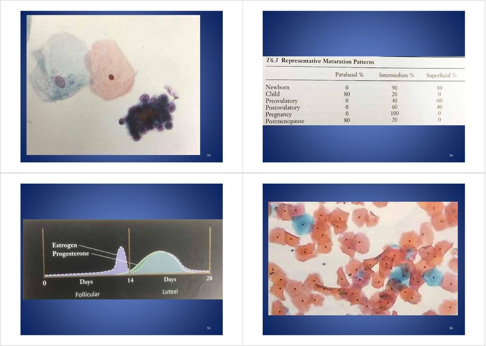
4 Structure of Stratified Squamous Epithelium Structure of Stratified Squamous Epithelium Germinal layer single, small, regular, undifferentiated cells BASAL cells. Next layer PARABASAL cells immature, crowded, two three deep. INTERMEDIATE layer variable thickness more cytoplasm; nuclei still show recognizable chromatin. Superficial cells are actually dead or dying & exfoliate spontaneously. Mucosal thickness depends on hormonal status all layers respond. Under oestrogen influence, superficial layer develops in about four days Normal Cellular Components Superficial cells Intermediate cells Parabasal cells Squamous metaplastic cells (mature vs, immature) Endocervical cells Inflammatory cells +/ Basal cells/ reserve cells +/ Endometrial cells 15 16

5 Cytology Epithelial Cells Pap stain : 2 components are cytoplasmic stains: eosin superficial cells pink or orange light green cytoplasm of less mature. Nuclei are stained by haematoxklin. Good fixation for good staining quality BASAL Cells Small primitive cells difficult to recognize Rarely sampled deep position. Short rows of small regular cells with sparse green cytoplasm, oval nuclei & high N/C ratio but chromatin pattern is fine & several chromocentres may be present PARABASAL Cells Round to oval, fairly dense green cytoplasm although if smear not well fixed cytoplasm may take up pinkish stain of eosin. Nuclei occupy about one half of cell, fine chromatin pattern

6 21 22 PARABASAL Cells Less mature parabasal cells in sheets. More mature usually dissociate. Usually predominate in PM smears. young women postnatally during lactational amenorrhoea or under abnormal conditions of inflammation or oestrogen deficiency 23 24

7 INTERMEDIATE Cells Polygonal shape, larger than parabasal Pale green cytoplasm peripheral fold Cytoplasm may stain with eosin esp. if poor fixation eosinophilic intermediate Low N / C ratio. Nucleus round / ovoid, fine chromatin. INTERMEDIATE Cells Tight groups or discrete depend upon hormonal state for 14 days. In 2 nd half of cycle, ragged cytoplasm may disintegrate > bare nuclei. With high progesterone, accumulate glycogen an irregular central deposit of pale yellow stained materal INTERMEDIATE Squamous Cells Nuclear diameter: 5 6 micron in diameter Mean nuclear area: 36 um

8 Superficial Cells Large & polygonal; pink to orange flat cytoplasm, rarely show folding as in intermediate cells Slightly larger than intermediate cells Nuclei small & condensed or pyknotic Almost always discrete compare with intermediate cells Superficial Cells Granular cell layer cells show small dark blue granules in cytoplasm. Nests of benign squamous cells epithelial pearls sometimes seen in normal smears. Cells may also be artefactually squashed & distorted in smear taking

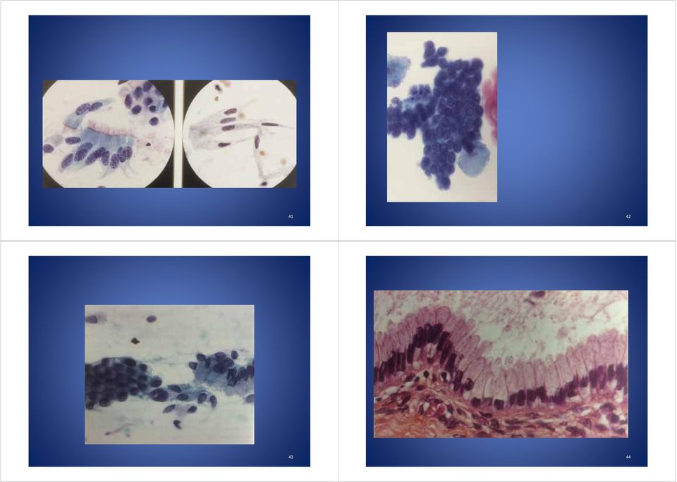
9 33 34 Anucleate Squames Mature superficial squamous cells with loss of nuclei. Polygonal shape; often stain with dimer of eosin >> orange or yellow cytoplasm. Anucleate squames in combination with granular cells > completion of keratinization process. Endocervical cells 1 Small sheets/ groups, less often single. From above > honeycomb; side on > picketfence. Cyanophilic cytoplasm translucent or vacuolated prone to degeneration. Fine chromation pattern; one or more small nucleoli may be identified

10 Endocervical cells 2 Occas. pink cilia are visible more common PM Mucin secreting goblet cells infrequent usually reactive feature. Endocervical cells 3 Nuclear size may vary considerably within a group. Multinucleation not uncommon esp. in inflammation or injury. Smear taking method affects yield: Brush >> Aylesbury >> Ayre spatula

11
12 Metaplastic Cells 1 Normal constituent once tranformation zone develops Immature: don t exfoliate spontaneously Mature: resemble original ectocervix intermediate cells & superficial cells so can t recognize as separate population Identifiable squamous metaplastic cells are size of parabasal & early intermediate cells Endometrial Cells 2 Early in menstruation, well formed, tight. 3 D clusters epithelial cell rim & central stromal cell core. Soon degenerative nuclear crumpling & disorganization of cells > small clusters of densely hyperchromatic crowded cells. Polymorphs often in endometrial clusters

13 Endometrial Cells Mean nuclear area: 37 um

14
15
16 Assessment of Squamous Cellularity Requires > 5,000 well visualized & wellpreserved squamous cells ThinPrep (preparation diameter = 20mm): > 3 4 squamous cells per 40 x HPFs SurePath (preparation diameter = 13mm): > 8 9 squamous cells per 40 x HPFs A minimum of 10 microscopic fields should be assessed Low Squamous Cellularity Obscuring Inflammation Liquid based preparations with 5,000 20,000 squamous cells Specify if 50 75% of squamous cells obscured Considered as unsatisfactory if > 75 % squamous cells obscured 63 64

17

18
19 End 73

INFLAMMATION AND REACTIVE CHANGES IN CERVICAL EPITHELIUM
 INFLAMMATION AND REACTIVE CHANGES IN CERVICAL EPITHELIUM Inflammation is a response of a tissue to injury, often caused by invading microorganisms. The suffix which indicates inflammation is "-itis" (the
worry When to Cervical Abnormalities CME Workshop What s the situation? What are the trends? By Dianne Miller, MD, FRCSC In this article:
 CME Workshop When to worry Cervical Abnormalities By Dianne Miller, MD, FRCSC What s the situation? Over 600,000 Papanicolaou s (Pap) smears were performed in British Columbia in 2000. Approximately 13,400
CERVICAL AND VAGINAL CYTOLOGY Edmund S. Cibas
 S2638-01.qxd 12/11/02 11:42 Page 1 1 Edmund S. Cibas THE HISTORY OF THE PAP TEST SAMPLING AND PREPARATION METHODS Conventional Smears Liquid-based Preparations ThinPrep SurePath AUTOMATED SCREENING Historical
Diagnostic Challenge. Department of Pathology,
 Cytology of Pleural Fluid as a Diagnostic Challenge Paavo Pääkkö,, MD, PhD Chief Physician and Head of the Department Department of Pathology, Oulu University Hospital,, Finland Oulu University Hospital
EDUCATIONAL COMMENTARY - GRANULOCYTE FORMATION AND CHRONIC MYELOCYTIC LEUKEMIA
 LEUKEMIA Educational commentary is provided through our affiliation with the American Society for Clinical Pathology (ASCP). To obtain FREE CME/CMLE credits click on Earn CE Credits under Continuing Education
Cytology of Lymph Nodes
 Indications Cytology of Lymph Nodes Lymph node enlargement That was easy Mary Anna Thrall Don Meuten Indications Lymph node enlargement Suspect metastasis Normal sized lymph nodes are Normal Do NOT aspirate
Cytology : first alert of mesothelioma? Professor B. Weynand, UCL Yvoir, Belgium
 Cytology : first alert of mesothelioma? Professor B. Weynand, UCL Yvoir, Belgium Introduction 3 cavities with the same embryologic origin the mesoderme Pleura Exudates Pleura Peritoneum Pericardium 22%
Preparation of Blood Films
 Preparation of Blood Films Principle: Blood film enables us to evaluate WBC, RBC, and PLT morphology, also, allows us to perform differential WBC count, furthermore estimation of WBC and platelets counts
Granulocytes vs. Agranulocytes
 Leukocytes are white blood cells (AKA colorless (non-pigmented) blood cells). (Much) smaller in number than RBCs. Unlike RBCs, there are several different types of WBCs. All contain a visible nucleus.
Small & Large Intestines
 Small & Large Intestines Small Intestine: principal site for digestion of food and absorption of the products of digestion Large Intestine: reabsorption of water and elimination of undigested food and
Effusions: Mesothelioma and Metastatic Cancers
 Effusions: Mesothelioma and Metastatic Cancers Malignant Mesothelioma Incidence: 2,500 cases/year ~60-80% pts with pleural MM relationship with asbestos exposure Other risk factors: radiation, other carcinogens,
Explanation of your PAP smear
 Explanation of your PAP smear Approximately 5-10% of PAP smears in the United States are judged to be abnormal. Too often, the woman who receives this news worries that she already has, or will develop,
Creation Date: 12/24/2008. Effective Date: 07/14/2009 Date of Revision/Review: 07/14/2009 Version #:1 Date of Next Review: 07/14/2010
 Site: Fremont Rideout Health Group Laboratory Services Policy and Procedure Creation Date: 12/24/2008 Subject/Title: Collection of Specimens for Conventional & ThinPrep Pap Tests, HPV Document Owner: Rogers,
Lab Exercise 4. Epithelial Tissues. Connective Tissue Proper. What you need to be able to do on the exam after completing this lab exercise:
 Lab Exercise 4 Epithelial Tissues Connective Tissue Proper Textbook Reference: See Chapter 4 What you need to be able to do on the exam after completing this lab exercise: Be able to identify each type
Liquid-based cytology
 Liquid-based cytology Claire Bourgain UZBrussel Introduction LBC for gynaecological cytology has been introduced in Belgium a decade ago There is no consensus On performance compared to conventional cytology
Screening for Cancer of the Cervix
 Screening for Cancer of the Cervix An Office Manual for Health Professionals Cervical Cancer Screening Program tenth Edition 2013 Contact Information Cervical Cancer Screening Program (CCSP) Administration
Guidelines for reporting histopathology of cervical carcinoma
 Guidelines for reporting histopathology of cervical carcinoma Naveena Singh, Consultant Pathologist Introduction Cancer management is multidisciplinary Histopathology report has a MAJOR impact on management
Carcinosarcoma of the Ovary
 Carcinosarcoma of the Ovary A Rare Finding Presented By: Kathryn Kiely Anisa I. Kanbour School of Cytotechnology of the University of Pittsburgh Medical Center Pittsburgh, PA Patient History 55 year old
Classification of squamous cell cervical cytology. Luz Helena Camargo Casallas
 Classification of squamous cell cervical cytology Luz Helena Camargo Casallas Universidad Nacional de Colombia Faculty of Medicine - Engineering Faculty Bogotá D.C. Colombia 2012 Classification of squamous
STAINING OF PBF AND INTERPRETATION OF NORMAL AND ABNORMAL RED CELL MORPHOLOGY
 9 STAINING OF PBF AND INTERPRETATION OF NORMAL AND ABNORMAL RED CELL MORPHOLOGY 9.1 INTRODUCTION A peripheral blood smear (peripheral blood film) is a glass microscope slide coated on one side with a thin
CHEM 107. Hair handout. Basic Structure of Hair. 3-22-05 and 3-24-05
 CHEM 107 Hair handout. 3-22-05 and 3-24-05 Basic Structure of Hair A hair can be defined as a slender, thread-like outgrowth from a follicle in the skin of mammals. Composed mainly of keratin, it has three
Cervical Cancer Screening Guideline
 Cervical Cancer Screening Guideline Prevention 2 Abbreviations Used 2 Specimen Collection Techniques 3 Screening 4 Management Women 21 Years and Older Pap results 5 findings: ASC-US and LSIL 6 findings:
White Blood Cells (WBCs) or Leukocytes
 or Leukocytes") Lec.5 Z.H.Al-Zubaydi Medical Physiology White Blood Cells (WBCs) or Leukocytes Although leukocytes are far less numerous than red blood cells, they are important to body defense against disease. On average,
Lec.5 Z.H.Al-Zubaydi Medical Physiology White Blood Cells (WBCs) or Leukocytes Although leukocytes are far less numerous than red blood cells, they are important to body defense against disease. On average,
Hematology Morphology Critique
 Survey Slide: History: 60-year-old female presenting with pneumonia Further Laboratory Data: Hgb : 90 g/l RBC : 2.92 10 12 /L Hct : 0.25 L/L MCV : 87 fl MCH : 30.8 pg MCHC : 355 g/l RDW : 17.7 % WBC :
Histopathology of Major Salivary Gland Neoplasms
 Histopathology of Major Salivary Gland Neoplasms Sam J. Cunningham, MD, PhD Faculty Advisor: Shawn D. Newlands, MD, PhD Faculty Advisor: David C. Teller, MD The University of Texas Medical Branch, Department
Male. Female. Death rates from lung cancer in USA
 Male Female Death rates from lung cancer in USA Smoking represents an interesting combination of an entrenched industry and a clearly drug-induced cancer Tobacco Use in the US, 1900-2000 5000 100 Per Capita
ALTHOUGH excellent accounts have been published in recent years of
 THE EXFOLIATIVE CYTOLOGY OF DIFFUSE MALIGNANT MESOTHELIOMA BERNARD NAYLOR Department of Pathology, University of Michigan, Ann Arbor, Michigan, U.S.A. PLATE~ LXXXVI-XCI ALTHOUGH excellent accounts have
Immunohistochemistry on cytology specimens from pleural and peritoneal fluid
 Immunohistochemistry on cytology specimens from pleural and peritoneal fluid Dr Naveena Singh Consultant Pathologist Bart health NHS Trust London United Kingdom Disclosures and Acknowledgements I have
Effusion cytology. Dr Alpha Tsui Royal Melbourne Hospital 2008
 Effusion cytology Dr Alpha Tsui Royal Melbourne Hospital 2008 General points: -large unilateral effusion (>1 litre) in the elderly is highly suspicious for malignancy -effusions associated with malignancies
Pap smears, cytology and CCHC lab work and follow up
 Pap smears, cytology and CCHC lab work and follow up What is a Pap Smear? A Pap smear (also known as the Pap test) is a medical procedure in which a sample of cells from a woman's cervix (the end of the
R-16: Chronic nonspecific cervisit
 R-16: Chronic nonspecific cervisit Ectoservikal squamous epithelium Endoservical columnar epithelium Dilated cystic endoservical glands lymphoplasmocytes R18:Squamous cell carcinoma insitu Neoplastic epithelium
DESMOPLASTIC SMALL ROUND CELL TUMOR: A RARE PATHOLOGY PUZZLE
 DESMOPLASTIC SMALL ROUND CELL TUMOR: A RARE PATHOLOGY PUZZLE Ryan Granger University of Rhode Island Cytotechnology program May 2, 2015 ASCT Annual Meeting Nashville, Tennessee DESMOPLASTIC SMALL ROUND
NHS Cervical Screening Programme Achievable standards, Benchmarks for reporting, and Criteria for evaluating cervical cytopathology
 NHS Cervical Screening Programme Achievable standards, Benchmarks for reporting, and Criteria for evaluating cervical cytopathology third edition including revised performance indicators NHSCSP PubliCatioN
Animal Tissues. I. Epithelial Tissue
 Animal Tissues There are four types of tissues found in animals: epithelial tissue, connective tissue, muscle tissue, and nervous tissue. In this lab you will learn the major characteristics of each tissue
Chapter 3. Testing and reporting the results of visual inspection with Lugol's iodine (VILI)
") Chapter 3 Testing and reporting the results of visual inspection with Lugol's iodine (VILI) Instruments and materials required: Examination table with knee crutches or leg rests or stirrups; Good light
Chapter 3 Testing and reporting the results of visual inspection with Lugol's iodine (VILI) Instruments and materials required: Examination table with knee crutches or leg rests or stirrups; Good light
SEED Haematology. Sysmex Educational Enhancement and Development February 2013
 SEED Haematology Sysmex Educational Enhancement and Development February 2013 The role of the peripheral blood smear in the modern haematology laboratory Automated haematology cell counting The laboratory
Urinalysis and Body Fluids CRg
 Urinalysis and Body Fluids CRg Unit 2; Session 1 Urine Microscopic Examination The Complete Urinalysis Physical properties already covered Chemical analysis in the next unit Microscopic our current focus
Touch DNA and DNA Recovery. H. Miller Coyle
 Touch DNA and DNA Recovery 1 2 What is the link between cell biology & forensic science? Cells are the trace substances left behind that can identify an individual. Cells contain DNA. There are two forms
Management of Abnormal PAP Smears. K Chacko, MD, FACP 2010 GIM Conference
 Management of Abnormal PAP Smears K Chacko, MD, FACP 2010 GIM Conference Scope of the Problem About 7-10% 7 of PAPs will come back abnormal 3.5 to 4 million in the US each year Approximate 4000 deaths
ATLAS OF HEAD AND NECK PATHOLOGY THYROID PAPILLARY CARCINOMA
 Papillary carcinoma is the most common of thyroid malignancies and occurs in all age groups but particularly in women under 45 years of age. There is a high rate of cervical metastatic disease and yet
Blood. Functions of Blood. Components of Blood. Transporting. Distributing body heat. A type of connective tissue. Formed elements.
 Blood Functions of Blood Transporting nutrients respiratory gases waste products Distributing body heat Components of Blood A type of connective tissue Formed elements Living blood cells Plasma Nonliving
Cardiovascular System. Blood Components
 Cardiovascular System Blood Components 1 Components of Blood Formed elements: erythrocytes, leukocytes, platelets Plasma: water, proteins, other solutes The components of blood can be divided into two
Plasma Membrane hydrophilic polar heads
 The Parts of the Cell 3 main parts in ALL cells: plasma membrane, cytoplasm, genetic material this is about the parts of a generic eukaryotic cell Plasma Membrane -is a fluid mosaic model membrane is fluid
Cells. Introduction WSBCTC 1
 Cells Cells are the fundamental unit of life. All living things are composed of cells. While there are several characteristics that are common to all cells, such as the presence of a cell membrane, cytoplasm,
h. Large intestine 3
 (1) General features (a) Large intestine is last organ of digestive tract proper divided into 3 or 4 regions cecum appendix in humans colon rectum 1 b) No villi lumenal epithelium has microvilli This brush
SCREENING FOR CANCER OF THE CERVIX
 SCREENING FOR CANCER OF THE CERVIX An Office Manual for Health Professionals This manual has been prepared by the Cervical Cancer Screening Program of the BC Cancer Agency to support effective use of the
Management of Abnormal Pap Smear Clinical Practice Guideline
 Management of Abnormal Pap Smear Clinical Guideline General Principles: The Papanicolaou (Pap) smear is widely credited with reducing mortality from cervical cancer, and remains the single best method
An abnormal Pap smear - what does it mean?
 An abnormal Pap smear - what does it mean? It is natural to feel worried if you have just found out that your Pap smear result is not normal (abnormal). Around 1 in 10 Pap smears will show changes in the
Cervical Cancer The Importance of Cervical Screening and Vaccination
 Cervical Cancer The Importance of Cervical Screening and Vaccination Cancer Cells Cancer begins in cells, the building blocks that make up tissues. Tissues make up the organs of the body. Sometimes, this
Human Papilloma Virus (HPV)
") Human Papilloma Virus (HPV) A Sexually Transmitted Disease (STD) which can lead to Cervical, Penile and Anal Cancer What Adolescents Need to Know! Most people have heard of HIV/AIDS; however, most people
Human Papilloma Virus (HPV) A Sexually Transmitted Disease (STD) which can lead to Cervical, Penile and Anal Cancer What Adolescents Need to Know! Most people have heard of HIV/AIDS; however, most people
Abnormal Uterine Bleeding FAQ Sheet
 Abnormal Uterine Bleeding FAQ Sheet What is abnormal uterine bleeding? Under normal circumstances, a woman's uterus sheds a limited amount of blood during each menstrual period. Bleeding that occurs between
Specimen collection and transport for Chlamydia trachomatis and Neisseria gonorrhoeae testing
 Specimen collection and transport for Chlamydia trachomatis and Neisseria gonorrhoeae testing Overview Chlamydia trachomatis (CT) and Neisseria gonorrhoeae (NG) infections are two of the most common sexually
Anatomy PHL 212. By Dr Tajdar Husain Khan
 Anatomy PHL 212 By Dr Tajdar Husain Khan Overview of Anatomy Anatomy(from the Greek word anatome,"dissection") is a branch of natural science dealing with the structural organization of living things The
HISTOLOGY LABORATORY. Microscope Orientation and Blood Smear Lab
 HISTOLOGY LABORATORY Microscope Orientation and Blood Smear Lab For practicing how to use the microscope DO NOT use the blood smear slide (it is too boring for the lower mags). Use a slide from the white
Histology. Epithelial Tissue
 Histology Epithelial Tissue Epithelial Tissue Lines internal and external body surfaces Forms glands Epithelial Tissue Little extracellular matrix Attached on one side Avascular Basement membrane Apical
The Huntington Library, Art Collections, and Botanical Gardens
 The Huntington Library, Art Collections, and Botanical Gardens Rooting for Mitosis Overview Students will fix, stain, and make slides of onion root tips. These slides will be examined for the presence
Polyps. Hyperplasias. CAP 2011: Course AP104. The High Risk Benign Endometrium. Mutter and Nucci 1
 Course AP104 Endometrial Hyperplasia A morphologic Definition Hyperplasias Hormonal Effect or Precancer? George L. Mutter, MD Harvard Medical School and Brigham and Women s Hospital Boston, MA Endometrial
PROPERTY OF ELSEVIER SAMPLE CONTENT - NOT FINAL ABNORMAL PAP SMEAR (ABNORMAL CERVICAL CYTOLOGIC FINDINGS) Kathleen Dor
 Kathleen Dor") 1 ABNORMAL PAP SMEAR (ABNORMAL CERVICAL CYTOLOGIC FINDINGS) Kathleen Dor Cervical cytology screening has significantly decreased rates of mortality from cervical cancer; however, 400 women die each year
1 ABNORMAL PAP SMEAR (ABNORMAL CERVICAL CYTOLOGIC FINDINGS) Kathleen Dor Cervical cytology screening has significantly decreased rates of mortality from cervical cancer; however, 400 women die each year
An introduction to invasive cancer of the uterine cervix
 An introduction to invasive cancer of the uterine cervix Preclinical invasive cancer refers to early cervical cancer, with minimal stromal invasion, often without any symptoms or clinical features. As
Outline. Workup for metastatic breast cancer. Metastatic breast cancer
 Metastatic breast cancer Immunostain Update: Diagnosis of metastatic breast carcinoma, emphasizing distinction from GYN primary 1/3 of breast cancer patients will show metastasis 1 st presentation or 20-30
RAD 223. Radiography physiology. Lecture Notes. First lecture: Cell and Tissue
 RAD 223 Radiography physiology Lecture Notes First lecture: Cell and Tissue Physiology: the word physiology derived from a Greek word for study of nature. It is the study of how the body and its part work
SEMESTER VI 3 RD YEAR PATHOLOGY KIDNEY TUMORS
 SEMESTER VI 3 RD YEAR PATHOLOGY KIDNEY TUMORS LEARNING OBJECTIVES At the end of the lecture, students should be able to: Know the pathology of renal tumors. RENAL TUMORS RENAL PAPILLARY ADENOMA Common
Tissue Types. 1. Epithelial Tissue (or epithelium) is the lining, covering, and glandular tissue of the body
 is the lining, covering, and glandular tissue of the body") Tissue Types A. Tissues 1. Tissues: groups of cells similar in structure and function 2. Four Types of Tissues: a. Epithelium: for covering b. Connective Tissue: for support c. Muscle: for movement d.
Tissue Types A. Tissues 1. Tissues: groups of cells similar in structure and function 2. Four Types of Tissues: a. Epithelium: for covering b. Connective Tissue: for support c. Muscle: for movement d.
ORIGINAL ARTICLES. Materials and Methods
 ORIGINAL ARTICLES Cytomorphologic Features of Metastatic Urothelial Carcinoma in Serous Effusions Cheng Cheng Huang, M.D., PH.D., 1 Anoja Attele, M.D., 1 and Claire W. Michael, M.D. 2 * Metastatic urothelial
The Cell: Organelle Diagrams
 The Cell: Organelle Diagrams Fig 7-4. A prokaryotic cell. Lacking a true nucleus and the other membrane-enclosed organelles of the eukaryotic cell, the prokaryotic cell is much simpler in structure. Only
The Diagnosis of Cancer in the Pathology Laboratory
 The Diagnosis of Cancer in the Pathology Laboratory Dr Edward Sheffield Christmas Select 74 Meeting, Queen s Hotel Cheltenham, 3 rd December 2014 Agenda Overview of the pathology of cancer How specimens
HPV OncoTect E6, E7 mrna Kit A highly specific molecular test for early detection of cervical cancer
 Revolutionizing healthcare one cell at a time HPV OncoTect E6, E7 mrna Kit A highly specific molecular test for early detection of cervical cancer Numerous studies confirm that the presence of HR HPV DNA
UNIT 1 - Living Organisms and the Environment Situations. Cells
 Lesson Summaries HUMAN AND SOCIAL BIOLOGY UNIT 1 - Living Organisms and the Environment Situations Lesson 2 Cells OBJECTIVES At the end of this lesson you will be able to: a) Describe the structure of
OBJECTIVES PROCEDURE. Lab 2- Bio 160. Name:
 Lab 2- Bio 160 Name: Prokaryotic and Eukaryotic Cells OBJECTIVES To explore cell structure and morphology in prokaryotes and eukaryotes. To gain more experience using the microscope. To obtain a better
Colposcopy and Treatment of Cervical Intraepithelial Neoplasia:
 Colposcopy and Treatment of Cervical Intraepithelial Neoplasia World Health Organization - International Agency for Research on Cancer (IARC) World Health Organization Regional Office for Africa (AFRO)
PathoBasic - Vulva, Vagina, Cervix. E.Obermann
 PathoBasic - Vulva, Vagina, Cervix E.Obermann Vulva Inflammatory Changes Non-neoplastic lesions «Skin type lesions» Lichen sclerosus Infection: HSV, Candida Fibroepithelial stroma polyp Cutaneous Neoplasia
EIN. (Endometrial Intraepithelial Neoplasia): Improved Criteria for diagnosing endometrial precancer. Stanley J. Robboy, MD, FCAP,
: Improved Criteria for diagnosing endometrial precancer. Stanley J. Robboy, MD, FCAP,") EIN (Endometrial Intraepithelial Neoplasia): Improved Criteria for diagnosing endometrial precancer Stanley J. Robboy, MD, FCAP, FFPath FRCPI (Hon), FRCPath (UK, Hon) Professor of Pathology, Duke University
EIN (Endometrial Intraepithelial Neoplasia): Improved Criteria for diagnosing endometrial precancer Stanley J. Robboy, MD, FCAP, FFPath FRCPI (Hon), FRCPath (UK, Hon) Professor of Pathology, Duke University
Glossary. amenorrhea, primary - from the beginning and lifelong; menstruation never begins at puberty.
 Glossary amenorrhea - absence or cessation of menstrual periods. amenorrhea, primary - from the beginning and lifelong; menstruation never begins at puberty. A amenorrhea, secondary - due to some physical
Human Anatomy & Physiology I with Dr. Hubley. Practice Exam 1
 Human Anatomy & Physiology I with Dr. Hubley Practice Exam 1 1. Which definition is the best definition of the term gross anatomy? a. The study of cells. b. The study of tissues. c. The study of structures
FRIEND TO FRIEND CPT CODES 2015 2016. Diagnostic digital breast tomosynthesis, unilateral (list separately in addition to code for primary procedure)
") FRIEND TO FRIEND CPT CODES 2015 2016 CPT CODE SERVICE DESCRIPTION FEE EFFECTIVE G0101 Screening pelvic examination $36.69 01 Jan 16 G0202 Mammography, screening, digital, bilateral (2 view film study of
FRIEND TO FRIEND CPT CODES 2015 2016 CPT CODE SERVICE DESCRIPTION FEE EFFECTIVE G0101 Screening pelvic examination $36.69 01 Jan 16 G0202 Mammography, screening, digital, bilateral (2 view film study of
LABORATORY 2 Staining and processing of blood parasites Differential counts of leukocytes (giemsa stains)
") LABORATORY 2 Staining and processing of blood parasites Differential counts of leukocytes (giemsa stains) SPECIMENS TO BE STAINED 1. Thin and thick blood smears from a patient returning from Africa - case
LABORATORY 2 Staining and processing of blood parasites Differential counts of leukocytes (giemsa stains) SPECIMENS TO BE STAINED 1. Thin and thick blood smears from a patient returning from Africa - case
Cervical Cancer Screening and Management Guidelines: Changing Again, Huh?
 Cervical Cancer Screening and Management Guidelines: Changing Again, Huh? Summary of 2013 recommendations from ASC (American Cancer Society), ASCCP (American Society for Colposcopy and Cervical Pathology),
3.1 Cells and cell function
 BTEC s own resources 3.1 Cells and cell function In this section: P1 How you are made Key terms Tissue a group of similar cells acting together to perform a particular function. Epithelial cells one of
Cytopathology Case Presentation #8
 Cytopathology Case Presentation #8 Emily E. Volk, MD William Beaumont Hospital, Troy, MI Jonathan H. Hughes, MD Laboratory Medicine Consultants, Las Vegas, Nevada Clinical History 44 year old woman presents
The Cervical Screening Manual
 The Cervical Screening Manual A Guide for Health Departments and Providers Collaboration Partners: Chronic Disease and Injury Section Breast and Cervical Cancer Control Program Women s and Children s Health
Prokaryotic and Eukaryotic Cells
 Lab 2- Bio 201 Prokaryotic and Eukaryotic Cells Name: OBJECTIVES To explore cell structure and morphology in prokaryotes and eukaryotes. To gain more experience using the microscope, and in particular,
ABO-Rh Blood Typing Using Neo/BLOOD
 ABO-Rh Blood Typing Using Neo/BLOOD Objectives Determine the ABO and Rh blood type of unknown simulated blood samples. Prepare a simulated blood smear. Examine a prepared blood smear under the microscope
EXTRACTION OF DNA FROM CALF THYMUS CELLS Revised 2/1/96 Introduction
 Revised 2/1/96 Introduction Cells may be classified into two primary types depending on whether they have a discrete nucleus (eukaryotic) or do not (prokaryotic). Prokaryotes include bacteria, such as
A Practical Manual on Visual Screening for Cervical Neoplasia
 A Practical Manual on Visual Screening for Cervical Neoplasia IARCPress World Health Organization - International Agency for Research on Cancer (IARC) World Health Organization Regional Office for Africa
Exercise 9: Blood. Readings: Silverthorn 5 th ed, 547 558, 804 805; 6 th ed, 545 557, 825 826.
 Exercise 9: Blood Readings: Silverthorn 5 th ed, 547 558, 804 805; 6 th ed, 545 557, 825 826. Blood Typing The membranes of human red blood cells (RBCs) contain a variety of cell surface proteins called
BLOOD FILM STAINING EFFECTS
 An Educational Supplement prepared by ALQEP May 2004 Introduction The stained peripheral blood film is one of the world s most widely and frequently used tests. Since its introduction in the late nineteenth
Introduction: Tumor Swelling / new growth / mass. Two types of growth disorders: Non-Neoplastic. Secondary / adaptation due to other cause.
 Disorders of Growth Introduction: Tumor Swelling / new growth / mass Two types of growth disorders: Non-Neoplastic Secondary / adaptation due to other cause. Neoplastic. Primary growth abnormality. Non-Neoplastic
Two main classes: Epithelial Connective (synovial) Epithelial. Cutaneous Mucous Serous
 Epithelial. Cutaneous Mucous Serous") Two main classes: Epithelial Connective (synovial) Epithelial Cutaneous Mucous Serous Epithelial Membranes = sheet of epithelia + connective tissue base 1. Cutaneous membrane: outer skin layer (stratified
Two main classes: Epithelial Connective (synovial) Epithelial Cutaneous Mucous Serous Epithelial Membranes = sheet of epithelia + connective tissue base 1. Cutaneous membrane: outer skin layer (stratified
2012 Updated Consensus Guidelines for the Management of Abnormal Cervical Cancer Screening Tests and Cancer Precursors
 2012 Updated Consensus Guidelines for the Management of Abnormal Cervical Cancer Screening Tests and Cancer Precursors L. Stewart Massad, MD, Mark H. Einstein, MD, Warner K. Huh, MD, Hormuzd A. Katki,
A Statistical Analysis of Rescreening Alarms
 0 1986 Alan R. Liss, Inc. Cytometry 7205-211 (1986) A Statistical Analysis of Rescreening Alarms in a Population of Normal and Abnormal A Gynecologic Specimens' L.L. Wheeless, R.D. Robinson, C. Cox, T.K.
Cell Cycle in Onion Root Tip Cells (IB)
") Cell Cycle in Onion Root Tip Cells (IB) A quick overview of cell division The genetic information of plants, animals and other eukaryotic organisms resides in several (or many) individual DNA molecules,
Cell Cycle in Onion Root Tip Cells (IB) A quick overview of cell division The genetic information of plants, animals and other eukaryotic organisms resides in several (or many) individual DNA molecules,
Pancreas 23 rd Annual Seminar in Pathology Pittsburgh, PA. Disclosures. Outline
 Pancreas 23 rd Annual Seminar in Pathology Pittsburgh, PA Gladwyn Leiman Director of Cytopathology, Fletcher Allen Health Care Professor of Pathology, University of Vermont Disclosures None Outline Very
Information Model Requirements of Post-Coordinated SNOMED CT Expressions for Structured Pathology Reports
 Information Model Requirements of Post-Coordinated SNOMED CT Expressions for Structured Pathology Reports W. Scott Campbell, Ph.D., MBA James R. Campbell, MD Acknowledgements Steven H. Hinrichs, MD Chairman
Biology 13A Lab #3: Cells and Tissues
 Biology 13A Lab #3: Cells and Tissues Lab #3 Table of Contents: Expected Learning Outcomes.... 28 Introduction...... 28 Activity 1: Eukaryotic Cell Structure... 29 Activity 2: Perspectives on Tissue Preparations.
Video Microscopy Tutorial 5
 Video Microscopy Tutorial 5 Lool Alikes in Effusion Cytology:Review of Diagnostic Challenges Claire Michael, MD There are no disclosures necessary. Look-Alikes in Effusion Cytology: Review of Diagnostic
Gynecologic Pathology I Pathology of the Cervix, Vagina, and Vulva
 Carey Z. August, M.D. Gynecologic Pathology I UIC College of Medicine Attending Pathologist, Advocate Illinois Masonic Medical M2 Pathology Course Center Lecture #53 Clinical Assistant Professor of Pathology,
The Tissue Level of Organization
 The Tissue Level of Organization Tissues A groups of similar cells, usually having similar embryonic origin and specialized function Histology: the study of tissues Four general types Epithelial Muscle
Pre-Lab Questions. 1. What is cell theory? 2. What do all cells contain? 3. What is a prokaryote? 4. What is a eukaryote? 5. What is an organelle?
 Name: TOC# Background Ever since the first microscope was used, biologists have been interested in studying the cellular organization of all living things. After hundred s of years of observations by many
Cervical Cancer Prevention and Early Detection What is cervical cancer?
 Cervical Cancer Prevention and Early Detection What is cervical cancer? Cervical cancer starts in cells lining the cervix. The cervix is the lower part of the uterus (womb). It is sometimes called the
Something Old, Something New.
 Something Old, Something New. Michelle A. Fajardo, D.O. Loma Linda University Medical Center Clinical Presentation 6 year old boy, presented with hematuria Renal mass demonstrated by ultrasound & CT scan