Histopathology of Major Salivary Gland Neoplasms
|
|
|
- Cuthbert Tyler
- 9 years ago
- Views:
Transcription
1 Histopathology of Major Salivary Gland Neoplasms Sam J. Cunningham, MD, PhD Faculty Advisor: Shawn D. Newlands, MD, PhD Faculty Advisor: David C. Teller, MD The University of Texas Medical Branch, Department of Otolaryngology Grand Rounds Presentation November 16, 2005
2 Introduction Neoplasms of the major salivary glands constitute minor portion of head and neck neoplasms Less than 2% are malignant Most neoplasms in parotid 75%, 0.8% in sublingual glands Remainder equally distributed between submandibular gland and minor salivary glands
3 Introduction Incidence rises at age 15 and peaks at Incidence of malignant neoplasms increases after 4 th and 5 th decades and peaks years. Benign neoplasms present slightly earlier Malignant neoplasms occur most often in men.
4 Introduction Cancers of the salivary glands account for only 6% of H&N cancers Only 0.3% of all cancers Proportion of malignant and benign varies with the gland of origin.
5 Introduction
6 Salivary Gland Microanatomy Saliva transported from central structure (acini) in complex ductal system to the oral cavity System is a bilayer with internal luminal layer and external reserve layer. Internal layer forms acini and ductal epithelium External layer forms myoepithelium and reserve cells
7 Salivary Gland Microanatomy
8
9 Bicellular Theory Intercalated Ducts Pleomorphic adenoma Warthin s tumor Oncocytoma Acinic cell Adenoid cystic Excretory Ducts Squamous cell Mucoepidermoid
10 Multicellular Theory Striated duct oncocytic tumors Acinar cells acinic cell carcinoma Excretory Duct squamous cell and mucoepidermoid carcinoma Intercalated duct and myoepithelial cells pleomorphic tumors
11 Classification of Salivary Gland Neoplasms WHO Adenomas Carcinomas Nonepithelial Tumors Malignant lymphomas Secondary tumors Unclassified tumors Tumor-like lesions
12 Classification of Salivary Gland Neoplasms Armed Forces Institute of Pathology Benign Epithelial Neoplasms Malignant Epithelial Neoplasms Mesenchymal Neoplasms Malignant Lymphomas Metastatic Tumors Nonneoplastic Tumor-like Conditions
13 Benign Neoplasms Pleomorphic Adenoma Warthin s Tumor Basal Cell Adenoma Oncocytoma Canalicular Adenoma Myoepithelioma

14 Pleomorphic Adenoma Histology Mixture of epithelial, myopeithelial and stromal components Epithelial cells: nests, sheets, ducts, trabeculae Stroma: myxoid, chrondroid, fibroid, osteoid No true capsule Tumor pseudopods
15 Pleomorphic Adenoma Necrosis and mitosis rare IHC profile consistent with dual architecture Glandular areas stain with CEA and S-100, actin, epithelial membrane antigen Mesemchymal areas stain with S-100 and actin only
16 Histology Papillary projections into cystic spaces surrounded by lymphoid stroma Epithelium: double cell layer Luminal cells Basal cells Stroma: mature lymphoid follicles with germinal centers Warthin s Tumor
17 Warthin s Tumor
18 Basal Cell Adenoma Solid nests of cells with scant cytoplasm and hyperchromatic nuclei Tendency for peripheral pallisading.
19 Basal Cell Adenoma Solid Most common Solid nests of tumor cells Uniform, hyperchromatic, round nuclei, indistinct cytoplasm Peripheral nuclear palisading Scant stroma
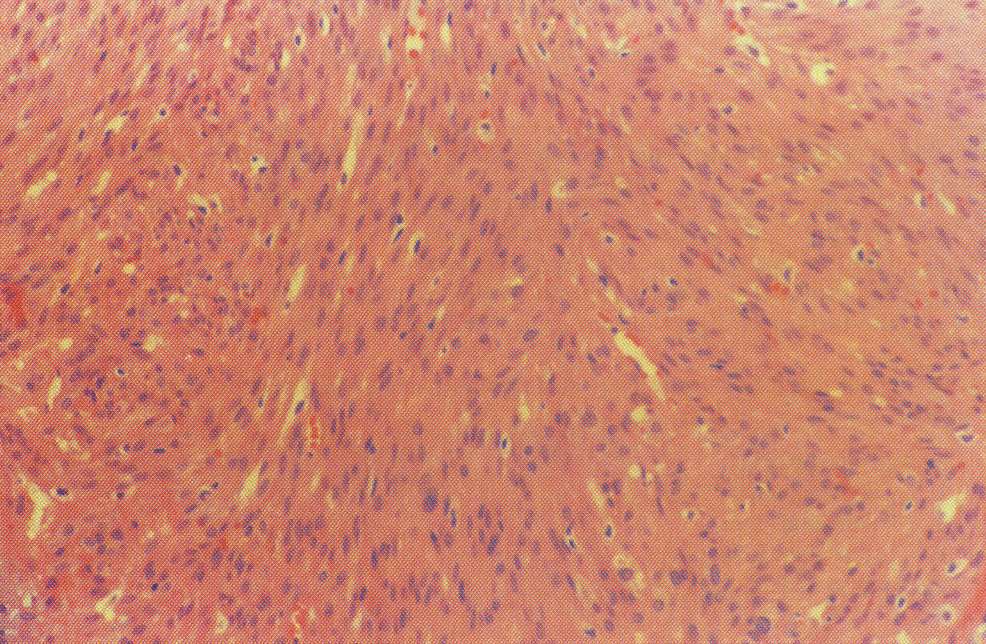
20 Basal Cell Adenoma Trabecular Cells in elongated trabecular pattern Vascular stroma
21 Basal Cell Adenoma Tubular Multiple duct-like structures Columnar cell lining Vascular stroma
22 Basal Cell Adenoma Membranous Thick eosinophilic hyaline membranes surrounding nests of tumor cells jigsaw-puzzle appearance

23 Basal Cell Adenoma

24 Histology Cords of uniform cells and thin fibrous stroma Large polyhedral cells Distinct cell membrane Granular, eosinophilic cytoplasm Central, round, vesicular nucleus Oncocytoma
25 Positive staining for phosphotungstic acid:hematoxylin, cytokeratin, epithelial membrane antigen Negative for S-100 glial fibrillary, smooth muscle actin Oncocytoma

26 Canalicular Adenoma Histology Well-circumscribed Multiple foci Tubular structures line by columnar or cuboidal cells Vascular stroma
27 Myoepithelioma Histology Spindle cell More common Parotid Uniform, central nuclei Eosinophilic granular or fibrillar cytoplasm Plasmacytoid cell Polygonal Eccentric oval nuclei
28 Myoepithelioma
29 Malignant Neoplasms Mucoepidermoid Carcinoma Adenoid Cystic Carcinoma Polymorphous Low-Grade Adenocarcinoma Acinic Cell Carcinoma Adenocarcinoma Malignant Mixed Tumor Epithelial-Myoepithelial Carcinoma Salivary Duct Carcinoma Squamous Cell Carcinoma Undifferentiated Carcinoma

30 Mucoepidermoid Carcinoma Histology Lowgrade Mucus cell > epidermoid cells Prominent cysts Mature cellular elements
31 Mucoepidermoid Carcinoma Histology Intermediate- grade Mucus = epidermoid Fewer and smaller cysts Increasing pleomorphism and mitotic figures
32 Mucoepidermoid Carcinoma Histology Highgrade Epidermoid > mucus Solid tumor cell proliferation Mistaken for SCCA Mucin staining
33 Low Grade Mucoepidermoid Carcinoma

34 High Grade Mucoepidermoid Carcinoma
35 Adenoid Cystic Carcinoma Histology cribriform pattern Most common swiss cheese appearance
36 Adenoid Cystic Carcinoma Histology tubular pattern Layered cells forming duct-like structures Basophilic mucinous substance Histology solid pattern Solid nests of cells without cystic or tubular spaces

37 Adenoid Cystic Carcinoma

38 Polymorphous Low-Grade Histology Adenocarcinoma Isomorphic cells, indistinct borders, uniform nuclei Peripheral Indianfile pattern

39 Polymorphous Low-Grade Markedly positive staining for S-100, epithelial membrane antigen, and cytokeratins. Less predictable with CEA and musclespecific actin Adenocarcinoma
40 Acinic Cell Carcinoma Histology Solid and microcystic patterns Most common Solid sheets Numerous small cysts Polyhedral cells Small, dark, eccentric nuclei Basophilic granular cytoplasm

41 Acinic Cell Carcinoma Positive staining with cytokeratins and CEA, mixed results with others Vacuolated cells with eccentrically located nuclei and granular, basophilic cytoplasm, scant stroma
42 Adenocarcinoma Histology Heterogeneity Presence of glandular structures and absence of epidermoid component Requires exclusion of other specific salivary gland carcinomas

43 Adenocarcinoma
44 Malignant Mixed Tumors Carcinoma ex-pleomorphic adenoma Carcinoma developing in the epithelial component of preexisting pleomorphic adenoma Carcinosarcoma True malignant mixed tumor carcinomatous and sarcomatous components Metastatic mixed tumor Metastatic deposits of otherwise typical pleomorphic adenoma

45 Carcinoma Ex-Pleomorphic Adenoma Histology Malignant cellular change adjacent to typical pleomorphic adenoma Carcinomatous component Adenocarcinoma Undifferentiated

46 Carcinosarcoma Histology Biphasic appearance Sarcomatous component Dominant chondrosarcoma Carinomatous component Moderately to poorly differentiated ductal carcinoma Undifferentiated

47 Malignant Mixed Tumor


48 Epithelial-Myoepithelial Carcinoma Dual epithelial component Irregular, eccentric nuclei w vacuolated cytoplasm IHC reveals dual cell origin epithelial:cytokeratins Myoep:S-100, actin

49 Epithelial-Myoepithelial Carcinoma Tumor cell nests Two cell types Thickened basement membrane

50 Salivary Duct Carcinoma Large polygonal cells w well defined borders Pleomorphic nuclei w prominent nucleoli and granular, eosinophilic cytoplasm IHC patterns similar to breast CA except neg for estrogen CEA, epithelial membrane + S-100, cytokeratins -
51 Squamous Cell Carcinoma Histology Infiltrating Nests of tumor cells Well differentiated Keratinization Moderately-well differentiated Poorly differentiated No keratinization

52 Squamous Cell Carcinoma

53 Undifferentiated Carcinoma High grade, high mitotic activity, scant cytoplasm, hyperchromatic nuclei IHC:cytokeratins, epithelial membrane antigen +/- neuroendocrine
54 References Seifert, Diseases of the Salivary Glands. Thieme Publishers, NY Otolaryngologic clinics of North America. Salivary Gland Disorders. WB Saunders, Phila, PA Oct Ellis, Surgerical Pathology of the Salivary Glands. WB Saunders, Phila PA, Salivary Gland Neoplasms: A Clinicopathologic Approach to Treatment. 3 rd ed. American Academy of Otolaryngology, Head and Neck Surgery Foundation Inc Bailey, Head and Neck Surgery-Otolaryngology. Lippencott, Williams, Wilkins. 3 rd ed Rosen, Salivary Gland Neoplasms. Dr. Quinns online textbook of Otolaryngology Cummings, Otolaryngology Head and Neck Surgery. Elsiever and Mosby
55 Question 1 The highlighted area represents: a. the acini b. the intercalated duct c. the striated duct d. the excretory duct

56 Question 2 The highlighted area represents: a. the acini b. the intercalated duct c. the striated duct d. the excretory duct

57 Question 3 The highlighted area represents: a. the acini b. the intercalated duct c. the striated duct d. the excretory duct
58 Question 4 The highlighted area represents: a. the acini b. the intercalated duct c. the striated duct d. the excretory duct
59 Question 5 The parotid gland neoplasms are: a.) Mostly Benign b.) Mostly Malignant c.) About equal distribution, benign=malignant
60 Question 6 The submandibular gland neoplasms are: a.) Mostly Benign b.) Mostly Malignant c.) About equal distribution, benign=malignant
61 Question 7 The sublingual gland neoplasms are: a.) Mostly Benign b.) Mostly Malignant c.) About equal distribution, benign=malignant

62 Question 8 Identify the neoplasm:

63 Question 9 Identify the neoplasm:
64 Question 10 Identify the neoplasm:
Diagnostic Challenge. Department of Pathology,
 Cytology of Pleural Fluid as a Diagnostic Challenge Paavo Pääkkö,, MD, PhD Chief Physician and Head of the Department Department of Pathology, Oulu University Hospital,, Finland Oulu University Hospital
ATLAS OF HEAD AND NECK PATHOLOGY THYROID PAPILLARY CARCINOMA
 Papillary carcinoma is the most common of thyroid malignancies and occurs in all age groups but particularly in women under 45 years of age. There is a high rate of cervical metastatic disease and yet
SEMESTER VI 3 RD YEAR PATHOLOGY KIDNEY TUMORS
 SEMESTER VI 3 RD YEAR PATHOLOGY KIDNEY TUMORS LEARNING OBJECTIVES At the end of the lecture, students should be able to: Know the pathology of renal tumors. RENAL TUMORS RENAL PAPILLARY ADENOMA Common
R-16: Chronic nonspecific cervisit
 R-16: Chronic nonspecific cervisit Ectoservikal squamous epithelium Endoservical columnar epithelium Dilated cystic endoservical glands lymphoplasmocytes R18:Squamous cell carcinoma insitu Neoplastic epithelium
MALIGNANT MESOTHELIOMA UPDATE ON PATHOLOGY AND IMMUNOHISTOCHEMISTRY
 MALIGNANT MESOTHELIOMA UPDATE ON PATHOLOGY AND IMMUNOHISTOCHEMISTRY Sisko Anttila, MD, PhD Jorvi Hospital Laboratory of Pathology Helsinki University Hospital Espoo, Finland 2nd Nordic Conference on Applied
Something Old, Something New.
 Something Old, Something New. Michelle A. Fajardo, D.O. Loma Linda University Medical Center Clinical Presentation 6 year old boy, presented with hematuria Renal mass demonstrated by ultrasound & CT scan
MALIGNANT MESOTHELIOMA UPDATE ON PATHOLOGY AND IMMUNOHISTOCHEMISTRY
 MALIGNANT MESOTHELIOMA CLASSIFICATION MALIGNANT MESOTHELIOMA UPDATE ON PATHOLOGY AND IMMUNOHISTOCHEMISTRY Sisko Anttila, MD, PhD Jorvi Hospital Laboratory of Pathology Helsinki University Hospital Espoo,
Histologic Subtypes of Renal Cell Carcinoma
 Histologic Subtypes of Renal Cell Carcinoma M. Scott Lucia, MD Associate Professor Chief of Genitourinary and Renal Pathology Director, Prostate Diagnostic Laboratory Dept. of Pathology University of Colorado
Histopathology and prognosis in renal cancer
 Histopathology and prognosis in renal cancer Granular cell Granular cell Granular cell Granular cell Clear cell Chromophobe cell Papillary type 2 Luca Mazzucchelli Istituto cantonale di patologia, Locarno
TUMORS OF THE TESTICULAR ADNEXA and SPERMATIC CORD
 TUMORS OF THE TESTICULAR ADNEXA and SPERMATIC CORD Victor E. Reuter, MD Memorial Sloan-Kettering Cancer Center [email protected] 66 th Annual Pathology Seminar California Society of Pathologists Short
Basic Professional Training Program for Associate Medical Technologist
 Basic Professional Training Program for Associate Medical Technologist Basic Cytology Part 2 (Preparartion and normal morphology) Normal Morphology in Liquid based Gynecologic Cytology Speaker: Mr. Fung
Immunohistochemical differentiation of metastatic tumours
 Immunohistochemical differentiation of metastatic tumours Dr Abi Wheal ST1. TERA 3/2/14 Key points from a review article written by Daisuke Nonaka Intro Metastatic disease is the initial presentation in
PRIMARY SEROUS CARCINOMA OF PERITONEUM: A CASE REPORT
 PRIMARY SEROUS CARCINOMA OF PERITONEUM: A CASE REPORT Dott. Francesco Pontieri (*) U.O. di Anatomia Patologica P.O. di Rossano (CS) Dott. Gian Franco Zannoni Anatomia Patologica Facoltà di Medicina e Chirurgia
Introduction: Tumor Swelling / new growth / mass. Two types of growth disorders: Non-Neoplastic. Secondary / adaptation due to other cause.
 Disorders of Growth Introduction: Tumor Swelling / new growth / mass Two types of growth disorders: Non-Neoplastic Secondary / adaptation due to other cause. Neoplastic. Primary growth abnormality. Non-Neoplastic
Thymic carcinoma: update of current diagnostic criteria and histologic types
 Seminars in Diagnostic Pathology 22, 198-212 Thymic carcinoma: update of current diagnostic criteria and histologic types Saul Suster, MD From the Department of Pathology, The Ohio State University and
Cytopathology Case Presentation #8
 Cytopathology Case Presentation #8 Emily E. Volk, MD William Beaumont Hospital, Troy, MI Jonathan H. Hughes, MD Laboratory Medicine Consultants, Las Vegas, Nevada Clinical History 44 year old woman presents
DESMOPLASTIC SMALL ROUND CELL TUMOR: A RARE PATHOLOGY PUZZLE
 DESMOPLASTIC SMALL ROUND CELL TUMOR: A RARE PATHOLOGY PUZZLE Ryan Granger University of Rhode Island Cytotechnology program May 2, 2015 ASCT Annual Meeting Nashville, Tennessee DESMOPLASTIC SMALL ROUND
The menstrual cycle Hypophysis
 Normal endometrium Tumors of the uterine corpus by MB The endometrium comprises the zona functionalis zf (superficial two thirds) ) and the zona basalis zb (deep one third) The zf responds to hormonal
Immunohistochemistry of soft tissue tumors
 Immunohistochemistry of soft tissue tumors Immunohistochemistry Major advances : antigen retrieval techniques (HIER) sensitive detection systems numerous antibodies of good quality Standardization : automated
Index. F Factor VIII-related antigen, see VWF FactorXIIIa, for dermatofibroma, 272-275 5-HT, see Serotonin
 A Acantholytic squamous cell carcinoma vs epithelioid angiosarcoma, 56-57 Acinic cell carcinoma of pancreas, 76-77 vs ductal adenocarcinoma, 74-75 vs islet cell tumor, 78-81 Adenomatoid tumor vs hemangioma,
Immunohistochemistry on cytology specimens from pleural and peritoneal fluid
 Immunohistochemistry on cytology specimens from pleural and peritoneal fluid Dr Naveena Singh Consultant Pathologist Bart health NHS Trust London United Kingdom Disclosures and Acknowledgements I have
HKCPath Anatomical Pathology Peer Review and Scores : PDF version for download
 AP2003R1 http://hkcpath.org. Correspondence: [email protected] 1of 10 07/08/2003 HKCPath Anatomical Pathology Peer Review and Scores : PDF version for download AP141 Bone Marrow: Metastatic Carcinoma from
Gladwyn Leiman, MCCCh, FIAC, FRCPath Scott Anderson, MD
 Cytology Works shop #5 Gladwyn Leiman, MCCCh, FIAC, FRCPath Scott Anderson, MD Disclosur re information The speakers have no relationship that represents a possible conflict of interest with respect to
PATHOLOGY OF THE PLEURA: Mesothelioma and mimickers Necessity of Immunohistochemistry. M. Praet
 PATHOLOGY OF THE PLEURA: Mesothelioma and mimickers Necessity of Immunohistochemistry M. Praet Pathology of the Pleura Normal serosa: visceral and parietal layers Inflammation Neoplasia: Primary: mesothelioma
Pathology of lung cancer
 Pathology of lung cancer EASO COURSE ON LUNG CANCER AND MESOTHELIOMA DAMASCUS (SYRIA), MAY 3-4, 2007 Gérard ABADJIAN MD Pathologist Associate Professor, Saint Joseph University Pathology Dept. Hôtel-Dieu
Renal Cell Carcinoma: Advances in Diagnosis B. Iványi, MD
 Renal Cell Carcinoma: Advances in Diagnosis B. Iványi, MD Department of Pathology University of Szeged, Hungary ISUP Vancouver Classification of Renal Neoplasia Am J Surg Pathol 37:14691489, 2013 13 histologic
Cytology of Lymph Nodes
 Indications Cytology of Lymph Nodes Lymph node enlargement That was easy Mary Anna Thrall Don Meuten Indications Lymph node enlargement Suspect metastasis Normal sized lymph nodes are Normal Do NOT aspirate
Update on Mesothelioma
 November 8, 2012 Update on Mesothelioma Intro incidence and nomenclature Update on Classification Diagnostic specimens Morphologic features Epithelioid Histology Biphasic Histology Immunohistochemical
Animal Tissues. I. Epithelial Tissue
 Animal Tissues There are four types of tissues found in animals: epithelial tissue, connective tissue, muscle tissue, and nervous tissue. In this lab you will learn the major characteristics of each tissue
A. Pericardial smear. Examination of the pericardial aspirate can provide useful diagnostic information.
 5. PERICARDIUM Heart is encased by the pericardium which has a visceral layer (a) covering the heart and the parietal layer (b). In normal states it is thin, transparent and the myocardium can be seen
MODERN IMMUNOHISTOCHEMISTRY
 MODERN IMMUNOHISTOCHEMISTRY Cambridge Illustrated Surgical Pathology Peiguo G. Chu City of Hope National Medical Center, Duarte, California Lawrence M. Weiss City of Hope National Medical Center, Duarte,
Renal Tumors with Eosinophilic Cytoplasm: 2013 Classification. Jesse K. McKenney, MD Associate Head, Surgical Pathology
 Renal Tumors with Eosinophilic Cytoplasm: 2013 Classification Jesse K. McKenney, MD Associate Head, Surgical Pathology Renal Epithelial Neoplasia History 1981: WHO Classification of Renal Neoplasms 1.
Pediatric Oncology for Otolaryngologists
 Pediatric Oncology for Otolaryngologists Frederick S. Huang, M.D. Division of Hematology/Oncology Department of Pediatrics The University of Texas Medical Branch Grand Rounds Presentation to Department
The Tissue Level of Organization
 The Tissue Level of Organization Tissues A groups of similar cells, usually having similar embryonic origin and specialized function Histology: the study of tissues Four general types Epithelial Muscle
Guidelines for reporting histopathology of cervical carcinoma
 Guidelines for reporting histopathology of cervical carcinoma Naveena Singh, Consultant Pathologist Introduction Cancer management is multidisciplinary Histopathology report has a MAJOR impact on management
Disclosures. Learning Objectives. Effusion = Confusion. Diagnosis Of Serous Cavity Effusions - Beware The Mesothelial Cell!
 Disclosures Diagnosis Of Serous Cavity Effusions - Beware The Mesothelial Cell! No Relevant Financial Relationships with Commercial Interests Syed Z. Ali, M.D. Syed Z. Ali, M.D. Associate Professor of
Male. Female. Death rates from lung cancer in USA
 Male Female Death rates from lung cancer in USA Smoking represents an interesting combination of an entrenched industry and a clearly drug-induced cancer Tobacco Use in the US, 1900-2000 5000 100 Per Capita
THE PATHOLOGY OF MEDIASTINAL MASSES. Anatomic Distribution of Mediastinal Masses
 Page 1 of 5 THE PATHOLOGY OF MEDIASTINAL MASSES Dr. S. Boag 549-6666 Ext. 4190, mailto:%[email protected] 1. INTRODUCTION A wide range of process, neoplastic and non-neoplastic, can produce
Effusions: Mesothelioma and Metastatic Cancers
 Effusions: Mesothelioma and Metastatic Cancers Malignant Mesothelioma Incidence: 2,500 cases/year ~60-80% pts with pleural MM relationship with asbestos exposure Other risk factors: radiation, other carcinogens,
Académie internationale de Pathologie - Division arabe XX ème congrès 24-26 novembre 2008 Alger. Immunohistochemistry in malignant mesotheliomas
 Académie internationale de Pathologie - Division arabe XX ème congrès 24-26 novembre 2008 Alger Immunohistochemistry in malignant mesotheliomas Françoise Thivolet-Béjui Groupement Hospitalier Est Lyon-Bron
Emerging Subtypes in Renal Cancer. Donna E. Hansel, MD PhD Professor of Pathology, UC San Diego Division Chief, Anatomic Pathology dhansel@ucsd.
 Emerging Subtypes in Renal Cancer Donna E. Hansel, MD PhD Professor of Pathology, UC San Diego Division Chief, Anatomic Pathology [email protected] Some General Comments Fuhrman nuclear grading clear cell
Tubulocystic Carcinoma of the Kidney, a Rare Distinct Entity
 Tubulocystic Carcinoma of the Kidney, a Rare Distinct Entity 2 Shreenath Bishu, Laurie J. Eisengart and Ximing J. Yang * Department of Pathology, Northwestern University, Feinberg School of Medicine, Feinberg,
Cytology : first alert of mesothelioma? Professor B. Weynand, UCL Yvoir, Belgium
 Cytology : first alert of mesothelioma? Professor B. Weynand, UCL Yvoir, Belgium Introduction 3 cavities with the same embryologic origin the mesoderme Pleura Exudates Pleura Peritoneum Pericardium 22%
Carcinosarcoma of the Ovary
 Carcinosarcoma of the Ovary A Rare Finding Presented By: Kathryn Kiely Anisa I. Kanbour School of Cytotechnology of the University of Pittsburgh Medical Center Pittsburgh, PA Patient History 55 year old
How To Test For Cancer
 Diagnosis Of Serous Cavity Effusions - Beware The Mesothelial Cell! Effusion = Confusion Syed Z. Ali, M.D. Professor of Pathology and Radiology The Johns Hopkins Hospital Baltimore, Maryland Diagnostic
Outline. Workup for metastatic breast cancer. Metastatic breast cancer
 Metastatic breast cancer Immunostain Update: Diagnosis of metastatic breast carcinoma, emphasizing distinction from GYN primary 1/3 of breast cancer patients will show metastasis 1 st presentation or 20-30
Patologia neoplastica borderline della mammella"
 Patologia neoplastica borderline della mammella" Anna Sapino Department of Medical Sciences University of Torino (Italy) B3 Lesion of uncertain malignant potential This category mainly consists of lesions
Renal Pathology Update. Sundus Hussein MD, FRCPC
 Renal Pathology Update Sundus Hussein MD, FRCPC Case History A 45 year old male with incidentally discovered a 3.5 x 3.9 x 2.7 cm renal mass Handling partial nephrectomy Handling partial nephrectomy
The Mainz Classification of Renal Cell Tumors
 The Mainz classification is effective in distinguishing the histopathologic and cytogenetic features of various types of renal cell carcinoma. Virginia S. King. Costa del Sol. Watercolor, 18 24. The Mainz
Epithelial Tumors of the Kidney Diagnostic Problems and Recently Described Entities
 Pathology of Renal Neoplasia Epithelial Tumors of the Kidney Diagnostic Problems and Recently Described Entities Wael A Sakr, MD Wayne State University School of Medicine CURRENT CLASSIFICATION = EPITHELIAL
Lip Cancer: Treatment & Reconstruction
 Lip Cancer: Treatment & Reconstruction GBMC - Head & Neck Cancer Grand Rounds Elizabeth E. Redd, M.D. With the assistance of Ira Papel, M.D. Patrick Byrne, M.D. Lip Cancer: Treatment & Reconstruction Anatomic
FNA Cytology of Mediastinal Lesions. Presenters: Xiaoqi Lin, M.D., Ph.D. Ritu Nayar, M.D.
 Disclosure information The speakers have no relationship that represents a possible conflict of interest with respect to the content of this presentation. FNA Cytology of Mediastinal Lesions Presenters:
STUDY ON MORPHOLOGICAL FEATURES OF NODULAR HIDRADENOMA B. Pushpa 1, K. Duraisamy 2, Gayathri 3
 STUDY ON MORPHOLOGICAL FEATURES OF NODULAR HIDRADENOMA B. Pushpa 1, K. Duraisamy 2, Gayathri 3 HOW TO CITE THIS ARTICLE: B. Pushpa, K. Duraisamy, Gayathri. Study on Morphological Features of Nodular Hidradenoma.
MUSCLE TISSUE. Larry Johnson Texas A&M University
 MUSCLE TISSUE Larry Johnson Texas A&M University Objectives Histologically identify and functionally characterize each of the 3 types of muscle tissues. Describe the organization of the sarcomere as seen
Malignant Peritoneal Mesothelioma in Women A Study of 75 Cases With Emphasis on Their Morphologic Spectrum and Differential Diagnosis
 Anatomic Pathology / MALIGNANT PERITONEAL MESOTHELIOMA IN WOMEN Malignant Peritoneal Mesothelioma in Women A Study of 75 Cases With Emphasis on Their Morphologic Spectrum and Differential Diagnosis Patricia
Tissues (Histology) Ch. 3 Human Anatomy lecture
 Ch. 3 Human Anatomy lecture") I. Histology the study of tissues A. 4 basic tissue types epithelial connective muscle nervous Tissues (Histology) Ch. 3 Human Anatomy lecture B. Usually found in combinations to form organs. C. As you
I. Histology the study of tissues A. 4 basic tissue types epithelial connective muscle nervous Tissues (Histology) Ch. 3 Human Anatomy lecture B. Usually found in combinations to form organs. C. As you
Lung Cancer. Ossama Tawfik, MD, PhD Professor, Vice Chairman Director of Anatomic &Surgical Pathology University of Kansas School of Medicine
 Lung Cancer Ossama Tawfik, MD, PhD Professor, Vice Chairman Director of Anatomic &Surgical Pathology University of Kansas School of Medicine Alexandria, Egypt July 1-1 3, 2008 OBJECTIVES Describe and
YOUR LUNG CANCER PATHOLOGY REPORT
 UNDERSTANDING YOUR LUNG CANCER PATHOLOGY REPORT 1-800-298-2436 LungCancerAlliance.org A GUIDE FOR THE PATIENT 1 CONTENTS What is a Pathology Report?...3 The Basics...4 Sections of a Pathology Report...7
Contents. 1. Introduction and Approach to Fine Needle Aspiration Cytology... 1. 2. Head, Neck, Orbit and Salivary Glands... 12
 Contents 1. Introduction and Approach to Fine Needle Aspiration Cytology... 1 Complications 1 Fine Needle Aspiration Technique 1 Evaluation of FNAC Smear 4 Cell Morphology 4 Nucleus 4 Cytoplasm 6 Background
Case presentation: Mesothelioma of the tunica vaginalis. Dr Ben Shepherd Pathology Queensland Princess Alexandra Hospital Brisbane
 Case presentation: Mesothelioma of the tunica vaginalis Dr Ben Shepherd Pathology Queensland Princess Alexandra Hospital Brisbane A 76 year old man presented June 2011 with a 6 month history of painless
Information Model Requirements of Post-Coordinated SNOMED CT Expressions for Structured Pathology Reports
 Information Model Requirements of Post-Coordinated SNOMED CT Expressions for Structured Pathology Reports W. Scott Campbell, Ph.D., MBA James R. Campbell, MD Acknowledgements Steven H. Hinrichs, MD Chairman
Primary -Benign - Malignant Secondary
 TUMOURS OF THE LUNG Primary -Benign - Malignant Secondary The incidence of lung cancer has been increasing almost logarithmically and is now reaching epidemic levels. The overall cure rate is very low
The develpemental origin of mesothelium
 Mesothelioma Tallinn 14.12.06 Henrik Wolff Finnish Institute of Occupational Health The develpemental origin of mesothelium Mesodermal cavities (pleura, peritoneum and pericardium ) are lined with mesenchymal
CASE OF THE MONTH AUGUST-2015 DR. GURUDUTT GUPTA HEAD HISTOPATHOLOGY
 CASE OF THE MONTH AUGUST-2015 DR. GURUDUTT GUPTA HEAD HISTOPATHOLOGY CASE HISTORY 52Y MALE RIGHT RADICAL NEPHERECTOMY Case of right renal mass with IVC thrombus. History of surgery and RT for right occipital
Intraobserver and Interobserver Reproducibility of WHO and Gleason Histologic Grading Systems in Prostatic Adenocarcinomas
 International Urology and Nephrology 28 (1), pp. 73-77 (1996) Intraobserver and Interobserver Reproducibility of WHO and Gleason Histologic Grading Systems in Prostatic Adenocarcinomas $. O. OZDAMAR,*
Changes in Breast Cancer Reports After Second Opinion. Dr. Vicente Marco Department of Pathology Hospital Quiron Barcelona. Spain
 Changes in Breast Cancer Reports After Second Opinion Dr. Vicente Marco Department of Pathology Hospital Quiron Barcelona. Spain Second Opinion in Breast Pathology Usually requested when a patient is referred
METASTATIC CLEAR CELL RENAL CELL CARCINOMA TO THE SUBCUTANEOUS AREA IN ILLIAC FOSSA AND ADRENAL GLAND WITHOUT AN IDENTIFIABLE PRIMARY TUMOR
 Indian J.Sci.Res. 5(1) : 121-125, 2014 METASTATIC CLEAR CELL RENAL CELL CARCINOMA TO THE SUBCUTANEOUS AREA IN ILLIAC FOSSA AND ADRENAL GLAND WITHOUT AN IDENTIFIABLE PRIMARY TUMOR a1 b c d REETA DHAR, SHILPI
Practical Effusion Cytology
 Practical Effusion Cytology A Community Pathologist s Approach to Immunocytochemistry in Body Fluid Cytology Emily E. Volk, MD William Beaumont Hospital Troy, MI College of American Pathologists 2004.
Biology 105 Human Biology PRACTICE MIDTERM EXAM 1. Essentials of Anatomy and Physiology, 5e (Martini/Nath) Chapter 4 The Tissue Level of Organization
 Chapter 4 The Tissue Level of Organization") Essentials of Anatomy and Physiology, 5e (Martini/Nath) Chapter 4 The Tissue Level of Organization Multiple-Choice Questions 1) The four main types of tissues include A) epithelium. B) muscle. C) nerve.
Essentials of Anatomy and Physiology, 5e (Martini/Nath) Chapter 4 The Tissue Level of Organization Multiple-Choice Questions 1) The four main types of tissues include A) epithelium. B) muscle. C) nerve.
MRI of the Uterus BENIGN. Jeffrey C. Weinreb, M.D. FACR [email protected] Yale University School of Medicine
 MRI of the Uterus BENIGN Jeffrey C. Weinreb, M.D. FACR [email protected] Yale University School of Medicine Normal Anatomy M Junctional JZ Zone EE Junctional Zone is the inner layer or the myometrium
Salivary Gland Cancer
 What is cancer? Salivary Gland Cancer The body is made up of trillions of living cells. Normal body cells grow, divide to make new cells, and die in an orderly way. During the early years of a person s
Today s Topics. Tumors of the Peritoneum in Women
 Today s Topics Tumors of the Peritoneum in Women Charles Zaloudek, M.D. Department of Pathology 505 Parnassus Ave., M563 University of California, San Francisco San Francisco, CA USA [email protected]
The evolving pathology of solitary fibrous tumours. Luciane Dreher Irion MREH / CMFT / NSOPS
 The evolving pathology of solitary fibrous tumours Luciane Dreher Irion MREH / CMFT / NSOPS Historical review Haemangiopericytoma (HPC) first described primarily as a soft tissue vascular tumour of pericytic
The Male Breast: Masses, Malignancies and More
 The Male Breast: Masses, Malignancies and More Monique Marie Tyminski, DO, R Hultman, DO, J Watkins, MD, T Stockl, MD, E T Ghosh, MD, S A MacMaster, MD Teaching Points: Understand male breast anatomy and
Ovarian tumors Ancillary methods
 Ovarian tumors Ancillary methods Ovarian tumor course Oslo, 24-25/11/14 Prof. Ben Davidson, MD PhD Department of Pathology, Norwegian Radium Hospital, Oslo University Hospital, Oslo, Norway Division of
The WHO Classification of Renal Tumors and Common Issues in TNM Staging for Renal Cell Carcinoma
 The WHO Classification of Renal Tumors and Common Issues in TNM Staging for Renal Cell Carcinoma Steven Shen, M.D.,Ph.D. Staff Pathologist and Assistant Member The Methodsit Hospital and Research Institute
Multiple Primary and Histology Coding Rules
 Multiple Primary and Histology Coding Rules January 10, 2008 National Cancer Institute Surveillance Epidemiology and End Results Program Bethesda, MD PLEASE NOTE This PDF of the 2007 Multiple Primaries
Unit 1 Higher Human Biology Summary Notes
 Unit 1 Higher Human Biology Summary Notes a. Cells tissues organs body systems Division of labour occurs in multicellular organisms (rather than each cell carrying out every function) Most cells become
Histopathology of Colorectal Cancer after Neoadjuvant Chemoradiation Therapy
 The Open Pathology Journal, 2009, 3, 91-98 91 Open Access Histopathology of Colorectal Cancer after Neoadjuvant Chemoradiation Therapy Maura O Neil * and Ivan Damjanov Department of Pathology and Laboratory
New Concepts and Refinements to. Existing WHO (2004) Renal Cell. Tumor Categories In Adults
 Renal Cell. Tumor Categories In Adults") New Concepts and Refinements to Existing WHO (2004) Renal Cell Tumor Categories In Adults Dr Varsha Manucha Assistant Professor Department of Pathology and Laboratory Medicine Temple University Hospital
New Concepts and Refinements to Existing WHO (2004) Renal Cell Tumor Categories In Adults Dr Varsha Manucha Assistant Professor Department of Pathology and Laboratory Medicine Temple University Hospital
EIN. (Endometrial Intraepithelial Neoplasia): Improved Criteria for diagnosing endometrial precancer. Stanley J. Robboy, MD, FCAP,
: Improved Criteria for diagnosing endometrial precancer. Stanley J. Robboy, MD, FCAP,") EIN (Endometrial Intraepithelial Neoplasia): Improved Criteria for diagnosing endometrial precancer Stanley J. Robboy, MD, FCAP, FFPath FRCPI (Hon), FRCPath (UK, Hon) Professor of Pathology, Duke University
EIN (Endometrial Intraepithelial Neoplasia): Improved Criteria for diagnosing endometrial precancer Stanley J. Robboy, MD, FCAP, FFPath FRCPI (Hon), FRCPath (UK, Hon) Professor of Pathology, Duke University
Section B: Epithelial Tissue 1. Where are epithelial tissues found within the body? 2. What are the functions of the epithelial tissues?
 Tissue worksheet Name Section A: Intro to Histology Cells are the smallest units of life. In complex organisms, cells group together with one another based on similar structure and function to form tissues.
Polyps. Hyperplasias. CAP 2011: Course AP104. The High Risk Benign Endometrium. Mutter and Nucci 1
 Course AP104 Endometrial Hyperplasia A morphologic Definition Hyperplasias Hormonal Effect or Precancer? George L. Mutter, MD Harvard Medical School and Brigham and Women s Hospital Boston, MA Endometrial
RENAL CANCER PATHOLOGY WHAT REALLY MATTERS? STEWART FLEMING UNIVERSITY OF DUNDEE
 RENAL CANCER PATHOLOGY WHAT REALLY MATTERS? STEWART FLEMING UNIVERSITY OF DUNDEE MAJOR PARADIGM SHIFT IN EARLY 1990S IN UNDERSTANDING RENAL CANCER Molecular differential pathology of renal cell tumours
Renal Cell Carcinoma Associated with Xp11.2 Translocation: Clinicopathologic and Immunohistochemical Findings of 4 Cases
 The Korean Journal of Pathology 2005; 39: 406-11 Renal Cell Carcinoma Associated with Xp11.2 Translocation: Clinicopathologic and Immunohistochemical Findings of 4 Cases Sanghui Park Ji-Eun Kwon Yeon-Lim
LYMPHOMA. BACHIR ALOBEID, M.D. HEMATOPATHOLOGY DIVISION PATHOLOGY DEPARTMENT Columbia University/ College of Physicians & Surgeons
 LYMPHOMA BACHIR ALOBEID, M.D. HEMATOPATHOLOGY DIVISION PATHOLOGY DEPARTMENT Columbia University/ College of Physicians & Surgeons Normal development of lymphocytes Lymphocyte proliferation and differentiation:
Lung Carcinomas New 2015 WHO Classification. Spasenija Savic Pathology
 Lung Carcinomas New 2015 WHO Classification Spasenija Savic Pathology ***EXPECTED SPRING 2015*** This authoritative, concise reference book provides an international standard for oncologists and pathologists
NATIONAL INSTITUTES OF HEALTH National Cancer Institute. Neoplasms
 NATIONAL INSTITUTES OF HEALTH National Cancer Institute to Neoplasms CONVERSION of NEOPLASMS BY TOPOGRAPHY AND MORPHOLOGY from the INTERNATIONAL CLASSIFICATION OF DISEASES FOR ONCOLOGY, SECOND EDITION
Efficient Tumor Immunohistochemistry A Differential Diagnosis-Driven Approach
 Efficient Tumor Immunohistochemistry A Differential Diagnosis-Driven Approach Publishing Team Erik Tanck (production manager/designer) Joshua Weikersheimer (publisher) Copyright 2006 by the American Society
Effusion cytology. Dr Alpha Tsui Royal Melbourne Hospital 2008
 Effusion cytology Dr Alpha Tsui Royal Melbourne Hospital 2008 General points: -large unilateral effusion (>1 litre) in the elderly is highly suspicious for malignancy -effusions associated with malignancies
Ovarian mucinous lesions. Ovarian mucinous lesions: Common diagnostic dilemmas. Ovarian mucinous lesions: problematic issues
 Ovarian mucinous lesions Ovarian mucinous lesions: Common diagnostic dilemmas Karuna Garg, MD University of California San Francisco Intestinal or usual type Seromucinous (Endocervical mucinous or Mullerian
Notice of Faculty Disclosure
 The Diagnosis of Malignant Mesothelioma Andrew Churg, MD Department of Pathology University of British Columbia Vancouver, BC, Canada [email protected] Notice of Faculty Disclosure In accordance with