Diagnostic Challenge. Department of Pathology,
|
|
|
- Lydia Newton
- 10 years ago
- Views:
Transcription
1 Cytology of Pleural Fluid as a Diagnostic Challenge Paavo Pääkkö,, MD, PhD Chief Physician and Head of the Department Department of Pathology, Oulu University Hospital,, Finland

2 Oulu University Hospital

3 General overview of cytology of pleural effusions! Exfoliative cytological examination of pleural effusions common method for determining whether effusion is benign or malignant! Effusions subdivided into transudates and exudates depending on the protein content! Transudates result from alterations in hydrostatic or oncotic pressure, often due to systemic factors, e.g. congestive heart failure! Exudates result from pathological processes localized to the serosal membranes => higher protein content and cellularity increased compared to transudates, e.g. infections

4 Utility and limitations of cytology of pleural effusions! Cells exfoliated into effusion fluid can be examined as cytology smears, liquid-based preparations, cytospin preparations, or cell blocks! Most of the exudates benign! The absence of malignant cells does not rule out malignancy! Only % of malignant mesotheliomas,, and % of cancers metastatic to the pleura diagnosed by exfoliative cytology

5 Cytology vs. histology! Exfoliative cytology has of limited usefulness in diagnosing malignant mesothelioma! Benign reactive mesothelial cells may have features that mimic malignancy,, and malignant mesotheliomas may be cytologically bland! Without evidence of invasion of underlying tissues, diagnosis of mesothelioma on cytologic grounds maybe difficult! Sarcomatous mesotheliomas typically do not shed cells into effusion

6 Cytological features of malignancy! Cancer cells as individual cells, sheets of cohesive cells, and three-dimensional spherical clusters, called morulae! Papillary or acinar structures! Usually the number of malignant cells high, rarely a few! Presence of psammoma bodies! Enlarged cells with enlarged nuclei, coarse chromatin, prominent nucleoli! Mitoses, atypical mitoses,, and necrotic debris

7 Reactive atypia of mesothelial cells! Benign mesothelial cells exfoliate easily and display a spectrum of reactive changes from minimal reactive change to highly atypical reactive change, mimicking malignancy! Reactive mesothelial cells shed as invidual cells, in clusters or sheets, with adjacent cells separated from one another by spaces referred to as windows

8 Cytological features of reactive mesothelial cells! Nuclei round or oval with distict nuclear membranes, chromatin vesicular or finely granular,, and cytoplasm adundant and darkly-stained! Peripheral cytoplasm stains darker than central cytoplasm,, and microvilli around the periphery result in fuzzy rim or border! Binucleation or multinucleation frequent! Cytoplasmic vacuoles may compress the nucleus, suggesting the signet ring cells of adenocarcinoma

9 Reactive mesothelial cells

10 Cytological features of malignant mesothelioma! Malignant mesotheliomas cause <1 % of malignant pleural effusions,, and only epithelial malignant mesotheliomas likely exfoliate cells into effusion fluids! Malignant mesothelioma cells lack the significant degree of cytological pleomorphism! Cells arranged in sheets, clusters, morulae or papillary structures

11 Hints for correct interpretation! Atypical reactive mesothelial cells blend with cells with lesser degrees of reactive atypia within a benign effusion, giving the impression of one population of cells! In cases of malignancy, reactive mesothelial cells and malignant cells often appear as two separate and distinct population of cells! It is necessary to first determine if cells in a fluid are truly malignant before secondarily determining the type of malignancy! Immunohistochemical confirmation that atypical cells are mesothelial in origin does not help to distinguish between reactive mesothelial cell proliferation and mesothelioma

12 Case 1! 60-year old man! Exposed to asbestos! Hydrothorax and pleural plaques! Cytological specimen taken from pleural cavity

13

14

15 Calretinin CK 5/6 CK7 EMA

16 Conclusion from Case 1! Strong suspicion for malignancy, fitting rather for malignant mesothelioma than metastatic adenocarcinoma! The diagnosis of malignant mesothelioma was later confirmed by histological sample

17 Calretinin WT1 EMA

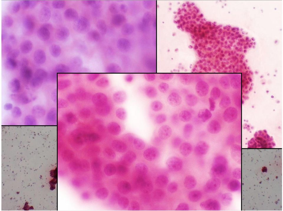
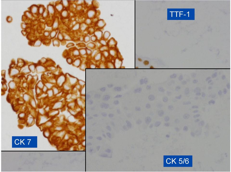
18 Case 2! 76-year old male! Suspicion for lung malignancy! Hydrothorax! An aspiration from left pleural cavity performed, and cytological analyses requested

19
20 TTF-1 CK 7 CK 5/6
21 EMA E-Cadherin Calretinin
22 Conclusion from Case 2! Strong suspicion for a metastatic carcinoma! TTF-1 positivity suggests for pulmonary origin of the carcinoma
23 Case 3! 76-year old female! Breast carcinoma operated 6 years ago! Fluid in the pleural cavity and both lungs contain tumour infiltrations! Pleural cytology requested
24
25 PAN-CK CK 5/6 Estrogen receptor
26 Connclusion from Case 3! Strong suspicion for a metastatic carcinoma! Estrogen receptor-positivity suggests for a metastatic breast carcinoma
27 Case 4! 76-year old male! Laryngeal squamous cell carcinoma operated 1993! COPD! Heavy smoker! Abundant fluid in the left pleural cavity
28
29
30 CK 7 CK 7 TTF-1 Calretinin CK 5/6
31 Conclusion from Case 4! Mild suspicion for a malignancy, origin of which possibly in the lungs! In addition,, the cell population contained atypical mesothelial cell proliferation
32 Summary and conclusions! Most of the exudates benign! The absence of malignant cells does not rule out malignancy! Malignant mesotheliomas cause <1 % of malignant pleural effusions,, and only epithelial malignant mesotheliomas likely exfoliate cells into effusion fluids! It is necessary to first determine if cells in a fluid are truly malignant before secondarily determining the type of malignancy! Immunohistochemistry of histological sections from a cell block may help to determine the type of malignancy
33
Cytology : first alert of mesothelioma? Professor B. Weynand, UCL Yvoir, Belgium
 Cytology : first alert of mesothelioma? Professor B. Weynand, UCL Yvoir, Belgium Introduction 3 cavities with the same embryologic origin the mesoderme Pleura Exudates Pleura Peritoneum Pericardium 22%
Immunohistochemistry on cytology specimens from pleural and peritoneal fluid
 Immunohistochemistry on cytology specimens from pleural and peritoneal fluid Dr Naveena Singh Consultant Pathologist Bart health NHS Trust London United Kingdom Disclosures and Acknowledgements I have
How To Diagnose And Treat A Tumour In An Effusion
 Effusions of the Serous Cavities Annika Dejmek Professor/Consultant in Cytopathology Clinical Pathology; Department of Laboratory Medicine, Malmö, Lund University 5th EFCS Tutorial Trondheim 2012 Pleura
Cytopathology Case Presentation #8
 Cytopathology Case Presentation #8 Emily E. Volk, MD William Beaumont Hospital, Troy, MI Jonathan H. Hughes, MD Laboratory Medicine Consultants, Las Vegas, Nevada Clinical History 44 year old woman presents
Effusions: Mesothelioma and Metastatic Cancers
 Effusions: Mesothelioma and Metastatic Cancers Malignant Mesothelioma Incidence: 2,500 cases/year ~60-80% pts with pleural MM relationship with asbestos exposure Other risk factors: radiation, other carcinogens,
MALIGNANT MESOTHELIOMA UPDATE ON PATHOLOGY AND IMMUNOHISTOCHEMISTRY
 MALIGNANT MESOTHELIOMA UPDATE ON PATHOLOGY AND IMMUNOHISTOCHEMISTRY Sisko Anttila, MD, PhD Jorvi Hospital Laboratory of Pathology Helsinki University Hospital Espoo, Finland 2nd Nordic Conference on Applied
MALIGNANT MESOTHELIOMA UPDATE ON PATHOLOGY AND IMMUNOHISTOCHEMISTRY
 MALIGNANT MESOTHELIOMA CLASSIFICATION MALIGNANT MESOTHELIOMA UPDATE ON PATHOLOGY AND IMMUNOHISTOCHEMISTRY Sisko Anttila, MD, PhD Jorvi Hospital Laboratory of Pathology Helsinki University Hospital Espoo,
PATHOLOGY OF THE PLEURA: Mesothelioma and mimickers Necessity of Immunohistochemistry. M. Praet
 PATHOLOGY OF THE PLEURA: Mesothelioma and mimickers Necessity of Immunohistochemistry M. Praet Pathology of the Pleura Normal serosa: visceral and parietal layers Inflammation Neoplasia: Primary: mesothelioma
Outline. Workup for metastatic breast cancer. Metastatic breast cancer
 Metastatic breast cancer Immunostain Update: Diagnosis of metastatic breast carcinoma, emphasizing distinction from GYN primary 1/3 of breast cancer patients will show metastasis 1 st presentation or 20-30
Practical Effusion Cytology
 Practical Effusion Cytology A Community Pathologist s Approach to Immunocytochemistry in Body Fluid Cytology Emily E. Volk, MD William Beaumont Hospital Troy, MI College of American Pathologists 2004.
The develpemental origin of mesothelium
 Mesothelioma Tallinn 14.12.06 Henrik Wolff Finnish Institute of Occupational Health The develpemental origin of mesothelium Mesodermal cavities (pleura, peritoneum and pericardium ) are lined with mesenchymal
Case of the. Month October, 2012
 Case of the Month October, 2012 Case The patient is a 47-year-old male with a 3-week history of abdominal pain. A CT scan of the abdomen revealed a suggestion of wall thickening at the tip of the appendix
Video Microscopy Tutorial 5
 Video Microscopy Tutorial 5 Lool Alikes in Effusion Cytology:Review of Diagnostic Challenges Claire Michael, MD There are no disclosures necessary. Look-Alikes in Effusion Cytology: Review of Diagnostic
Update on Mesothelioma
 November 8, 2012 Update on Mesothelioma Intro incidence and nomenclature Update on Classification Diagnostic specimens Morphologic features Epithelioid Histology Biphasic Histology Immunohistochemical
Cytology of Effusion Fluids. Cytology of Effusion Fluids. Types of Effusion Fluids. Anatomy. Causes of Effusions. Sampling of Effusion Fluids
 Cytology of Effusion Fluids John W. Wong, MD, FRCPC Sunnybrook Health Sciences Centre Assistant Professor, Laboratory Medicine and Pathobiology Faculty of Medicine, University of Toronto November 10, 2012
Carcinosarcoma of the Ovary
 Carcinosarcoma of the Ovary A Rare Finding Presented By: Kathryn Kiely Anisa I. Kanbour School of Cytotechnology of the University of Pittsburgh Medical Center Pittsburgh, PA Patient History 55 year old
Malignant Mesothelioma in Body Fluids - with Special Reference to Differential Diagnosis from Metastatic Adenocarcinoma -
 The Korean Journal of Pathology 2009; 43: 458-66 DOI: 10.4132/KoreanJPathol.2009.43.5.458 Malignant Mesothelioma in Body Fluids - with Special Reference to Differential Diagnosis from Metastatic Adenocarcinoma
ORIGINAL ARTICLES. Materials and Methods
 ORIGINAL ARTICLES Cytomorphologic Features of Metastatic Urothelial Carcinoma in Serous Effusions Cheng Cheng Huang, M.D., PH.D., 1 Anoja Attele, M.D., 1 and Claire W. Michael, M.D. 2 * Metastatic urothelial
ALTHOUGH excellent accounts have been published in recent years of
 THE EXFOLIATIVE CYTOLOGY OF DIFFUSE MALIGNANT MESOTHELIOMA BERNARD NAYLOR Department of Pathology, University of Michigan, Ann Arbor, Michigan, U.S.A. PLATE~ LXXXVI-XCI ALTHOUGH excellent accounts have
Diagnosis of Mesothelioma Pitfalls and Practical Information
 Diagnosis of Mesothelioma Pitfalls and Practical Information Mary Beth Beasley, M.D. Mt Sinai Medical Ctr Dept of Pathology One Gustave L Levy Place New York, NY 10029 (212) 241-5307 [email protected]
PRIMARY SEROUS CARCINOMA OF PERITONEUM: A CASE REPORT
 PRIMARY SEROUS CARCINOMA OF PERITONEUM: A CASE REPORT Dott. Francesco Pontieri (*) U.O. di Anatomia Patologica P.O. di Rossano (CS) Dott. Gian Franco Zannoni Anatomia Patologica Facoltà di Medicina e Chirurgia
Notice of Faculty Disclosure
 The Diagnosis of Malignant Mesothelioma Andrew Churg, MD Department of Pathology University of British Columbia Vancouver, BC, Canada [email protected] Notice of Faculty Disclosure In accordance with
Académie internationale de Pathologie - Division arabe XX ème congrès 24-26 novembre 2008 Alger. Immunohistochemistry in malignant mesotheliomas
 Académie internationale de Pathologie - Division arabe XX ème congrès 24-26 novembre 2008 Alger Immunohistochemistry in malignant mesotheliomas Françoise Thivolet-Béjui Groupement Hospitalier Est Lyon-Bron
Effusion cytology. Dr Alpha Tsui Royal Melbourne Hospital 2008
 Effusion cytology Dr Alpha Tsui Royal Melbourne Hospital 2008 General points: -large unilateral effusion (>1 litre) in the elderly is highly suspicious for malignancy -effusions associated with malignancies
The Diagnosis of Cancer in the Pathology Laboratory
 The Diagnosis of Cancer in the Pathology Laboratory Dr Edward Sheffield Christmas Select 74 Meeting, Queen s Hotel Cheltenham, 3 rd December 2014 Agenda Overview of the pathology of cancer How specimens
Disclosures. Learning Objectives. Effusion = Confusion. Diagnosis Of Serous Cavity Effusions - Beware The Mesothelial Cell!
 Disclosures Diagnosis Of Serous Cavity Effusions - Beware The Mesothelial Cell! No Relevant Financial Relationships with Commercial Interests Syed Z. Ali, M.D. Syed Z. Ali, M.D. Associate Professor of
Male. Female. Death rates from lung cancer in USA
 Male Female Death rates from lung cancer in USA Smoking represents an interesting combination of an entrenched industry and a clearly drug-induced cancer Tobacco Use in the US, 1900-2000 5000 100 Per Capita
How To Test For Cancer
 Diagnosis Of Serous Cavity Effusions - Beware The Mesothelial Cell! Effusion = Confusion Syed Z. Ali, M.D. Professor of Pathology and Radiology The Johns Hopkins Hospital Baltimore, Maryland Diagnostic
HKCPath Anatomical Pathology Peer Review and Scores : PDF version for download
 AP2003R1 http://hkcpath.org. Correspondence: [email protected] 1of 10 07/08/2003 HKCPath Anatomical Pathology Peer Review and Scores : PDF version for download AP141 Bone Marrow: Metastatic Carcinoma from
Seattle. Case Presentations. Case 1. 76 year old female with a history of breast cancer 12 years ago. Now presents with a pleural effusion.
 Seattle Montreal IAP September 2006 Case Presentations Allen M. Gown, M.D. Medical Director and Chief Pathologist PhenoPath Laboratories Clinical Professor of Pathology University of British Columbia Case
ASBESTOS EXPOSURE AND SARCOMATOID MALIGNANT PLEURAL MESOTHELIOMA Gorantla Sambasivarao 1, Namballa Usharani 2, Tupakula Suresh Babu 3
 ASBESTOS EXPOSURE AND SARCOMATOID MALIGNANT PLEURAL MESOTHELIOMA Gorantla Sambasivarao 1, Namballa Usharani 2, Tupakula Suresh Babu 3 HOW TO CITE THIS ARTICLE: Gorantla Sambasivarao, Namballa Usharani,
A 70-year old Man with Pleural Effusion
 Mesothelioma Diagnosis: Pitfalls and Latest Updates S Klebe and DW Henderson Recommendations Indisputable malignant cells on cytomorphological criteria which demonstrate a mesothelial phenotype, which
20 Diagnostic Cytopathology, Vol 36, No 1 ' 2007 WILEY-LISS, INC.
 Utility of WT-1, p63, MOC31, Mesothelin, and Cytokeratin (K903 and CK5/6) Immunostains in Differentiating Adenocarcinoma, Squamous Cell Carcinoma, and Malignant Mesothelioma in Effusions Robert T. Pu,
Fine Needle Aspiration Cytologic Features of Well-Differentiated Papillary Mesothelioma in the Pleura
 The Korean Journal of Pathology 2009; 43: 583-8 DOI: 10.4132/KoreanJPathol.2009.43.6.583 Fine Needle Aspiration Cytologic Features of Well-Differentiated Papillary Mesothelioma in the Pleura - A Case Report
DESMOPLASTIC SMALL ROUND CELL TUMOR: A RARE PATHOLOGY PUZZLE
 DESMOPLASTIC SMALL ROUND CELL TUMOR: A RARE PATHOLOGY PUZZLE Ryan Granger University of Rhode Island Cytotechnology program May 2, 2015 ASCT Annual Meeting Nashville, Tennessee DESMOPLASTIC SMALL ROUND
The Use of Immunohistochemistry to Distinguish Reactive Mesothelial Cells From Malignant Mesothelioma in Cytologic Effusions
 The Use of Immunohistochemistry to Distinguish Reactive Mesothelial Cells From Malignant Mesothelioma in Cytologic Effusions Farnaz Hasteh, MD 1 ; Grace Y. Lin, MD, PhD 1 ; Noel Weidner, MD 1 ; and Claire
ThinPrep Non-Gyn Lecture Series. Body Fluid Cytology
 ThinPrep Non-Gyn Lecture Series Body Fluid Cytology Benefits of ThinPrep Technology The use of ThinPrep Non-Gyn for body fluid specimens: Optimizes cell preservation Standardizes specimen preparation Simplifies
The diagnostic usefulness of tumour markers CEA and CA-125 in pleural effusion
 Malaysian J Path01 2002; 24(1) : 53-58 The diagnostic usefulness of tumour markers CEA and CA-125 in pleural effusion Pavai STHANESHWAR MD, Sook-Fan YAP FRCPath, FRCPA and Gita JAYARAM MDPath, MRCPath
A. Pericardial smear. Examination of the pericardial aspirate can provide useful diagnostic information.
 5. PERICARDIUM Heart is encased by the pericardium which has a visceral layer (a) covering the heart and the parietal layer (b). In normal states it is thin, transparent and the myocardium can be seen
INFLAMMATORY PLEURAL EFFUSION
 PLEURA- LESIONS LESIONS OF PLEURA Primary Intra pleural bacterial infections Neoplasm (mesothelioma) Secondary A complication of some underlying disease PLEURAL EFFUSION Common manifestation of both primary
YOUR LUNG CANCER PATHOLOGY REPORT
 UNDERSTANDING YOUR LUNG CANCER PATHOLOGY REPORT 1-800-298-2436 LungCancerAlliance.org A GUIDE FOR THE PATIENT 1 CONTENTS What is a Pathology Report?...3 The Basics...4 Sections of a Pathology Report...7
Cytology of Malignant Mesothelioma
 31 Cytology of Malignant Mesothelioma Richard M. DeMay Because patients with mesotheliomas frequently present with effusions, cytologic examination of the effusion fluid may be the first diagnostic study.
Silent Time-Bomb, Mesothelioma
 Silent Time-Bomb, Mesothelioma ーClinical Pathology Shotaro Maeda Tama-Nagayama Hospita,lNippon Medical School JAPAN Mesothelioma 1 General remarks 2 Pathology 3 Cytology 4 6 cases of the mesothelioma diagnosed
Neoplasms of the LUNG and PLEURA
 Neoplasms of the LUNG and PLEURA 2015-2016 FCDS Educational Webcast Series Steven Peace, BS, CTR September 19, 2015 2015 Focus o Anatomy o SSS 2000 o MPH Rules o AJCC TNM 1 Case 1 Case Vignette HISTORY:
Case presentation. Awatif Al-Nafussi
 Case presentation Awatif Al-Nafussi Case History 49 year old DVT & small PE June 08, Pelvic mass Ca125 33 Laparotomy-TAHBSO, drainage of ascites Ovarian carcinoma Clinical diagnosis Multiple specimens
Surgeons Role in Symptom Management. A/Prof Cliff K. C. Choong Consultant Thoracic Surgeon Latrobe Regional Hospital GIPPSLAND
 Surgeons Role in Symptom Management A/Prof Cliff K. C. Choong Consultant Thoracic Surgeon Latrobe Regional Hospital GIPPSLAND Conditions PLEURAL Pleural effusion Pneumothorax ENDOBRONCHIAL Haemoptysis
The Diagnostic Value of Pleural Fluid Cytology in Benign and Malignant Pleural Effusions
 Med. J. Cairo Univ., Vol. 80, No. 2, June: 95-103, 2012 www.medicaljournalofcairouniversity.com The Diagnostic Value of Pleural Fluid Cytology in Benign and Malignant Pleural Effusions SAMAR A. EL-SHEIKH,
Information Model Requirements of Post-Coordinated SNOMED CT Expressions for Structured Pathology Reports
 Information Model Requirements of Post-Coordinated SNOMED CT Expressions for Structured Pathology Reports W. Scott Campbell, Ph.D., MBA James R. Campbell, MD Acknowledgements Steven H. Hinrichs, MD Chairman
TUMORS OF THE TESTICULAR ADNEXA and SPERMATIC CORD
 TUMORS OF THE TESTICULAR ADNEXA and SPERMATIC CORD Victor E. Reuter, MD Memorial Sloan-Kettering Cancer Center [email protected] 66 th Annual Pathology Seminar California Society of Pathologists Short
Diseases. Inflammations Non-inflammatory pleural effusions Pneumothorax Tumours
 Pleura Visceral pleura covers lungs and extends into fissures Parietal pleura limits mediastinum and covers dome of diaphragm and inner aspect of chest wall. Two layers between them (pleural cavity) contains
Case based applications part III
 Case based applications part III Los Angeles Society Of Pathologists January 25, 2014 Sanja Dacic, MD, PhD University of Pittsburgh Medical Center 1 CASE 1 A 44-year-old woman with multiple lung nodules.
Ovarian tumors Ancillary methods
 Ovarian tumors Ancillary methods Ovarian tumor course Oslo, 24-25/11/14 Prof. Ben Davidson, MD PhD Department of Pathology, Norwegian Radium Hospital, Oslo University Hospital, Oslo, Norway Division of
ESSENTIALS OF FLUID CYTOLOGY
 ESSENTIALS OF FLUID CYTOLOGY Gia-Khanh Nguyen 2009 ESSENTIALS OF FLUID CYTOLOGY Gia-Khanh Nguyen, M.D. Professor Emeritus Department of Laboratory Medicine and Pathology Faculty of Medicine and Dentistry
Changes in Breast Cancer Reports After Second Opinion. Dr. Vicente Marco Department of Pathology Hospital Quiron Barcelona. Spain
 Changes in Breast Cancer Reports After Second Opinion Dr. Vicente Marco Department of Pathology Hospital Quiron Barcelona. Spain Second Opinion in Breast Pathology Usually requested when a patient is referred
3-F. Pathology of Mesothelioma
 3-F. Pathology of Mesothelioma Kouki Inai Professor of Department of Pathology, Graduate School of Biomedical Science, Hiroshima University Introduction Mesothelioma is a peculiar type of malignancy, which
Pathology of lung cancer
 Pathology of lung cancer EASO COURSE ON LUNG CANCER AND MESOTHELIOMA DAMASCUS (SYRIA), MAY 3-4, 2007 Gérard ABADJIAN MD Pathologist Associate Professor, Saint Joseph University Pathology Dept. Hôtel-Dieu
Immunohistochemical differentiation of metastatic tumours
 Immunohistochemical differentiation of metastatic tumours Dr Abi Wheal ST1. TERA 3/2/14 Key points from a review article written by Daisuke Nonaka Intro Metastatic disease is the initial presentation in
ATLAS OF HEAD AND NECK PATHOLOGY THYROID PAPILLARY CARCINOMA
 Papillary carcinoma is the most common of thyroid malignancies and occurs in all age groups but particularly in women under 45 years of age. There is a high rate of cervical metastatic disease and yet
Case Report A Cause of Bilateral Chylothorax: A Case of Mesothelioma without Pleural Involvement during Initial Diagnosis
 Case Reports in Pulmonology Volume 2015, Article ID 962504, 4 pages http://dx.doi.org/10.1155/2015/962504 Case Report A Cause of Bilateral Chylothorax: A Case of Mesothelioma without Pleural Involvement
Today s Topics. Tumors of the Peritoneum in Women
 Today s Topics Tumors of the Peritoneum in Women Charles Zaloudek, M.D. Department of Pathology 505 Parnassus Ave., M563 University of California, San Francisco San Francisco, CA USA [email protected]
The Value of Thyroid Transcription Factor-1 in Cytologic Preparations as a Marker for Metastatic Adenocarcinoma of Lung Origin
 Anatomic Pathology / TTF-1 IN CYTOLOGY OF BODY FLUIDS The Value of Thyroid Transcription Factor-1 in Cytologic Preparations as a Marker for Metastatic Adenocarcinoma of Lung Origin Jonathan L. Hecht, MD,
Primary -Benign - Malignant Secondary
 TUMOURS OF THE LUNG Primary -Benign - Malignant Secondary The incidence of lung cancer has been increasing almost logarithmically and is now reaching epidemic levels. The overall cure rate is very low
Uses and Abuses of Pathology in Asbestos-exposed Populations
 Uses and Abuses of Pathology in Asbestos-exposed Populations Jerrold L. Abraham, MD Department of Pathology State University of New York Upstate Medical University Syracuse, NY, 13210 USA The term: Asbestosis,
How To Defend The Mesothelioma Case
 Defending the Mesothelioma Case: Types, Alternative Causes, and Working Up the Case Mary Price Birk Baker & Hostetler, LLP 303 East 17 th Avenue, Suite 1100 Denver, CO 80202 (303) 764-4041 (303) 861-7805
Histopathology of Major Salivary Gland Neoplasms
 Histopathology of Major Salivary Gland Neoplasms Sam J. Cunningham, MD, PhD Faculty Advisor: Shawn D. Newlands, MD, PhD Faculty Advisor: David C. Teller, MD The University of Texas Medical Branch, Department
A Cytokeratin- and Calretinin-negative Staining Sarcomatoid Malignant Mesothelioma
 A Cytokeratin- and Calretinin-negative Staining Sarcomatoid Malignant Mesothelioma MICHAEL G. HURTUK and MICHELE CARBONE Cardinal Bernadin Cancer Center, Cancer Immunology Program, Department of Pathology,
INFLAMMATION AND REACTIVE CHANGES IN CERVICAL EPITHELIUM
 INFLAMMATION AND REACTIVE CHANGES IN CERVICAL EPITHELIUM Inflammation is a response of a tissue to injury, often caused by invading microorganisms. The suffix which indicates inflammation is "-itis" (the
Rare Thoracic Tumours
 Rare Thoracic Tumours 1. Epithelial Tumour of Trachea 1 1.1 General Results Table 1. Epithelial Tumours of Trachea: Incidence, Trends, Survival Flemish Region 2001-2010 Both Sexes Incidence Trend EAPC
Malignant Mesothelioma Diagnosed by Bronchoscopic Biopsy
 CASE REPORT http://dx.doi.org/10.4046/trd.2015.78.3.297 ISSN: 1738-3536(Print)/2005-6184(Online) Tuberc Respir Dis 2015;78:297-301 Malignant Mesothelioma Diagnosed by Bronchoscopic Biopsy Yeon-Hee Park,
Accuracy of pleural effusion cytopathology. Fatemeh Samiee Rad
 Accuracy of pleural effusion cytopathology Fatemeh Samiee Rad Assistant professor of Pathology, Qazvin Metabolic Disease Research Center, Faculty of Medicine, Qazvin University of Medical Sciences, Qazvin,
MALIGNANT MESOTHELIOMA: A TYPICAL PRESENTATION IN AN ATYPICAL PATIENT
 MALIGNANT MESOTHELIOMA: A TYPICAL PRESENTATION IN AN ATYPICAL PATIENT Written by: Karyn Varley MS, SCT(ASCP) The donating laboratory would like to remain anonymous. PATIENT HISTORY 28 year old female Lived
Basic Professional Training Program for Associate Medical Technologist
 Basic Professional Training Program for Associate Medical Technologist Basic Cytology Part 2 (Preparartion and normal morphology) Normal Morphology in Liquid based Gynecologic Cytology Speaker: Mr. Fung
Case presentation: Mesothelioma of the tunica vaginalis. Dr Ben Shepherd Pathology Queensland Princess Alexandra Hospital Brisbane
 Case presentation: Mesothelioma of the tunica vaginalis Dr Ben Shepherd Pathology Queensland Princess Alexandra Hospital Brisbane A 76 year old man presented June 2011 with a 6 month history of painless
FNA Cytology of Mediastinal Lesions. Presenters: Xiaoqi Lin, M.D., Ph.D. Ritu Nayar, M.D.
 Disclosure information The speakers have no relationship that represents a possible conflict of interest with respect to the content of this presentation. FNA Cytology of Mediastinal Lesions Presenters:
Cytology of Lymph Nodes
 Indications Cytology of Lymph Nodes Lymph node enlargement That was easy Mary Anna Thrall Don Meuten Indications Lymph node enlargement Suspect metastasis Normal sized lymph nodes are Normal Do NOT aspirate
Breast Fine Needle Aspiration Cytology Reporting : A Study of Application of Probabilistic Approach
 54 Original Study Indian Medical Gazette FEBRUARY 2013 Breast Fine Needle Aspiration Cytology Reporting : A Study of Application of Probabilistic Approach Amrish N. Pandya, Professor & Head, IHBT Department,
Malignant Mesothelioma Electron Microscopy
 33 Malignant Mesothelioma Electron Microscopy Raoul Fresco In spite of recent advances in immunocytochemistry, electron microscopy continues to be the gold standard for the differential diagnosis of mesothelioma
Introduction: Tumor Swelling / new growth / mass. Two types of growth disorders: Non-Neoplastic. Secondary / adaptation due to other cause.
 Disorders of Growth Introduction: Tumor Swelling / new growth / mass Two types of growth disorders: Non-Neoplastic Secondary / adaptation due to other cause. Neoplastic. Primary growth abnormality. Non-Neoplastic
Pleural Mesothelioma: An Institutional Experience of 66 Cases
 The Korean Journal of Pathology 2014; 48: 91-99 ORIGINAL ARTICLE Pleural Mesothelioma: An Institutional Experience of 66 Cases Soomin Ahn In Ho Choi Joungho Han Jhingook Kim 1 Myung-Ju Ahn 2 Departments
WORKPLACE SAFETY AND INSURANCE APPEALS TRIBUNAL DECISION NO. 1557/14
 WORKPLACE SAFETY AND INSURANCE APPEALS TRIBUNAL DECISION NO. 1557/14 BEFORE: M. Crystal: Vice-Chair HEARING: August 20, 2014 at Toronto Written DATE OF DECISION: December 4, 2014 NEUTRAL CITATION: 2014
Distinguishing benign from malignant mesothelial
 ORIGINAL ARTICLE IMP3 and GLUT-1 Immunohistochemistry for Distinguishing Benign From Malignant Mesothelial Proliferations Anna F. Lee, MDCM, PhD,*w Allen M. Gown, MD,wz and Andrew Churg, MD*w Abstract:
Clinical cases in Malignant Pleural Mesothelioma: Adherence to the ESMO Clinical Practice Guidelines
 Clinical cases in Malignant Pleural Mesothelioma: Adherence to the ESMO Clinical Practice Guidelines Wieneke Buikhuisen The Netherlands Cancer Institute Amsterdam The Netherlands Case (1) Male, 56 year
PROTOCOL OF THE RITA DATA QUALITY STUDY
 PROTOCOL OF THE RITA DATA QUALITY STUDY INTRODUCTION The RITA project is aimed at estimating the burden of rare malignant tumours in Italy using the population based cancer registries (CRs) data. One of
Cytopathology of Pleural Mesotheliomas
 Cytopathology of Pleural Mesotheliomas Gia-Khanh Nguyen, MD Key Words: Pleural mesothelioma; Epithelial mesothelioma; Sarcomatous mesothelioma; Mixed mesothelioma; Exfoliative cytology; Fineneedle aspiration
WORKPLACE SAFETY AND INSURANCE APPEALS TRIBUNAL DECISION NO. 171/08
 WORKPLACE SAFETY AND INSURANCE APPEALS TRIBUNAL DECISION NO. 171/08 BEFORE: M.F. Keil : Vice-Chair M. Christie: Member Representative of Employers M. Ferrari: Member Representative of Workers HEARING:
Nilesh P. Patel, MD, 1 Cullen A. Taylor, MD, 1* Edward A. Levine, MD, 2 Jacqueline K. Trupiano, MD, 1* and Kim R. Geisinger, MD 1.
 Anatomic Pathology / PRIMARY PERITONEAL MESOTHELIOMA Cytomorphologic Features of Primary Peritoneal Mesothelioma in Effusion, Washing, and Fine-Needle Aspiration Biopsy Specimens Examination of 49 Cases
J of Evidence Based Med & Hlthcare, pissn- 2349-2562, eissn- 2349-2570/ Vol. 2/Issue 33/Aug. 17, 2015 Page 5063
 PERITONEAL MALIGNANT MESOTHELIOMA: A RARE S. R. Dhamotharan 1, S. Shanthi Nirmala 2, F. Celine Foustina Mary 3, M. Arul Raj Kumar 4, R. Vinothprabhu 5 HOW TO CITE THIS ARTICLE: S. R. Dhamotharan, S. Shanthi
Survey of Mesothelioma Associated with Asbestos Exposure in Japan
 The research and development and the dissemination projects related to the 13 fields of occupational injuries and illnesses Survey of Mesothelioma Associated with Asbestos Exposure in Japan Clinical characteristics
Gladwyn Leiman, MCCCh, FIAC, FRCPath Scott Anderson, MD
 Cytology Works shop #5 Gladwyn Leiman, MCCCh, FIAC, FRCPath Scott Anderson, MD Disclosur re information The speakers have no relationship that represents a possible conflict of interest with respect to
Multiple Primary and Histology Site Specific Coding Rules KIDNEY. FLORIDA CANCER DATA SYSTEM MPH Kidney Site Specific Coding Rules
 Multiple Primary and Histology Site Specific Coding Rules KIDNEY 1 Prerequisites 2 Completion of Multiple Primary and Histology General Coding Rules 3 There are many ways to view the Multiple l Primary/Histology
Deciduoid Mesothelioma:
 Deciduoid Mesothelioma: Cytologic Presentation and Diagnostic Pitfalls Cheng Cheng Huang, M.D. and Claire W. Michael, M.D.* We report two cases of malignant deciduoid mesothelioma (MDM), a very rare variant
HEALTH CARE FOR EXPOSURE TO ASBESTOS. 2010 The SafetyNet Centre for Occupational Health and Safety Research Memorial University www.safetynet.mun.
 HEALTH CARE FOR PATIENTS WITH EXPOSURE TO ASBESTOS 2010 The SafetyNet Centre for Occupational Health and Safety Research Memorial University www.safetynet.mun.ca HEALTH CARE FOR PATIENTS WITH EXPOSURE
Protocol applies to all primary borderline and malignant epithelial tumors, and malignant mesothelial neoplasms of the peritoneum.
 Peritoneum Protocol applies to all primary borderline and malignant epithelial tumors, and malignant mesothelial neoplasms of the peritoneum. Protocol revision date: January 2004 No AJCC/UICC staging system