TUMORS OF THE TESTICULAR ADNEXA and SPERMATIC CORD
|
|
|
- Claud Anthony
- 8 years ago
- Views:
Transcription
1 TUMORS OF THE TESTICULAR ADNEXA and SPERMATIC CORD Victor E. Reuter, MD Memorial Sloan-Kettering Cancer Center 66 th Annual Pathology Seminar California Society of Pathologists Short Course in Genitourinary Pathology MSKCC Disclosures: none

2 TESTICULAR APPENDAGES and CORD LESIONS Outline: Rete testis Tubuli efferentes/epididymis Mesothelium Soft tissue Metastatic disease

3 When confronted with an adnexal testicular mass, what should I be thinking about? Neoplastic or not? Reactive process, infection, infarct If neoplastic, primary or not? If primary, what type? Are there ancillary studies I can depend on? After I type it, what factors must I evaluate to predict progression and how do I stage the tumor? If metastatic, what are the more likely sources? Clinical history

4 METASTATIC DISEASE TO THE TESTIS Rarely is it the initial presentation of disease Epithelial tumors predominate Prostate Colon Kidney Melanoma Lymphoma Sarcoma

5 RETE TESTIS Benign Lesions Classification Hyperplasia Adenomatous hyperplasia Adenoma - Sertoliform adenoma Morphologic criteria Circumscribed Cystic or solid Cytologically banal For adenoma; not associated with other neoplasm

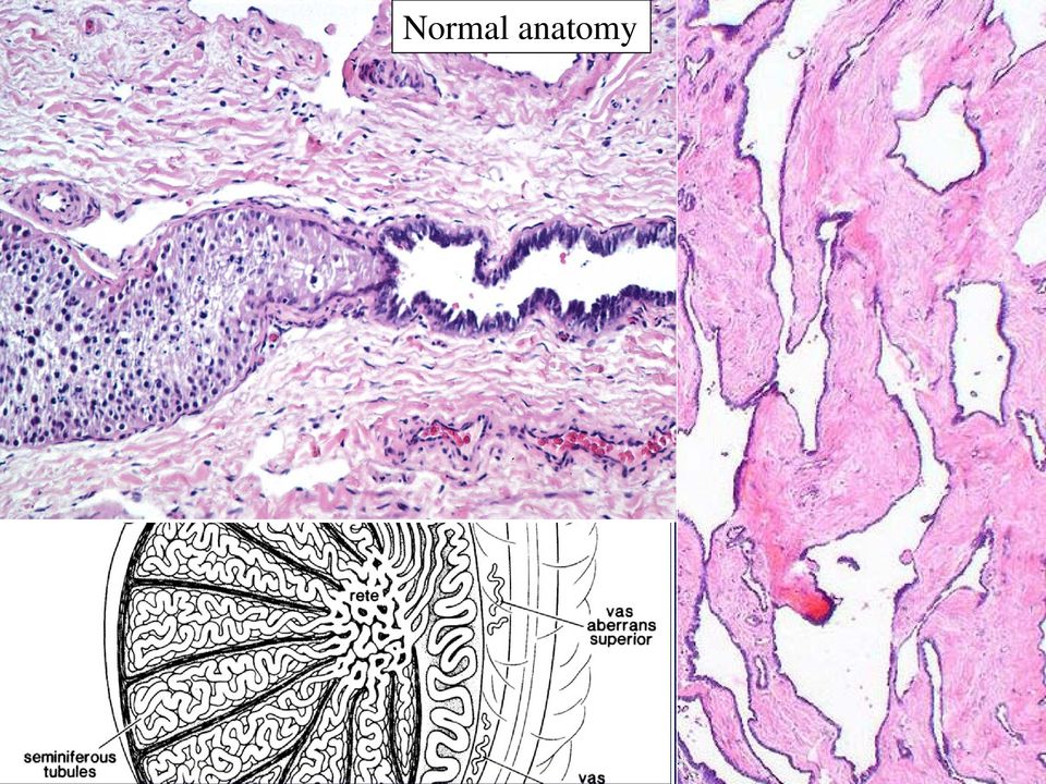
6 Normal anatomy
7 Pagetoid spread of ITGCN with reactive papillary hyperplasia of the rete

8 Reactive changes in the rete testis

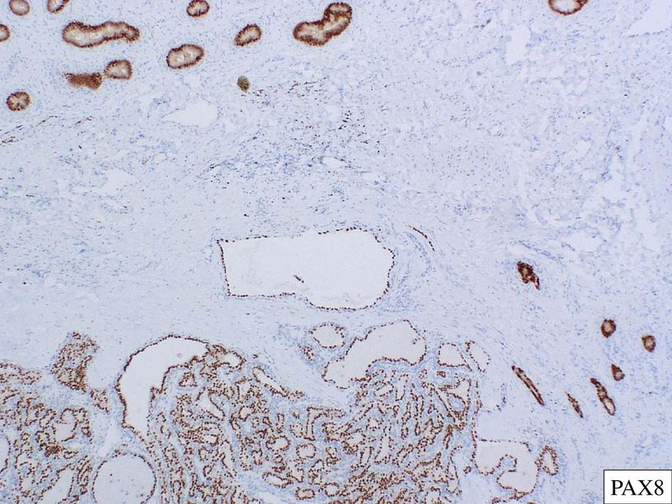
9 Hilum Well circumscribed Banal appearing Inhibin /+ Sertoliform adenoma of the rete

10 PAX8
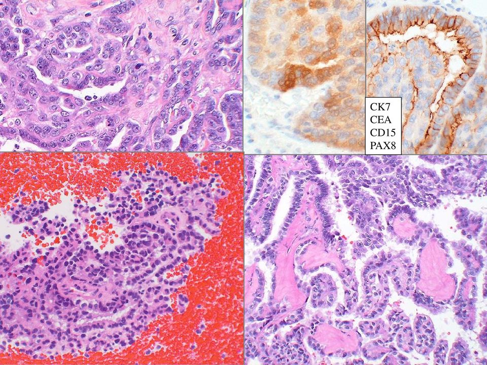
11 RETE TESTIS Adenocarcinoma Lesion centered in the hilum* Transition from normal to neoplastic epithelium* Absence of primary elsewhere Morphology incompatible with any other intrascrotal primary Proper immunohistochemical panel excluding other primaries *may be difficult to assess

12 RETE TESTIS Adenocarcinoma Clinical scenario: Adult males Unilateral painful testicular swelling With or without hydrocele Poor survival Pathology: Solid or cystic Mostly tubulopapillary (solid/spindled/reteform) Cuboidal to columnar cells Eosinophilic to basophilic cytoplasm
 Cuboidal to columnar cells Eosinophilic to")
13
14

15 CK7 CEA CD15 PAX8
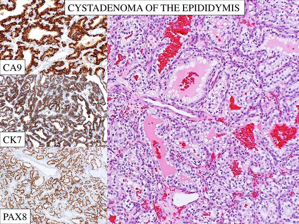
16 CYSTADENOMA OF THE EPIDIDYMIS vhl syndrome Sporadic Painless, movable mass Wide age distribution Variable size No hydrocele Microscopy: Papillary/tubulopapillary growth Clear cytoplasm Low grade nuclei Nuclei polarized towards the luminal surface

17 CYSTADENOMA OF THE EPIDIDYMIS CA9 CK7 PAX8
18 MÜLLERIAN-TYPE EPITHELIAL TUMORS Clinical scenario: Wide age range (adults) No specific medical history Painful testicular swelling Carcinomas may recur and metastasize Pathology: Solid or cystic Wide range of Mülleriantype morphologies Serous features dominate (endometrioid, clear,mucinous, Brenner) Entire histologic spectrum (cystadenomaborderline-carcinoma)
 Entire histologic spectrum")
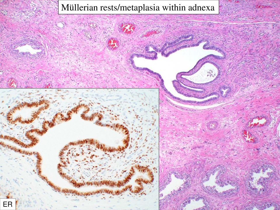
19 Müllerian rests/metaplasia

20 ER Müllerian rests/metaplasia within adnexa
21 Cystadenoma Serous borderline tumor
22 Papillary serous carcinoma
23 PARATESTICULAR SEROUS PAPILLARY CARCINOMA Cases 6 Age (yrs) 31 (16-42) Size (cm) 2 ( ) Presentation Mass 5 Hydrocele 1 Jones et al. Am J Surg Pathol.1995;19:
24 PARATESTICULAR SEROUS PAPILLARY CARCINOMA: Testicular groove* 4 Paratesticular soft tissue 1 Visceral tunica vaginalis 1 * two cases had extensive visceral tunica vaginalis involvement with invasion of the tunica albuginea Jones et al.am J Surg Pathol.1995;19:
25 Endometrioid carcinoma
26 ER WT-1 Positive: PR, CA125, CK7, CD15 Negative: CK20, Calretinin, CEA
27 LESIONS OF MESOTHELIAL ORIGIN Reactive changes Hyperplasia, cyst Mesothelioma Adenomatoid tumor
28 When confronted with a hydrocele sac, what should I be thinking about? If the urologist decides to send it to pathology What does the specimen look like? Thin or thick? Smooth or rough surface? What should I submit for morphology? Submit in toto or simply sample? What did the cyst fluid look like? What is the clinical history?
29 Reactive changes: Thickening Inflammation Mesothelial proliferation parallel to mesothelial surface
30 Reactive mesothelial hyperplasia Reactive changes: Thickening Inflammation Mesothelial proliferation parallel to mesothelial surface
31 Reactive mesothelial hyperplasia Respects the underlying muscular coat / no vertical growth
32 Mesothelioma Haphazard arrangement of cells Nodular of diffuse thickening Vertical growth
33 Mesothelioma CK 5/6 Calretinin WT-1 Seminiferous tubules
34 Mesothelioma; morphologic spectrum
35 What about benign papillary mesothelioma versus WD papillary mesothelioma? Beware of the word benign Minute, focal, cytologically banal, single cell layer, limited to the surface Complexity, solid component, and cytologic atypia warrant a diagnosis of mesothelioma
36 Papillary mesothelioma
37 Papillary mesothelioma
38 MESOTHELIOMA OF TUNICA VAGINALIS Clinicopathologic Features Cases 63 Age (yrs) mean 53.5, range 7-80 Presentation Asbestos exposure 48% Histology Local invasion hydrocele and/or mass 75% epithelial 25% biphasic skin of scrotum/penis, epididymis/testis, spermatic cord Jones et al. Am J Surg Pathol.1995;19:
39
40 MESOTHELIOMA OF TUNICA VAGINALIS Sites of Metastasis Site presentation later total (%) Lymph nodes retroperitoneum (38) inguinal (15) mediastinal 2 2 (4) Lung 8 8 (17) Peritoneum 3 3 ( 6) Liver 2 2 ( 4) Pelvic soft tissue 3 3 ( 6) Jones et al. Am J Surg Pathol.1995;19:
41 MESOTHELIOMA OF TUNICA VAGINALIS Outcome of Patients With Follow-up F/U (yrs) Patients DOD AWD NED < < < < < > Total 52 23(44%) 9(17%) 20(39%) Jones et al.am J Surg Pathol.1995;19:
42
43 WT1 Calretinin Also: CK5/6 D2-40
44
45 ADENOMATOID TUMOR - Well circumscribed, mobile - Not encapsulated Microscopy: - Epithelioid or spindled - Cords, nests, tubules - Microcystic, signet ring - No mitotic activity - May have intervening stroma or adnexal structure - May infarct - atypical myofibroblastic reaction Immunohistochemistry: - Like mesothelioma Differential Diagnosis: Mesothelioma, Adenoca.,YST
46 ADENOMATOID TUMOR Calretinin WT-1
47 ADENOMATOID TUMOR
48 MESENCHYMAL LESIONS OF THE SPERMATIC CORD Fibrous reactive versus neoplastic Lipomatous lipoma liposarcoma dedifferentiated Smooth muscle Squeletal muscle To also consider: Angiolipoma Angiofibroma Angiomyoblastoma Angiomyxoma
49 NODULAR PERIORCHITIS paratesticular fibrous pseudotumor Non neoplastic Nodular masses single, multiple, confluent Rarely diffuse Plaque-like May be massive and disfiguring Usually unilateral Gross features: Firm to hard, white
50 NODULAR PERIORCHITIS Microscopic features: Densely hyalinized collagen Clacification Well circumscribed May be somewhat cellular Inflammation Granulation tissue Differential diagnosis: Inflammatory pseudotumor Fibromatosis
51 Nodular periorchitis Fibrous Pseudotumor
52 LIPOMA OF THE SPERMATIC CORD
53 LIPOSARCOMA OF THE CORD - Most common sarcoma at this site - WD liposarcoma / atypical lipomatous tumor Gross appearance: - Variable, based of fatty, fibrous, or dedifferentiated component Microscopy: - Mature appearing adipocytes of varying size - Lipoblasts can be rare - Spindle and florrette-like cells - Fibrous bands are common - Dedifferentiation low grade high grade - Smooth muscle differentiation
54 Well differentiated Liposarcoma
55
56 Dedifferentiated liposarcoma, low grade
57 Dedifferentiated liposarcoma, high grade
58 Dedifferentiated Liposarcoma, low grade CDK4 MDM2
59 Retroperitoneal recurrence Dedifferentiated liposarcoma, now high grade CDK4 MDM2
60 Adnexal tumors: TESTICULAR APPENDAGES and CORD LESIONS Summary Rare; benign and reactive more common than malignant If epithelioid and malignant, consider a metastasis If primary, location, histology and IHC can help Adenocarcinoma (CEA, CD15, etc) Müllerian (ER, PAX8, WT1, CA125, etc) Mesothelioma (Calretinin, WT1, CK 5/6, Podoplanin) Soft tissue tumors: Beware of the subtle features of WD and low grade dedifferentiated liposarcoma Beware of benign mimics of liposarcoma and embryonal RMS
Case presentation: Mesothelioma of the tunica vaginalis. Dr Ben Shepherd Pathology Queensland Princess Alexandra Hospital Brisbane
 Case presentation: Mesothelioma of the tunica vaginalis Dr Ben Shepherd Pathology Queensland Princess Alexandra Hospital Brisbane A 76 year old man presented June 2011 with a 6 month history of painless
Update on Mesothelioma
 November 8, 2012 Update on Mesothelioma Intro incidence and nomenclature Update on Classification Diagnostic specimens Morphologic features Epithelioid Histology Biphasic Histology Immunohistochemical
MALIGNANT MESOTHELIOMA UPDATE ON PATHOLOGY AND IMMUNOHISTOCHEMISTRY
 MALIGNANT MESOTHELIOMA CLASSIFICATION MALIGNANT MESOTHELIOMA UPDATE ON PATHOLOGY AND IMMUNOHISTOCHEMISTRY Sisko Anttila, MD, PhD Jorvi Hospital Laboratory of Pathology Helsinki University Hospital Espoo,
MALIGNANT MESOTHELIOMA UPDATE ON PATHOLOGY AND IMMUNOHISTOCHEMISTRY
 MALIGNANT MESOTHELIOMA UPDATE ON PATHOLOGY AND IMMUNOHISTOCHEMISTRY Sisko Anttila, MD, PhD Jorvi Hospital Laboratory of Pathology Helsinki University Hospital Espoo, Finland 2nd Nordic Conference on Applied
PATHOLOGY OF THE PLEURA: Mesothelioma and mimickers Necessity of Immunohistochemistry. M. Praet
 PATHOLOGY OF THE PLEURA: Mesothelioma and mimickers Necessity of Immunohistochemistry M. Praet Pathology of the Pleura Normal serosa: visceral and parietal layers Inflammation Neoplasia: Primary: mesothelioma
Today s Topics. Tumors of the Peritoneum in Women
 Today s Topics Tumors of the Peritoneum in Women Charles Zaloudek, M.D. Department of Pathology 505 Parnassus Ave., M563 University of California, San Francisco San Francisco, CA USA charles.zaloudek@ucsf.edu
Outline. Workup for metastatic breast cancer. Metastatic breast cancer
 Metastatic breast cancer Immunostain Update: Diagnosis of metastatic breast carcinoma, emphasizing distinction from GYN primary 1/3 of breast cancer patients will show metastasis 1 st presentation or 20-30
Case of the. Month October, 2012
 Case of the Month October, 2012 Case The patient is a 47-year-old male with a 3-week history of abdominal pain. A CT scan of the abdomen revealed a suggestion of wall thickening at the tip of the appendix
The develpemental origin of mesothelium
 Mesothelioma Tallinn 14.12.06 Henrik Wolff Finnish Institute of Occupational Health The develpemental origin of mesothelium Mesodermal cavities (pleura, peritoneum and pericardium ) are lined with mesenchymal
Changes in Breast Cancer Reports After Second Opinion. Dr. Vicente Marco Department of Pathology Hospital Quiron Barcelona. Spain
 Changes in Breast Cancer Reports After Second Opinion Dr. Vicente Marco Department of Pathology Hospital Quiron Barcelona. Spain Second Opinion in Breast Pathology Usually requested when a patient is referred
Immunohistochemistry on cytology specimens from pleural and peritoneal fluid
 Immunohistochemistry on cytology specimens from pleural and peritoneal fluid Dr Naveena Singh Consultant Pathologist Bart health NHS Trust London United Kingdom Disclosures and Acknowledgements I have
Protocol for the Examination of Specimens From Patients With Tumors of the Peritoneum
 Protocol for the Examination of Specimens From Patients With Tumors of the Peritoneum Protocol applies to all primary borderline and malignant epithelial tumors and malignant mesothelial neoplasms of the
PRIMARY SEROUS CARCINOMA OF PERITONEUM: A CASE REPORT
 PRIMARY SEROUS CARCINOMA OF PERITONEUM: A CASE REPORT Dott. Francesco Pontieri (*) U.O. di Anatomia Patologica P.O. di Rossano (CS) Dott. Gian Franco Zannoni Anatomia Patologica Facoltà di Medicina e Chirurgia
Effusions: Mesothelioma and Metastatic Cancers
 Effusions: Mesothelioma and Metastatic Cancers Malignant Mesothelioma Incidence: 2,500 cases/year ~60-80% pts with pleural MM relationship with asbestos exposure Other risk factors: radiation, other carcinogens,
The evolving pathology of solitary fibrous tumours. Luciane Dreher Irion MREH / CMFT / NSOPS
 The evolving pathology of solitary fibrous tumours Luciane Dreher Irion MREH / CMFT / NSOPS Historical review Haemangiopericytoma (HPC) first described primarily as a soft tissue vascular tumour of pericytic
Diagnosis of Mesothelioma Pitfalls and Practical Information
 Diagnosis of Mesothelioma Pitfalls and Practical Information Mary Beth Beasley, M.D. Mt Sinai Medical Ctr Dept of Pathology One Gustave L Levy Place New York, NY 10029 (212) 241-5307 mbbeasleymd@yahoo.com
Diagnostic Challenge. Department of Pathology,
 Cytology of Pleural Fluid as a Diagnostic Challenge Paavo Pääkkö,, MD, PhD Chief Physician and Head of the Department Department of Pathology, Oulu University Hospital,, Finland Oulu University Hospital
Ovarian tumors Ancillary methods
 Ovarian tumors Ancillary methods Ovarian tumor course Oslo, 24-25/11/14 Prof. Ben Davidson, MD PhD Department of Pathology, Norwegian Radium Hospital, Oslo University Hospital, Oslo, Norway Division of
Something Old, Something New.
 Something Old, Something New. Michelle A. Fajardo, D.O. Loma Linda University Medical Center Clinical Presentation 6 year old boy, presented with hematuria Renal mass demonstrated by ultrasound & CT scan
Notice of Faculty Disclosure
 The Diagnosis of Malignant Mesothelioma Andrew Churg, MD Department of Pathology University of British Columbia Vancouver, BC, Canada achurg@mail.ubc.ca Notice of Faculty Disclosure In accordance with
Protocol applies to all primary borderline and malignant epithelial tumors, and malignant mesothelial neoplasms of the peritoneum.
 Peritoneum Protocol applies to all primary borderline and malignant epithelial tumors, and malignant mesothelial neoplasms of the peritoneum. Protocol revision date: January 2004 No AJCC/UICC staging system
Diseases. Inflammations Non-inflammatory pleural effusions Pneumothorax Tumours
 Pleura Visceral pleura covers lungs and extends into fissures Parietal pleura limits mediastinum and covers dome of diaphragm and inner aspect of chest wall. Two layers between them (pleural cavity) contains
3-F. Pathology of Mesothelioma
 3-F. Pathology of Mesothelioma Kouki Inai Professor of Department of Pathology, Graduate School of Biomedical Science, Hiroshima University Introduction Mesothelioma is a peculiar type of malignancy, which
Introduction: Tumor Swelling / new growth / mass. Two types of growth disorders: Non-Neoplastic. Secondary / adaptation due to other cause.
 Disorders of Growth Introduction: Tumor Swelling / new growth / mass Two types of growth disorders: Non-Neoplastic Secondary / adaptation due to other cause. Neoplastic. Primary growth abnormality. Non-Neoplastic
Cytology : first alert of mesothelioma? Professor B. Weynand, UCL Yvoir, Belgium
 Cytology : first alert of mesothelioma? Professor B. Weynand, UCL Yvoir, Belgium Introduction 3 cavities with the same embryologic origin the mesoderme Pleura Exudates Pleura Peritoneum Pericardium 22%
Pathology of the Female Peritoneum, Common and Uncommon Problems
 Pathology of the Female Peritoneum, Common and Uncommon Problems An Update on Gynecologic Pathology Florence, Italy Anaís Malpica, M.D. Professor of Pathology Pathology of the Female Peritoneum Keratin
Académie internationale de Pathologie - Division arabe XX ème congrès 24-26 novembre 2008 Alger. Immunohistochemistry in malignant mesotheliomas
 Académie internationale de Pathologie - Division arabe XX ème congrès 24-26 novembre 2008 Alger Immunohistochemistry in malignant mesotheliomas Françoise Thivolet-Béjui Groupement Hospitalier Est Lyon-Bron
SEMESTER VI 3 RD YEAR PATHOLOGY KIDNEY TUMORS
 SEMESTER VI 3 RD YEAR PATHOLOGY KIDNEY TUMORS LEARNING OBJECTIVES At the end of the lecture, students should be able to: Know the pathology of renal tumors. RENAL TUMORS RENAL PAPILLARY ADENOMA Common
Practical Effusion Cytology
 Practical Effusion Cytology A Community Pathologist s Approach to Immunocytochemistry in Body Fluid Cytology Emily E. Volk, MD William Beaumont Hospital Troy, MI College of American Pathologists 2004.
INFLAMMATORY PLEURAL EFFUSION
 PLEURA- LESIONS LESIONS OF PLEURA Primary Intra pleural bacterial infections Neoplasm (mesothelioma) Secondary A complication of some underlying disease PLEURAL EFFUSION Common manifestation of both primary
Ovarian mucinous lesions. Ovarian mucinous lesions: Common diagnostic dilemmas. Ovarian mucinous lesions: problematic issues
 Ovarian mucinous lesions Ovarian mucinous lesions: Common diagnostic dilemmas Karuna Garg, MD University of California San Francisco Intestinal or usual type Seromucinous (Endocervical mucinous or Mullerian
A. Pericardial smear. Examination of the pericardial aspirate can provide useful diagnostic information.
 5. PERICARDIUM Heart is encased by the pericardium which has a visceral layer (a) covering the heart and the parietal layer (b). In normal states it is thin, transparent and the myocardium can be seen
Case Report Mesothelioma of the tunica vaginalis testis with prominent adenomatoid features: a case report
 Int J Clin Exp Pathol 2014;7(10):7082-7087 www.ijcep.com /ISSN:1936-2625/IJCEP0001992 Case Report Mesothelioma of the tunica vaginalis testis with prominent adenomatoid features: a case report Lian-He
How To Diagnose And Treat A Tumour In An Effusion
 Effusions of the Serous Cavities Annika Dejmek Professor/Consultant in Cytopathology Clinical Pathology; Department of Laboratory Medicine, Malmö, Lund University 5th EFCS Tutorial Trondheim 2012 Pleura
Case presentation. Awatif Al-Nafussi
 Case presentation Awatif Al-Nafussi Case History 49 year old DVT & small PE June 08, Pelvic mass Ca125 33 Laparotomy-TAHBSO, drainage of ascites Ovarian carcinoma Clinical diagnosis Multiple specimens
Seattle. Case Presentations. Case 1. 76 year old female with a history of breast cancer 12 years ago. Now presents with a pleural effusion.
 Seattle Montreal IAP September 2006 Case Presentations Allen M. Gown, M.D. Medical Director and Chief Pathologist PhenoPath Laboratories Clinical Professor of Pathology University of British Columbia Case
MAJOR PARADIGM SHIFT IN EARLY 1990S IN UNDERSTANDING RENAL CANCER
 Renal tumours WHO 4 MAJOR PARADIGM SHIFT IN EARLY 1990S IN UNDERSTANDING RENAL CANCER Molecular differential pathology of renal cell tumours G. KOVACS A CLASSIFICATION BASED ON UNDERSTANDING THE GENETIC
Frozen Section Diagnosis
 Frozen Section Diagnosis Dr Catherine M Corbishley Honorary Consultant Histopathologist St George s Healthcare NHS Trust and lead examiner final FRCPath Practical 2008-2011 Frozen Section Diagnosis The
Renal Tumors with Eosinophilic Cytoplasm: 2013 Classification. Jesse K. McKenney, MD Associate Head, Surgical Pathology
 Renal Tumors with Eosinophilic Cytoplasm: 2013 Classification Jesse K. McKenney, MD Associate Head, Surgical Pathology Renal Epithelial Neoplasia History 1981: WHO Classification of Renal Neoplasms 1.
DESMOPLASTIC SMALL ROUND CELL TUMOR: A RARE PATHOLOGY PUZZLE
 DESMOPLASTIC SMALL ROUND CELL TUMOR: A RARE PATHOLOGY PUZZLE Ryan Granger University of Rhode Island Cytotechnology program May 2, 2015 ASCT Annual Meeting Nashville, Tennessee DESMOPLASTIC SMALL ROUND
Carcinosarcoma of the Ovary
 Carcinosarcoma of the Ovary A Rare Finding Presented By: Kathryn Kiely Anisa I. Kanbour School of Cytotechnology of the University of Pittsburgh Medical Center Pittsburgh, PA Patient History 55 year old
Neoplasms of the LUNG and PLEURA
 Neoplasms of the LUNG and PLEURA 2015-2016 FCDS Educational Webcast Series Steven Peace, BS, CTR September 19, 2015 2015 Focus o Anatomy o SSS 2000 o MPH Rules o AJCC TNM 1 Case 1 Case Vignette HISTORY:
Male. Female. Death rates from lung cancer in USA
 Male Female Death rates from lung cancer in USA Smoking represents an interesting combination of an entrenched industry and a clearly drug-induced cancer Tobacco Use in the US, 1900-2000 5000 100 Per Capita
Immunohistochemical differentiation of metastatic tumours
 Immunohistochemical differentiation of metastatic tumours Dr Abi Wheal ST1. TERA 3/2/14 Key points from a review article written by Daisuke Nonaka Intro Metastatic disease is the initial presentation in
J of Evidence Based Med & Hlthcare, pissn- 2349-2562, eissn- 2349-2570/ Vol. 2/Issue 33/Aug. 17, 2015 Page 5063
 PERITONEAL MALIGNANT MESOTHELIOMA: A RARE S. R. Dhamotharan 1, S. Shanthi Nirmala 2, F. Celine Foustina Mary 3, M. Arul Raj Kumar 4, R. Vinothprabhu 5 HOW TO CITE THIS ARTICLE: S. R. Dhamotharan, S. Shanthi
More than 2,500 people are diagnosed with mesothelioma in the UK each year.
 This information is an extract from the booklet Understanding mesothelioma. You may find the full booklet helpful. We can send you a free copy see page 5. Contents Introduction Pleural mesothelioma Peritoneal
The Diagnosis of Cancer in the Pathology Laboratory
 The Diagnosis of Cancer in the Pathology Laboratory Dr Edward Sheffield Christmas Select 74 Meeting, Queen s Hotel Cheltenham, 3 rd December 2014 Agenda Overview of the pathology of cancer How specimens
Disclosures. Learning Objectives. Effusion = Confusion. Diagnosis Of Serous Cavity Effusions - Beware The Mesothelial Cell!
 Disclosures Diagnosis Of Serous Cavity Effusions - Beware The Mesothelial Cell! No Relevant Financial Relationships with Commercial Interests Syed Z. Ali, M.D. Syed Z. Ali, M.D. Associate Professor of
Pathology of lung cancer
 Pathology of lung cancer EASO COURSE ON LUNG CANCER AND MESOTHELIOMA DAMASCUS (SYRIA), MAY 3-4, 2007 Gérard ABADJIAN MD Pathologist Associate Professor, Saint Joseph University Pathology Dept. Hôtel-Dieu
R-16: Chronic nonspecific cervisit
 R-16: Chronic nonspecific cervisit Ectoservikal squamous epithelium Endoservical columnar epithelium Dilated cystic endoservical glands lymphoplasmocytes R18:Squamous cell carcinoma insitu Neoplastic epithelium
MALIGNANT MESOTHELIOMA: A TYPICAL PRESENTATION IN AN ATYPICAL PATIENT
 MALIGNANT MESOTHELIOMA: A TYPICAL PRESENTATION IN AN ATYPICAL PATIENT Written by: Karyn Varley MS, SCT(ASCP) The donating laboratory would like to remain anonymous. PATIENT HISTORY 28 year old female Lived
Epithelial Tumors of the Kidney Diagnostic Problems and Recently Described Entities
 Pathology of Renal Neoplasia Epithelial Tumors of the Kidney Diagnostic Problems and Recently Described Entities Wael A Sakr, MD Wayne State University School of Medicine CURRENT CLASSIFICATION = EPITHELIAL
Cytopathology Case Presentation #8
 Cytopathology Case Presentation #8 Emily E. Volk, MD William Beaumont Hospital, Troy, MI Jonathan H. Hughes, MD Laboratory Medicine Consultants, Las Vegas, Nevada Clinical History 44 year old woman presents
General Rules SEER Summary Stage 2000. Objectives. What is Staging? 5/8/2014
 General Rules SEER Summary Stage 2000 Linda Mulvihill Public Health Advisor NCRA Annual Meeting May 2014 National Center for Chronic Disease Prevention and Health Promotion Division of Cancer Prevention
CASE OF THE MONTH AUGUST-2015 DR. GURUDUTT GUPTA HEAD HISTOPATHOLOGY
 CASE OF THE MONTH AUGUST-2015 DR. GURUDUTT GUPTA HEAD HISTOPATHOLOGY CASE HISTORY 52Y MALE RIGHT RADICAL NEPHERECTOMY Case of right renal mass with IVC thrombus. History of surgery and RT for right occipital
PROTOCOL OF THE RITA DATA QUALITY STUDY
 PROTOCOL OF THE RITA DATA QUALITY STUDY INTRODUCTION The RITA project is aimed at estimating the burden of rare malignant tumours in Italy using the population based cancer registries (CRs) data. One of
HKCPath Anatomical Pathology Peer Review and Scores : PDF version for download
 AP2003R1 http://hkcpath.org. Correspondence: pkhui@ha.org.hk 1of 10 07/08/2003 HKCPath Anatomical Pathology Peer Review and Scores : PDF version for download AP141 Bone Marrow: Metastatic Carcinoma from
Immunohistochemistry of soft tissue tumors
 Immunohistochemistry of soft tissue tumors Immunohistochemistry Major advances : antigen retrieval techniques (HIER) sensitive detection systems numerous antibodies of good quality Standardization : automated
POSTMENOPAUSAL ASSESS AND WHAT TO DO
 POSTMENOPAUSAL OVARIAN CYSTS:HOW TO ASSESS AND WHAT TO DO Steven R. Goldstein, MD Professor of Obstetrics and Gynecology Director of Gynecologic Ultrasound Co-Director, Bone Densitometry New York University
How To Test For Cancer
 Diagnosis Of Serous Cavity Effusions - Beware The Mesothelial Cell! Effusion = Confusion Syed Z. Ali, M.D. Professor of Pathology and Radiology The Johns Hopkins Hospital Baltimore, Maryland Diagnostic
Primary -Benign - Malignant Secondary
 TUMOURS OF THE LUNG Primary -Benign - Malignant Secondary The incidence of lung cancer has been increasing almost logarithmically and is now reaching epidemic levels. The overall cure rate is very low
Histopathology of Major Salivary Gland Neoplasms
 Histopathology of Major Salivary Gland Neoplasms Sam J. Cunningham, MD, PhD Faculty Advisor: Shawn D. Newlands, MD, PhD Faculty Advisor: David C. Teller, MD The University of Texas Medical Branch, Department
Primary malignant gonadal mesotheliomas and asbestos
 Histopathology 2000, 37, 150±159 Primary malignant gonadal mesotheliomas and asbestos R L Attanoos & A R Gibbs Department of Histopathology, University Hospital of Wales and Llandough Hospital, Cardiff,
Case Report Predominantly Fibrous Malignant Mesothelioma in a Cat
 SAGE-Hindawi Access to Research Volume 2010, Article ID 396794, 4 pages doi:10.4061/2010/396794 Case Report Predominantly Fibrous Malignant Mesothelioma in a Cat Alexander Th. A. Weiss, Afonso B. da Costa,
Malignant mesothelioma of the pleura: relation
 Thorax 1982;37:810-815 Malignant mesothelioma of the pleura: relation between histological type and clinical behaviour MR LAW, MARGARET E HODSON, BE HEARD From the Cardiothoracic Institute and Brompton
Malignant Peritoneal Mesothelioma in Women A Study of 75 Cases With Emphasis on Their Morphologic Spectrum and Differential Diagnosis
 Anatomic Pathology / MALIGNANT PERITONEAL MESOTHELIOMA IN WOMEN Malignant Peritoneal Mesothelioma in Women A Study of 75 Cases With Emphasis on Their Morphologic Spectrum and Differential Diagnosis Patricia
METASTATIC CLEAR CELL RENAL CELL CARCINOMA TO THE SUBCUTANEOUS AREA IN ILLIAC FOSSA AND ADRENAL GLAND WITHOUT AN IDENTIFIABLE PRIMARY TUMOR
 Indian J.Sci.Res. 5(1) : 121-125, 2014 METASTATIC CLEAR CELL RENAL CELL CARCINOMA TO THE SUBCUTANEOUS AREA IN ILLIAC FOSSA AND ADRENAL GLAND WITHOUT AN IDENTIFIABLE PRIMARY TUMOR a1 b c d REETA DHAR, SHILPI
Cystic Neoplasms of the Pancreas: A multidisciplinary approach to the prevention and early detection of invasive pancreatic cancer.
 This lecture is drawn from the continuing medical education program Finding Hope: Prevention, Early Detection and Treatment of Pancreatic Cancer, Nov, 2011. Robert P. Jury, MD Cystic Neoplasms of the Pancreas:
Renal Cell Carcinoma: Advances in Diagnosis B. Iványi, MD
 Renal Cell Carcinoma: Advances in Diagnosis B. Iványi, MD Department of Pathology University of Szeged, Hungary ISUP Vancouver Classification of Renal Neoplasia Am J Surg Pathol 37:14691489, 2013 13 histologic
Case Report Malignant Mesothelioma Mimicking Invasive Mammary Carcinoma in a Male Breast
 Case Reports in Oncological Medicine Volume 2015, Article ID 298523, 4 pages http://dx.doi.org/10.1155/2015/298523 Case Report Malignant Mesothelioma Mimicking Invasive Mammary Carcinoma in a Male Breast
YOUR LUNG CANCER PATHOLOGY REPORT
 UNDERSTANDING YOUR LUNG CANCER PATHOLOGY REPORT 1-800-298-2436 LungCancerAlliance.org A GUIDE FOR THE PATIENT 1 CONTENTS What is a Pathology Report?...3 The Basics...4 Sections of a Pathology Report...7
Plueral Malignancy: Radiologic-pathologic
 Plueral Malignancy: Radiologic-pathologic Correlation Ritu R. Gill, MD Pleural Malignancies: Radiologic-Pathologic Correlation Ritu R Gill MD Brigham and Women s Hospital Boston, Massachusetts Pleural
The menstrual cycle Hypophysis
 Normal endometrium Tumors of the uterine corpus by MB The endometrium comprises the zona functionalis zf (superficial two thirds) ) and the zona basalis zb (deep one third) The zf responds to hormonal
Histologic Subtypes of Renal Cell Carcinoma
 Histologic Subtypes of Renal Cell Carcinoma M. Scott Lucia, MD Associate Professor Chief of Genitourinary and Renal Pathology Director, Prostate Diagnostic Laboratory Dept. of Pathology University of Colorado
- Slide Seminar - Endocrine pathology in non-endocrine organs. Case 11. Stefano La Rosa, Gioacchino D Ambrosio, Fausto Sessa
 - Slide Seminar - Endocrine pathology in non-endocrine organs Case 11 Stefano La Rosa, Gioacchino D Ambrosio, Fausto Sessa Dept. of Pathology, Multimedica, Milan, Italy Dept. of Surgical and Morphological
CHAPTER 2. Neoplasms (C00-D49) March 2014. 2014 MVP Health Care, Inc.
 March 2014. 2014 MVP Health Care, Inc.") Neoplasms (C00-D49) March 2014 2014 MVP Health Care, Inc. CHAPTER SPECIFIC CATEGORY CODE BLOCKS C00-C14 Malignant neoplasms of lip, oral cavity and pharynx C15-C26 Malignant neoplasms of digestive organs
Neoplasms (C00-D49) March 2014 2014 MVP Health Care, Inc. CHAPTER SPECIFIC CATEGORY CODE BLOCKS C00-C14 Malignant neoplasms of lip, oral cavity and pharynx C15-C26 Malignant neoplasms of digestive organs
Testicular Mesothelioma
 5 Testicular Mesothelioma Alexander N. Zubritsky Moscow Russian Federation 1. Introduction The testicular mesothelioma is one of the rare tumor from other types of mesotheliomas (less than 1%)[9, 43, 81,
Emerging Subtypes in Renal Cancer. Donna E. Hansel, MD PhD Professor of Pathology, UC San Diego Division Chief, Anatomic Pathology dhansel@ucsd.
 Emerging Subtypes in Renal Cancer Donna E. Hansel, MD PhD Professor of Pathology, UC San Diego Division Chief, Anatomic Pathology dhansel@ucsd.edu Some General Comments Fuhrman nuclear grading clear cell
Video Microscopy Tutorial 5
 Video Microscopy Tutorial 5 Lool Alikes in Effusion Cytology:Review of Diagnostic Challenges Claire Michael, MD There are no disclosures necessary. Look-Alikes in Effusion Cytology: Review of Diagnostic
Diagnostic Pitfalls In Thoracic Tumors
 1376 Diagnostic Pitfalls In Thoracic Tumors Neda Kalhor, MD Cesar A. Moran, MD, FASCP WEEKEND OF PATHOLOGY AMERICAN SOCIETY FOR CLINICAL PATHOLOGY 33 W Monroe Ste 1600 Chicago, IL 60603 Program Content
FRCPath Part 2 Histopathology Short Cases, autumn 2014
 FRCPath Part 2 Histopathology Short Cases, autumn 2014 Commentary Case 1 Female age 52: palpable lesion, left breast Breast, fat necrosis. Average: 2.6/5 This case was chosen as a good example of fat necrosis
This module consists of four units which will provide the user a basic knowledge of cancer as a disease.
 Module 5: What is Cancer? This module consists of four units which will provide the user a basic knowledge of cancer as a disease. After completing this module, cancer abstractors will be able to: Define
BIOBANK LPCE-NICE CHEST
 BIOBANK LE-NIE HEST athologist :. BUTORI 12/09/2013 LE / HU Unit atient : N LH13-3603 N LB 13-0691 ID : RO A onsent : YES Age : 54 Diagnosis and staging : chronic pleuresia 5x1000µL BIOBANK LE-NIE HEST
Objectives. Mylene T. Truong, MD. Malignant Pleural Mesothelioma Background
 Imaging of Pleural Tumors Mylene T. Truong, MD Imaging of Pleural Tumours Mylene T. Truong, M. D. University of Texas M.D. Anderson Cancer Center, Houston, TX Objectives To review tumors involving the
BIOBANK LPCE-NICE CHEST
 BIOBANK LE-NIE HEST athologist : S. LASSALLE 01/03/2011 Time for frozen procedure : 10 mn LE / HU Unit atient : N LH 11-0004 N LB 11-0002 onsent : YES Age : 37 ID : TH ER Diagnosis and staging : Hodgkin
ASBESTOS EXPOSURE AND SARCOMATOID MALIGNANT PLEURAL MESOTHELIOMA Gorantla Sambasivarao 1, Namballa Usharani 2, Tupakula Suresh Babu 3
 ASBESTOS EXPOSURE AND SARCOMATOID MALIGNANT PLEURAL MESOTHELIOMA Gorantla Sambasivarao 1, Namballa Usharani 2, Tupakula Suresh Babu 3 HOW TO CITE THIS ARTICLE: Gorantla Sambasivarao, Namballa Usharani,
OBJECTIVES By the end of this segment, the community participant will be able to:
 Cancer 101: Cancer Diagnosis and Staging Linda U. Krebs, RN, PhD, AOCN, FAAN OCEAN Native Navigators and the Cancer Continuum (NNACC) (NCMHD R24MD002811) Cancer 101: Diagnosis & Staging (Watanabe-Galloway
CT and MRI features of the Pathologic Subtypes of Papillary Renal Cell Carcinoma. Melissa Price, MD Aoife Kilcoyne, MD Mukesh G.
 CT and MRI features of the Pathologic Subtypes of Papillary Renal Cell Carcinoma Melissa Price, MD Aoife Kilcoyne, MD Mukesh G. Harisinghani, MD Disclosures Neither I nor my immediate family members have
NEOPLASMS C00 D49. Presented by Jan Halloran CCS
 NEOPLASMS C00 D49 Presented by Jan Halloran CCS 1 INTRODUCTION A neoplasm is a new or abnormal growth. In the ICD-10-CM classification system, neoplastic disease is classified in categories C00 through
Index. F Factor VIII-related antigen, see VWF FactorXIIIa, for dermatofibroma, 272-275 5-HT, see Serotonin
 A Acantholytic squamous cell carcinoma vs epithelioid angiosarcoma, 56-57 Acinic cell carcinoma of pancreas, 76-77 vs ductal adenocarcinoma, 74-75 vs islet cell tumor, 78-81 Adenomatoid tumor vs hemangioma,
Translocation Renal Cell Carcinomas
 Translocation Renal Cell Carcinomas Cora N. Sternberg, MD, FACP Chair, Department of Medical Oncology San Camillo and Forlanini Hospitals Rome, Italy Kidney cancer is not a single disease Clear cell (75%)
ATLAS OF HEAD AND NECK PATHOLOGY THYROID PAPILLARY CARCINOMA
 Papillary carcinoma is the most common of thyroid malignancies and occurs in all age groups but particularly in women under 45 years of age. There is a high rate of cervical metastatic disease and yet
Report series: General cancer information
 Fighting cancer with information Report series: General cancer information Eastern Cancer Registration and Information Centre ECRIC report series: General cancer information Cancer is a general term for
THE PATHOLOGY OF MEDIASTINAL MASSES. Anatomic Distribution of Mediastinal Masses
 Page 1 of 5 THE PATHOLOGY OF MEDIASTINAL MASSES Dr. S. Boag 549-6666 Ext. 4190, mailto:%20boag@cliff.path.queensu.ca 1. INTRODUCTION A wide range of process, neoplastic and non-neoplastic, can produce
Mesothelioma: Questions and Answers
 CANCER FACTS N a t i o n a l C a n c e r I n s t i t u t e N a t i o n a l I n s t i t u t e s o f H e a l t h D e p a r t m e n t o f H e a l t h a n d H u m a n S e r v i c e s Mesothelioma: Questions
Abstracts and References
 Abstracts and References Soft Tissue Pathology Professor Cyril Fisher, Royal Marsden Hospital Learning points CD34 and CK positivity coexist in epithelioid sarcoma and epithelioid endothelial tumours.
BAP1 germline mutations A new Cutaneous Nevus Melanoma Syndrome. Thomas Wiesner
 BAP1 germline mutations A new Cutaneous Nevus Melanoma Syndrome Thomas Wiesner Disclosure Listed as co-inventor US patent application US 61/463,389 BAP1 mutational analysis in determining susceptibility
MODERN IMMUNOHISTOCHEMISTRY
 MODERN IMMUNOHISTOCHEMISTRY Cambridge Illustrated Surgical Pathology Peiguo G. Chu City of Hope National Medical Center, Duarte, California Lawrence M. Weiss City of Hope National Medical Center, Duarte,
Benign Liver Tumors. Cameron Schlegel PGY-1 3/6/2013
 Benign Liver Tumors Cameron Schlegel PGY-1 3/6/2013 Outline Benign Liver Tumors are, in general. Asymptomatic Diagnosed: imaging Treatment: Do no harm Unless Malignant potential Causing symptoms Differential
Rare Thoracic Tumours
 Rare Thoracic Tumours 1. Epithelial Tumour of Trachea 1 1.1 General Results Table 1. Epithelial Tumours of Trachea: Incidence, Trends, Survival Flemish Region 2001-2010 Both Sexes Incidence Trend EAPC