Renal Cell Carcinoma: Advances in Diagnosis B. Iványi, MD
|
|
|
- Rosa Pearson
- 10 years ago
- Views:
Transcription
1 Renal Cell Carcinoma: Advances in Diagnosis B. Iványi, MD Department of Pathology University of Szeged, Hungary

2 ISUP Vancouver Classification of Renal Neoplasia Am J Surg Pathol 37: , histologic types of RCC Aim: review of the cytomorphologic features and the immunoprofile of 8 types of RCC and aspects of grading

3 Immunoprofile of RCCs1,2 Onc Clear cell T1 Papill T2 Papill Chrom Coll duct Muc tub spindle Clear cell papill Xp11 Acquired CDassoc. CAIX No data CD10 CK7 focal AMACR CD117 no data TFE3 CAIX: carbonix anhydrase IX AMACR: αmethylacyl Coenzyme A Racemase 1AlAhmadie et al. Role of immunohistochemistry in the evaluation of needle core biopsies in adult renal cortical tumors. Am J Surg Pathol 35:949961, Tan et al. Renal tumors. Diagnostic and biomarkers. Am J Surg Pathol 37:

4 Clear cell RCC. CAIX: circumferential membranous expression; CD10: membranous and cytoplasmic expression CAIX CD10

5 Immunoprofile of clear cell RCC Onc Clear cell T1 Papill T2 Papill Chrom Coll duct Muc tub spindle Clear cell papill Xp11 Acquired CDassoc. CAIX No data CD10 CK7 focal AMACR CD117 no data TFE3 We routinely evaluate CAIX and CK7 stainings in high grade tumors and in clear cell tumors with nonconventional features

6 High grade tumor cells with eosinophilic cytoplasm. Positive for CAIX and negative for CK7 clear cell RCC CAIX

7 If tumor giant cells display circumferential membrane staining with CAIX and are negative for CK7 clear cell RCC CAIX

8 Clear cell tumors with nonconventional features: subnuclear clearing tubulopapillary structure sheetlike growth pattern require immunostainings

9 Evaluation of a case with subnuclear clearing

10 Basolateral (cupshaped) CAIXpositivity, diffuse CK7positivity, and CD10negativity clear cell tubulopapillary RCC CK7 CAIX

11 Immunoprofile of clear cell (tubulo)papillary RCC Onc Clear cell T1 Papill T2 Papill Chrom Coll duct Muc tub spindle Clear cell papill Xp11 Acquired CDassoc. CAIX No data CD10 CK7 focal AMACR CD117 no data TFE3

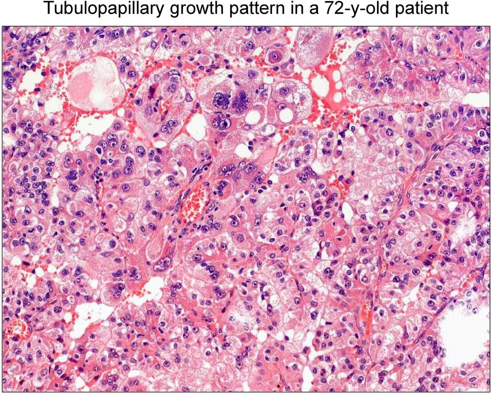
12 Tubulopapillary growth pattern in a 72yold patient
13 RCC with clear cells and papillary growth pattern in a 39yold pt

14 Papillary architecture and clear cells in a 31yo pt Consultation case; courtesy of G. Cserni, Kecskemét, Hungary

15 40yold pt. Sheetlike growth pattern and clear cells

16 Diffuse nuclear TFE3protein positivity 3rd case 1st case 2nd case 4th case

17 Immunoprofile of TFE3 (Xp11) translocation RCC Clear cell T1 Papill T2 Papill Chrom Coll duct Muc tub spindle Clear cell papill Xp111 Acquired CDassoc. CAIX No data CD10 CK7 focal AMACR CD117 no data TFE3 1Some Xp11 translocation RCCs can express MelanA, HMB45 and cathepsink Onc

18 Papillary RCC. Type 1: small cells with scanty cytoplasm; Type 2: larger cells with eo cytoplasm, higher nuclear grade Type 1 Type 2

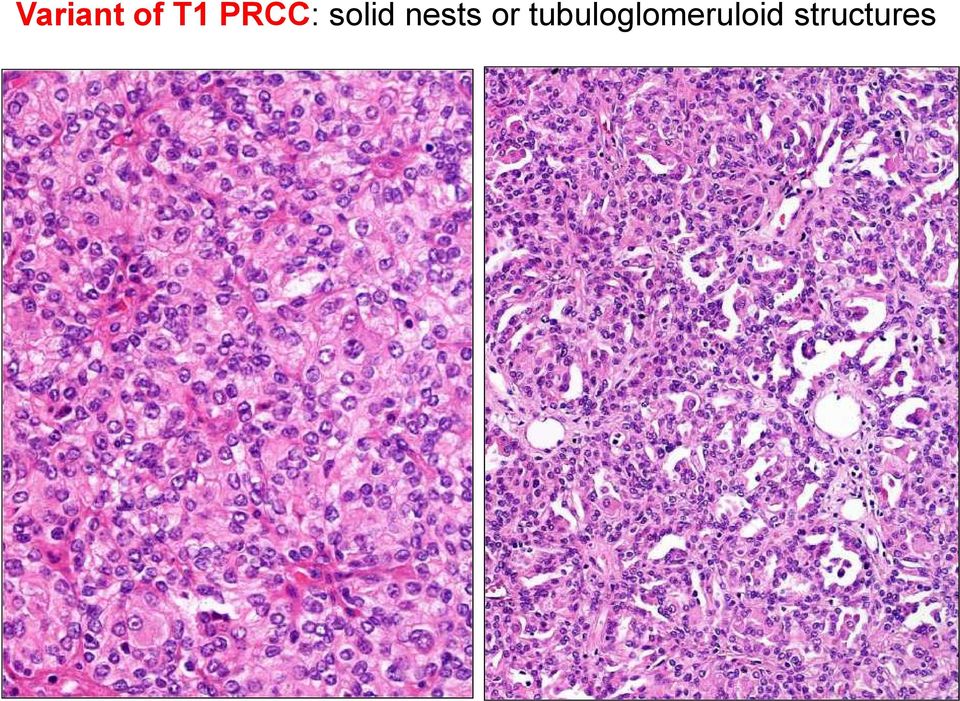
19 Variant of T1 PRCC: solid nests or tubuloglomeruloid structures
20 Type 1 PRCC: positive for CK7 and AMACR AMACR CK7

21 Immunoprofile of papillary RCC Onc Clear cell T1 Papill T2 Papill Chrom Coll duct Muc tub spindle Clear cell papill Xp11 Acquired CDassoc. CAIX No data CD10 CK7 focal AMACR CD117 no data TFE3 Type 2 PRCC has variable staining patterns because this is likely a heterogeneous category rather than a distinct entity
22 Mucinous tubular and spindle cell RCC: tubules lined by bland cuboidal cells together with spindle cells and intercellular mucin Consultation case; courtesy of I. Pálka, Kiskunhalas, Hungary
23 Immunoprofile of mucinous tubular and spindle cell RCC Onc Clear cell T1 Papill T2 Papill Chrom Coll duct Muc tub spindle Clear cell papill Xp11 Acquired CDassoc. CAIX No data CD10 CK7 focal AMACR CD117 no data TFE3 Some cases cannot be distinguished from type 1 papillary RCC
24 Collecting duct RCC: 1) high grade tumor; 2) tubular or tubulopapillary growth; 3) desmoplasia; 4) medullary location
25 Diff. dg.: CDRCC can resemble to type 2 papillary RCC
26 Diff. dg.: urothelial carcinoma invading the kidney
27 Immunoprofile of collecting duct RCC Onc Clear cell T1 Papill T2 Papill Chrom Coll duct Muc tub spindle Clear cell papill Xp11 Acquired CDassoc. CAIX No data CD10 CK7 focal AMACR CD117 no data TFE3 Overlap between CDRCC and urothelial carcinoma: PAX 8 can be positive in UCC, and p63 can be positive in CDRCC
28 CDRCC vs UCC: several cuts from the renal pelvis
29 Chromophobe RCC: chromophobe cells are arranged along vascular channels
30 Hale s colloidal iron: diffuse cytoplasmic staining (eosinophilic variant can be negative)
31 Ultrastructure: microvesicles in the cytoplasm
32 Immunoprofile of chromophobe RCC: CK7, CD117 CK7 CD117
33 Immunoprofile of chromophobe RCC Onc Clear cell T1 Papill T2 Papill Chrom Coll duct Muc tub spindle Clear cell papill Xp11 Acquired CDassoc. CAIX No data CD10 CK7 focal AMACR CD117 no data TFE3
34 Diff. dg.: eosinophilic variant vs oncocytoma RO RO
35 Immunoprofile of RO: CK7, CD117 RO, CK7 RO, CD117
36 Incidences of RCC types in Szeged (Investigators: B. Ivanyi, L. Kuthi, A. Jenei) The key histologic features were sought in HE sections of 612 consecutive RCCs Tissue microarray blocks from 96 cases were analyzed with an antibody panel of CAIX, CD10, CK7, AMACR, ckit1, vimentin and TFE3 Reading: no (<10%), focal (<75%) or diffuse staining
37 Results 85% clear cell 6.8% papillary 4.7% chromophobe 1.8% unclassified 0.3% Xp11 0.3% clear cell (tubulo)papillary 0.3% collecting duct 0.16% hybrid oncocytic/chromophobe tumor
38 Diagnosis of metastasis of renal origin The application of CK7, CD10, CAIX1, vimentin, Pax 2/Pax 8 in various combinations is usually conclusive 1CAIX can be expressed by carcinomas of endometrium, stomach, cervix, breast, lung, and liver tumors, neuroendocrine tumors, and mesotheliomas. Truong LD, Shen SS. Immunohistochemical diagnosis of renal neoplasms. Arch Pathol Lab Med 135;92109, 2011
39 Bx from the pancreas: high grade carcinoma; Negative for CK20, CK7, MUC5AC
40 Diffuse, membranosus CAIX and CD10 positivity CAIX CD10
41 CK7 Vimentin
42 Diagnosis: invasion of pancreas by high grade clear cell RCC Search for clinical data: nephrectomy performed not in Szeged because of high grade RCC 2 years earlier
43 The ISUP Grading System for Renal Cell Carcinoma and Other Prognostic Parameters Am J Surg Pathol 2013;37: The main morphotypes of RCC have prognostic significance Excellent prognosis Multilocular cystic RCC (multilocular cystic renal neoplasm of low malignant potential) Good prognosis Extensively cystic CCRCC Clear cell (tubulopapillary) RCC, MTSRCC Bad prognosis CDRCC
44 Sarcomatoid component, rhabdoid differentiation, tumor necrosis have prognostic impact. Reporting is recommended
45 ISUP grading system for clear cell and papillary RCC Grade I inconspicous or absent nucleoli at 400x Grade II distinctly visible nucleoli at 400x Grade III distinctly visible nucleoli at 100x Gr IV tumors with sarcomatoid or rhabdoid differentiation or with extreme nuclear pleiomorphism or with tumor giant cells
46 Take home message Utilizing immunostains improves classification of renal tumors Both extent and patterns must be considered for a definitive diagnosis Clear cell carcinomas with subnuclear clearing or tubulopapillary architecture should raise the possibility of clear cell (tubulo)papillary RCC or translocation RCC
RENAL CELL CARCINOMA EPIDEMIOLOGY
 RENAL CELL CARCINOMA: THE 2012 ISUP VANCOUVER CLASSIFICATION David Grignon MD, Indiana University, Indianapolis, IN RENAL CELL CARCINOMA EPIDEMIOLOGY Siegel et al. CaA Cancer J Clin 63:11-30, 2013 1 EPITHELIAL
Renal Pathology Update. Sundus Hussein MD, FRCPC
 Renal Pathology Update Sundus Hussein MD, FRCPC Case History A 45 year old male with incidentally discovered a 3.5 x 3.9 x 2.7 cm renal mass Handling partial nephrectomy Handling partial nephrectomy
Epithelial Tumors of the Kidney Diagnostic Problems and Recently Described Entities
 Pathology of Renal Neoplasia Epithelial Tumors of the Kidney Diagnostic Problems and Recently Described Entities Wael A Sakr, MD Wayne State University School of Medicine CURRENT CLASSIFICATION = EPITHELIAL
Emerging Subtypes in Renal Cancer. Donna E. Hansel, MD PhD Professor of Pathology, UC San Diego Division Chief, Anatomic Pathology dhansel@ucsd.
 Emerging Subtypes in Renal Cancer Donna E. Hansel, MD PhD Professor of Pathology, UC San Diego Division Chief, Anatomic Pathology [email protected] Some General Comments Fuhrman nuclear grading clear cell
Renal Tumors with Eosinophilic Cytoplasm: 2013 Classification. Jesse K. McKenney, MD Associate Head, Surgical Pathology
 Renal Tumors with Eosinophilic Cytoplasm: 2013 Classification Jesse K. McKenney, MD Associate Head, Surgical Pathology Renal Epithelial Neoplasia History 1981: WHO Classification of Renal Neoplasms 1.
Histologic Subtypes of Renal Cell Carcinoma
 Histologic Subtypes of Renal Cell Carcinoma M. Scott Lucia, MD Associate Professor Chief of Genitourinary and Renal Pathology Director, Prostate Diagnostic Laboratory Dept. of Pathology University of Colorado
The WHO Classification of Renal Tumors and Common Issues in TNM Staging for Renal Cell Carcinoma
 The WHO Classification of Renal Tumors and Common Issues in TNM Staging for Renal Cell Carcinoma Steven Shen, M.D.,Ph.D. Staff Pathologist and Assistant Member The Methodsit Hospital and Research Institute
Gladwyn Leiman, MCCCh, FIAC, FRCPath Scott Anderson, MD
 Cytology Works shop #5 Gladwyn Leiman, MCCCh, FIAC, FRCPath Scott Anderson, MD Disclosur re information The speakers have no relationship that represents a possible conflict of interest with respect to
New Concepts and Refinements to. Existing WHO (2004) Renal Cell. Tumor Categories In Adults
 Renal Cell. Tumor Categories In Adults") New Concepts and Refinements to Existing WHO (2004) Renal Cell Tumor Categories In Adults Dr Varsha Manucha Assistant Professor Department of Pathology and Laboratory Medicine Temple University Hospital
New Concepts and Refinements to Existing WHO (2004) Renal Cell Tumor Categories In Adults Dr Varsha Manucha Assistant Professor Department of Pathology and Laboratory Medicine Temple University Hospital
Something Old, Something New.
 Something Old, Something New. Michelle A. Fajardo, D.O. Loma Linda University Medical Center Clinical Presentation 6 year old boy, presented with hematuria Renal mass demonstrated by ultrasound & CT scan
RENAL CANCER PATHOLOGY WHAT REALLY MATTERS? STEWART FLEMING UNIVERSITY OF DUNDEE
 RENAL CANCER PATHOLOGY WHAT REALLY MATTERS? STEWART FLEMING UNIVERSITY OF DUNDEE MAJOR PARADIGM SHIFT IN EARLY 1990S IN UNDERSTANDING RENAL CANCER Molecular differential pathology of renal cell tumours
Histopathology and prognosis in renal cancer
 Histopathology and prognosis in renal cancer Granular cell Granular cell Granular cell Granular cell Clear cell Chromophobe cell Papillary type 2 Luca Mazzucchelli Istituto cantonale di patologia, Locarno
The role of Immunohistochemistry in diagnosis of renal cell carcinoma subtypes.
 Asian Archives of Pathology 2013; Vol. 9 No.1, 9-16 Original articles The role of Immunohistochemistry in diagnosis of renal cell carcinoma subtypes. Sutthiporn Namnak, BSc *, Wipawee Kittikowit, MD *,
Seattle. Case Presentations. Case 1. 76 year old female with a history of breast cancer 12 years ago. Now presents with a pleural effusion.
 Seattle Montreal IAP September 2006 Case Presentations Allen M. Gown, M.D. Medical Director and Chief Pathologist PhenoPath Laboratories Clinical Professor of Pathology University of British Columbia Case
MALIGNANT MESOTHELIOMA UPDATE ON PATHOLOGY AND IMMUNOHISTOCHEMISTRY
 MALIGNANT MESOTHELIOMA CLASSIFICATION MALIGNANT MESOTHELIOMA UPDATE ON PATHOLOGY AND IMMUNOHISTOCHEMISTRY Sisko Anttila, MD, PhD Jorvi Hospital Laboratory of Pathology Helsinki University Hospital Espoo,
Diagnosis of Mesothelioma Pitfalls and Practical Information
 Diagnosis of Mesothelioma Pitfalls and Practical Information Mary Beth Beasley, M.D. Mt Sinai Medical Ctr Dept of Pathology One Gustave L Levy Place New York, NY 10029 (212) 241-5307 [email protected]
MALIGNANT MESOTHELIOMA UPDATE ON PATHOLOGY AND IMMUNOHISTOCHEMISTRY
 MALIGNANT MESOTHELIOMA UPDATE ON PATHOLOGY AND IMMUNOHISTOCHEMISTRY Sisko Anttila, MD, PhD Jorvi Hospital Laboratory of Pathology Helsinki University Hospital Espoo, Finland 2nd Nordic Conference on Applied
Classificazione anatomo-patologica nei RCC Matteo Brunelli. Department of Pathology and Diagnostic, University di Verona, Italy
 Classificazione anatomo-patologica nei RCC Matteo Brunelli Department of Pathology and Diagnostic, University di Verona, Italy WHO 2004 AFIP 2004 = ISUP Vancouver Classification 2013 5 newentities 3 emerging
Tubulocystic Carcinoma of the Kidney, a Rare Distinct Entity
 Tubulocystic Carcinoma of the Kidney, a Rare Distinct Entity 2 Shreenath Bishu, Laurie J. Eisengart and Ximing J. Yang * Department of Pathology, Northwestern University, Feinberg School of Medicine, Feinberg,
Outline. Workup for metastatic breast cancer. Metastatic breast cancer
 Metastatic breast cancer Immunostain Update: Diagnosis of metastatic breast carcinoma, emphasizing distinction from GYN primary 1/3 of breast cancer patients will show metastasis 1 st presentation or 20-30
Outcome prediction for renal cell carcinoma (RCC)
") SPECIAL ARTICLE The International Society of Urological Pathology (ISUP) Grading System for Renal Cell Carcinoma and Other Prognostic Parameters Brett Delahunt, MD,* John C. Cheville, MD,w Guido Martignoni,
SPECIAL ARTICLE The International Society of Urological Pathology (ISUP) Grading System for Renal Cell Carcinoma and Other Prognostic Parameters Brett Delahunt, MD,* John C. Cheville, MD,w Guido Martignoni,
Emerging and Newly Recognized Subtypes of Renal Tumors: Keys to Recognition Session 1006
 Emerging and Newly Recognized Subtypes of Renal Tumors: Keys to Recognition Session 1006 Sean R. Williamson, MD Senior Staff Pathologist, Henry Ford Health System Clinical Assistant Professor of Pathology,
The develpemental origin of mesothelium
 Mesothelioma Tallinn 14.12.06 Henrik Wolff Finnish Institute of Occupational Health The develpemental origin of mesothelium Mesodermal cavities (pleura, peritoneum and pericardium ) are lined with mesenchymal
Renal Cell Carcinoma Associated with Xp11.2 Translocation: Clinicopathologic and Immunohistochemical Findings of 4 Cases
 The Korean Journal of Pathology 2005; 39: 406-11 Renal Cell Carcinoma Associated with Xp11.2 Translocation: Clinicopathologic and Immunohistochemical Findings of 4 Cases Sanghui Park Ji-Eun Kwon Yeon-Lim
Immunohistochemical differentiation of metastatic tumours
 Immunohistochemical differentiation of metastatic tumours Dr Abi Wheal ST1. TERA 3/2/14 Key points from a review article written by Daisuke Nonaka Intro Metastatic disease is the initial presentation in
Update on Mesothelioma
 November 8, 2012 Update on Mesothelioma Intro incidence and nomenclature Update on Classification Diagnostic specimens Morphologic features Epithelioid Histology Biphasic Histology Immunohistochemical
CT and MRI features of the Pathologic Subtypes of Papillary Renal Cell Carcinoma. Melissa Price, MD Aoife Kilcoyne, MD Mukesh G.
 CT and MRI features of the Pathologic Subtypes of Papillary Renal Cell Carcinoma Melissa Price, MD Aoife Kilcoyne, MD Mukesh G. Harisinghani, MD Disclosures Neither I nor my immediate family members have
The Mainz Classification of Renal Cell Tumors
 The Mainz classification is effective in distinguishing the histopathologic and cytogenetic features of various types of renal cell carcinoma. Virginia S. King. Costa del Sol. Watercolor, 18 24. The Mainz
PATHOLOGY OF THE PLEURA: Mesothelioma and mimickers Necessity of Immunohistochemistry. M. Praet
 PATHOLOGY OF THE PLEURA: Mesothelioma and mimickers Necessity of Immunohistochemistry M. Praet Pathology of the Pleura Normal serosa: visceral and parietal layers Inflammation Neoplasia: Primary: mesothelioma
Renal Neoplasms: An Update on Immunohistochemical and Histochemical Features
 Application Renal Neoplasms: An Update on Immunohistochemical and Histochemical Features Stephen M. Bonsib, MD Ami Bhalodia, MD Albert and Harriet Smith Professor and Chair Louisiana State University Health
Translocation Renal Cell Carcinomas
 Translocation Renal Cell Carcinomas Cora N. Sternberg, MD, FACP Chair, Department of Medical Oncology San Camillo and Forlanini Hospitals Rome, Italy Kidney cancer is not a single disease Clear cell (75%)
Immunohistochemical Analysis of Chromophobe Renal Cell Carcinoma, Renal Oncocytoma, and Clear Cell Carcinoma
 Immunohistochemical Analysis of Chromophobe Renal Cell Carcinoma, Renal Oncocytoma, and Clear Cell Carcinoma An Optimal and Practical Panel for Differential Diagnosis Lina Liu, MD; Junqi Qian, MD; Harpreet
SEMESTER VI 3 RD YEAR PATHOLOGY KIDNEY TUMORS
 SEMESTER VI 3 RD YEAR PATHOLOGY KIDNEY TUMORS LEARNING OBJECTIVES At the end of the lecture, students should be able to: Know the pathology of renal tumors. RENAL TUMORS RENAL PAPILLARY ADENOMA Common
Académie internationale de Pathologie - Division arabe XX ème congrès 24-26 novembre 2008 Alger. Immunohistochemistry in malignant mesotheliomas
 Académie internationale de Pathologie - Division arabe XX ème congrès 24-26 novembre 2008 Alger Immunohistochemistry in malignant mesotheliomas Françoise Thivolet-Béjui Groupement Hospitalier Est Lyon-Bron
Practical Effusion Cytology
 Practical Effusion Cytology A Community Pathologist s Approach to Immunocytochemistry in Body Fluid Cytology Emily E. Volk, MD William Beaumont Hospital Troy, MI College of American Pathologists 2004.
KIDNEY CARCINOMA ASSOCIATED WITH XP11.2 TRANSLOCATION / TFE3 (ASPL-TFE3) GENE FUSION
 GENE FUSION") Case Report International Braz J Urol Official Journal of the Brazilian Society of Urology KIDNEY CA AND Xp11.2 TRANSLOCATION Vol. 31 (3): 251-255, May - June, 2005 KIDNEY CARCINOMA ASSOCIATED WITH XP11.2
Case Report International Braz J Urol Official Journal of the Brazilian Society of Urology KIDNEY CA AND Xp11.2 TRANSLOCATION Vol. 31 (3): 251-255, May - June, 2005 KIDNEY CARCINOMA ASSOCIATED WITH XP11.2
CASE OF THE MONTH AUGUST-2015 DR. GURUDUTT GUPTA HEAD HISTOPATHOLOGY
 CASE OF THE MONTH AUGUST-2015 DR. GURUDUTT GUPTA HEAD HISTOPATHOLOGY CASE HISTORY 52Y MALE RIGHT RADICAL NEPHERECTOMY Case of right renal mass with IVC thrombus. History of surgery and RT for right occipital
METASTATIC CLEAR CELL RENAL CELL CARCINOMA TO THE SUBCUTANEOUS AREA IN ILLIAC FOSSA AND ADRENAL GLAND WITHOUT AN IDENTIFIABLE PRIMARY TUMOR
 Indian J.Sci.Res. 5(1) : 121-125, 2014 METASTATIC CLEAR CELL RENAL CELL CARCINOMA TO THE SUBCUTANEOUS AREA IN ILLIAC FOSSA AND ADRENAL GLAND WITHOUT AN IDENTIFIABLE PRIMARY TUMOR a1 b c d REETA DHAR, SHILPI
Today s Topics. Tumors of the Peritoneum in Women
 Today s Topics Tumors of the Peritoneum in Women Charles Zaloudek, M.D. Department of Pathology 505 Parnassus Ave., M563 University of California, San Francisco San Francisco, CA USA [email protected]
HKCPath Anatomical Pathology Peer Review and Scores : PDF version for download
 AP2003R1 http://hkcpath.org. Correspondence: [email protected] 1of 10 07/08/2003 HKCPath Anatomical Pathology Peer Review and Scores : PDF version for download AP141 Bone Marrow: Metastatic Carcinoma from
Lung Carcinomas New 2015 WHO Classification. Spasenija Savic Pathology
 Lung Carcinomas New 2015 WHO Classification Spasenija Savic Pathology ***EXPECTED SPRING 2015*** This authoritative, concise reference book provides an international standard for oncologists and pathologists
Changes in Breast Cancer Reports After Second Opinion. Dr. Vicente Marco Department of Pathology Hospital Quiron Barcelona. Spain
 Changes in Breast Cancer Reports After Second Opinion Dr. Vicente Marco Department of Pathology Hospital Quiron Barcelona. Spain Second Opinion in Breast Pathology Usually requested when a patient is referred
RENAL PARENCHYMAL MALIGNANCY (RENAL CELL CARCINOMA) STRUCTURED REPORTING PROTOCOL. (1st EDITION 2011)
 STRUCTURED REPORTING PROTOCOL. (1st EDITION 2011)") RENAL PARENCHYMAL MALIGNANCY (RENAL CELL CARCINOMA) STRUCTURED REPORTING PROTOCOL (1st EDITION 2011) Core Document versions: AJCC Cancer Staging Manual 7 th edition (including errata corrected with 5th
RENAL PARENCHYMAL MALIGNANCY (RENAL CELL CARCINOMA) STRUCTURED REPORTING PROTOCOL (1st EDITION 2011) Core Document versions: AJCC Cancer Staging Manual 7 th edition (including errata corrected with 5th
Ovarian tumors Ancillary methods
 Ovarian tumors Ancillary methods Ovarian tumor course Oslo, 24-25/11/14 Prof. Ben Davidson, MD PhD Department of Pathology, Norwegian Radium Hospital, Oslo University Hospital, Oslo, Norway Division of
Pathology of lung cancer
 Pathology of lung cancer EASO COURSE ON LUNG CANCER AND MESOTHELIOMA DAMASCUS (SYRIA), MAY 3-4, 2007 Gérard ABADJIAN MD Pathologist Associate Professor, Saint Joseph University Pathology Dept. Hôtel-Dieu
PRIMARY SEROUS CARCINOMA OF PERITONEUM: A CASE REPORT
 PRIMARY SEROUS CARCINOMA OF PERITONEUM: A CASE REPORT Dott. Francesco Pontieri (*) U.O. di Anatomia Patologica P.O. di Rossano (CS) Dott. Gian Franco Zannoni Anatomia Patologica Facoltà di Medicina e Chirurgia
TUMORS OF THE TESTICULAR ADNEXA and SPERMATIC CORD
 TUMORS OF THE TESTICULAR ADNEXA and SPERMATIC CORD Victor E. Reuter, MD Memorial Sloan-Kettering Cancer Center [email protected] 66 th Annual Pathology Seminar California Society of Pathologists Short
DESMOPLASTIC SMALL ROUND CELL TUMOR: A RARE PATHOLOGY PUZZLE
 DESMOPLASTIC SMALL ROUND CELL TUMOR: A RARE PATHOLOGY PUZZLE Ryan Granger University of Rhode Island Cytotechnology program May 2, 2015 ASCT Annual Meeting Nashville, Tennessee DESMOPLASTIC SMALL ROUND
ATLAS OF HEAD AND NECK PATHOLOGY THYROID PAPILLARY CARCINOMA
 Papillary carcinoma is the most common of thyroid malignancies and occurs in all age groups but particularly in women under 45 years of age. There is a high rate of cervical metastatic disease and yet
A 70-year old Man with Pleural Effusion
 Mesothelioma Diagnosis: Pitfalls and Latest Updates S Klebe and DW Henderson Recommendations Indisputable malignant cells on cytomorphological criteria which demonstrate a mesothelial phenotype, which
Multiple Primary and Histology Site Specific Coding Rules KIDNEY. FLORIDA CANCER DATA SYSTEM MPH Kidney Site Specific Coding Rules
 Multiple Primary and Histology Site Specific Coding Rules KIDNEY 1 Prerequisites 2 Completion of Multiple Primary and Histology General Coding Rules 3 There are many ways to view the Multiple l Primary/Histology
Carcinoma papilar renal, cromófobo y otras histologías. Maria José Méndez Vidal Servicio de oncología Medica Hospital Reina Sofía Córdoba
 Carcinoma papilar renal, cromófobo y otras histologías. Maria José Méndez Vidal Servicio de oncología Medica Hospital Reina Sofía Córdoba Europe 121 629 new cases RCC 2012, 75 676 affected men Slide 3
2009 update on the classification of renal epithelial tumors in adults
 International Journal of Urology (2009) 16, 432 443 doi: 10.1111/j.1442-2042.2009.02302.x Review Article 2009 update on the classification of renal epithelial tumors in adults Antonio Lopez-Beltran, 1
Case presentation. Awatif Al-Nafussi
 Case presentation Awatif Al-Nafussi Case History 49 year old DVT & small PE June 08, Pelvic mass Ca125 33 Laparotomy-TAHBSO, drainage of ascites Ovarian carcinoma Clinical diagnosis Multiple specimens
The evolving pathology of solitary fibrous tumours. Luciane Dreher Irion MREH / CMFT / NSOPS
 The evolving pathology of solitary fibrous tumours Luciane Dreher Irion MREH / CMFT / NSOPS Historical review Haemangiopericytoma (HPC) first described primarily as a soft tissue vascular tumour of pericytic
Diagnostic Challenge. Department of Pathology,
 Cytology of Pleural Fluid as a Diagnostic Challenge Paavo Pääkkö,, MD, PhD Chief Physician and Head of the Department Department of Pathology, Oulu University Hospital,, Finland Oulu University Hospital
The Role of Genetic Testing in the Evaluation of Thyroid Nodules. Thyroid Cancer and FNA. Thyroid Cancer. Pure Follicular Cancers.
 Where does Molecular Analysis of FNA Specimens fit into the evaluation of thyroid nodules? The Role of Genetic Testing in the Evaluation of Thyroid Nodules Ultrasound TSH Risk factors Jill E. Langer, MD
Immunohistochemistry on cytology specimens from pleural and peritoneal fluid
 Immunohistochemistry on cytology specimens from pleural and peritoneal fluid Dr Naveena Singh Consultant Pathologist Bart health NHS Trust London United Kingdom Disclosures and Acknowledgements I have
RENAL EPITHELIAL TUMORS 2009: THE ROLE OF ELECTRON MICROSCOPY IN UNDERSTANDING PATHOGENESIS, DIAGNOSIS, AND CLASSIFICATION.
 RENAL EPITHELIAL TUMORS 2009: THE ROLE OF ELECTRON MICROSCOPY IN UNDERSTANDING PATHOGENESIS, DIAGNOSIS, AND CLASSIFICATION. Guillermo A. Herrera MD Nephrocor, Tempe, Arizona Epithelial renal cell tumors
Role of histopatological assessment of renal cancer specimen after radical and partial nephrectomy
 Role of histopatological assessment of renal cancer specimen after radical and partial nephrectomy Dr hab. n. med. Agnieszka Korolczuk Joanna Irla Natalia Pieszek Monika Orzeł Dr hab. n. med. Justyna Szumiło
XP11.2 Translocation renal cell carcinoma: Clinical experience of Taipei Veterans General Hospital
 Available online at www.sciencedirect.com Journal of the Chinese Medical Association 74 (2011) 500e504 Original Article XP11.2 Translocation renal cell carcinoma: Clinical experience of Taipei Veterans
Case of the. Month October, 2012
 Case of the Month October, 2012 Case The patient is a 47-year-old male with a 3-week history of abdominal pain. A CT scan of the abdomen revealed a suggestion of wall thickening at the tip of the appendix
CASE REVIEW. Oncocytic Renal Neoplasms. A Molecular Approach to a Common Diagnostic Challenge
 CASE REVIEW Oncocytic Renal Neoplasms A Molecular Approach to a Common Diagnostic Challenge Federico A. Monzon, MD,* Steven S. Shen, MD, PhD,* Bonnie Kemp, MD, Karla Alvarez, BSc, and Alberto G. Ayala,
Notice of Faculty Disclosure
 The Diagnosis of Malignant Mesothelioma Andrew Churg, MD Department of Pathology University of British Columbia Vancouver, BC, Canada [email protected] Notice of Faculty Disclosure In accordance with
Thymic carcinoma: update of current diagnostic criteria and histologic types
 Seminars in Diagnostic Pathology 22, 198-212 Thymic carcinoma: update of current diagnostic criteria and histologic types Saul Suster, MD From the Department of Pathology, The Ohio State University and
Four Important Facts about Kidney Cancer
 Volume Article.13-11 Publish Date: 24th June 2013 Author(s): MPUH - CRS Team Four Important Facts about Kidney Cancer ARTICLE hhh Muljibhai Patel Urological Hospital (MPUH) Centre For Robotic Surgery (CRS)
Efficient Tumor Immunohistochemistry A Differential Diagnosis-Driven Approach
 Efficient Tumor Immunohistochemistry A Differential Diagnosis-Driven Approach Publishing Team Erik Tanck (production manager/designer) Joshua Weikersheimer (publisher) Copyright 2006 by the American Society
Information Model Requirements of Post-Coordinated SNOMED CT Expressions for Structured Pathology Reports
 Information Model Requirements of Post-Coordinated SNOMED CT Expressions for Structured Pathology Reports W. Scott Campbell, Ph.D., MBA James R. Campbell, MD Acknowledgements Steven H. Hinrichs, MD Chairman
PathMD: Board Review Letter TM
 Questions 1. Mantle Cell Lymphoma is associated with which of the following: A. t(11;14) B. t(8;14) C. t(2;5) D. t(11;18) E. t(14;18) 2. A case of anaplastic large cell lymphoma (ALCL) with t(1;2) is stained
How To Test For Cancer
 Diagnosis Of Serous Cavity Effusions - Beware The Mesothelial Cell! Effusion = Confusion Syed Z. Ali, M.D. Professor of Pathology and Radiology The Johns Hopkins Hospital Baltimore, Maryland Diagnostic
Introduction. Wu A, Kunju L P, Cheng L & Shah R B (2008) Histopathology 53, 533 544
 Histopathology 53, 533 544") Histopathology 2008, 53, 533 544. DOI: 10.1111/j.1365-2559.2008.03151.x Renal cell carcinoma in children and young adults: analysis of clinicopathological, immunohistochemical and molecular characteristics
Histopathology 2008, 53, 533 544. DOI: 10.1111/j.1365-2559.2008.03151.x Renal cell carcinoma in children and young adults: analysis of clinicopathological, immunohistochemical and molecular characteristics
Effusions: Mesothelioma and Metastatic Cancers
 Effusions: Mesothelioma and Metastatic Cancers Malignant Mesothelioma Incidence: 2,500 cases/year ~60-80% pts with pleural MM relationship with asbestos exposure Other risk factors: radiation, other carcinogens,
HER2 Status: What is the Difference Between Breast and Gastric Cancer?
 Ask the Experts HER2 Status: What is the Difference Between Breast and Gastric Cancer? Bharat Jasani MBChB, PhD, FRCPath Marco Novelli MBChB, PhD, FRCPath Josef Rüschoff, MD Robert Y. Osamura, MD, FIAC
Title: Immunohistochemical staining of radixin and moesin in prostatic adenocarcinoma
 Author's response to reviews Title: Immunohistochemical staining of radixin and moesin in prostatic adenocarcinoma Authors: Tanner L Bartholow ([email protected]) Uma R Chandran ([email protected])
The Diagnosis of Cancer in the Pathology Laboratory
 The Diagnosis of Cancer in the Pathology Laboratory Dr Edward Sheffield Christmas Select 74 Meeting, Queen s Hotel Cheltenham, 3 rd December 2014 Agenda Overview of the pathology of cancer How specimens
LYMPHOMA. BACHIR ALOBEID, M.D. HEMATOPATHOLOGY DIVISION PATHOLOGY DEPARTMENT Columbia University/ College of Physicians & Surgeons
 LYMPHOMA BACHIR ALOBEID, M.D. HEMATOPATHOLOGY DIVISION PATHOLOGY DEPARTMENT Columbia University/ College of Physicians & Surgeons Normal development of lymphocytes Lymphocyte proliferation and differentiation:
Histopathology of Major Salivary Gland Neoplasms
 Histopathology of Major Salivary Gland Neoplasms Sam J. Cunningham, MD, PhD Faculty Advisor: Shawn D. Newlands, MD, PhD Faculty Advisor: David C. Teller, MD The University of Texas Medical Branch, Department
Protocol for the Examination of Specimens From Patients With Invasive Carcinoma of Renal Tubular Origin
 Protocol for the Examination of Specimens From Patients With Invasive Carcinoma of Renal Tubular Origin Wilms tumors and tumors of urothelial origin are not included. Based on AJCC/UICC TNM, 7th edition
بسم هللا الرحمن الرحيم
 بسم هللا الرحمن الرحيم Updates in Mesothelioma By Samieh Amer, MD Professor of Cardiothoracic Surgery Faculty of Medicine, Cairo University History Wagner and his colleagues (1960) 33 cases of mesothelioma
Ovarian mucinous lesions. Ovarian mucinous lesions: Common diagnostic dilemmas. Ovarian mucinous lesions: problematic issues
 Ovarian mucinous lesions Ovarian mucinous lesions: Common diagnostic dilemmas Karuna Garg, MD University of California San Francisco Intestinal or usual type Seromucinous (Endocervical mucinous or Mullerian
Disclosures. Learning Objectives. Effusion = Confusion. Diagnosis Of Serous Cavity Effusions - Beware The Mesothelial Cell!
 Disclosures Diagnosis Of Serous Cavity Effusions - Beware The Mesothelial Cell! No Relevant Financial Relationships with Commercial Interests Syed Z. Ali, M.D. Syed Z. Ali, M.D. Associate Professor of
KIDNEY FUNCTION RELATION TO SIZE OF THE TUMOR IN RENAL CELL CANCINOMA
 KIDNEY FUNCTION RELATION TO SIZE OF THE TUMOR IN RENAL CELL CANCINOMA O.E. Stakhvoskyi, E.O. Stakhovsky, Y.V. Vitruk, O.A. Voylenko, P.S. Vukalovich, V.A. Kotov, O.M. Gavriluk National Canсer Institute,
Am J Clin Exp Urol 2014;2(3):266-272 www.ajceu.us /ISSN:2330-1910/AJCEU0000945. Longwen Chen 1, Fang-Ming Deng 2, Jonathan Melamed 2, Ming Zhou 2
:266-272 www.ajceu.us /ISSN:2330-1910/AJCEU0000945. Longwen Chen 1, Fang-Ming Deng 2, Jonathan Melamed 2, Ming Zhou 2") Am J Clin Exp Urol 2014;2(3):266-272 www.ajceu.us /ISSN:2330-1910/AJCEU0000945 Case Report Differential diagnosis of renal tumors with tubulopapillary architecture in children and young adults: a case
Am J Clin Exp Urol 2014;2(3):266-272 www.ajceu.us /ISSN:2330-1910/AJCEU0000945 Case Report Differential diagnosis of renal tumors with tubulopapillary architecture in children and young adults: a case
The Use of Immunohistochemistry to Distinguish Reactive Mesothelial Cells From Malignant Mesothelioma in Cytologic Effusions
 The Use of Immunohistochemistry to Distinguish Reactive Mesothelial Cells From Malignant Mesothelioma in Cytologic Effusions Farnaz Hasteh, MD 1 ; Grace Y. Lin, MD, PhD 1 ; Noel Weidner, MD 1 ; and Claire
Kidney Cancer OVERVIEW
 Kidney Cancer OVERVIEW Kidney cancer is the third most common genitourinary cancer in adults. There are approximately 54,000 new cancer cases each year in the United States, and the incidence of kidney
Pathology of Renal Cell Carcinoma
 Pathology of Renal Cell Carcinoma Ming Zhou and Huiying He 2 Arising from the renal tubular epithelial cells, renal cell carcinoma (RCC) accounts for more than 90% of primary kidney tumors in adults. It
Index. F Factor VIII-related antigen, see VWF FactorXIIIa, for dermatofibroma, 272-275 5-HT, see Serotonin
 A Acantholytic squamous cell carcinoma vs epithelioid angiosarcoma, 56-57 Acinic cell carcinoma of pancreas, 76-77 vs ductal adenocarcinoma, 74-75 vs islet cell tumor, 78-81 Adenomatoid tumor vs hemangioma,