Pathology of the Female Peritoneum, Common and Uncommon Problems
|
|
|
- Bethany Joan Burns
- 10 years ago
- Views:
Transcription


1 Pathology of the Female Peritoneum, Common and Uncommon Problems An Update on Gynecologic Pathology Florence, Italy Anaís Malpica, M.D. Professor of Pathology

2 Pathology of the Female Peritoneum Keratin Granulomas Endometriosis Histiocytic Aggregates Multilocular Peritoneal Inclusion Cysts Well Differentiated Papillary Mesothelioma Malignant Mesothelioma Localized Diffuse

3 Keratin Granulomas Keratin deposits, sometimes with ghost squamous cells, surrounded by foreign-body type giant cells

4 Keratin granulomas can be detected in Endometrial endometrioid Ca with squamous differentiation Ovarian endometrioid Ca with squamous differentiation Atypical polypoid adenomyoma Cervical squamous carcinoma

5 Gross: Keratin Granulomas Granules, flecks or small nodules Yellow, yellow-gray, brown or cream-colored Size: 0.3 cm to 1 cm Location: Ovarian surface with or without involvement of the adjacent stroma Fallopian tube serosa with or without keratin clumps in the fallopian tubes lumens Uterine serosa

6 Location: Keratin Granulomas Omentum and parietal peritoneum Appendiceal serosa Serosa of sigmoid, small bowel or cul-de-sac It is important to ensure a thorough microscopic examination to rule out metastatic tumor Keratin granulomas in the peritoneum have no prognostic significance Kim K-R and Scully RE, 1990

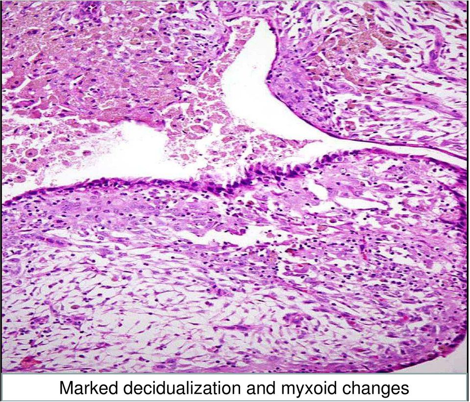
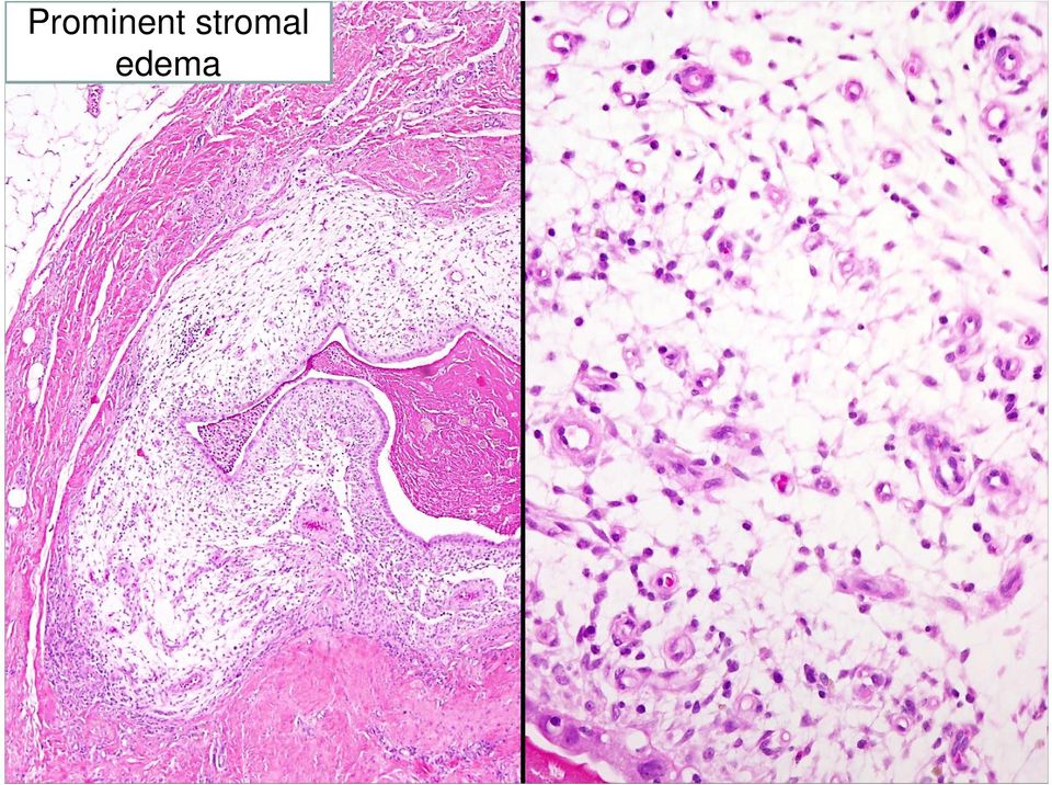
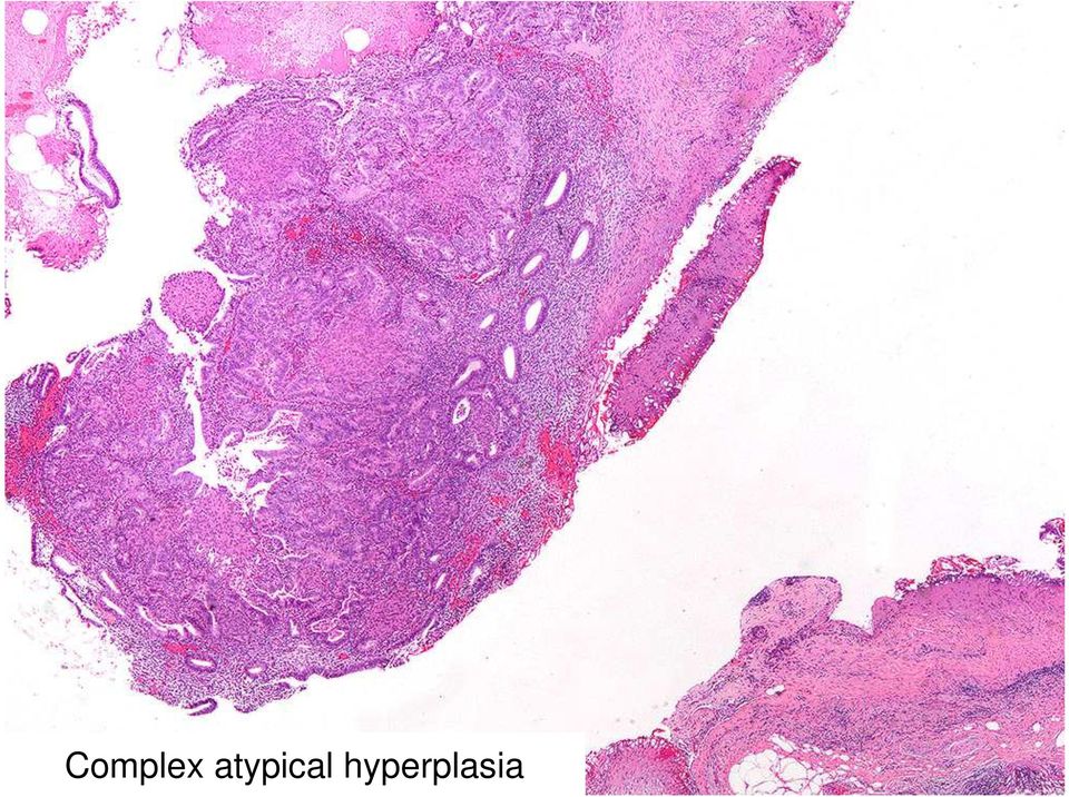
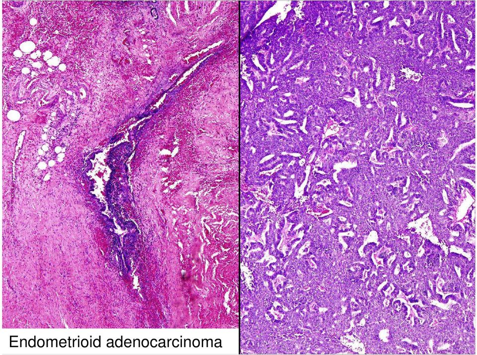
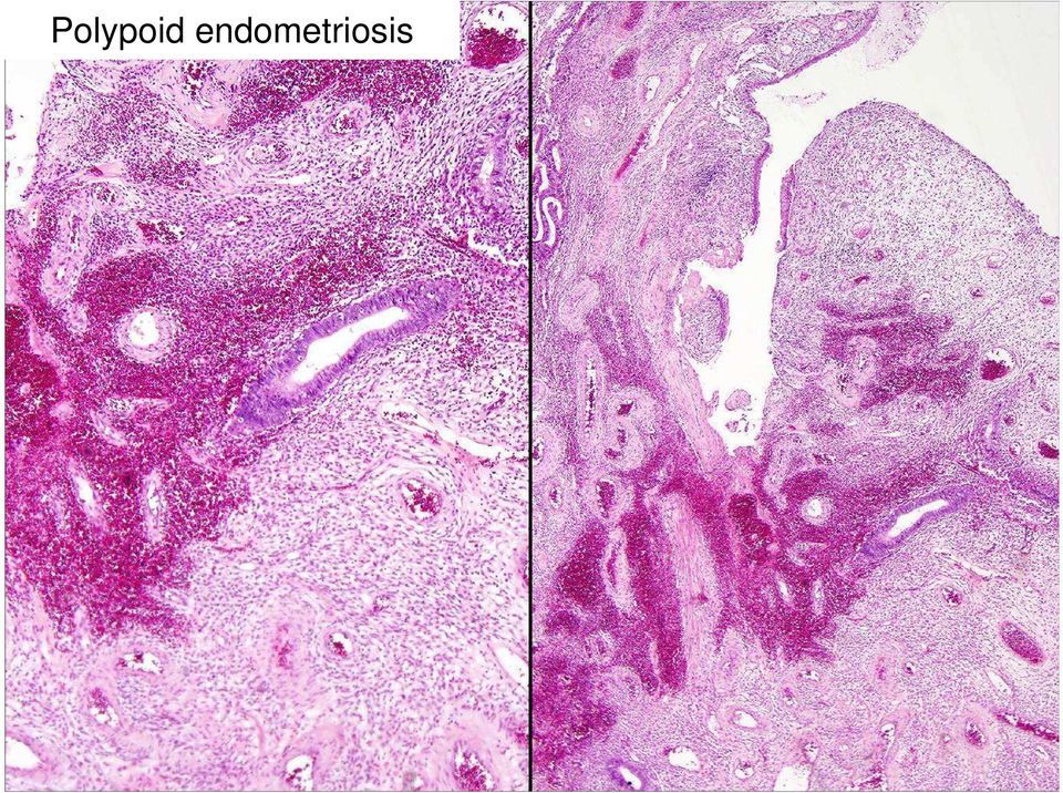
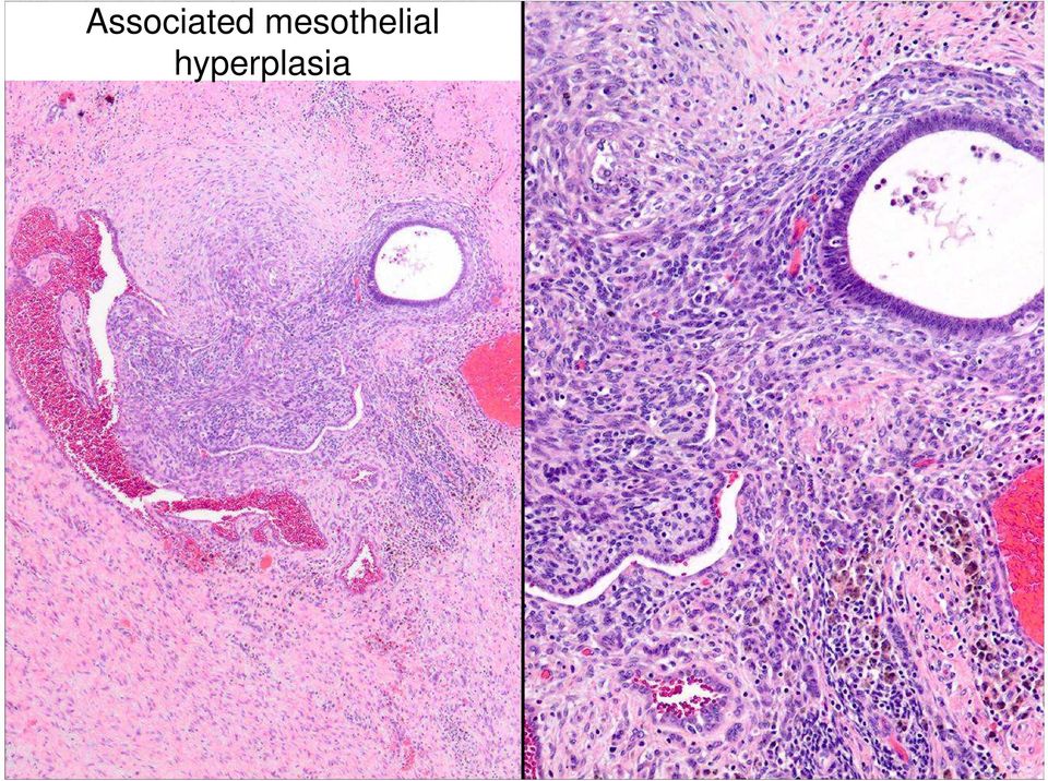
7 Endometriosis Some of the problems: Stromal changes Hyperplasia and carcinoma Polypoid endometriosis Associated mesothelial hyperplasia

8 Marked decidualization and myxoid changes
9 Prominent stromal edema
10 Complex atypical hyperplasia
11
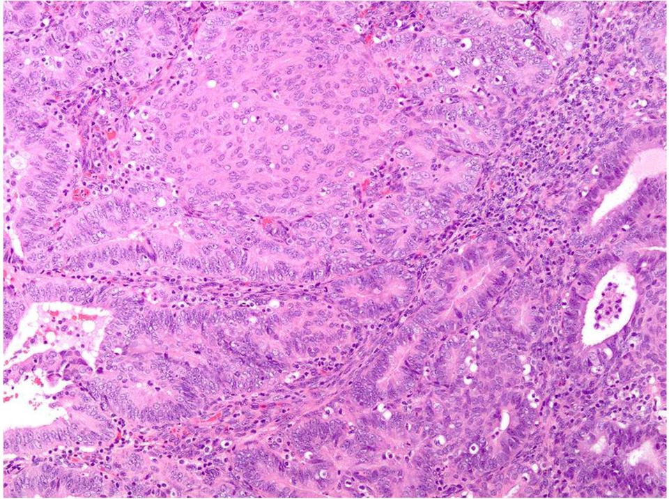
12 Endometrioid adenocarcinoma
13 Polypoid endometriosis

14 Polypoid endometriosis
15 Associated mesothelial hyperplasia
16
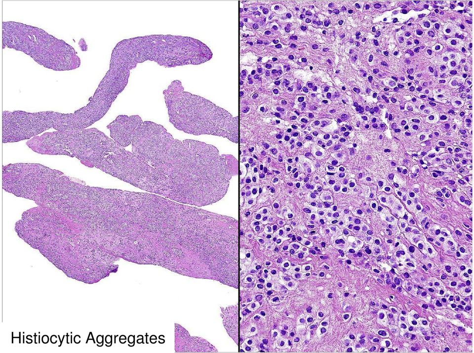
17 Histiocytic Aggregates
18 KP-1 + Inhibin - N
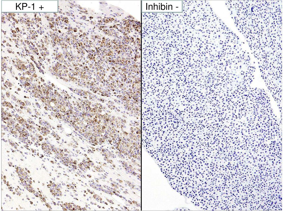
19 Histiocytic Aggregates Non-specific peritoneal inflammatory response Usually a microscopic finding Occasionally, they can be seen grossly Microscopic features: Aggregates, nodules or plaques of histiocytes Some of them with nuclear grooves Not to be mistaken for granulosa cell tumor They can be admixed with mesothelial cells

20 Multilocular Peritoneal Inclusion Cysts Uncommon lesion More frequent in women in their 20 s and 30 s Age range, 15 to 92 years Less frequent in men Rare in children In rare cases, patients were related (mother/daughter, two sisters)

21 Multilocular Peritoneal Inclusion Cysts Origin Reactive versus Neoplastic Hormonal influence: rapid growth during pregnancy A rare case reported to be associated with asbestos exposure Associated with: Previous abdominal/pelvic surgery Pelvic inflammatory disease Endometriosis
22 Multilocular Peritoneal Inclusion Cysts Symptoms Abdominal / pelvic pain Abdominal or pelvic mass Inguinal or incisional hernia Non-specific symptoms: dyspareunia, constipation, uterine bleeding Absent (10% of cases detected incidentally) Usually not associated with ascites
23 Multilocular Peritoneal Inclusion Cysts Predominantly in the pelvis Involving the surface of the uterus fallopian tube ovary Sites of Involvement
24 Multilocular Peritoneal Inclusion Cysts Sites of Involvement Predominantly in the pelvis Involving the surface of the cul de sac bladder rectum pelvic wall
25 Multilocular Peritoneal Inclusion Cysts Sites of Involvement Abdominal structures Surface of the small and large bowel Omentum Anterior abdominal wall Stomach Liver
26 Multilocular Peritoneal Inclusion Cysts Sites of Involvement Retroperitoneum Pancreas Spleen Pericardium Pleura
27 Multilocular Peritoneal Inclusion Cysts Distribution Solitary Localized Multiple, noncontiguous lesions confined to a localized peritoneal area (e.g., left lower quadrant) Diffuse
28 Multilocular Peritoneal Inclusion Cysts Gross Cyst (s) ranging in size from a few mm to more than 20 cm filled with clear/yellow serous or blood-tinged fluid, less frequently gelatinous A rare case has been found as a freefloating pelvic cyst
29 Multilocular Peritoneal Inclusion Cysts
30
31
32
33 Metaplastic changes
34 Hobnail cells
35 Reactive changes
36 Adenomatoid areas
37 Calretinin (+)
38 Keratin 5/6 (+)
39 MOC-31 and Ber-EP4 (-)
40 Multilocular Peritoneal Inclusion Cysts Immunoperoxidase studies for estrogen and progesterone receptors 14 cases Estrogen receptor (+, diffusely) 1 case Progesterone receptor (+, focally) 1 case Estrogen and progesterone receptors (+, focally) 1 case Sawh R, Malpica A, Deavers MT, et al. 2003
41 Multilocular Peritoneal Inclusion Cysts Differential Diagnosis Mesothelioma It can have areas resembling a benign cystic mesothelioma. Therefore, thorough sampling is of utmost importance to make an accurate diagnosis
42 Lymphangioma Multicystic abdominal lymphangiomas Mostly in males, < 5 year-old In adults, often an incidental finding They can reach a large size (omentum, mesentery, retropertioneum, mesocolon) Para-ovarian lymphangioma
43 The cyst wall contains bundles of smooth muscle and aggregates of lymphoid tissue
44 Lymphangioma Immunohistochemical studies: CD31(+) CD34 (+) D2-40 (+) Factor VIII (+)
45 Cystic Endosalpingiosis Tubal-type epithelium It can have blunt papillae and psammoma bodies IHC Calretinin (-) Keratin 5/6 (-)
46 Endometriosis with Stromal Changes
47 Clear Cell Ca Cystic areas Flattened epithelium Polygonal cells with atypia and mitosis
48 Clear Cell Carcinoma Immunohistochemical studies: HNF-1 β, Leu-M1, Ber-EP4, MOC-31 (+) Calretinin and keratin 5/6 (-)
49 Unilocular Cyst of Mesothelial Origin Single cyst Children or adults Origin: developmental Location: mesocolon, mesentery of small intestine, omentum, retroperitoneum
50 Multilocular Peritoneal Inclusion Cysts Treatment Surgical Hormone therapy Gonadotropin-releasing hormone agonist (leuprolide) Tamoxifen Megestrol
51 Multilocular Peritoneal Inclusion Cysts Treatment Other (limited experience) Observation Image-guided aspiration and sclerotherapy Laser ablation Hyperthermic peritoneal perfusion in patients with recurrences Excision of all visible lesions and peritonectomy
52 Multilocular Peritoneal Inclusion Cysts Prognosis Benign behavior Without treatment patients can die of disease due to local progression A rare case has been reported as having malignant transformation within a period of 10 years diagnosis was incidentally made, but soon after the patient was found to have multiple lesions? adequate sampling? accurate diagnosis
53 Multilocular Peritoneal Inclusion Cysts Recurrences can be seen in up to 50% of the cases Recurrences can be seen up to 20 years after the initial diagnosis
54 Well Differentiated Papillary Mesothelioma Uncommon mesothelial tumor mostly seen in women over a wide age range (18 to 75 years of age) However, patients are usually in their 30 s and 40 s Rare in men
55 Well Differentiated Papillary Mesothelioma It is infrequently seen at other anatomical sites: Pleura Pericardium Tunica vaginalis Epididymis Rare cases associated with asbestos exposure
56 Well Differentiated Papillary Mesothelioma Usually, it is an incidental finding in patients who undergo surgery for endometriosis, neoplasms or infertility Some cases with: Acute abdomen due to bleeding or torsion Abdomino/pelvic pain
57 Well Differentiated Papillary Mesothelioma Gross Single or multiple lesions A few mm up to 2 cm (Malpica et al, 2012) Up to 5 cm (Chen et al, 2013)
58
59 Bland mesothelial cells with no mitosis or up to 1 mitosis per 10 HPFs
60
61 seedling < 0.5 mm
62 WDPM in the fimbrial end of the fallopian tube Calretinin +
63 Well Differentiated Papillary Mesothelioma Differential Diagnosis Mesothelial hyperplasia Papillae usually with only mesothelial cells or very little fibroconnective tissue core Inflammatory/reactive changes in the vicinity Malignant mesothelioma Clinicopathologic correlation Serous tumor of low malignant potential Cells are mostly columnar with occasional cuboidal cells, with the reverse being true for well differentiated papillary mesothelioma
64 Well Differentiated Papillary Mesothelioma Recurrence can be seen occasionally In our study, 1/26 cases recurred In Chen s study, no recurrences In Daya s study, 1 patient alive with disease 4 years after diagnosis At the present time, it is prudent to consider this lesion as a tumor of uncertain malignant potential that requires follow-up
65 Malignant Peritoneal Mesothelioma It accounts for 17-32% of mesotheliomas in females Age range, 17 to 92 years of age (mean age, 47 years) Baker P, et al, to 85 years of age (median age, 49 years) Malpica A, et al, 2014
66
67 5 female patients, age range 8 to 15 years Three case reports with younger patients: 2, 3 and 5 years of age
68 Malignant Peritoneal Mesothelioma Etiologic Factors: Asbestos exposure (+/-) Exposure to erionite (a mineral fiber found in Turkey) Therapeutic irradiation Exposure to simian virus 40 (SV 40) Chronic peritoneal irritation
69 Malignant Peritoneal Mesothelioma A family history of carcinoma detected in 30/42 cases Malpica A, et al, 2014
70 Malignant Peritoneal Mesothelioma Clinical Presentation Abdominal pain or discomfort Abdominal distension Nausea Anorexia Weight loss Abdominal or pelvic mass Bowel obstruction CA-125 can be elevated Incidental
71 In 13 cases the dx of mesothelioma was an incidental operative finding
72 Malignant Peritoneal Mesothelioma Gross Features Multiple or solitary Firm/soft/friable Gray, pink, brown, tan, white Nodules, plaques, granules, adhesions or papillary excrescences along the peritoneal surfaces Circumscribed mass
73 Epithelioid Type
74 Sarcomatoid Type
75 Biphasic Type
76 Variable nuclear atypia
77 Papillary
78 Tubular
79 Tubulopapillary
80 Solid
81 Cords
82 Single cells
83 Deciduoid
84 Abundant Foamy Histiocytes
85 Localized Mesothelioma
86 Mesothelioma with Mucin
87 Alcian blue at ph 2.5 (+)
88 Calretinin (+) Cytokeratin 5/6 (+)
89 MOC-31 (-) Ber-EP4 (-)
90 CEA (-) Leu- M1 (CD 15) (-)
91 Histochemical Studies Most cases of epithelioid mesothelioma Mucicarmine (-) Rare mucicarmine (+) PAS (+), PAS-D (-) Glycogen Rare mesothelioma, PAS-D (+) Alcian blue, hyaluronidase-sensitive (+) Hyaluronic acid (acid mucin) Adenocarcinomas contain neutral mucin, PAS-D (+) and Mucicarmin (+)
92 Immunohistochemical Studies Gyn Section/MDACC traditional panel 2 positive markers for mesothelioma Calretinin Keratin 5/6 Thrombomodulin 2 negative markers for mesothelioma PAX-8 Estrogen receptor Ber-EP4 MOC-31 (or B72.3)
93
94 PAX-8, Serous Ca Claudin 4, Serous Ca PAX-8 and Claudin-4 appear to be the best positive markers for carcinoma
95 Malignant Mesothelioma- IHC Do We Ever Get Conflicting Results?
96 Calretinin (+)
97 ER(+)
98 B72.3 (-) CA19-9 (-) MOC-31 (-) Ber-EP4 (-)
99 Other Immunoperoxidase Studies Keratin 5/6, focally positive PR (-)
100 EM Long, slender,non-branching, bushy microvilli
101 PAX-8 in Mesothelial Lesions PAX-8 + Malignant mesothelioma, 0/33 cases WDPM, 9/13 cases polyclonal antibody showed more diffuse staining than monoclonal antibody Peritoneal inclusion cyst, 1/5 cases PAX-8 + (monoclonal antibody) Malignant mesothelioma, 5/14 cases WDPM, 1/9 cases Peritoneal inclusion cyst, 11/16 cases Banet N, et al, 2014 Pandya D, et al, 2014
102 Malignant Mesothelioma Do we always need to see invasion? Not always However, either a mass or diffuse involvement of the peritoneal surface needs to be seen
103 9 cm cul de sac mass
104 Calretinin (+) CK 5/6 (+)
105 Focal invasion
106 Focal invasion
107 Mesothelial Hyperplasia
108 Mesothelial Hyperplasia
109 Mesothelial Hyperplasia Gross description is important Microscopically: it shows no evidence of invasion into the adipose, fibroconnective or fibromuscular tissue as usually seen in malignant mesothelioma However, malignant mesothelioma can grow diffusely without invasion
110 Mesothelial Hyperplasia IHC Mesothelial hyperplasia cells: desmin (+, 85%), and EMA (-,80%), p53 (-, 100%), GLUT -1 (-, 87-97%), and IMP3 (-, %) Malignant mesothelioma: desmin (-, 90%) and EMA (+, 80%), GLUT-1 (+, 60-67%), p53 (+, 45%), IMP3 (+, 53-73%) However, exceptions to the these results occur, limiting the value of the studies
111 Well Differentiated Papillary Mesothelioma 1. There is no infiltration of underlying tissue 2. No complexity of the papillary growth
112 Low Grade Serous Carcinoma
113 High Grade Serous Carcinoma
114 Clear Cell Carcinoma
115 Histiocytic reaction/aggregates KP-1 (CD68) (+)
116 Metastatic Adenocarcinoma
117 Differential Diagnosis Carcinosarcoma Ectopic decidua Sex cord stromal tumor (adult granulosa cell tumor or Sertoli-Leydig cell tumor)
118 Treatment No standard treatment Cytoreductive surgery with hyperthermic intraperitoneal chemotherapy with or without postoperative chemotherapy
119 Prognosis Many tumors are highly aggressive Some tumors have a relatively indolent course The following features have been associated with a worse outcome: Diffuse growth pattern Biphasic and sarcomatoid histology Increased mitotic activity (> 4 mitoses per 10 HPFs) p16 loss
120 Summary Multilocular Peritoneal Inclusion Cysts Thorough sampling is required to ensure a correct diagnosis It can recur
121 Summary Well Differentiated Papillary Mesothelioma Thorough sampling is required to ensure a correct diagnosis It can recur
122 Summary Malignant Mesothelioma In women, this disease can have an indolent course Invasion can be focal and not required for diagnosis if there is a mass
123
Today s Topics. Tumors of the Peritoneum in Women
 Today s Topics Tumors of the Peritoneum in Women Charles Zaloudek, M.D. Department of Pathology 505 Parnassus Ave., M563 University of California, San Francisco San Francisco, CA USA [email protected]
MALIGNANT MESOTHELIOMA UPDATE ON PATHOLOGY AND IMMUNOHISTOCHEMISTRY
 MALIGNANT MESOTHELIOMA CLASSIFICATION MALIGNANT MESOTHELIOMA UPDATE ON PATHOLOGY AND IMMUNOHISTOCHEMISTRY Sisko Anttila, MD, PhD Jorvi Hospital Laboratory of Pathology Helsinki University Hospital Espoo,
MALIGNANT MESOTHELIOMA UPDATE ON PATHOLOGY AND IMMUNOHISTOCHEMISTRY
 MALIGNANT MESOTHELIOMA UPDATE ON PATHOLOGY AND IMMUNOHISTOCHEMISTRY Sisko Anttila, MD, PhD Jorvi Hospital Laboratory of Pathology Helsinki University Hospital Espoo, Finland 2nd Nordic Conference on Applied
Case presentation. Awatif Al-Nafussi
 Case presentation Awatif Al-Nafussi Case History 49 year old DVT & small PE June 08, Pelvic mass Ca125 33 Laparotomy-TAHBSO, drainage of ascites Ovarian carcinoma Clinical diagnosis Multiple specimens
Effusions: Mesothelioma and Metastatic Cancers
 Effusions: Mesothelioma and Metastatic Cancers Malignant Mesothelioma Incidence: 2,500 cases/year ~60-80% pts with pleural MM relationship with asbestos exposure Other risk factors: radiation, other carcinogens,
Outline. Workup for metastatic breast cancer. Metastatic breast cancer
 Metastatic breast cancer Immunostain Update: Diagnosis of metastatic breast carcinoma, emphasizing distinction from GYN primary 1/3 of breast cancer patients will show metastasis 1 st presentation or 20-30
MALIGNANT MESOTHELIOMA: A TYPICAL PRESENTATION IN AN ATYPICAL PATIENT
 MALIGNANT MESOTHELIOMA: A TYPICAL PRESENTATION IN AN ATYPICAL PATIENT Written by: Karyn Varley MS, SCT(ASCP) The donating laboratory would like to remain anonymous. PATIENT HISTORY 28 year old female Lived
Case of the. Month October, 2012
 Case of the Month October, 2012 Case The patient is a 47-year-old male with a 3-week history of abdominal pain. A CT scan of the abdomen revealed a suggestion of wall thickening at the tip of the appendix
Update on Mesothelioma
 November 8, 2012 Update on Mesothelioma Intro incidence and nomenclature Update on Classification Diagnostic specimens Morphologic features Epithelioid Histology Biphasic Histology Immunohistochemical
TUMORS OF THE TESTICULAR ADNEXA and SPERMATIC CORD
 TUMORS OF THE TESTICULAR ADNEXA and SPERMATIC CORD Victor E. Reuter, MD Memorial Sloan-Kettering Cancer Center [email protected] 66 th Annual Pathology Seminar California Society of Pathologists Short
Protocol for the Examination of Specimens From Patients With Tumors of the Peritoneum
 Protocol for the Examination of Specimens From Patients With Tumors of the Peritoneum Protocol applies to all primary borderline and malignant epithelial tumors and malignant mesothelial neoplasms of the
JOURNAL OF CLINICAL AND DIAGNOSTIC RESEARCH
 JOURNAL OF CLINICAL AND DIAGNOSTIC RESEARCH How to cite this article: DEBNATH S, MISRA V, SINGH PA, SINGH M. LOW GRADE CYSTIC MESOTHELIOMA OF RECTUS SHEATH.Journal of Clinical and Diagnostic Research [serial
The develpemental origin of mesothelium
 Mesothelioma Tallinn 14.12.06 Henrik Wolff Finnish Institute of Occupational Health The develpemental origin of mesothelium Mesodermal cavities (pleura, peritoneum and pericardium ) are lined with mesenchymal
PRIMARY SEROUS CARCINOMA OF PERITONEUM: A CASE REPORT
 PRIMARY SEROUS CARCINOMA OF PERITONEUM: A CASE REPORT Dott. Francesco Pontieri (*) U.O. di Anatomia Patologica P.O. di Rossano (CS) Dott. Gian Franco Zannoni Anatomia Patologica Facoltà di Medicina e Chirurgia
Protocol applies to all primary borderline and malignant epithelial tumors, and malignant mesothelial neoplasms of the peritoneum.
 Peritoneum Protocol applies to all primary borderline and malignant epithelial tumors, and malignant mesothelial neoplasms of the peritoneum. Protocol revision date: January 2004 No AJCC/UICC staging system
Case presentation: Mesothelioma of the tunica vaginalis. Dr Ben Shepherd Pathology Queensland Princess Alexandra Hospital Brisbane
 Case presentation: Mesothelioma of the tunica vaginalis Dr Ben Shepherd Pathology Queensland Princess Alexandra Hospital Brisbane A 76 year old man presented June 2011 with a 6 month history of painless
J of Evidence Based Med & Hlthcare, pissn- 2349-2562, eissn- 2349-2570/ Vol. 2/Issue 33/Aug. 17, 2015 Page 5063
 PERITONEAL MALIGNANT MESOTHELIOMA: A RARE S. R. Dhamotharan 1, S. Shanthi Nirmala 2, F. Celine Foustina Mary 3, M. Arul Raj Kumar 4, R. Vinothprabhu 5 HOW TO CITE THIS ARTICLE: S. R. Dhamotharan, S. Shanthi
Malignant Peritoneal Mesothelioma in Women A Study of 75 Cases With Emphasis on Their Morphologic Spectrum and Differential Diagnosis
 Anatomic Pathology / MALIGNANT PERITONEAL MESOTHELIOMA IN WOMEN Malignant Peritoneal Mesothelioma in Women A Study of 75 Cases With Emphasis on Their Morphologic Spectrum and Differential Diagnosis Patricia
MESOTHELIAL LESIONS OF THE PERITONEUM
 1 MESOTHELIAL LESIONS OF THE PERITONEUM Philip B. Clement, MD Departments of Pathology, Vancouver General Hospital and the University of British Columbia 2 Mesothelial lesions are commonly encountered
Frequently Asked Questions About Ovarian Cancer
 Media Contact: Gerri Gomez Howard Cell: 303-748-3933 [email protected] Frequently Asked Questions About Ovarian Cancer What is ovarian cancer? Ovarian cancer is a cancer that forms in tissues
Académie internationale de Pathologie - Division arabe XX ème congrès 24-26 novembre 2008 Alger. Immunohistochemistry in malignant mesotheliomas
 Académie internationale de Pathologie - Division arabe XX ème congrès 24-26 novembre 2008 Alger Immunohistochemistry in malignant mesotheliomas Françoise Thivolet-Béjui Groupement Hospitalier Est Lyon-Bron
INFLAMMATORY PLEURAL EFFUSION
 PLEURA- LESIONS LESIONS OF PLEURA Primary Intra pleural bacterial infections Neoplasm (mesothelioma) Secondary A complication of some underlying disease PLEURAL EFFUSION Common manifestation of both primary
3-F. Pathology of Mesothelioma
 3-F. Pathology of Mesothelioma Kouki Inai Professor of Department of Pathology, Graduate School of Biomedical Science, Hiroshima University Introduction Mesothelioma is a peculiar type of malignancy, which
Practical Effusion Cytology
 Practical Effusion Cytology A Community Pathologist s Approach to Immunocytochemistry in Body Fluid Cytology Emily E. Volk, MD William Beaumont Hospital Troy, MI College of American Pathologists 2004.
The menstrual cycle Hypophysis
 Normal endometrium Tumors of the uterine corpus by MB The endometrium comprises the zona functionalis zf (superficial two thirds) ) and the zona basalis zb (deep one third) The zf responds to hormonal
Ovarian tumors Ancillary methods
 Ovarian tumors Ancillary methods Ovarian tumor course Oslo, 24-25/11/14 Prof. Ben Davidson, MD PhD Department of Pathology, Norwegian Radium Hospital, Oslo University Hospital, Oslo, Norway Division of
Diagnosis of Mesothelioma Pitfalls and Practical Information
 Diagnosis of Mesothelioma Pitfalls and Practical Information Mary Beth Beasley, M.D. Mt Sinai Medical Ctr Dept of Pathology One Gustave L Levy Place New York, NY 10029 (212) 241-5307 [email protected]
Immunohistochemistry on cytology specimens from pleural and peritoneal fluid
 Immunohistochemistry on cytology specimens from pleural and peritoneal fluid Dr Naveena Singh Consultant Pathologist Bart health NHS Trust London United Kingdom Disclosures and Acknowledgements I have
Diseases of peritoneum Lect. Al Qassim University, Faculty of Medicine Phase II Year III, CMD 332 Pathology Department 31-32
 Diseases of peritoneum Lect Al Qassim University, Faculty of Medicine Phase II Year III, CMD 332 Pathology Department 31-32 Describe the etiology, pathogenesis and types of peritonitis Define ascites and
Notice of Faculty Disclosure
 The Diagnosis of Malignant Mesothelioma Andrew Churg, MD Department of Pathology University of British Columbia Vancouver, BC, Canada [email protected] Notice of Faculty Disclosure In accordance with
PATHOLOGY OF THE PLEURA: Mesothelioma and mimickers Necessity of Immunohistochemistry. M. Praet
 PATHOLOGY OF THE PLEURA: Mesothelioma and mimickers Necessity of Immunohistochemistry M. Praet Pathology of the Pleura Normal serosa: visceral and parietal layers Inflammation Neoplasia: Primary: mesothelioma
Luis D. Carcorze Soto, MD PGY-3
 Luis D. Carcorze Soto, MD PGY-3 Peritoneal Surface Malignancies Peritoneum Patient Selection Operative Technique HIPEC EPIC Primary: Primary Peritoneal Carcinoma Malignant Peritoneal Mesothelioma Metastatic:
Disclosures. Learning Objectives. Effusion = Confusion. Diagnosis Of Serous Cavity Effusions - Beware The Mesothelial Cell!
 Disclosures Diagnosis Of Serous Cavity Effusions - Beware The Mesothelial Cell! No Relevant Financial Relationships with Commercial Interests Syed Z. Ali, M.D. Syed Z. Ali, M.D. Associate Professor of
Surgical Staging of Endometrial Cancer
 Surgical Staging of Endometrial Cancer I. Endometrial Cancer Surgical Staging Overview Uterine cancer types: carcinomas type I and type II, sarcomas, carcinosarcomas Hysterectomy with BSO Surgical Staging
How To Treat A Uterine Sarcoma
 EVERYONE S GUIDE FOR CANCER THERAPY Malin Dollinger, MD, Ernest H. Rosenbaum, MD, Margaret Tempero, MD, and Sean Mulvihill, MD 4 th Edition 2001 Uterus: Uterine Sarcomas Jeffrey L. Stern, MD Uterine sarcomas
Cytoreductive Surgery and Hyperthermic Intraperitoneal Chemotherapy (HIPEC): Now and the Future
: Now and the Future") Cytoreductive Surgery and Hyperthermic Intraperitoneal Chemotherapy (HIPEC): Now and the Future Mazin Al-kasspooles, MD Associate Professor of Surgery Division of Surgical Oncology Director, Regional Therapy
Cytoreductive Surgery and Hyperthermic Intraperitoneal Chemotherapy (HIPEC): Now and the Future Mazin Al-kasspooles, MD Associate Professor of Surgery Division of Surgical Oncology Director, Regional Therapy
Clinical Commissioning Policy: Cytoreductive Surgery and Hyperthermic Intraperitoneal Chemotherapy for Peritoneal Mesothelioma
 Clinical Commissioning Policy: Cytoreductive Surgery and Hyperthermic Intraperitoneal Chemotherapy for Peritoneal Mesothelioma Reference: NHS England B03/P/a 1 Information Reader Box (IRB) to be inserted
Gynecology Abnormal Physiology of the ovaries. Simple Cystic Masses
 Gynecology Abnormal Physiology of the ovaries (Effective February 2007) pediatric, reproductive, and perimenopausal/postmenopausal (24-28 %) Simple Cystic Masses ovary s function is to mature oocytes until
DESMOPLASTIC SMALL ROUND CELL TUMOR: A RARE PATHOLOGY PUZZLE
 DESMOPLASTIC SMALL ROUND CELL TUMOR: A RARE PATHOLOGY PUZZLE Ryan Granger University of Rhode Island Cytotechnology program May 2, 2015 ASCT Annual Meeting Nashville, Tennessee DESMOPLASTIC SMALL ROUND
Diagnostic Challenge. Department of Pathology,
 Cytology of Pleural Fluid as a Diagnostic Challenge Paavo Pääkkö,, MD, PhD Chief Physician and Head of the Department Department of Pathology, Oulu University Hospital,, Finland Oulu University Hospital
Introduction Ovarian cysts are a very common female condition. An ovarian cyst is a fluid-filled sac on an ovary in the female reproductive system.
 Ovarian Cysts Introduction Ovarian cysts are a very common female condition. An ovarian cyst is a fluid-filled sac on an ovary in the female reproductive system. Most women have ovarian cysts sometime
OVARIAN CYSTS. Types of Ovarian Cysts There are many types of ovarian cysts and these can be categorized into functional and nonfunctional
 OVARIAN CYSTS Follicular Cyst Ovarian cysts are fluid-filled sacs that form within or on the ovary. The majority of these cysts are functional meaning they usually form during a normal menstrual cycle.
Omental mesothelioma as a diagnostic and therapeutic challenge: A case report
 www.edoriumjournals.com Case in Images OPEN ACCESS Omental mesothelioma as a diagnostic and therapeutic challenge: A case report Cihan Akgul Ozmen, Yekta Tuzun, Hatice Ozturkmen Akay, Hasan Nazaroglu ABSTRACT
Cytology : first alert of mesothelioma? Professor B. Weynand, UCL Yvoir, Belgium
 Cytology : first alert of mesothelioma? Professor B. Weynand, UCL Yvoir, Belgium Introduction 3 cavities with the same embryologic origin the mesoderme Pleura Exudates Pleura Peritoneum Pericardium 22%
How To Test For Cancer
 Diagnosis Of Serous Cavity Effusions - Beware The Mesothelial Cell! Effusion = Confusion Syed Z. Ali, M.D. Professor of Pathology and Radiology The Johns Hopkins Hospital Baltimore, Maryland Diagnostic
Something Old, Something New.
 Something Old, Something New. Michelle A. Fajardo, D.O. Loma Linda University Medical Center Clinical Presentation 6 year old boy, presented with hematuria Renal mass demonstrated by ultrasound & CT scan
ProSono Copyright 2006. Ovarian Pathology
 Ovarian Pathology Physiologic cysts: Functional cysts Pathology: A simple cyst is a sac containing fluid or semi-solid material. Physiologic cysts are generic types of hormonally active cysts that result
WOMENCARE A Healthy Woman is a Powerful Woman (407) 898-1500. Endometriosis
 898-1500. Endometriosis") Endometriosis WOMENCARE A Healthy Woman is a Powerful Woman (407) 898-1500 The lining of the uterus is called the endometrium. Sometimes, endometrial tissue grows elsewhere in the body. When this happens
Endometriosis WOMENCARE A Healthy Woman is a Powerful Woman (407) 898-1500 The lining of the uterus is called the endometrium. Sometimes, endometrial tissue grows elsewhere in the body. When this happens
Ovarian mucinous lesions. Ovarian mucinous lesions: Common diagnostic dilemmas. Ovarian mucinous lesions: problematic issues
 Ovarian mucinous lesions Ovarian mucinous lesions: Common diagnostic dilemmas Karuna Garg, MD University of California San Francisco Intestinal or usual type Seromucinous (Endocervical mucinous or Mullerian
Male. Female. Death rates from lung cancer in USA
 Male Female Death rates from lung cancer in USA Smoking represents an interesting combination of an entrenched industry and a clearly drug-induced cancer Tobacco Use in the US, 1900-2000 5000 100 Per Capita
From the Archives of the AFIP
 Note: This copy is for your personal non-commercial use only. To order presentation-ready copies for distribution to your colleagues or clients, contact us at www.rsna.org/rsnarights. AFIP ARCHIVES 583
HKCPath Anatomical Pathology Peer Review and Scores : PDF version for download
 AP2003R1 http://hkcpath.org. Correspondence: [email protected] 1of 10 07/08/2003 HKCPath Anatomical Pathology Peer Review and Scores : PDF version for download AP141 Bone Marrow: Metastatic Carcinoma from
Ovarian cysts Diagnosis and Management
 Ovarian cysts Diagnosis and Management Mr P K Athanasias MRCOG Consultant Gynaecologist St Anthony s Hospital [email protected] Introduction ovary is an ovum-producing reproductive organ located in
Ovarian Cancer. in Georgia, 1999-2003. Georgia Department of Human Resources Division of Public Health
 Ovarian Cancer in Georgia, 1999-23 Georgia Department of Human Resources Division of Public Health Acknowledgments Georgia Department of Human Resources......B. J. Walker, Commissioner Division of Public
Cystic mesothelioma of the peritoneum
 Pathology Feature William R. Hart, M.D. Section Editor Cystic mesothelioma of the peritoneum Report of a case with multiple recurrences and review of the literature 1 Janet M. Miles, M.D. William R. Hart,
How To Diagnose And Treat A Tumour In An Effusion
 Effusions of the Serous Cavities Annika Dejmek Professor/Consultant in Cytopathology Clinical Pathology; Department of Laboratory Medicine, Malmö, Lund University 5th EFCS Tutorial Trondheim 2012 Pleura
Understanding Your Diagnosis of Endometrial Cancer A STEP-BY-STEP GUIDE
 Understanding Your Diagnosis of Endometrial Cancer A STEP-BY-STEP GUIDE Introduction This guide is designed to help you clarify and understand the decisions that need to be made about your care for the
بسم هللا الرحمن الرحيم
 بسم هللا الرحمن الرحيم Updates in Mesothelioma By Samieh Amer, MD Professor of Cardiothoracic Surgery Faculty of Medicine, Cairo University History Wagner and his colleagues (1960) 33 cases of mesothelioma
Gynecology Abnormal Pelvic Anatomy and Physiology: Cervix. Cervix. Nabothian cysts. cervical polyps. leiomyomas. Cervical stenosis
 Gynecology Abnormal Pelvic Anatomy and Physiology: (Effective February 2007) pediatric, reproductive, and perimenopausal/postmenopausal (24-28 %) Cervix Nabothian cysts result from chronic cervicitis most
Endometriosis: An Overview
 Endometriosis: An Overview www.bcwomens.ca Welcome to the BC Women s Centre for Pelvic Pain and Endometriosis. This handout will give you some basic information about endometriosis. It will also explain
NEOPLASMS C00 D49. Presented by Jan Halloran CCS
 NEOPLASMS C00 D49 Presented by Jan Halloran CCS 1 INTRODUCTION A neoplasm is a new or abnormal growth. In the ICD-10-CM classification system, neoplastic disease is classified in categories C00 through
Index. F Factor VIII-related antigen, see VWF FactorXIIIa, for dermatofibroma, 272-275 5-HT, see Serotonin
 A Acantholytic squamous cell carcinoma vs epithelioid angiosarcoma, 56-57 Acinic cell carcinoma of pancreas, 76-77 vs ductal adenocarcinoma, 74-75 vs islet cell tumor, 78-81 Adenomatoid tumor vs hemangioma,
Diseases. Inflammations Non-inflammatory pleural effusions Pneumothorax Tumours
 Pleura Visceral pleura covers lungs and extends into fissures Parietal pleura limits mediastinum and covers dome of diaphragm and inner aspect of chest wall. Two layers between them (pleural cavity) contains
A. Pericardial smear. Examination of the pericardial aspirate can provide useful diagnostic information.
 5. PERICARDIUM Heart is encased by the pericardium which has a visceral layer (a) covering the heart and the parietal layer (b). In normal states it is thin, transparent and the myocardium can be seen
Immunohistochemical differentiation of metastatic tumours
 Immunohistochemical differentiation of metastatic tumours Dr Abi Wheal ST1. TERA 3/2/14 Key points from a review article written by Daisuke Nonaka Intro Metastatic disease is the initial presentation in
Corporate Medical Policy
 Corporate Medical Policy Hyperthermic Intraperitoneal Chemotherapy File Name: Origination: Last CAP Review: Next CAP Review: Last Review: hyperthermic_intraperitoneal_chemotherapy 5/19/2005 3/2016 3/2017
The Adnexal Mass and Early Ovarian Cancer
 The Adnexal Mass and Early Ovarian Cancer Fred Ueland, MD University of Kentucky Gynecologic Oncology Never give in. Never give in. Never, never, never, never- in nothing great or small, large or petty-
Carcinosarcoma of the Ovary
 Carcinosarcoma of the Ovary A Rare Finding Presented By: Kathryn Kiely Anisa I. Kanbour School of Cytotechnology of the University of Pittsburgh Medical Center Pittsburgh, PA Patient History 55 year old
A succesfull case of HIPEC in a peritoneal mesothelioma patient
 A succesfull case of HIPEC in a peritoneal mesothelioma patient Firmino, NLJ¹²; Miranda, E¹³; Oliveira, DA ²; Lima, MBA ¹²; Diniz, AF ¹²; Gomes, GES ¹²; Azevedo, LW ¹²; Soares,MC¹²; Gomes, ASA³. ¹Pernambuco
ISOLATED PANCREATIC METASTASIS OF A MALIGNANT PLEURAL MESOTHELIOMA
 ISOLATED PANCREATIC METASTASIS OF A MALIGNANT PLEURAL MESOTHELIOMA Yi-Ting Lin, 1 Bing-Shiun Wu, 2 Sheau-Fang Yang, 3 and Huang-Chi Chen 4 Departments of 1 Family Medicine, 2 Internal Medicine, and 3 Pathology,
Tumour Markers. What are Tumour Markers? How Are Tumour Markers Used?
 Dr. Anthony C.H. YING What are? Tumour markers are substances that can be found in the body when cancer is present. They are usually found in the blood or urine. They can be products of cancer cells or
Renal Pathology Update. Sundus Hussein MD, FRCPC
 Renal Pathology Update Sundus Hussein MD, FRCPC Case History A 45 year old male with incidentally discovered a 3.5 x 3.9 x 2.7 cm renal mass Handling partial nephrectomy Handling partial nephrectomy
Ovarian Cancer: A Case Report
 Ovarian Cancer: A Case Report Abstract Ovarian cancer is a very common cancer among women. It is an extremely diverse disease requiring several treatment options. Occasionally ovarian cancer is diagnosed
Mesothelioma: Questions and Answers
 CANCER FACTS N a t i o n a l C a n c e r I n s t i t u t e N a t i o n a l I n s t i t u t e s o f H e a l t h D e p a r t m e n t o f H e a l t h a n d H u m a n S e r v i c e s Mesothelioma: Questions
Fallopian Tube Cancer
 Fallopian Tube Cancer Fred Ueland, MD University of Kentucky Gynecologic Oncology Anatomy Three layers of the fallopian tube: Internal mucosa (endosalpinx) Intermediate muscular layer (myosalpinx( myosalpinx)
POSTMENOPAUSAL ASSESS AND WHAT TO DO
 POSTMENOPAUSAL OVARIAN CYSTS:HOW TO ASSESS AND WHAT TO DO Steven R. Goldstein, MD Professor of Obstetrics and Gynecology Director of Gynecologic Ultrasound Co-Director, Bone Densitometry New York University
INTRAPERITONEAL HYPERTHERMIC CHEMOTHERAPY (IPHC) FOR PERITONEAL CARCINOMATOSIS AND MALIGNANT ASCITES. INFORMATION FOR PATIENTS AND FAMILY MEMBERS
 FOR PERITONEAL CARCINOMATOSIS AND MALIGNANT ASCITES. INFORMATION FOR PATIENTS AND FAMILY MEMBERS") INTRAPERITONEAL HYPERTHERMIC CHEMOTHERAPY (IPHC) FOR PERITONEAL CARCINOMATOSIS AND MALIGNANT ASCITES. INFORMATION FOR PATIENTS AND FAMILY MEMBERS Description of Treatment A major difficulty in treating
INTRAPERITONEAL HYPERTHERMIC CHEMOTHERAPY (IPHC) FOR PERITONEAL CARCINOMATOSIS AND MALIGNANT ASCITES. INFORMATION FOR PATIENTS AND FAMILY MEMBERS Description of Treatment A major difficulty in treating
Renal Cell Carcinoma: Advances in Diagnosis B. Iványi, MD
 Renal Cell Carcinoma: Advances in Diagnosis B. Iványi, MD Department of Pathology University of Szeged, Hungary ISUP Vancouver Classification of Renal Neoplasia Am J Surg Pathol 37:14691489, 2013 13 histologic
Emerging Subtypes in Renal Cancer. Donna E. Hansel, MD PhD Professor of Pathology, UC San Diego Division Chief, Anatomic Pathology dhansel@ucsd.
 Emerging Subtypes in Renal Cancer Donna E. Hansel, MD PhD Professor of Pathology, UC San Diego Division Chief, Anatomic Pathology [email protected] Some General Comments Fuhrman nuclear grading clear cell
Malignant Mesothelioma
 Malignant Malignant mesothelioma is a tumour originating from mesothelial cells. 85 95% of mesotheliomas are caused by asbestos exposure. It occurs much more commonly in the chest (malignant pleural mesothelioma)
Malignant Mesothelioma
 Malignant mesothelioma is a tumour originating from mesothelial cells. 85 95% of mesotheliomas are caused by asbestos exposure. It occurs much more commonly in the chest (malignant pleural mesothelioma)
National Coverage Determination (NCD) for Tumor Antigen by Immunoassay - CA 125 (190.28)
 for Tumor Antigen by Immunoassay - CA 125 (190.28)") National Coverage Determination (NCD) for Tumor Antigen by Immunoassay - CA 125 (190.28) Tracking Information Publication Number Manual Section Number 100-3 190.28 Manual Section Title Tumor Antigen by
National Coverage Determination (NCD) for Tumor Antigen by Immunoassay - CA 125 (190.28) Tracking Information Publication Number Manual Section Number 100-3 190.28 Manual Section Title Tumor Antigen by
Benign Ovarian Masses
 Benign Ovarian Masses Anthony Hanbidge Learning Objectives Describe technique for assessment of ovarian masses Explain importance of transvaginal scan List the common benign masses Specify distinguishing
Primary malignant gonadal mesotheliomas and asbestos
 Histopathology 2000, 37, 150±159 Primary malignant gonadal mesotheliomas and asbestos R L Attanoos & A R Gibbs Department of Histopathology, University Hospital of Wales and Llandough Hospital, Cardiff,
Ovarian Cancer 101 Jessica McAlpine, MD
 Ovarian Cancer 101 Jessica McAlpine, MD Different types of ovarian cancer: Outline: Ovarian Cancer Presentation, behavior, site of origin Primary treatment: surgery, chemo, +/-radiation Role of genetics
A 70-year old Man with Pleural Effusion
 Mesothelioma Diagnosis: Pitfalls and Latest Updates S Klebe and DW Henderson Recommendations Indisputable malignant cells on cytomorphological criteria which demonstrate a mesothelial phenotype, which
Metastatic Cervical Cancer s/p Radiation Therapy, Radical Hysterectomy and Attempted Modified Internal Hemipelvectomy
 Metastatic Cervical Cancer s/p Radiation Therapy, Radical Hysterectomy and Attempted Modified Internal Hemipelvectomy Sarah Hutto,, MSIV Marc Underhill, M.D. January 27, 2009 Past History 45 yo female
ATLAS OF HEAD AND NECK PATHOLOGY THYROID PAPILLARY CARCINOMA
 Papillary carcinoma is the most common of thyroid malignancies and occurs in all age groups but particularly in women under 45 years of age. There is a high rate of cervical metastatic disease and yet
Adjuvant Therapy for Breast Cancer: Questions and Answers
 CANCER FACTS N a t i o n a l C a n c e r I n s t i t u t e N a t i o n a l I n s t i t u t e s o f H e a l t h D e p a r t m e n t o f H e a l t h a n d H u m a n S e r v i c e s Adjuvant Therapy for Breast
What is Mesothelioma?
 What is Mesothelioma? Mesothelioma is a rare type of cancer that develops in the mesothelial cells found in one s body. These cells form membranous linings that surround and protect the body s organs and
Peritoneal Carcinosis
 Peritoneal Carcinosis What is it and how to cure it Peritoneum Peritoneum is a thin and transparent membrane that covers the internal part of the abdominal and pelvic cavity and all the viscera contained
SEMESTER VI 3 RD YEAR PATHOLOGY KIDNEY TUMORS
 SEMESTER VI 3 RD YEAR PATHOLOGY KIDNEY TUMORS LEARNING OBJECTIVES At the end of the lecture, students should be able to: Know the pathology of renal tumors. RENAL TUMORS RENAL PAPILLARY ADENOMA Common
Primary -Benign - Malignant Secondary
 TUMOURS OF THE LUNG Primary -Benign - Malignant Secondary The incidence of lung cancer has been increasing almost logarithmically and is now reaching epidemic levels. The overall cure rate is very low
Endometriosis Obstetrics & Gynaecology Women and Children s Group
 Endometriosis Obstetrics & Gynaecology Women and Children s Group This leaflet has been designed to give you important information about your condition / procedure, and to answer some common queries that
Case based applications part III
 Case based applications part III Los Angeles Society Of Pathologists January 25, 2014 Sanja Dacic, MD, PhD University of Pittsburgh Medical Center 1 CASE 1 A 44-year-old woman with multiple lung nodules.
Understanding CA 125 Levels A GUIDE FOR OVARIAN CANCER PATIENTS. foundationforwomenscancer.org
 Understanding CA 125 Levels A GUIDE FOR OVARIAN CANCER PATIENTS foundationforwomenscancer.org Contents Introduction...1 CA 125................................... 1 The CA 125 Test...2 The Use of the CA
Endometrial Cancer Treatment
 Endometrial Cancer Treatment January 2006 By Shelly Smits, RHIT, CCS, CTR mary by Ian Thompson, MD Data Source: Cancer registry information on uterine cancer diagnosed 1/1/2000 to 12/31/2004. Reason for
Cardiac Masses and Tumors
 Cardiac Masses and Tumors Question: What is the diagnosis? A. Aortic valve myxoma B. Papillary fibroelastoma C. Vegetation from Infective endocarditis D. Thrombus in transit E. None of the above Answer: