RACE I Rapid Assessment by Cardiac Echo. Intensive Care Training Program Radboud University Medical Centre NIjmegen
|
|
|
- Gwendolyn Price
- 7 years ago
- Views:
Transcription
1 RACE I Rapid Assessment by Cardiac Echo Intensive Care Training Program Radboud University Medical Centre NIjmegen

2 RACE Goal-directed study with specific questions Excludes Doppler ultrasound Perform 50 full and complete RACE examiminations and discuss with supervisor RACE is not a comprehensive echocardiographic examination

3 Always... Place patient in left lateral position with left arm elevated Choose the correct transducer Enter patient s details in the machine Connect the ECG

4 Standard views Parasternal long axis (PLAX) Parasternal short axis (PSAX) Aortic, Basal (MV), Mid (Papillary), Apical Apical four chamber (A4C) Apical five chamber (A5C) Apical two chamber (A2C) Subcostal (SC) Inferior Vena Cava (IVC)
 Subcostal (SC) Inferior Vena Cava")
5 Parasternal - long axis Patient slightly turned toward left 3 d or 4 th intercostal space Scan plane from right shoulder left kidney Transducer index mark directed towards right shoulder (11 o clock)

6 Parasternal - long axis

7 Parasternal - long axis RV LV Ao LA

8 Parasternal - short axis Patient slightly turned toward left 3 d or 4 th intercostal space Turn transducer from long axis position 90 0 clockwise Transducer index mark directed toward mid left clavicle As much as possible parasternal Four planes: Aortic - Basal - Mid - Apical

9 PSAX - Aortic Tilt transducer superiorly Angle medially

10 PSAX - Aortic RVout RA LA PA

11 PSAX - Basal right ventricle Septal Anterior Anterior mitral leaflet Posterior Posterior mitral leaflet Lateral

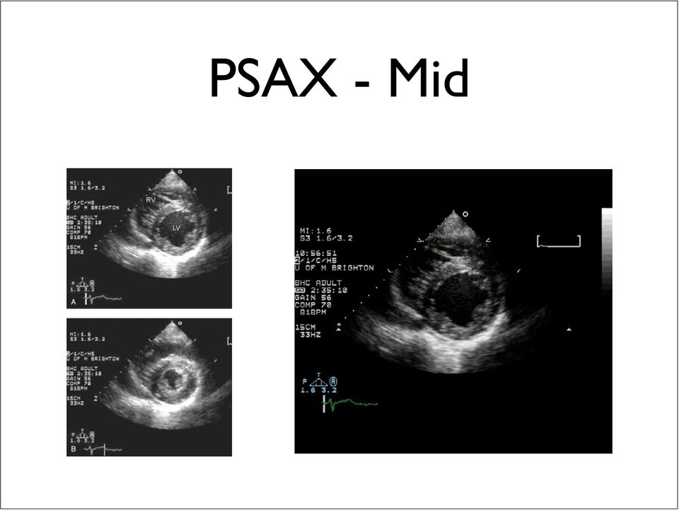
12 PSAX - Mid
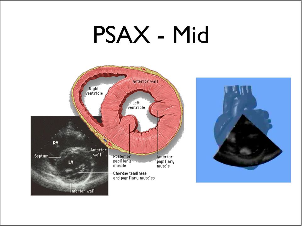
13 PSAX - Mid
14 Apical 4-chamber view Patient slightly turned toward left Transducer on apex - usually 5 th intercostal space in anterior axillary line Axis pointed towards right shoulder with index mark towards left axilla (3 o clock) Tilt transducer superiorly
 Tilt")
15 Apical 4-chamber view

16 Apical 4-chamber view RV LV RA LA

17 Apical 5-chamber view Av Aorta From 4-chamber view Tilt transducer anteriorly

18 Apical 2-chamber view Patient slightly turned toward left Transducer on apex From 4-chamber view rotate 90 0 counter clockwise with index mark pointing upwards (12 o clock)

19 Apical 2-chamber view

20 Apical 2-chamber view Anterior Inferior LV LA

21 Subcostal view Patient on back Transducer just right from xyphoid pointed toward left midclavicular region Index mark pointed towards left hip Push transducer downwards in horizontal plane - tilt anteriorly
22 Subcostal view
23 Subcostal - long axis view
24 Subcostal - IVC view Patient on back From subcostal view turn 90 0 counter clockwise with index mark upwards - sweep towards patient s right side - tilt slightly posteriorly
25 Subcostal - IVC view
26 Left Heart Assessment Size: small - normal - dilated? Left atrial size Segmental wall defects Contractility: normal - decreased - hyperdynamic Left ventricular wall thickness Aortic and mitral valve appearance
27 Chamber Size Objective M-mode Cursor through tip of mitral leaflet in diastole perpendicular to LV axis Normal dimensions LIVDd: mm LIVDs: mm IVS and PW: < 11 mm
28 Chamber Size Objective PLAX: cineloop at end-expiration. Playback and freeze at end-diastole and peak-systole respectively. Measure LV dimension. A4C: idem. LV dimension at end-diastole at 1/3 from MV leaflet tips to apex. Length from apex to MV leaflet tips.
29 Chamber Size Objective
30 PLAX Subjective A4C PsAX A2C Crude review - small - normal - dilated
31 LV contraction Objective M-mode: systolic wall thickening in PLAX (IVS and posterior wall) and PSAX (anterior and posterior wall) Simpsons method - need A4C and A2C views
32
33 PLAX Subjective A4C PsAX A2C Low - normal - hyperdynamic - symmetrical?
34 Asymmetrical contraction
35 Valvular abnormalities Aortic valve: PLAX-PSAX-A5C-A2C Movement and thickness of cusps - abnormal masses Measure diameter aorta above coronary sinuses - PLAX Mitral valve: PLAX-PSAX-A4C-A2C-SC Movement and thickness of leaflets - abnormal masses
36 Valvular abnormalities Aortic valve stenosis?
37 Valvular abnormalities AV and MV endocarditis Aortic ascending aneurysm
38 Valvular abnormalities Mitral valve stenosis?
39 Left atrial size PLAX: M-mode through aortic root and left atrium (diameter ) normal mm A4C: diameter (diagonal and cross section in mid chamber position), area, volume (Simpsons method)
40 Left atrial size PLAX - Dilated cardiomyopathy A4C - Mitral stenosis
41 RACE II Rapid Assessment by Cardiac Echo Intensive Care Training Program Radboud University Medical Centre NIjmegen
42 Right heart assessment Size: small, normal, dilated Thickness Elevated RV pressure Tricuspid valve appearance Intracardiac masses RA size Contractility: decreased, normal, hyperdynamic
43 RV size Subjective: PLAX, PSAX, A4C, SC
44 RV size Objective Compare with LV: normal ratio < severely dilated > 1
45 RV contractility Objective: TAPSE M-mode Tricuspid Annular Plane Systolic Excursion Peak-to-peak displacement normally > 2.0 cm
46 RV contractility Subjective: PLAX, PSAX, A4C, SC
47 RV wall thickness SC, A4C, PLAX Obtain still frame - measure diastolic thickness > 5 mm abnormal
48 Elevated RV pressure Dilated RA and RV Hyperdynamic RV Paradoxical septal motion PSAX and A4C Systolic flattening - pressure overload Diastolic flattening - volume overload Fixed flattening - D shaped LV: both
49 Valvular abnormalities Tricuspid valve (PSAX, A4C, SC): movement an thickness of cusps - abnormal masses Tricuspid valve prolaps Tricuspid valve endocarditis
50 Intracardiac masses Right atrial myxoma Right ventricular thrombus
51 RA size A4C and SC Subjective - high error rate Objective - A4C: AP and XS dimensions, area RA compression by hematoma
52 Pericardial assessment Pericardium Pericardial space Right atrium Right ventricle Inferior vena cava
53 Thickening Calcification Pericardium PLAX, PSAX, A4C Posterior pericardial calcification
54 Pericardial space Pericardial effusion - PLAX Minimal Small 5 mm Moderate/ Severe 5-10 mm Large swinging heart Exclude fat pad and pleural effusion
55 Tamponade Right atrium Early systolic collapse Right ventricle Early diastolic collapse Right ventricle Early diastolic collapse With tamponade IVC is dilated and/or fixed
56
57 Preload assessment Inferior vena cava Right atrium Right ventricle Left ventricle Coronary sinus
58 Inferior vena cava Subcostal - IVC view Measure diameter either with 2-D or M- mode within 2 cm from the IVC-RA junction - with M-mode make sniff manoeuvre
59 Estimation of RAP In spontaneously breathing patients IVC size at RA-IVC J/C Respiratory change RAP (mm Hg) Small (< 1.5 cm) Decrease > 50% or Collapse 0-5 mm Hg Normal ( cm) Decrease > 50% 5-10 mm Hg Normal ( cm) Decrease < 50% mm Hg Dilated (> 2.3 cm) Decrease < 50% mm Hg Dilated (> 2.3 cm) No change > 20 mm Hg
60 IVC at RA junction
61 Right atrium A4C Enlarged RA + leftward septal bowing points to elevated RAP
62 Right ventricle PSAX and A4C Diameter (A4C) - see RV size IV septum (PSAX) - shape and paradoxical motion
63 Left ventricle PLAX - PSAX - A4C Wall kissing unless significant LV hypertrophy
64 Coronary sinus PLAX If dilated consider RAP elevation, PH or persistent left superior vena cava
65 Other pathologies Type A aortic dissection
66 At the end... What is left heart function like? What is right heart function like? Is there a hemodynamically important pericardial effusion? Is significant hypovolemia present? Are there other relevant findings?
Section Four: Pulmonary Artery Waveform Interpretation
 Section Four: Pulmonary Artery Waveform Interpretation All hemodynamic pressures and waveforms are generated by pressure changes in the heart caused by myocardial contraction (systole) and relaxation/filling
What is echo? CHAPTER 1 1.1 BASIC NOTIONS. Ultrasound production and detection
 What is echo? CHAPTER 1 1.1 BASIC NOTIONS Echocardiography (echo) the use of ultrasound to examine the heart is a safe, powerful, non-invasive and painless technique. Echo is easy to understand as many
Administrative. Patient name Date compare with previous Position markers R-L, upright, supine Technical quality
 CHEST X-RAY Administrative Patient name Date compare with previous Position markers R-L, upright, supine Technical quality AP or PA ( with x-ray beam entering from back of patient, taken at 6 feet) Good
020 // Congenital Heart Disease
 020 // Congenital Heart Disease CONTENTS 188 Basics 188 Atrial Septal Defect (ASD) 191 Patent Foramen Ovale (PFO) 192 Ventricular Septal Defects (VSD) 194 Patent Ductus Arteriosus (PDA) 195 Coronary Fistulas
Practical class 3 THE HEART
 Practical class 3 THE HEART OBJECTIVES By the time you have completed this assignment and any necessary further reading or study you should be able to:- 1. Describe the fibrous pericardium and serous pericardium,
Case III. Disscussion. the UHP ultrasound protocol. Novel Ultrasound Approach to the Empiric Evaluation of the Undifferentiated Hypotensive Patient
 The UHP Ultrasound Protocol: A Novel Ultrasound Approach to the Empiric Evaluation of the Undifferentiated Hypotensive Patient JOHN S. ROSE, MD,* AARON E. BAIR, MD,* DIKU MANDAVIA, MD, AND DONNA J. KINSER,
Example Theory Multiple Choice Questions
 Example Theory Multiple Choice Questions Q a). In an ultrasound imaging system: Sector width, sector depth and frame rate can all be controlled independently b) Frame rate falls as sector width increases
Heart Murmurs. Outline. Basic Pathophysiology
 Heart Murmurs David Leder Outline I. Basic Pathophysiology II. Describing murmurs III. Systolic murmurs IV. Diastolic murmurs V. Continuous murmurs VI. Summary Basic Pathophysiology Murmurs = Math Q =
Heart Sounds & Murmurs
 Cardiovascular Physiology Heart Sounds & Murmurs Dr. Abeer A. Al-Masri MBBS, MSc, PhD Associate Professor Consultant Cardiovascular Physiologist Faculty of Medicine, KSU Detected over anterior chest wall
Normal & Abnormal Intracardiac. Lancashire & South Cumbria Cardiac Network
 Normal & Abnormal Intracardiac Pressures Lancashire & South Cumbria Cardiac Network Principle Pressures recorded from catheter tip Electrical transducer - wheatstone bridge mechanical to electrical waveform
Read It, Code It, See It
 Read It, Code It, See It Richard L. Prager, M.D. University of Michigan Ann Arbor, Michigan Dorothy Latham, R.N. Port Huron Hospital Port Huron, Michigan Nothing to Disclose Disclosure Preoperative diagnosis:
ECHOCARDIOGRAPHY PROPERTY OF ELSEVIER SAMPLE CONTENT - NOT FINAL CHAPTER 6. Hisham Dokainish, MD, FACC, FASE
 CHAPTER 6 ECHOCARDIOGRAPHY Hisham Dokainish, MD, FACC, FASE 1. How does echocardiography work? Echocardiography uses transthoracic and transesohageal probes that emit ultrasound directed at cardiac structures.
Normal Intracardiac Pressures. Lancashire & South Cumbria Cardiac Network
 Normal Intracardiac Pressures Lancashire & South Cumbria Cardiac Network Principle Pressures recorded from catheter tip Electrical transducer - wheatstone bridge mechanical to electrical waveform display
Chapter 2 Cardiac Interpretation of Pediatric Chest X-Ray
 Chapter 2 Cardiac Interpretation of Pediatric Chest X-Ray Ra-id Abdulla and Douglas M. Luxenberg Key Facts The cardiac silhouette occupies 50 55% of the chest width on an anterior posterior chest X-ray
Guidelines for the Echocardiographic Assessment of the Right Heart in Adults: A Report from the American Society of Echocardiography
 GUIDELINES AND STANDARDS Guidelines for the Echocardiographic Assessment of the Right Heart in Adults: A Report from the American Society of Echocardiography Endorsed by the European Association of Echocardiography,
Electrophysiology Introduction, Basics. The Myocardial Cell. Chapter 1- Thaler
 Electrophysiology Introduction, Basics Chapter 1- Thaler The Myocardial Cell Syncytium Resting state Polarized negative Membrane pump Depolarization fundamental electrical event of the heart Repolarization
Auscultation of the Heart
 Review of Clinical Signs uscultation of the Heart Series Editor: Bernard Karnath, MD Bernard Karnath, MD William Thornton, MD uscultation of the heart can provide clues to the diagnosis of many cardiac
How To Teach An Integrated Ultrasound
 University of South Carolina School of Medicine Integrated Ultrasound Curriculum iusc Richard Hoppmann The Integrated Ultrasound Curriculum Initiated 2006 First (M1) and Second (M2) Year Medical Students
Echocardiography has become the primary imaging
 Guidelines and Standards for Performance of a Pediatric Echocardiogram: A Report from the Task Force of the Pediatric Council of the American Society of Echocardiography Wyman W. Lai, MD, MPH, FASE, Tal
Heart valve repair and replacement
 16 Heart valve repair and replacement 222 Valvular heart disease can be treated in a variety of ways: valve replacement, in which an artificial (prosthetic) heart valve is implanted surgically to replace
12-Lead EKG Interpretation. Judith M. Haluka BS, RCIS, EMT-P
 12-Lead EKG Interpretation Judith M. Haluka BS, RCIS, EMT-P ECG Grid Left to Right = Time/duration Vertical measure of voltage (amplitude) Expressed in mm P-Wave Depolarization of atrial muscle Low voltage
Teaching Med-5 Students Point-of-Care Transthoracic Echocardiography
 Teaching Med-5 Students Point-of-Care Transthoracic Echocardiography Anthony M.-H. Ho, Lester A. H. Critchley, Patricia Kan, Sylvia Au, Siu Keung Ng, Simon K. C. Chan, Philip Lam, Gordon Choi, Alex Lee,
5. Management of rheumatic heart disease
 5. Management of rheumatic heart disease The fundamental goal in the long-term management of RHD is to prevent ARF recurrences, and therefore, prevent the progression of RHD, and in many cases allow for
Since the introduction of transesophageal echocardiography
 POSITION PAPER ASE/SCA Guidelines for Performing a Comprehensive Intraoperative Multiplane Transesophageal Echocardiography Examination: Recommendations of the American Society of Echocardiography Council
Distance Learning Program Anatomy of the Human Heart/Pig Heart Dissection Middle School/ High School
 Distance Learning Program Anatomy of the Human Heart/Pig Heart Dissection Middle School/ High School This guide is for middle and high school students participating in AIMS Anatomy of the Human Heart and
Cardiology Fellowship Manual. Goals & Objectives -Cardiac Imaging- 1 Page
 Cardiology Fellowship Manual Goals & Objectives -Cardiac Imaging- 1 Page 2015-2016 UNIV. OF NEBRASKA CHILDREN S HOSPITAL & MEDICAL CENTER DIVISION OF CARDIOLOGY FELLOWSHIP PROGRAM CARDIAC IMAGING ROTATION
Echocardiography can noninvasively
 Bedside echocardiography in the assessment of the critically ill Yanick Beaulieu, MD, FRCPC Advances in ultrasound technology continue to enhance its diagnostic applications in daily medical practice.
AMERICAN SOCIETY OF ECHOCARDIOGRAPHY
 AMERICAN SOCIETY OF ECHOCARDIOGRAPHY RECOMMENDATIONS FOR A STANDARDIZED REPORT FOR ADULT TRANSTHORACIC ECHOCARDIOGRAPHY From the American Society of Echocardiography s Nomenclature and Standards Committee
Carcinoid Hjärtsjukdom
 Carcinoid Hjärtsjukdom CARCINOID TUMORS 20/milj/år FORE-GUT 10% bronchial pancreatic gastric duodenal MID-GUT 70% 40% appendiceal jejunal 30% (6/m/år) ileal prox colonic HIND-GUT 20% distal colonic rectal
Anatomy Review. Heart Murmurs. Surface Topography of the Heart 7/19/2011. The Base of the Heart and Erb s Point
 James A Mathey PA C, MPA CAPA WORKSHOP 2010 Heart Murmurs Anatomy Review 4 Classic Auscultatory Areas: Aortic 2ICS R SB Pulmonic 2ICS L SB Tricuspid 4 th L Lower SB Mitral 5ICS MCL Surface Topography of
Note: The left and right sides of the heart must pump exactly the same volume of blood when averaged over a period of time
 page 1 HEART AS A PUMP A. Functional Anatomy of the Heart 1. Two pumps, arranged in series a. right heart: receives blood from the systemic circulation (via the great veins and vena cava) and pumps blood
New insights in the assessment of right ventricular function: an echocardiographic study. Avin Calcutteea
 New insights in the assessment of right ventricular function: an echocardiographic study Avin Calcutteea Department of Public Health and Clinical Medicine Umeå University, 2013 New insights in the assessment
Heart and Vascular System Practice Questions
 Heart and Vascular System Practice Questions Student: 1. The pulmonary veins are unusual as veins because they are transporting. A. oxygenated blood B. de-oxygenated blood C. high fat blood D. nutrient-rich
How To Understand What You Know
 Heart Disorders Glossary ABG (Arterial Blood Gas) Test: A test that measures how much oxygen and carbon dioxide are in the blood. Anemia: A condition in which there are low levels of red blood cells in
HISTORY. Questions: 1. What diagnosis is suggested by this history? 2. How do you explain her symptoms during pregnancy?
 HISTORY 33-year-old woman. CHIEF COMPLAINT: months duration. Dyspnea, fatigue and nocturnal wheezing of six PRESENT ILLNESS: At ages 5 and 9, she had migratory arthritis. At age 29, in the third trimester
Echocardiographic Evaluation of Pericardial Disease
 Echocardiographic Evaluation of Pericardial Disease Edwin G. Avery, IV, M.D. Chief, Division of Cardiac Anesthesia University Hospitals Case Medical Center Associate Professor of Anesthesiology Case Western
Fellow TEE Review Workshop Hemodynamic Calculations 2013. Director, Intraoperative TEE Program. Johns Hopkins School of Medicine
 Fellow TEE Review Workshop Hemodynamic Calculations 2013 Mary Beth Brady, MD, FASE Director, Intraoperative TEE Program Johns Hopkins School of Medicine At the conclusion of the workshop, the participants
Dysfunction of aortic valve prostheses
 Dysfunction of aortic valve prostheses Kai Andersen Oslo University Hospital Rikshospitalet, Norway Dysfunction of aortic valve prostheses Kai Andersen Oslo University Hospital Rikshospitalet, Norway No
Cardiac Masses and Tumors
 Cardiac Masses and Tumors Question: What is the diagnosis? A. Aortic valve myxoma B. Papillary fibroelastoma C. Vegetation from Infective endocarditis D. Thrombus in transit E. None of the above Answer:
Chapter 20: The Cardiovascular System: The Heart
 Chapter 20: The Cardiovascular System: The Heart Chapter Objectives ANATOMY OF THE HEART 1. Describe the location and orientation of the heart within the thorax and mediastinal cavity. 2. Describe the
Understanding the Electrocardiogram. David C. Kasarda M.D. FAAEM St. Luke s Hospital, Bethlehem
 Understanding the Electrocardiogram David C. Kasarda M.D. FAAEM St. Luke s Hospital, Bethlehem Overview 1. History 2. Review of the conduction system 3. EKG: Electrodes and Leads 4. EKG: Waves and Intervals
Epicardial Echocardiography and Epiaortic Ultrasonography
 Epicardial Echocardiography and Epiaortic Ultrasonography 20 Stanton K. Shernan and Kathryn E. Glas Despite its overwhelming popularity and favorable influence on perioperative clinical decision making
The RUSH Exam: Rapid Ultrasound in SHock in the Evaluation of the Critically lll
 The RUSH Exam: Rapid Ultrasound in SHock in the Evaluation of the Critically lll Phillips Perera, MD, RDMS, FACEP a, *, Thomas Mailhot, MD, RDMS b, David Riley, MD, MS, RDMS a, Diku Mandavia, MD, FACEP,
Introduction to CV Pathophysiology. Introduction to Cardiovascular Pathophysiology
 Introduction to CV Pathophysiology Munther K. Homoud, MD Tufts-New England Medical Center Spring 2008 Introduction to Cardiovascular Pathophysiology 1. Basic Anatomy 2. Excitation Contraction Coupling
The heart walls and coronary circulation
 CHAPTER 1 The heart walls and coronary circulation The heart is located in the central-left part of the thorax (lying on the diaphragm) and is oriented anteriorly, with the apex directed forward, downward,
Mr GH: Pericardial Window. Anaesthetic Management of Cardiac Tamponade
 Mr GH: Pericardial Window Anaesthetic Management of Cardiac Tamponade Mr GH 56 yo M HOPCx Asbestosis, adenoca R lung 8/52 6/52 cisplatin/ taxol chemo Weekly pleural taps for effusions Sent from Bendigo
Electrocardiography I Laboratory
 Introduction The body relies on the heart to circulate blood throughout the body. The heart is responsible for pumping oxygenated blood from the lungs out to the body through the arteries and also circulating
Dynamic Auscultation of Heart Sounds and Murmurs. Acknowledgement. Disclosures Real or Potential Conflicts of Interest
 Dynamic Auscultation of Heart Sounds and Murmurs W. Lane Edwards, Jr., MSN, ARNP, ANP Hospitalist Group of Southwest Florida Affiliate Professor of Nursing, University of Alaska at Anchorage Acknowledgement
Adult Cardiac Surgery ICD9 to ICD10 Crosswalks
 164.1 Malignant neoplasm of heart C38.0 Malignant neoplasm of heart 164.1 Malignant neoplasm of heart C45.2 Mesothelioma of pericardium 198.89 Secondary malignant neoplasm of other specified sites C79.89
Focused assessment of sonography in trauma
 Chapter1 Focused assessment of sonography in trauma Patricia Fermin and John Christian Fox Epicardial fat pad When imaging the heart, careful attention must be made in identifying any surrounding fluid.
Anatomi & Fysiologi 060301. The cardiovascular system (chapter 20) The circulation system transports; What the heart can do;
 The circulation system transports; What the heart can do;") The cardiovascular system consists of; The cardiovascular system (chapter 20) Principles of Anatomy & Physiology 2009 Blood 2 separate pumps (heart) Many blood vessels with varying diameter and elasticity
The cardiovascular system consists of; The cardiovascular system (chapter 20) Principles of Anatomy & Physiology 2009 Blood 2 separate pumps (heart) Many blood vessels with varying diameter and elasticity
Diagnostic and Therapeutic Procedures
 Diagnostic and Therapeutic Procedures Diagnostic and therapeutic cardiovascular s are central to the evaluation and management of patients with cardiovascular disease. Consistent with the other sections,
The P Wave: Indicator of Atrial Enlargement
 Marquette University e-publications@marquette Physician Assistant Studies Faculty Research and Publications Health Sciences, College of 8-12-2010 The P Wave: Indicator of Atrial Enlargement Patrick Loftis
Traumatic Cardiac Tamponade. Shane KF Seal 19 November 2003 POS
 Traumatic Cardiac Tamponade Shane KF Seal 19 November 2003 POS Objectives Definition Pathophysiology Diagnosis Treatment Cardiac Tamponade The decompensated phase of cardiac compression resulting from
Flash, Rocking on others Added value in DCM and CRT. C. Parsai Polyclinique des Fleurs France
 Flash, Rocking on others Added value in DCM and CRT C. Parsai Polyclinique des Fleurs France Cleland JGF et al. (2007) Nat Clin Pract Cardiovasc Med 4: 90 101 Predicting CRT Response Device Related Patient
Workshop B: Essentials of Neonatal Cardiology and CHD Anthony C. Chang, MD, MBA, MPH CARDIAC INTENSIVE CARE
 SHUNT LESIONS NEONATAL : CONGENITAL CARDIAC MALFORMATIONS AND CARDIAC SURGERY ANTHONY C. CHANG, MD, MBA, MPH CHILDREN S HOSPITAL OF ORANGE COUNTY ATRIAL SEPTAL DEFECT LEFT TO RIGHT SHUNT INCREASED PULMONARY
Ultrasound Simulators
 Ultrasound Simulators Tripp Bell, MD University of South Carolina School of Medicine Objectives Give a brief history of simulation in medicine Describe the types of ultrasound simulators Show how ultrasound
Guidelines for the Provision of Echocardiography in Canada
 Guidelines for the Provision of Echocardiography in Canada Recommendations of a Joint Canadian Cardiovascular Society and Canadian Society of Echocardiography Consensus Panel Guidelines for the Provision
17 Endocarditis. Infective endocarditis
 17 Endocarditis 234 Endocarditis refers to inflammation of the endocardium, the inner layer of the heart (including the heart valves). Endocarditis can be: infective (e.g. bacterial, fungal) non-infective
Guidelines for the Use of Echocardiography as a Monitor for Therapeutic Intervention in Adults: A Report from the American Society of Echocardiography
 ASE GUIDELINES AND STANDARDS Guidelines for the Use of Echocardiography as a Monitor for Therapeutic Intervention in Adults: A Report from the American Society of Echocardiography Thomas R. Porter, MD,
CT is great for detail and motion suppression, but lacks in tissue differentiation. MRI is just the opposite.
 RADT 4643 Compare and contrast the advantages and disadvantages of MRI imaging of the chest for diagnostic purposes. Advantages- Tissue contrast and ability to differentiate structures and pathology. Disadvantages-
Understanding your child s heart Atrial septal defect
 Understanding your child s heart Atrial septal defect About this factsheet This factsheet is for the parents of babies and children who have an atrial septal defect (ASD). It explains, what an atrial septal
Recommendations of the European Association of Echocardiography
 European Journal of Echocardiography (2011) 12, 339 353 doi:10.1093/ejechocard/jer051 RECOMMENDATIONS Recommendations of the European Association of Echocardiography How to use echo-doppler in clinical
CHEST Recent Advances in Chest Medicine
 CHEST Recent Advances in Chest Medicine Advanced Echocardiography for the Critical Care Physician Part 2 Mangala Narasimhan, DO, FCCP ; Seth J. Koenig, MD, FCCP ; and Paul H. Mayo, MD, FCCP This article
CONSTRICTIVE PERICARDITIS
 33 Profiles in Constrictive Pericarditis, Restrictive Cardiomyopathy, and Cardiac Tamponade Beverly H. Lorell and William Grossman BHL: Harvard Medical School, Hemodynamic and Molecular Physiology Research
Human Anatomy & Physiology II with Dr. Hubley
 Human Anatomy & Physiology II with Dr. Hubley Exam #1 Name: Instructions This exam consists of 40 multiple-choice questions. Each multiple-choice question answered correctly is worth one point, and the
Objectives. The ECG in Pulmonary and Congenital Heart Disease. Lead II P-Wave Amplitude during COPD Exacerbation and after Treatment (50 pts.
 The ECG in Pulmonary and Congenital Heart Disease Gabriel Gregoratos, MD Objectives Review the pathophysiology and ECG signs of pulmonary dysfunction Review the ECG findings in patients with: COPD (chronic
Recommendations for Evaluation of the Severity of Native Valvular Regurgitation with Two-dimensional and Doppler Echocardiography
 AMERICAN SOCIETY OF ECHOCARDIOGRAPHY REPORT Recommendations for Evaluation of the Severity of Native Valvular Regurgitation with Two-dimensional and Doppler Echocardiography A report from the American
THE HEART Dr. Ali Ebneshahidi
 THE HEART Dr. Ali Ebneshahidi Functions is of the heart & blood vessels 1. The heart is an essential pumping organ in the cardiovascular system where the right heart pumps deoxygenated blood (returned
Exchange solutes and water with cells of the body
 Chapter 8 Heart and Blood Vessels Three Types of Blood Vessels Transport Blood Arteries Carry blood away from the heart Transport blood under high pressure Capillaries Exchange solutes and water with cells
Cardiology. Anatomy and Physiology of the Heart.
 Cardiology Self Learning Package Module 1: Anatomy and Physiology of the Heart. Module 1: Anatomy and Physiology of the Heart Page 1 CONTENT Introduction Page 3 How to use the ECG Self Learning package.page
Ultrasonography for Dummies
 Ultrasonography for Dummies Thomas F. Morley, DO, FCCP, FACOI, FAASM Professor of Medicine Director of the Division of Pulmonary, Critical Care, and Sleep Medicine UMDNJ/SOM Echocardiography and Ultrasound
Electrodes placed on the body s surface can detect electrical activity, APPLIED ANATOMY AND PHYSIOLOGY. Circulatory system
 4 READING AND INTERPRETING THE ELECTROCARDIOGRAM Electrodes placed on the body s surface can detect electrical activity, which occurs in the heart. The recording of these electrical events comprises an
GERIATRYCZNE PROBLEMY KLINICZNE/GERIATRICS MEDICAL PROBLEMS
 65 G E R I A T R I A 2011; 5: 65-69 GERIATRYCZNE PROBLEMY KLINICZNE/GERIATRICS MEDICAL PROBLEMS Otrzymano/Submitted: 24.02.2011 Poprawiono/Corrected: 01.03.2011 Zaakceptowano/Accepted: 06.03.2011 Akademia
CTA OF THE EXTRACORONARY HEART
 CTA OF THE EXTRACORONARY HEART Charles White MD Director of Thoracic Imaging Department of Radiology University of Maryland NO DISCLOSURES CWHITE@UMM.EDU CARDIAC CASE DISTRIBUTION Coronary CTA 30% ED chest
Common types of congenital heart defects
 Common types of congenital heart defects Congenital heart defects are abnormalities that develop before birth. They can occur in the heart's chambers, valves or blood vessels. A baby may be born with only
Biol 111 Comparative & Human Anatomy Lab 9: Circulatory System of the Cat Spring 2014
 Biol 111 Comparative & Human Anatomy Lab 9: Circulatory System of the Cat Spring 2014 Philip J. Bergmann Lab Objectives 1. To learn how blood flows through a dual circuit circulation with lungs. 2. To
BIPOLAR LIMB LEADS UNIPOLAR LIMB LEADS PRECORDIAL (UNIPOLAR) LEADS VIEW OF EACH LEAD INDICATIVE ECG CHANGES
 LEADS VIEW OF EACH LEAD INDICATIVE ECG CHANGES") BIPOLAR LIMB LEADS Have both a distinctive positive and negative pole. Lead I LA (positive) RA (negative) Lead II LL (positive) RA (negative) Lead III LL (positive) LA (negative) UNIPOLAR LIMB LEADS Have
BIPOLAR LIMB LEADS Have both a distinctive positive and negative pole. Lead I LA (positive) RA (negative) Lead II LL (positive) RA (negative) Lead III LL (positive) LA (negative) UNIPOLAR LIMB LEADS Have
Heart Sounds and Murmurs. Objectives. Valves. Wright, 2012 1
 Heart Sounds and Murmurs Wendy L. Wright, MS, RN, ARNP, FNP, FAANP Family Nurse Practitioner Owner Wright & Associates Family Healthcare Partner Partners in Healthcare Education 1 Objectives Upon completion
Neal S. Gaither, MD, FACC, FSCAI. The Sonographer Knows
 Neal S. Gaither, MD, FACC, FSCAI The Sonographer Knows Definition of Stroke sudden death of brain cells in a localized area due to inadequate blood flow Annually, 500,000 new cases in U.S. one in three
Acute heart failure may be de novo or it may be a decompensation of chronic heart failure.
 Management of Acute Left Ventricular Failure Acute left ventricular failure presents as pulmonary oedema due to increased pressure in the pulmonary capillaries. It is important to realise though that left
Recommendations for the Evaluation of Left Ventricular Diastolic Function by Echocardiography
 GUIDELINES AND STANDARDS Recommendations for the Evaluation of Left Ventricular Diastolic Function by Echocardiography Sherif F. Nagueh, MD, Chair, Christopher P. Appleton, MD, Thierry C. Gillebert, MD,*
Convincing Deans that Ultrasound should be in the Medical Student Curriculum. Richard Hoppmann Dean University of South Carolina School of Medicine
 Convincing Deans that Ultrasound should be in the Medical Student Curriculum Richard Hoppmann Dean University of South Carolina School of Medicine Convincing Deans that Ultrasound should be in the Medical
How To Treat A Single Ventricle And Fontan
 COACH Columbus Ohio Adult Congenital Heart Disease Program The Heart Center at Nationwide Children s Hospital & The Ohio State University Single Ventricle Defects Normal Heart Structure The heart normally
Diastolic Heart Failure: Restrictive Cardiomyopathy, Constrictive Pericarditis, and Cardiac Tamponade: Clinical and Echocardiographic Evaluation
 Cardiology in Review Volume 10, Number 4, pp. 218 229 Copyright 2002 Lippincott Williams & Wilkins Diastolic Heart Failure: Restrictive Cardiomyopathy, Constrictive Pericarditis, and Cardiac Tamponade:
Patient Possible differentials Recommended diagnostics Puppy or kitten with a soft systolic murmur
 Cardiac Auscultation 101 Terri DeFrancesco, DVM, DACVIM (Cardiology), DACVECC Associate Professor in Cardiology and Critical Care NC State University College of Veterinary Medicine Email: teresa_defrancesco@ncsu.edu
Etiology and Diagnosis of Systolic Murmurs in Adults
 CLINICAL RESEARCH STUDY Etiology and Diagnosis of Systolic Murmurs in Adults Steven McGee, MD Primary and Specialty Medical Care, Department of Veterans Affairs Medical Center, Seattle, Wash; Department
Dr Richard Telford. Introduction
 Dr Richard Telford Valvular Heart Disease 1. You visit a patient who is due to have an orthopaedic procedure. He tells you he gets occasional chest pain and shortness of breath on exertion. You notice
Introduction to Electrocardiography. The Genesis and Conduction of Cardiac Rhythm
 Introduction to Electrocardiography Munther K. Homoud, M.D. Tufts-New England Medical Center Spring 2008 The Genesis and Conduction of Cardiac Rhythm Automaticity is the cardiac cell s ability to spontaneously
DETECTION OF LEFT-TO-RIGHT INTRACARDIAC SHUNTS
 10/5/00 3:35 PM 9 Shunt Detection and Quantification William Grossman University of California, San Francisco, School of Medicine; Division of Cardiology, University of California, San Francisco Medical
The road to mitral valve repair with live 3D transesophageal echocardiography (TEE)
") Clinical applications The road to mitral valve repair with live 3D transesophageal echocardiography (T) R.M. Lang I.S. Salgo A.C. Anyanwu D.H. Adams Professor of Medicine; Director, Noninvasive Cardiac
Clinical applications The road to mitral valve repair with live 3D transesophageal echocardiography (T) R.M. Lang I.S. Salgo A.C. Anyanwu D.H. Adams Professor of Medicine; Director, Noninvasive Cardiac
Echocardiographic assessment of valve stenosis: EAE/ASE recommendations for clinical practice
 European Journal of Echocardiography (2009) 10, 1 25 doi:10.1093/ejechocard/jen303 EAE/ASE RECOMMENDATIONS Echocardiographic assessment of valve stenosis: EAE/ASE recommendations for clinical practice
For more information about the use of the Propaq monitor, refer to the Propaq Directions For Use.
 Clinical Support 8500 S.W. Creekside Pl. Beaverton, OR 97008-7107 U.S.A. Telephone: 503-526-4200 Toll Free: 800-289-2500 clinicalsupport@protocol.com ELECTROCARDIOGRAPHY Introduction This article provides
NAME OF THE HOSPITAL: 1. Coronary Balloon Angioplasty: M7F1.1/ Angioplasty with Stent(PTCA with Stent): M7F1.3
: M7F1.3") 1. Coronary Balloon Angioplasty: M7F1.1/ Angioplasty with Stent(PTCA with Stent): M7F1.3 1. Name of the Procedure: Coronary Balloon Angioplasty 2. Select the Indication from the drop down of various indications
1. Coronary Balloon Angioplasty: M7F1.1/ Angioplasty with Stent(PTCA with Stent): M7F1.3 1. Name of the Procedure: Coronary Balloon Angioplasty 2. Select the Indication from the drop down of various indications
Quantification of cardiac chamber size, ventricular
 ASE COMMITTEE RECOMMENDATIONS Recommendations for Chamber Quantification: A Report from the American Society of Echocardiography s Guidelines and Standards Committee and the Chamber Quantification Writing
The pulse Tamás Fenyvesi
 The pulse Tamás Fenyvesi FT 3rd Dept.Med 1 The pulse has been studied for centuries Informations gained: 1. frequency, regularity 2. patency of peripheral arteries 3. characteristics of the arterial pressure
CorMatrix ECM Technology
 CorMatrix ECM Technology Rethink the treatment of a damaged heart REMODEL. REGROW. RESTORE. CorMatrix ECM Technology provides a natural bioscaffold matrix that enables the body s own cells to repair and
Left to Right Shunts and their Calculation. Ghada El Shahed, MD
 Left to Right Shunts and their Calculation Ghada El Shahed, MD Professor of Cardiology Ain Shams University Flow through systemic & pulmonary circulations is normally balanced and equal. Two circulations
HEART MURMURS THROUGHOUT CHILDHOOD
 HEART MURMURS THROUGHOUT CHILDHOOD Frances R. Zappalla, D.O. Nemours Cardiac Center A.I. du Pont Hospital for Children Wilmington, DE Definition: HEART MURMURS An extra abnormal heart sound usually detected
Management of the Patient with Aortic Stenosis undergoing Non-cardiac Surgery
 Management of the Patient with Aortic Stenosis undergoing Non-cardiac Surgery Srinivasan Rajagopal M.D. Assistant Professor Division of Cardiothoracic Anesthesia Objectives Describe the pathophysiology