The pulse Tamás Fenyvesi
|
|
|
- Maximillian Basil Morton
- 7 years ago
- Views:
Transcription
1 The pulse Tamás Fenyvesi FT 3rd Dept.Med 1

2 The pulse has been studied for centuries Informations gained: 1. frequency, regularity 2. patency of peripheral arteries 3. characteristics of the arterial pressure pulse wave Palpation and other techniques use local compression 2

3 Jan Sten The Sick Lady Mid 17th cent Rijksmuseum 3

4 George Washington In his last illness. cc

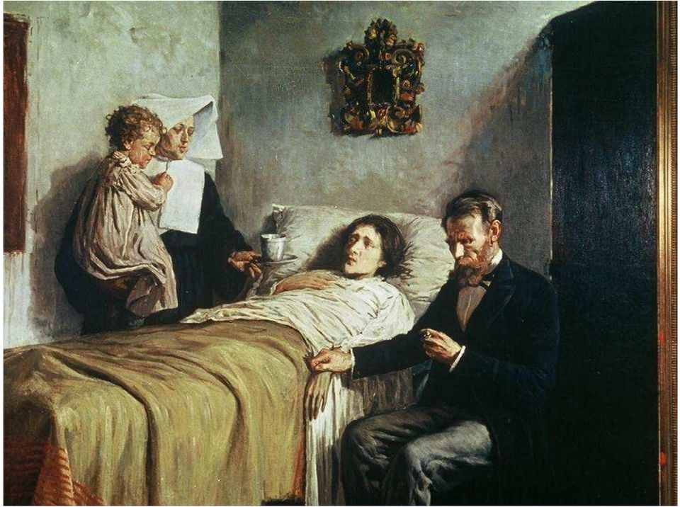
5 Frans van Miers,the Elder, The doctors visit 1657, Kunsthistorisches Museum, Wien 5

6 6

7 Picasso 1897, Ciencia y Caridad Museo Picasso,Barcelona 7

8 8
9 9
10 The arterial pulse contour changes to the periphery: damping different rate of transmission of components distorsion by reflected waves conversion of kinetic to potential energy 10

11 1.resistance: viscosity, vessel geometry opposes flow, HR independent 2.inertia: mass opposes rate of change of flow, HR dependent 3.compliance: distensibility opposes changes of blood volume, HR- dependent 11

12 anacrotic shoulder incisura 12

13 The anacrotic shoulder disappears to the periphery, the upstroke becomes steeper, starts later the incisura is characteristic for the carotid pulse contour it is gradually replaced by a later dicrotic notch and positive dicrotic wave 13

14 During palpation the auscultation of the heart serves for reference Factors influencing the pulse: stroke volume rate of ejection distensibility of peripheral arteries peripheral resistance pulse rate pulse pressure size of the vessel distance from the heart 14

15 15

16 16

17 The abnormal arterial pulse Hypokinetic pulse - small, weak low stroke volume, narrow pulse pressure, increased peripheral resistance left ventricular failure Ao valvular stenosis: pulsus parvus et tardus characteristic anacrotic notch 17

18 AI Pulsus tardus et parvus AS 18

19 The abnormal arterial pulse Hyperkinetic pulse strong,rapid upstroke water hammer rapid runoff peripheral shunts occasionally thrill on the carotid artery hyperkinetic circulation: anxiety, exercise, fever, hyperthyroidism 19

20 AI AS 20

21 Hyperkinetic pulse Aortic regurgitation pulsus celer et altus systolic collapse of the pulse Peripheral shunts: rapid runoff of blood from the arterial system Bradycardia 21
22 Specific abnormalities The twice beating pulse Dicrotic pulse: a second pulse wave is palpable during diastole, following S2 peripheral resistance diastolic BP fever, moderate AI 22
23 A.normal, B.outflow obst. C. bisferiens AI, D. Bisferiens HOCM E.bisferiens HF, septic shock 23
24 bisferiens 24 HOCM
25 The twice beating pulse Anacrotic pulse: a palpable double pulse both in systole, before S2 anacrotic notch on the upstroke AS Bisferiens pulse: in HOCM a very rapid initial upstroke the percussion wave is followed by a dip (the obstruction decelerates the ejection), this is followed by a second positive wave tidal wave 25
26 Pulsus alternans Regular pulse with an alternating height of the pressure pulse + often S3 : a sign of heart (LV) failure 26
27 Bigeminal pulse The pulse size alternates from beat to beat caused by bigeminal ventricular ectopy 27
28 Pulsus paradoxus In normal persons systolic BP by 3-10 mmhg during inspiration pooling of blood in the pulmonary vasculature if this is more than 10 mmhg p. p. cardiac tamponade constrictive pericarditis 28
29 29
30 LV close to the LV swinging away surface from the surface (Mayo Foundation for Medicac Education ) 30
31 Mayo 31
32 FIGURE Typical pulsed-wave Doppler pattern of tamponade recorded with a nasal respirometer. A, Mitral inflow velocity decreases (single arrowhead) after inspiration (Insp) and increases (double arrowheads) after expiration (Exp). B, Tricuspid inflow velocity has the opposite changes. E velocity increases (double arrowheads) after inspiration and decreases (single arrowhead) after expiration. (Modified from Oh JK, Hatle LK, Mulvagh SL, Tajik AJ: Transient constrictive pericarditis: Diagnosis by two-dimensional Doppler echocardiography. Mayo Clin Proc 68:1158, Used with permission of Mayo Foundation for Medical Education and 32 Research.)
33 Examination of the veins and their pulsations The normal venous pulse 3 positive waves a, c, v 2 negative x, y 33
34 34
35 a : the retrograde transmission of RA systole, at atrial relaxation it descends c : 1. Impact of the carotid artery 2. Retrograde bulging of the tricuspid valve in RV systole 35
36 x descent: 1. Displacement of the base of ventricles during systole 2. Right atrial relaxation V the tricuspid is closed blood is filling the venae cavae and right atrium in late ventricular systole 36
37 y descent: diastolic collapse, the tricuspid opens RA pressure rapidly Rapid filling of RA, the y nadir may coincide with a S3 37
38 the ascending limb of y wave depends on the rate of venous return. Long diastole plateau: h wave 38
39 Abnormalities a wave: absent in atrial fibrillation giant in tricuspid or pulm sten cannon waves - the right atrium contracts while the tricuspid is closed x wave: tricuspid regurgitation + r 39
40 40
41 Kata Tjuta in Ayers Rock, Australia 41
42 y wave: depends on the RV filling - compliance relation A slow y descent obstruction to RA emptying A sharp y descent shortly after S2 in constrictive pericarditis followed by a rapid ascent and plateau: dip and plateau 42
43 Venous pressure Estimated at the bedside 1. Veins of the hand: passive elevation to and above the sternal angle emptying 2. External jugular: trunk elevated to o occlude the e. j. by pressing with finger above the clavicle, it fills within s release and observe the fluid column 3. Paradox increase in venous distension during inspiration Kussmaul sign constrictive pericarditis 4. Hepatojugular reflux 43
44 44
45 FIGURE 12-3 Abnormal jugular venous waveforms. A, Large a waves associated with reduced RV compliance or elevated RV end-diastolic pressure. The phonocardiographic tracing (below) shows timing of the corresponding right-sided S 4. B, Normal jugular venous waveform (bottom), mild TR (middle), and severe TR (top), with corresponding phonocardiogram. With severe TR, there is ventricularization of the jugular venous waveform, with a prominent V wave and rapid Y descent. The X descent is absent. C, Jugular venous waveform in constrictive pericarditis with a prominent Y descent. Note the timing of the pericardial knock (K) relative to S 2. The abrupt rise in pressure after the nadir of the Y descent is caused by the rapid rise in venous pressure with ventricular filling. JVP = jugular venous pulse. (From Abrams J: Synopsis of Cardiac Physical Diagnosis. 2nd ed. Boston, Butterworth Heinemann, 2001, pp ) 45
46 46
47 47
48 48
49 49
50 TABLE THE CARDIAC CYCLE Left Ventricular Contraction Isovolumic contraction (b) Maximal ejection (c) Left Ventricular Relaxation Start of relaxation and reduced ejection (d) Isovolumic relaxation (e) LV filling: rapid phase (f) Slow LV filling (diastasis) (g) Atrial systole or booster (a) 50
51 FIGURE The mechanical events in the cardiac cycle were first assembled by Lewis in but first conceived by Wiggers in Note that mitral valve closure occurs after the crossover point of atrial and ventricular pressures at the start of systole. The visual phases of the ventricular cycle in the bottom panel are modified from Shepherd and Vanhoutte (Shepherd JT, Vanhoutte PM: The Human Cardiovascular System. New York, Raven Press, 1979, p 68.) For explanation of phases a to g, see Table ECG = electrocardiogram; JVP = jugular venous pressure; M1 = mitral component of first sound at time of mitral valve closure; T1 = tricuspid valve closure, second component of first heart sound; AO = aortic valve opening, normally inaudible; A2 = aortic valve closure, aortic component of second sound; P2 = pulmonary component of second sound, pulmonary valve closure; MO = mitral valve opening, which may be audible in mitral stenosis as the opening snap. S3 = third heart sound; S4 = fourth heart sound; a = wave produced by right atrial contraction; c = carotid wave artifact during rapid LV ejection phase; v = venous return wave that causes pressure to rise while tricuspid valve is closed. Cycle length of 800 milliseconds for 75 beats/min. (Modified from Opie LH: The Heart, Physiology, from Cell to Circulation. Philadelphia, Lippincott-Raven, Figure copyright L. H. Opie, 2001.) 51
52 TABLE THE CARDIAC CYCLE Left Ventricular Contraction Isovolumic contraction (b) Maximal ejection (c) Left Ventricular Relaxation Start of relaxation and reduced ejection (d) Isovolumic relaxation (e) LV filling: rapid phase (f) Slow LV filling (diastasis) (g) Atrial systole or booster (a) 52
53 53
54 54
55 55
Normal & Abnormal Intracardiac. Lancashire & South Cumbria Cardiac Network
 Normal & Abnormal Intracardiac Pressures Lancashire & South Cumbria Cardiac Network Principle Pressures recorded from catheter tip Electrical transducer - wheatstone bridge mechanical to electrical waveform
Normal Intracardiac Pressures. Lancashire & South Cumbria Cardiac Network
 Normal Intracardiac Pressures Lancashire & South Cumbria Cardiac Network Principle Pressures recorded from catheter tip Electrical transducer - wheatstone bridge mechanical to electrical waveform display
Section Four: Pulmonary Artery Waveform Interpretation
 Section Four: Pulmonary Artery Waveform Interpretation All hemodynamic pressures and waveforms are generated by pressure changes in the heart caused by myocardial contraction (systole) and relaxation/filling
Note: The left and right sides of the heart must pump exactly the same volume of blood when averaged over a period of time
 page 1 HEART AS A PUMP A. Functional Anatomy of the Heart 1. Two pumps, arranged in series a. right heart: receives blood from the systemic circulation (via the great veins and vena cava) and pumps blood
Mr GH: Pericardial Window. Anaesthetic Management of Cardiac Tamponade
 Mr GH: Pericardial Window Anaesthetic Management of Cardiac Tamponade Mr GH 56 yo M HOPCx Asbestosis, adenoca R lung 8/52 6/52 cisplatin/ taxol chemo Weekly pleural taps for effusions Sent from Bendigo
Heart Murmurs. Outline. Basic Pathophysiology
 Heart Murmurs David Leder Outline I. Basic Pathophysiology II. Describing murmurs III. Systolic murmurs IV. Diastolic murmurs V. Continuous murmurs VI. Summary Basic Pathophysiology Murmurs = Math Q =
Traumatic Cardiac Tamponade. Shane KF Seal 19 November 2003 POS
 Traumatic Cardiac Tamponade Shane KF Seal 19 November 2003 POS Objectives Definition Pathophysiology Diagnosis Treatment Cardiac Tamponade The decompensated phase of cardiac compression resulting from
Heart Sounds & Murmurs
 Cardiovascular Physiology Heart Sounds & Murmurs Dr. Abeer A. Al-Masri MBBS, MSc, PhD Associate Professor Consultant Cardiovascular Physiologist Faculty of Medicine, KSU Detected over anterior chest wall
RACE I Rapid Assessment by Cardiac Echo. Intensive Care Training Program Radboud University Medical Centre NIjmegen
 RACE I Rapid Assessment by Cardiac Echo Intensive Care Training Program Radboud University Medical Centre NIjmegen RACE Goal-directed study with specific questions Excludes Doppler ultrasound Perform 50
Auscultation of the Heart
 Review of Clinical Signs uscultation of the Heart Series Editor: Bernard Karnath, MD Bernard Karnath, MD William Thornton, MD uscultation of the heart can provide clues to the diagnosis of many cardiac
Exchange solutes and water with cells of the body
 Chapter 8 Heart and Blood Vessels Three Types of Blood Vessels Transport Blood Arteries Carry blood away from the heart Transport blood under high pressure Capillaries Exchange solutes and water with cells
Fellow TEE Review Workshop Hemodynamic Calculations 2013. Director, Intraoperative TEE Program. Johns Hopkins School of Medicine
 Fellow TEE Review Workshop Hemodynamic Calculations 2013 Mary Beth Brady, MD, FASE Director, Intraoperative TEE Program Johns Hopkins School of Medicine At the conclusion of the workshop, the participants
HEART HEALTH WEEK 3 SUPPLEMENT. A Beginner s Guide to Cardiovascular Disease HEART FAILURE. Relatively mild, symptoms with intense exercise
 WEEK 3 SUPPLEMENT HEART HEALTH A Beginner s Guide to Cardiovascular Disease HEART FAILURE Heart failure can be defined as the failing (insufficiency) of the heart as a mechanical pump due to either acute
5. Management of rheumatic heart disease
 5. Management of rheumatic heart disease The fundamental goal in the long-term management of RHD is to prevent ARF recurrences, and therefore, prevent the progression of RHD, and in many cases allow for
Direct Arterial Blood Pressure Monitoring Angel M. Rivera CVT, VTS (ECC) Animal Emergency Center Glendale, WI March 2003
 Animal Emergency Center Glendale, WI March 2003") Direct Arterial Blood Pressure Monitoring Angel M. Rivera CVT, VTS (ECC) Animal Emergency Center Glendale, WI March 2003 Introduction Direct measurement of arterial blood pressure is obtained via a peripheral
Direct Arterial Blood Pressure Monitoring Angel M. Rivera CVT, VTS (ECC) Animal Emergency Center Glendale, WI March 2003 Introduction Direct measurement of arterial blood pressure is obtained via a peripheral
Dynamic Auscultation of Heart Sounds and Murmurs. Acknowledgement. Disclosures Real or Potential Conflicts of Interest
 Dynamic Auscultation of Heart Sounds and Murmurs W. Lane Edwards, Jr., MSN, ARNP, ANP Hospitalist Group of Southwest Florida Affiliate Professor of Nursing, University of Alaska at Anchorage Acknowledgement
Chapter 20: The Cardiovascular System: The Heart
 Chapter 20: The Cardiovascular System: The Heart Chapter Objectives ANATOMY OF THE HEART 1. Describe the location and orientation of the heart within the thorax and mediastinal cavity. 2. Describe the
Heart and Vascular System Practice Questions
 Heart and Vascular System Practice Questions Student: 1. The pulmonary veins are unusual as veins because they are transporting. A. oxygenated blood B. de-oxygenated blood C. high fat blood D. nutrient-rich
HISTORY. Questions: 1. What diagnosis is suggested by this history? 2. How do you explain her symptoms during pregnancy?
 HISTORY 33-year-old woman. CHIEF COMPLAINT: months duration. Dyspnea, fatigue and nocturnal wheezing of six PRESENT ILLNESS: At ages 5 and 9, she had migratory arthritis. At age 29, in the third trimester
Introduction to CV Pathophysiology. Introduction to Cardiovascular Pathophysiology
 Introduction to CV Pathophysiology Munther K. Homoud, MD Tufts-New England Medical Center Spring 2008 Introduction to Cardiovascular Pathophysiology 1. Basic Anatomy 2. Excitation Contraction Coupling
Cardiovascular Physiology
 Cardiovascular Physiology Heart Physiology for the heart to work properly contraction and relaxation of chambers must be coordinated cardiac muscle tissue differs from smooth and skeletal muscle tissues
THE HEART Dr. Ali Ebneshahidi
 THE HEART Dr. Ali Ebneshahidi Functions is of the heart & blood vessels 1. The heart is an essential pumping organ in the cardiovascular system where the right heart pumps deoxygenated blood (returned
Edwards FloTrac Sensor & Edwards Vigileo Monitor. Understanding Stroke Volume Variation and Its Clinical Application
 Edwards FloTrac Sensor & Edwards Vigileo Monitor Understanding Stroke Volume Variation and Its Clinical Application 1 Topics System Configuration Pulsus Paradoxes Reversed Pulsus Paradoxus What is Stroke
The P Wave: Indicator of Atrial Enlargement
 Marquette University e-publications@marquette Physician Assistant Studies Faculty Research and Publications Health Sciences, College of 8-12-2010 The P Wave: Indicator of Atrial Enlargement Patrick Loftis
Section Two: Arterial Pressure Monitoring
 Section Two: Arterial Pressure Monitoring Indications An arterial line is indicated for blood pressure monitoring for the patient with any medical or surgical condition that compromises cardiac output,
Diastolic Heart Failure: Restrictive Cardiomyopathy, Constrictive Pericarditis, and Cardiac Tamponade: Clinical and Echocardiographic Evaluation
 Cardiology in Review Volume 10, Number 4, pp. 218 229 Copyright 2002 Lippincott Williams & Wilkins Diastolic Heart Failure: Restrictive Cardiomyopathy, Constrictive Pericarditis, and Cardiac Tamponade:
What is echo? CHAPTER 1 1.1 BASIC NOTIONS. Ultrasound production and detection
 What is echo? CHAPTER 1 1.1 BASIC NOTIONS Echocardiography (echo) the use of ultrasound to examine the heart is a safe, powerful, non-invasive and painless technique. Echo is easy to understand as many
Arterial pressure monitoring Direct arterial pressure monitoring permits continuous measurement of systolic, diastolic, and mean pressures and allows
 Arterial pressure monitoring Direct arterial pressure monitoring permits continuous measurement of systolic, diastolic, and mean pressures and allows arterial blood sampling. Because direct measurement
Anatomi & Fysiologi 060301. The cardiovascular system (chapter 20) The circulation system transports; What the heart can do;
 The circulation system transports; What the heart can do;") The cardiovascular system consists of; The cardiovascular system (chapter 20) Principles of Anatomy & Physiology 2009 Blood 2 separate pumps (heart) Many blood vessels with varying diameter and elasticity
The cardiovascular system consists of; The cardiovascular system (chapter 20) Principles of Anatomy & Physiology 2009 Blood 2 separate pumps (heart) Many blood vessels with varying diameter and elasticity
Electrocardiography I Laboratory
 Introduction The body relies on the heart to circulate blood throughout the body. The heart is responsible for pumping oxygenated blood from the lungs out to the body through the arteries and also circulating
The EasySense unit can detect that the Smart Q Heart Rate Sensor is connected and the range it is set to.
 Heart Rate Sensor Heart Rate Sensor (Product No PC-3147) Pulse rate Range: 0 to 200 bpm Resolution: 1 bpm Waveform Range: -2000 to 2000 mv Resolution: 1 mv Introduction The Smart Q Heart Rate Sensor monitors
Vascular System The heart can be thought of 2 separate pumps from the right ventricle, blood is pumped at a low pressure to the lungs and then back
 Vascular System The heart can be thought of 2 separate pumps from the right ventricle, blood is pumped at a low pressure to the lungs and then back to the left atria from the left ventricle, blood is pumped
Anatomy Review. Heart Murmurs. Surface Topography of the Heart 7/19/2011. The Base of the Heart and Erb s Point
 James A Mathey PA C, MPA CAPA WORKSHOP 2010 Heart Murmurs Anatomy Review 4 Classic Auscultatory Areas: Aortic 2ICS R SB Pulmonic 2ICS L SB Tricuspid 4 th L Lower SB Mitral 5ICS MCL Surface Topography of
Chapter 2 Cardiac Interpretation of Pediatric Chest X-Ray
 Chapter 2 Cardiac Interpretation of Pediatric Chest X-Ray Ra-id Abdulla and Douglas M. Luxenberg Key Facts The cardiac silhouette occupies 50 55% of the chest width on an anterior posterior chest X-ray
Circulatory System Review
 Circulatory System Review 1. Draw a table to describe the similarities and differences between arteries and veins? Anatomy Direction of blood flow: Oxygen concentration: Arteries Thick, elastic smooth
Question 1: Interpret the rhythm strip above (comment on regularity, rate, P wave, PR interval and QRS)?
?") It is your first month on your NICU rotation and you are prerounding on your patients. The nurse takes you aside and says she s been seeing something funny on the cardiac monitor. You walk to the bedside
It is your first month on your NICU rotation and you are prerounding on your patients. The nurse takes you aside and says she s been seeing something funny on the cardiac monitor. You walk to the bedside
Distance Learning Program Anatomy of the Human Heart/Pig Heart Dissection Middle School/ High School
 Distance Learning Program Anatomy of the Human Heart/Pig Heart Dissection Middle School/ High School This guide is for middle and high school students participating in AIMS Anatomy of the Human Heart and
Etiology and Diagnosis of Systolic Murmurs in Adults
 CLINICAL RESEARCH STUDY Etiology and Diagnosis of Systolic Murmurs in Adults Steven McGee, MD Primary and Specialty Medical Care, Department of Veterans Affairs Medical Center, Seattle, Wash; Department
Hemodynamic Monitoring: Principles to Practice M. L. Cheatham, MD, FACS, FCCM
 SUMMARY HEMODYNAMIC MONITORING: FROM PRINCIPLES TO PRACTICE Michael L. Cheatham, MD, FACS, FCCM Director, Surgical Intensive Care Units Orlando Regional Medical Center Orlando, Florida Fluid-filled catheters
Congestive heart failure (CHF) is a. Diastolic Heart Failure. By Michel D Astous, MD, FRCPC
 is a. Diastolic Heart Failure. By Michel D Astous, MD, FRCPC") Diastolic Heart Failure The evaluation of both systolic and diastolic functions is of great importance among patients presenting with signs of CHF, as the treatment may be quite different depending on
Diastolic Heart Failure The evaluation of both systolic and diastolic functions is of great importance among patients presenting with signs of CHF, as the treatment may be quite different depending on
Doc, I Am Fine, But I Have A Cardiac Condition
 Doc, I Am Fine, But I Have A Cardiac Condition Nevine Mahmoud, MD John Ludtke, MD Maj, USAFR, MC, FS RAM Class 2014 Wright State University Boonshoft School of Medicine Division of Aerospace Medicine Dayton,
How To Understand What You Know
 Heart Disorders Glossary ABG (Arterial Blood Gas) Test: A test that measures how much oxygen and carbon dioxide are in the blood. Anemia: A condition in which there are low levels of red blood cells in
CONSTRICTIVE PERICARDITIS
 33 Profiles in Constrictive Pericarditis, Restrictive Cardiomyopathy, and Cardiac Tamponade Beverly H. Lorell and William Grossman BHL: Harvard Medical School, Hemodynamic and Molecular Physiology Research
PHONOCARDIOGRAM ADDITIONAL PULMONARY VALVE CLOSURE CAUSED BY THE CATHETER AND DEMONSTRATED BY THE INTRACARDIAC
 Brit. Heart J., 1964, 26, 317. ADDITIONAL PULMONARY VALVE CLOSURE CAUSED BY THE CATHETER AND DEMONSTRATED BY THE INTRACARDIAC PHONOCARDIOGRAM BY LEON RESNEKOV AND JANE SOMERVILLE From the Institute of
Spurious systolic hypertension 143
 Spurious systolic hypertension in youth Michael F O Rourke a, Charalambos Vlachopoulos a and Robert M Graham b Abstract: Six young men diagnosed with systolic hypertension had normal carotid pressure wave
Ny teknologi: Fagdagene ved St. Olavs Hospital Lasse Løvstakken Dept. Circulation and Medical Imaging 11.06.2010
 1 Ny teknologi: Ultralyd måler m blodstrøm Fagdagene ved St. Olavs Hospital Lasse Løvstakken Dept. Circulation and Medical Imaging 11.06.2010 2 Conventional imaging methods of blood flow using ultrasound
Acute heart failure may be de novo or it may be a decompensation of chronic heart failure.
 Management of Acute Left Ventricular Failure Acute left ventricular failure presents as pulmonary oedema due to increased pressure in the pulmonary capillaries. It is important to realise though that left
SAM, Student Auscultation Manikin
 SAM, Student Auscultation Manikin Product: SAM, Student Auscultation Manikin Cat. No.: 718-9007 Price: (Call for latest pricing) 281-488-5901 or 1-800-364-5901 in US and Canada; Email: keith.johnson@cardionics.com
Common types of congenital heart defects
 Common types of congenital heart defects Congenital heart defects are abnormalities that develop before birth. They can occur in the heart's chambers, valves or blood vessels. A baby may be born with only
Cardiovascular System
 Topics to Review Diffusion Skeletal muscle fiber (cell) anatomy Membrane potential and action potentials Action potential propagation Excitation-contraction coupling in skeletal muscle skeletal muscle
ECHOCARDIOGRAPHY PROPERTY OF ELSEVIER SAMPLE CONTENT - NOT FINAL CHAPTER 6. Hisham Dokainish, MD, FACC, FASE
 CHAPTER 6 ECHOCARDIOGRAPHY Hisham Dokainish, MD, FACC, FASE 1. How does echocardiography work? Echocardiography uses transthoracic and transesohageal probes that emit ultrasound directed at cardiac structures.
Case III. Disscussion. the UHP ultrasound protocol. Novel Ultrasound Approach to the Empiric Evaluation of the Undifferentiated Hypotensive Patient
 The UHP Ultrasound Protocol: A Novel Ultrasound Approach to the Empiric Evaluation of the Undifferentiated Hypotensive Patient JOHN S. ROSE, MD,* AARON E. BAIR, MD,* DIKU MANDAVIA, MD, AND DONNA J. KINSER,
Electrodes placed on the body s surface can detect electrical activity, APPLIED ANATOMY AND PHYSIOLOGY. Circulatory system
 4 READING AND INTERPRETING THE ELECTROCARDIOGRAM Electrodes placed on the body s surface can detect electrical activity, which occurs in the heart. The recording of these electrical events comprises an
Overview of the Cardiovascular System
 Overview of the Cardiovascular System 2 vascular (blood vessel) loops: Pulmonary circulation: from heart to lungs and back) Systemic circulation: from heart to other organs and back Flow through systemic
Functions of Blood System. Blood Cells
 Functions of Blood System Transport: to and from tissue cells Nutrients to cells: amino acids, glucose, vitamins, minerals, lipids (as lipoproteins). Oxygen: by red blood corpuscles (oxyhaemoglobin - 4
Cardiovascular Biomechanics
 Cardiovascular Biomechanics Instructor Robin Shandas, Ph.D. Associate Professor of Pediatric Cardiology and Mechanical Engineering Robin.shandas@colorado.edu (303) 837-2586 (MWF) / (303) 492-0553 (T,Th)
Practical class 3 THE HEART
 Practical class 3 THE HEART OBJECTIVES By the time you have completed this assignment and any necessary further reading or study you should be able to:- 1. Describe the fibrous pericardium and serous pericardium,
Patient Possible differentials Recommended diagnostics Puppy or kitten with a soft systolic murmur
 Cardiac Auscultation 101 Terri DeFrancesco, DVM, DACVIM (Cardiology), DACVECC Associate Professor in Cardiology and Critical Care NC State University College of Veterinary Medicine Email: teresa_defrancesco@ncsu.edu
Workshop B: Essentials of Neonatal Cardiology and CHD Anthony C. Chang, MD, MBA, MPH CARDIAC INTENSIVE CARE
 SHUNT LESIONS NEONATAL : CONGENITAL CARDIAC MALFORMATIONS AND CARDIAC SURGERY ANTHONY C. CHANG, MD, MBA, MPH CHILDREN S HOSPITAL OF ORANGE COUNTY ATRIAL SEPTAL DEFECT LEFT TO RIGHT SHUNT INCREASED PULMONARY
Blood Vessels and Circulation
 13 Blood Vessels and Circulation FOCUS: Blood flows from the heart through the arterial blood vessels to capillaries, and from capillaries back to the heart through veins. The pulmonary circulation transports
Blood Pressure. Blood Pressure (mm Hg) pressure exerted by blood against arterial walls. Blood Pressure. Blood Pressure
 pressure exerted by blood against arterial walls. Blood Pressure. Blood Pressure") Blood Pressure Blood Pressure (mm Hg) pressure exerted by blood against arterial walls Systolic pressure exerted on arteries during systole Diastolic pressure in arteries during diastole 120/80 Borderline
Blood Pressure Blood Pressure (mm Hg) pressure exerted by blood against arterial walls Systolic pressure exerted on arteries during systole Diastolic pressure in arteries during diastole 120/80 Borderline
Equine Cardiovascular Disease
 Equine Cardiovascular Disease 3 rd most common cause of poor performance in athletic horses (after musculoskeletal and respiratory) Cardiac abnormalities are rare Clinical Signs: Poor performance/exercise
Diagnostic and Therapeutic Procedures
 Diagnostic and Therapeutic Procedures Diagnostic and therapeutic cardiovascular s are central to the evaluation and management of patients with cardiovascular disease. Consistent with the other sections,
Vtial sign #1: PULSE. Vital Signs: Assessment and Interpretation. Factors that influence pulse rate: Importance of Vital Signs
 Vital Signs: Assessment and Interpretation Elma I. LeDoux, MD, FACP, FACC Associate Professor of Medicine Vtial sign #1: PULSE Reflects heart rate (resting 60-90/min) Should be strong and regular Use 2
FVMA 2015: Diagnosis and Management of Pericardial Disease PERICARDIAL EFFUSION Pathophysiology of Cardiac Tamponade
 FVMA 2015: Diagnosis and Management of Pericardial Disease Jonathan A. Abbott, DVM, Dipl. ACVIM (Cardiology) VA-MD College of Veterinary Medicine Virginia Tech, Blacksburg, Virginia Pericardial disease
The heart then repolarises (or refills) in time for the next stimulus and contraction.
 in time for the next stimulus and contraction.") Atrial Fibrillation BRIEFLY, HOW DOES THE HEART PUMP? The heart has four chambers. The upper chambers are called atria. One chamber is called an atrium, and the lower chambers are called ventricles. In
Atrial Fibrillation BRIEFLY, HOW DOES THE HEART PUMP? The heart has four chambers. The upper chambers are called atria. One chamber is called an atrium, and the lower chambers are called ventricles. In
Cardiovascular Assessment
 Cardiovascular Assessment A Home study Course Offered by Nurses Research Publications P.O. Box 480 Hayward CA 94543-0480 Office: 510-888-9070 Fax: 510-537-3434 No unauthorized duplication photocopying
Haemodynamic Monitoring Learning Package. Name:. HORNSBY KU-RING-GAI HOSPITAL INTENSIVE CARE UNIT
 HORNSBY KU-RING-GAI HOSPITAL INTENSIVE CARE UNIT Haemodynamic Monitoring Learning Package Name:. Haemodynamic Learning package, Intensive Care Unit, Hornsby Hospital 2008 1 Package developed for use at
Cardiovascular diseases. pathology
 Cardiovascular diseases pathology Atherosclerosis Vascular diseases A disease that results in arterial wall thickens as a result of build- up of fatty materials such cholesterol, resulting in acute and
Adult Cardiac Surgery ICD9 to ICD10 Crosswalks
 164.1 Malignant neoplasm of heart C38.0 Malignant neoplasm of heart 164.1 Malignant neoplasm of heart C45.2 Mesothelioma of pericardium 198.89 Secondary malignant neoplasm of other specified sites C79.89
3 rd Russian-Bavarian Conference on Bio-Medical Engineering
 3 rd Russian-Bavarian Conference on Bio-Medical Engineering Blood Pressure Estimation based on Pulse Transit Time and Compensation of Vertical Position Dipl.-Inform. Med. Christian Douniama Dipl.-Ing.
SECTION III: HEMODYNAMIC PRINCIPLES
 10/5/00 3:36 PM 10 Calculation of Stenotic Valve Orifice Area Blase A. Carabello and William Grossman BAC: Baylor University, Department of Medicine, Houston Veterans Affairs Medical Center, Houston, Texas
Administrative. Patient name Date compare with previous Position markers R-L, upright, supine Technical quality
 CHEST X-RAY Administrative Patient name Date compare with previous Position markers R-L, upright, supine Technical quality AP or PA ( with x-ray beam entering from back of patient, taken at 6 feet) Good
Questions FOETAL CIRCULATION ANAESTHESIA TUTORIAL OF THE WEEK 91 18 TH MAY 2008
 FOETAL CIRCULATION ANAESTHESIA TUTORIAL OF THE WEEK 91 18 TH MAY 2008 Dr. S. Mathieu, Specialist Registrar in Anaesthesia Dr. D. J. Dalgleish, Consultant Anaesthetist Royal Bournemouth and Christchurch
Heart Sounds and Murmurs. Objectives. Valves. Wright, 2012 1
 Heart Sounds and Murmurs Wendy L. Wright, MS, RN, ARNP, FNP, FAANP Family Nurse Practitioner Owner Wright & Associates Family Healthcare Partner Partners in Healthcare Education 1 Objectives Upon completion
Factors Affecting Blood Pressure. Vessel Elasticity Blood Volume Cardiac Output
 Factors that Affect Pressure Graphics are used with permission of: Pearson Education Inc., publishing as Benjamin Cummings (http://www.aw-bc.com) Page 1. Introduction pressure is affected by several factors:
Human Anatomy & Physiology II with Dr. Hubley
 Human Anatomy & Physiology II with Dr. Hubley Exam #1 Name: Instructions This exam consists of 40 multiple-choice questions. Each multiple-choice question answered correctly is worth one point, and the
New insights in the assessment of right ventricular function: an echocardiographic study. Avin Calcutteea
 New insights in the assessment of right ventricular function: an echocardiographic study Avin Calcutteea Department of Public Health and Clinical Medicine Umeå University, 2013 New insights in the assessment
MISSING DATA ANALYSIS AMONG PATIENTS IN THE PINNACLE REGISTRY
 MISSING DATA ANALYSIS AMONG PATIENTS IN THE PINNACLE REGISTRY In order to improve the efficiency of PINNACLE Registry data analytics, a missing data analysis has been conducted on PINNACLE Registry data
Sign up to receive ATOTW weekly email worldanaesthesia@mac.com
 INTRODUCTION TO CARDIOVASCULAR PHYSIOLOGY ANAESTHESIA TUTORIAL OF THE WEEK 125 16 TH MARCH 2009 Toby Elkington, Specialist Registrar Carl Gwinnutt, Consultant Department of Anaesthesia, Salford Royal NHS
How To Teach An Integrated Ultrasound
 University of South Carolina School of Medicine Integrated Ultrasound Curriculum iusc Richard Hoppmann The Integrated Ultrasound Curriculum Initiated 2006 First (M1) and Second (M2) Year Medical Students
Blood Flow Hemodynamics, Cardiac Mechanics, and Doppler Echocardiography
 Blood Flow Hemodynamics, Cardiac Mechanics, and Doppler Echocardiography THE CARDIAC CYCLE Figure 4.1 The cardiac cycle showing superimposed hemodynamic and echocardiographic parameters. A4C: apical 4-chamber
Magnetic Resonance Quantitative Analysis. MRV MR Flow. Reliable analysis of heart and peripheral arteries in the clinical workflow
 Magnetic Resonance Quantitative Analysis MRV MR Flow Reliable analysis of heart and peripheral arteries in the clinical workflow CAAS MRV Functional Workflow Designed for imaging specialists, CAAS MRV
Blood vessels. transport blood throughout the body
 Circulatory System Parts and Organs Blood vessels transport blood throughout the body Arteries blood vessels that carry blood AWAY from the heart Pulmonary arteries carry the deoxygenated blood from heart
May 1 6, 2016 Loews Atlanta Hotel Atlanta, GA PRELIMINARY PROGRAM AT A GLANCE
 Sunday, May 1 7:00-8:30 pm Image Optimization Workshop: 2D Echocardiography Moderators: Fabio De Vasconelos Papa, MD; Mark A. Taylor, MD : Annemarie Thompson, MD Speakers: Gregg S. Hartman, MD Lori B.
Biol 111 Comparative & Human Anatomy Lab 9: Circulatory System of the Cat Spring 2014
 Biol 111 Comparative & Human Anatomy Lab 9: Circulatory System of the Cat Spring 2014 Philip J. Bergmann Lab Objectives 1. To learn how blood flows through a dual circuit circulation with lungs. 2. To
12-Lead EKG Interpretation. Judith M. Haluka BS, RCIS, EMT-P
 12-Lead EKG Interpretation Judith M. Haluka BS, RCIS, EMT-P ECG Grid Left to Right = Time/duration Vertical measure of voltage (amplitude) Expressed in mm P-Wave Depolarization of atrial muscle Low voltage
Superior Vena Cava and Hepatic Vein Doppler Echocardiography in Healthy Adults
 1032 JACC Vol. 10, No.5 November 1987:1032-9 Superior Vena Cava and Hepatic Vein Doppler Echocardiography in Healthy Adults CHRISTOPHER P. APPLETON, MD, LIV K. HATLE, MD, RICHARD L. POPP, MD, FACC Stanford,
PHARMACOLOGICAL Stroke Prevention in Atrial Fibrillation STROKE RISK ASSESSMENT SCORES Vs. BLEEDING RISK ASSESSMENT SCORES.
 PHARMACOLOGICAL Stroke Prevention in Atrial Fibrillation STROKE RISK ASSESSMENT SCORES Vs. BLEEDING RISK ASSESSMENT SCORES. Hossam Bahy, MD (1992 2012), 19 tools have been identified 11 stroke scores 1
Subclavian Steal Syndrome By Marta Thorup
 Subclavian Steal Syndrome By Marta Thorup Definition Subclavian steal syndrome (SSS), is a constellation of signs and symptoms that arise from retrograde flow of blood in the vertebral artery, due to proximal
For more information about the use of the Propaq monitor, refer to the Propaq Directions For Use.
 Clinical Support 8500 S.W. Creekside Pl. Beaverton, OR 97008-7107 U.S.A. Telephone: 503-526-4200 Toll Free: 800-289-2500 clinicalsupport@protocol.com ELECTROCARDIOGRAPHY Introduction This article provides
Doppler. Doppler. Doppler shift. Doppler Frequency. Doppler shift. Doppler shift. Chapter 19
 Doppler Doppler Chapter 19 A moving train with a trumpet player holding the same tone for a very long time travels from your left to your right. The tone changes relative the motion of you (receiver) and
Clinical revision for OSCE examinations. Please note that these notes cannot be printed
 Clinical revision for OSCE examinations Please note that these notes cannot be printed Copyright 1994-2009 Academic Medical Publishing. Tudor Lodge, Roseworth Crescent, Newcastle upon Tyne NE3 1NR. First
Heart Failure EXERCISES. Ⅰ. True or false questions (mark for true question, mark for false question. If it is false, correct it.
 Heart Failure EXERCISES Ⅰ. True or false questions (mark for true question, mark for false question. If it is false, correct it. ) 1. Heart rate increase is a kind of economic compensation, which should
Valve Disease and Diastology Summit
 Heart & Vascular Institute Valve Disease and Diastology Summit is offered in cooperation with the American Society of Echocardiography. Valve Disease and Diastology Summit March 4 6, 2016 Eden Roc Hotel
Heart valve repair and replacement
 16 Heart valve repair and replacement 222 Valvular heart disease can be treated in a variety of ways: valve replacement, in which an artificial (prosthetic) heart valve is implanted surgically to replace
AORTIC STENOSIS. Marie-Jeanne Bertrand MD MSc - 2015 1
 AORTIC STENOSIS TYPES OF AORTIC STENOSIS 1. Valvular AS - Congenital: bicuspid or unicuspid, young patients o Bicuspid valve 1-2% population 20% with CoAo (80% with CoAo have bicuspid valve) Fusion of
Electrocardiogram and Heart Sounds
 Electrocardiogram and Heart Sounds An introduction to the recording and analysis of electrocardiograms, and the sounds of the heart. Written by Staff of ADInstruments Introduction The beating of the heart
How To Treat A Single Ventricle And Fontan
 COACH Columbus Ohio Adult Congenital Heart Disease Program The Heart Center at Nationwide Children s Hospital & The Ohio State University Single Ventricle Defects Normal Heart Structure The heart normally
To provide the body (cells) with oxygen, and remove CO 2. To provide the body (cells) with nutrients and remove wastes.
 with oxygen, and remove CO 2. To provide the body (cells) with nutrients and remove wastes.") Circulatory system. Basic function: To provide the body (cells) with oxygen, and remove CO 2. To provide the body (cells) with nutrients and remove wastes. Not all organisms have a circulatory system -
Circulatory system. Basic function: To provide the body (cells) with oxygen, and remove CO 2. To provide the body (cells) with nutrients and remove wastes. Not all organisms have a circulatory system -