Conformational Properties of Polypeptide Chains
|
|
|
- Shon Baker
- 10 years ago
- Views:
Transcription
1 Conformational Properties of Polypeptide Chains

2 Levels of Organization Primary structure Amino acid sequence of the protein Secondary structure H bonds in the peptide chain backbone α helix and β sheets Tertiary structure Non covalent interactions between the R groups within the protein Quanternary structure Interaction between 2 polypeptide chains

3 Why protein structure? In the factory of living cells, proteins are the workers, performing a variety of biological tasks. Each protein has a particular 3 D structure that determines its function. Structure implies function. Structure is more conserved than sequence. Protein structure is central for understanding protein functions. Sequence Structure Function

4 The basics of protein Proteins have one or more polypeptide chains Building blocks: 20 amino acids. Length range from 10 to 1000 residues. Proteins fold into 3 D shape to perform biological functions.

5 Anfinsen s thermodynamic hypothesis The three dimensional structure of a native protein in its normal physiological milieu (solvent, ph, ionic strength, presence of other components such as metal ions or prosthetic groups, temperature, etc.) is the one in which the Gibbs free energy of the whole system is lowest; that is, that the native conformation is determined by the totality of interatomic interactions and hence by the amino acid sequence, in a given environment. --- Anfinsen s Nobel lecture, 1972

6 Gibbs free energy A thermodynamic potential that measures the "useful" or process-initiating work obtainable from an isothermal, isobaric thermodynamic system. The maximum amount of non-expansion work that can be extracted from a closed system Gibbs free energy (ΔG) or available energy is the chemical potential that is minimized when a system reaches equilibrium at constant pressure and temperature.
 or available energy is the chemical potential that is minimized when a system reaches")
7 Common structure of Amino Acid Cα is the chiral center R Side chain = H,CH 3, Atoms numbered β,γ,δ,ε,ζ.. Amino H H + N H C α H Atom lost during peptide bond formation Backbone C - O O Carboxylate

8 Amino Acids Hydrophilic Hydrophobic

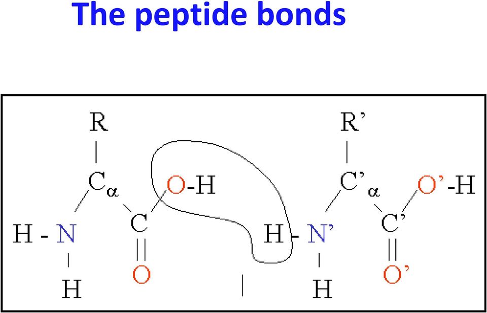
9 The peptide bonds
10 Coplanar atoms

11 Primary structure The amino acid sequences of polypeptides chains.

12 Shape = Amino Acid Sequence Proteins are made of 20 amino acids linked by peptide bonds Polypeptide backbone is the repeating sequence of the N C C N C C in the peptide bond The side chain or R group is not part of the backbone or the peptide bond The amino acid sequence (the primary structure) of a protein determines its three dimensional structure, which in turn determines its properties.
 of a protein determines its three dimensional structure, which in turn")
13 1 structure = sequence of amino acids

14 Secondary structure Local organization of protein backbone: α helix β strand (which assemble into β sheet) turn and interconnecting loop.
 turn and")
15 Protein Folding The peptide bond allows for rotation around it and therefore the protein can fold and orient the R groups in favorable positions Weak non covalent interactions will hold the protein in its functional shape these are weak and will take many to hold the shape

16 Protein Folding Proteins shape is determined by the sequence of the amino acids The final shape is called the conformation and has the lowest free energy possible Denaturation is the process of unfolding the protein Can be down with heat, ph or chemical compounds If the chemical compounds are removed, the proteins can be renatured or refold

17 Refolding Molecular chaperones are small proteins that help guide the folding and can help keep the new protein from associating with the wrong partner

18 What drives protein folding? Hydrophobic effects Hydrophobic residues tend to be buried inside Hydrophilic residues tend to be exposed to solvent Hydrogen bonds help to stabilize the structure.

19 Non covalent Bonds in Proteins

20 Globular Proteins The side chains will help determine the conformation in an aqueous solution

21 Hydrogen Bonds in Proteins H bonds form between 1) atoms involved in the peptide bond; 2) peptide bond atoms and R groups; 3) R groups
22 Protein Conformation Framework Bond rotation determines protein folding, 3D structure
23 Bond Rotation
24 Bond Rotation Determines Protein Folding
25 Protein Conformation Framework Bond rotation determines protein folding, 3D structure Torsion angle (dihedral angle) τ Measures orientation of four linked atoms in a molecule: A, B, C, D
26
27 Protein Conformation Framework Bond rotation determines protein folding, 3D structure Torsion angle (dihedral angle) τ Measures orientation of four linked atoms in a molecule: A, B, C, D τ ABCD defined as the angle between the normal to the plane of atoms A-B- C and normal to the plane of atoms B- C-D
28 Ethane Rotation A B D C A B C D
29 Protein Conformation Framework Bond rotation determines protein folding, 3D structure Torsion angle (dihedral angle) τ Measures orientation of four linked atoms in a molecule: A, B, C, D τ ABCD defined as the angle between the normal to the plane of atoms A-B- C and normal to the plane of atoms B- C-D Three repeating torsion angles along protein backbone: ω, φ, ψ
30 Backbone Torsion Angles
31 Backbone Torsion Angles φ and ψ Dihedral angle φ : rotation about the bond between N and C α Dihedral angle ψ : rotation about the bond between C α and the carbonyl carbon
32 φ and ψ angles
33 Backbone Torsion Angles ω Dihedral angle ω : rotation about the peptide bond, namely C α 1 -{C-N}- C α 2 ω angle tends to be planar (0º - cis, or 180 º - trans) due to delocalization of carbonyl π electrons and nitrogen lone pair
34 Backbone Torsion Angles φ and ψ are flexible, therefore rotation occurs here However, φ and ψ of a given amino acid residue are limited due to steric hindrance Only 10% of the {φ, ψ} combinations are generally observed for proteins First noticed by G.N. Ramachandran
35 Consequences of double bond character in the peptide bond trans peptide bond cis peptide bond Still another consequence: in the cis form, the R groups in adjacent residues tend to clash. Hence almost all peptide bonds in proteins are in the trans configuration.
36 Planarity of the peptide bond Psi (ψ) the angle of rotation about the Cα-C bond. Phi (φ) the angle of rotation about the N-Cα bond. The planar bond angles and bond lengths are fixed.
37 Side chain properties Carbon does not make hydrogen bonds with water easily hydrophobic O and N are generally more likely than C to H bond to water hydrophilic General amino acids groups: Hydrophobic Charged (positive/basic & negative/acidic) Polar 37
38 Unlike the peptide bond, free rotation occurs about the other two backbone bonds, but steric interactions within the polypeptide still severely limit plausible conformations, so that only certain combinations of phi and psi angles are allowed and between carbonyls between R groups or combinations of an R group, carbonyl or amide group. 180 allowed Ψ 0 not allowed φ 180
39 Steric Hindrance Interference to rotation caused by spatial arrangement of atoms within molecule Atoms cannot overlap Atom size defined by van der Waals radii Electron clouds repel each other
40 Steric Hindrance Most combinations of ψ and φ for an amino acid are not allowed because of steric collisions between the side chains and main chain. Theoretically if the torsion angles all are 180 o we have a purely trans system ( N, Cα, C ) if the torsion angles are all 0 we have the purely cis arrangement. Which is preferred? For the peptide bond (ω) [Ci Ni+1] The trans is preferred 1000:1
41 Certain side chain conformations are energetically favorable Ethane Valine Which valine stagger is energetically the best? The 1 st one Why?
42 G.N. Ramachandran Used computer models of small polypeptides to systematically vary φ and ψ with the objective of finding stable conformations For each conformation, the structure was examined for close contacts between atoms Atoms were treated as hard spheres with dimensions corresponding to their van der Waals radii Therefore, φ and ψ angles which cause spheres to collide correspond to sterically disallowed conformations of the polypeptide backbone
43 Ramachandran diagram Most angle combinations (75%) excluded by steric hindrance Dark green most favored Steric exclusion: powerful organizing principle Limited conformations favor protein folding, favorable entropy of too many conformations opposes folding
44 Ramachandran Plot QuickTime?and a TIFF (Uncompressed) decompressor are needed to see this picture. QuickTime?and a TIFF (Uncompressed) decompressor are needed to see this picture. G.N. Ramachandran, 1963
45 φ, ψ and Secondary Structure Name φ ψ Structure alpha-l left-handed alpha helix 3-10 Helix right-handed. π helix right-handed. Type II helices left-handed helices formed by polyglycine and polyproline. Collagen right-handed coil formed of three left handed helicies
46 The classical version of the Ramachandran plot for (a) alanine, (b) glycine. The fully allowed regions are shaded; the partially allowed regions are enclosed by a solid line. The connecting regions enclosed by the dashed lines are permissible with slight flexibility of bond angles (a,b). (c) for all 19 non-glycines and (d) for glycine. Most regions show a 45 degree slope rather than being parallel to any of the axes (c) For glycine (d), the beta sheet region is split into two distinct maxima and the two most populated regions (red), which were predicted to be only just permissible as shown in (b). There are five areas in the glycine plot; two with psi 0 and three with psi 180. Referenced from Hovmöller (2002)
47 Random Coil Following translation we have a primary amino acid sequence and it s random coil ~~~~~~~~~~~~ just flopping around folds to a 3D structure Uses van der waals interactions Hydrophobic interactions Salt Bridges H bonds Electrostatics forces
48 Motifs of Protein Structure Packing hydrophobic side chains into the interior of the molecule Creating a HYDROPHOBIC core and a HYDROPHILIC surface. The problem with creating such a hydrophobic core from a protein chain is to bring the side chains into the core the main chain must also come into the interior. The main chain is highly polar and therefore hydrophilic, with one hydrogen bond donor NH and one acceptor C = O for each peptide unit. In a hydrophobic environment these main chain polar groups must be neutralized by the formation of hydrogen bonds. This problem is solved very elegantly by the formation of regular secondary structure within the interior.
49 Secondary Structure The two very important secondary structures of proteins are: α helix β pleated sheet Both depend on hydrogen bonding between the amide H and the carbonyl O further down the chain or on a parallel chain. 5P2 49
50 Protein Folding 2 regular folding patterns have been identified formed between the bonds of the peptide backbone α helix protein turns like a spiral fibrous proteins (hair, nails, horns) β sheet protein folds back on itself as in a ribbon globular protein
51 α Helix 3.6 amino acids per turn of helix. The (p), distance per turn, is 0.54 nm, and the rise, distance between each atom is 0.15 (0.54/3.6). The N H group of an amino acid forms a hydrogen bond with the C=O group of the amino acid four residues earlier. This repeated i + 4 > i hydrogen bonding is the most prominent characteristic of an α helix
52 Characteristics of α Helix Although structurally ordered, isolated α helices are usually marginally stable in aqueous solution. A helix forms quickly (10 5 to 10 7 sec) but can unravel almost as quickly. Formation is generally independent of length, though unraveling isn t. The most common along the outside with one face out into solution and one into the hydrophobic core. Alpha helix have a hydrophobic and hydrophilic side Side chains to change from hydrophobic to hydrophilic with a periodicity of 3 4 residues.
53 α helix An α helix can, in theory, be right or left handed depending on the screw direction of the chain. Left handed ones don t exist!!! Because of steric interactions between side chains and CO groups ALWAYS right handed!
54 Properties of the alpha helix φ ψ 60 Hydrogen bonds between C=O of residue n, and NH of residue n residues/turn 1.5 Å/residue rise 100 /residue turn 54
55 Fig. 4-2a, p.84
56 Helices have electric dipoles.
57 The dipole moment in the α helix All the hydrogen bonds in an α helix point in the same direction along the helical axis. The dipole moments are also aligned along the helical axis. A partial net positive charge at the amino end and a negative charge at C terminal end. The α helix has a macrodipole moment of ~n * 3.5 Debye units. n = # residues
58 Preferred amino acids in α helices Different side chains have a weak but definite preference either for or against being in an α helix. Good helix formers: Ala, Glu, Leu, Met Poor helix formers: Ser Gly small, flexible doesn t like to be in fixed position Try big and bulky Pro can t enter the i i + 4 groups because can t H bond
59 Proline and α helix Amino acid side chains project out from the α helix and don t interfere with it except for proline. The last atom of the proline side chain is bonded to the main chain N atom which forms a ring. This prevents the N from contributing to a hydrogen bond an adding steric hindrance to the α helix conformation. Proline fits very well into the first (N term) of an α helix Proline in long α helices distorts the local geometry causing a BEND. Pro s are often referred to as helix breakers.
60 Helical wheel α helices are amphipathic Non polar side chains along one side of the helical cylinder and polar residues along the remainder of its surface. Such helices often aggregate with each other or with other non polar surfaces. The view is down the helix with the hydrophobic core being in the middle. The helix repeats itself after 5 turns or 18 residues so the residues are offset. Hydrophilic side Hydrophobic side
61 Variations on the classical α helix In natural proteins a slightly different geometry is seen. The CO groups tend to point out away from the helix axis and the H bonds are consequently not as straight So, φ ~ 62 o and ψ ~ 41 o instead of the classical 57 to 60 o and 47 to 50 o This geometry is more stable than the classical α helix because it permits each CO oxygen to H bond to the NH of the i + 4 residue. Occurs when the chain is more loosely or more tightly coiled With hydrogen bonds to residues i + 5 (π helix) or i + 3 (3 10 helix), respectively, NOT i + 4
62 3 10 helix and π helix Has 3 residues per turn and contains 10 atoms between the hydrogen bond donor and acceptor. Rare and usually occur at the ends of regular helices or as a single turn helices. Not energetically favorable for the most part The backbone atoms are packed too tight in the 3 10 or too loose in the π helix that there is a hole in the middle. Contrast of helix end views between α (squarish) vs 3-10 (triangular)
63
64 β sheet (pleated sheet) Two types of β sheet, parallel (same N to C direction) and anti parallel (opposite N to C direction) between polypeptide chains. β Sheet contains 2 amino acid residues per turn, and cannot form H bond between amide H and carboxyl O. Hydrogen bonds can only formed between adjacent polypeptide chains. Anti parallel form is more stable than parallel form. 64
65 The beta strand (& sheet) φ 135 ψ
66 H bond β pleated sheet
67 B Sheet: Lewis Structure CH C N HC C N CH C N O H H O H O N-term C-term C-term N-term CH C N HC C N CH C N O H H O H O Parallel sheet CH C N HC C N CH C N O H H O H O N-term C-term C-term N-term HC C N HC C N CH C N O H H O H O Antiparallel sheet
68 Topologies of β sheets
69 Properties of beta sheets Core of many proteins is the β sheet Form rigid structures with the H bond Formed of stretches of 5 10 residues in extended conformation Parallel/aniparallel, contiguous/non contiguous
70 Turns and Loops Secondary structure elements are connected by regions of turns and loops Turns short regions of non α, nonβ conformation Loops larger stretches with no secondary structure. Often disordered. Random coil Sequences vary much more than secondary structure regions Common AA found at turns are: glycine: small size allows a turn proline: geometry favors a turn
71 Proline C HN COOH H P
72 Globular Proteins Usually bind substrates (O2) within a hydrophobic cleft in the structure. Myoglobin and hemoglobin are typical examples of globular proteins. Both are hemoproteins and each is involved in oxygen metabolism. 5P2 72
73 Globular Protein Structure: Patterns of Folding Infinite number of globular protein folding, but some principles of protein folding are found. Made up of a number of domains a compact locally folded region of tertiary structure, which perform different functions of proteins. Two major kinds of folding patterns, α helic and β sheet. 73
74 Fig. 6.16
75 Myoglobin: 2 o and 3 o Aspects Globular myoglobin has 153 AA arranged in eight α helical regions labeled A H. The prosthetic heme group is necessary for its function, oxygen storage in mammalian muscle tissue. His E7 and F8 are important for locating the heme group within the protein and for binding oxygen. A representation of myoglobin follows with the helical regions shown as ribbons. 5P2 75
76 Myoglobin: 2 o and 3 o aspects Some helical regions Heme group with iron (orange) at the center 5P2 76
77 The Heme Group N of His F8 binds to fifth site on the iron. His E7 acts as a gate for oxygen. - OOC CH 2 CH 2 CH 2 CH 2 COO - H 3 C CH 3 N N H 2 Fe(II) C N N CH CH 3 Pyrrole ring CH 3 CH CH 2 5P2 77
78 Binding Site for Heme Lower His bonds covalently to iron(ii) Oxygen coordinates to sixth site on iron and the upper His acts as a gate for the oxygen. 5P2 78
79 3D Structure of Cytochrome c Cytochrome C 의 3차구조 Fig. 4-15, p.94
80 Fibrous Proteins Proteins with an elongated or filamentous form, often dominated by a single type of secondary structure over a large distance Fibrous proteins have a high concentration of α helix or β sheet. Examples include: a keratin collagen silk fibroin 5P2 80
81 Fibrous Proteins Structural Materials of Cells and Tissues More than 1/3 or more of the body protein in large vertebrates. Functions include: External protection such as skin, hair, feather and nail. Structure and support, shape and form, tendons, cartilage and bone. Native conformations of fibrous proteins are stable proteins after isolated and will not be denatured or unfolded. 81
82
83
84 Keratins Two classes of keratin, α and β keratins. α Keratins is the major fibrous proteins of hair and wool. Each α keratin molecule contains over 300 residues in length and all are α helical. Two α keratin helices bond together by their hydrophobic R side groups. The higher amounts of AAs are serine, glutamine, glutamic acid and cysteine. 84
85 Structure of α Keratin Adjacent polypeptide chains also crosslinked by disulfide bonds. Disulfide bond patterns between are what determine whether human hair is straight or curly.
86 Coiled Coil α Helical Dimer of α Keratin Amphipathic α helices: Residues a, d, a and d hydrophobic, other residues more hydrophilic
87 β-keratin Contains β-sheet structure. Are found mostly in birds and reptiles for feather and scales. 87
88 Fibroin They are all anti parallel pleated sheet structure. Comprise almost glycine, alanine and serine AA. Gly ala gly ala gly serine gly ala ala gly This alternation allows the β sheet to fit together. Strong and inextensible. No intrachain hydrogen bond, only interchain hydrogen bonds. No cystine cross linkage presence. The fibers are very flexible, e.g. silk. 88
89 Silk made by silkworms and spiders. Composed of microcrystalline array of antiparallel beta pleated sheets where each strand has alternating Gly and Ala or Ser residues. Structure of Silk Fibroin
90
91 Collagen Collagen is about one third of total protein in large animals, and is the major constituent of tendons and skin. Basic unit is tropocollagen which consists of three polypeptide chains in left handed helix tightly coiled into a three strand rope in right handed helix, arranged in head to tail. Tropocollagen have total about 3000 AA residues (each polypeptide has 1000 AA residues), having of molecule weight of 300,000. Every third residue can be only glycine because of the bulky effect. 91
92 Composition and structure of Collagen The tropocollagen subunit is a rod about 300 nm long and 1.5 nm in diameter, made up of three polypeptide strands, each of which is a left handed helix. Twisted together into a right handed coiled coil, a triple helix, a cooperative quaternary structure stabilized by numerous hydrogen bonds. Tropocollagen subunits spontaneously self assemble, with regularly staggered ends, into even larger arrays in the extracellular spaces of tissues. There is some covalent crosslinking within the triple helices, and a variable amount of covalent crosslinking between tropocollagen helices Prolines and hydroxyprolines cause it to be very tightly twisted and very rigid
93 Structure of Collagen Fibers Collagen is the most abundant vertebrate protein and the major stress-bearing component of connective tissue (bone, teeth, cartilage, tendon) and fibrous matrix of skin and blood vessels. 3 intertwined lefthanded helices 3.3 residues/turn Repeating Gly-X-Y (X often Pro, Y often Pro or hydroxypro)
94 Tropocollagen The 3 collagen molecules are crosslinked by : *H bonds very tight and do not stretch * Crosslinks between lysines, both interchain and intrachain, due to oxidation by enzyme LYSYL OXIDASE. Occurs only in collagen. Carbohydrates on hydroxylysines Tropocollagen molecules are head to tail and are staggered. More crosslinks occur with age.
95 Collagen Amino Acid Composition 1) Glycine (30%) 2) Proline 3) Modified amino acids hydroxyproline hydroxylysine 4) Glycosylation
96 Primary sequence of collagen Gly Pro Met Gly Pro Ser Gly Pro Arg Gly Leu Hyp Gly Pro Hyp Gly Ala Hyp Gly Pro Gln Gly Phe Gln Gly Pro Hyp Gly Glu Hyp Gly Glu Hyp Gly Ala Ser Gly Pro Met Gly Pro Arg Gly Pro Hyp Gly Pro Hyp Gly Lys Asn Gly Asp Asp
97 Glycosylation Sugars added to hydroxylysine or hydroxyproline Addition of disaccharides Glu Glu Gal Glu Gal Gal Glycosylation influences function Fibrils tendons low carbohydrate Sheets Lens cap, cornea High carbohydrate
98 Cross links between Tropocollagen 1) Between two lysine residues 1) Enzyme lysyl oxidase 2) Aldol condensation: enolate + carbonyl 3) Link Aldol cross link 2) Between two hydroxylysines and a lysine residue 1) Hydroxyprridinium cross link: increase heat stability Higher in older animals
99 Importance of Hydroxylation The principal residue to be hydroxylated in in proteins is proline. Lysine may also be hydroxylated. Catalyzed by prolyl 4 hydroxylase, prolyl 3 hydroxylase, and lysyl 5 hydroxylase. The reactions require iron (as well as molecular oxygen and α ketoglutarate) to carry out the oxidation Use vitamin C to return the iron to its oxidized state. Deprivation of ascorbate leads to deficiencies in proline hydroxylation Leads to less stable collagen, which can manifest itself as the disease scurvy.
100 Processes occuring within intracellular membrane of ER, golgi Synthesis of Pro α chain (RER) Hydroxylation of selected prolines and lysines Glycosylation of selected hydroxylysines Triple helix formation with 3 Pro α chains Secretion (collagen molecule)
101
102 Formation of Collagen Fiber
IV. -Amino Acids: carboxyl and amino groups bonded to -Carbon. V. Polypeptides and Proteins
 IV. -Amino Acids: carboxyl and amino groups bonded to -Carbon A. Acid/Base properties 1. carboxyl group is proton donor! weak acid 2. amino group is proton acceptor! weak base 3. At physiological ph: H
Peptide bonds: resonance structure. Properties of proteins: Peptide bonds and side chains. Dihedral angles. Peptide bond. Protein physics, Lecture 5
 Protein physics, Lecture 5 Peptide bonds: resonance structure Properties of proteins: Peptide bonds and side chains Proteins are linear polymers However, the peptide binds and side chains restrict conformational
Advanced Medicinal & Pharmaceutical Chemistry CHEM 5412 Dept. of Chemistry, TAMUK
 Advanced Medicinal & Pharmaceutical Chemistry CHEM 5412 Dept. of Chemistry, TAMUK Dai Lu, Ph.D. [email protected] Tel: 361-221-0745 Office: RCOP, Room 307 Drug Discovery and Development Drug Molecules Medicinal
Built from 20 kinds of amino acids
 Built from 20 kinds of amino acids Each Protein has a three dimensional structure. Majority of proteins are compact. Highly convoluted molecules. Proteins are folded polypeptides. There are four levels
The peptide bond is rigid and planar
 Level Description Bonds Primary Sequence of amino acids in proteins Covalent (peptide bonds) Secondary Structural motifs in proteins: α- helix and β-sheet Hydrogen bonds (between NH and CO groups in backbone)
Helices From Readily in Biological Structures
 The α Helix and the β Sheet Are Common Folding Patterns Although the overall conformation each protein is unique, there are only two different folding patterns are present in all proteins, which are α
Recap. Lecture 2. Protein conformation. Proteins. 8 types of protein function 10/21/10. Proteins.. > 50% dry weight of a cell
 Lecture 2 Protein conformation ecap Proteins.. > 50% dry weight of a cell ell s building blocks and molecular tools. More important than genes A large variety of functions http://www.tcd.ie/biochemistry/courses/jf_lectures.php
A. A peptide with 12 amino acids has the following amino acid composition: 2 Met, 1 Tyr, 1 Trp, 2 Glu, 1 Lys, 1 Arg, 1 Thr, 1 Asn, 1 Ile, 1 Cys
 Questions- Proteins & Enzymes A. A peptide with 12 amino acids has the following amino acid composition: 2 Met, 1 Tyr, 1 Trp, 2 Glu, 1 Lys, 1 Arg, 1 Thr, 1 Asn, 1 Ile, 1 Cys Reaction of the intact peptide
Structure of proteins
 Structure of proteins Primary structure: is amino acids sequence or the covalent structure (50-2500) amino acids M.Wt. of amino acid=110 Dalton (56 110=5610 Dalton). Single chain or more than one polypeptide
Proteins the primary biological macromolecules of living organisms
 Proteins the primary biological macromolecules of living organisms Protein structure and folding Primary Secondary Tertiary Quaternary structure of proteins Structure of Proteins Protein molecules adopt
Disulfide Bonds at the Hair Salon
 Disulfide Bonds at the Hair Salon Three Alpha Helices Stabilized By Disulfide Bonds! In order for hair to grow 6 inches in one year, 9 1/2 turns of α helix must be produced every second!!! In some proteins,
Introduction to Protein Folding
 Introduction to Protein Folding Chapter 4 Proteins: Three Dimensional Structure and Function Conformation - three dimensional shape Native conformation - each protein folds into a single stable shape (physiological
Amino Acids. Amino acids are the building blocks of proteins. All AA s have the same basic structure: Side Chain. Alpha Carbon. Carboxyl. Group.
 Protein Structure Amino Acids Amino acids are the building blocks of proteins. All AA s have the same basic structure: Side Chain Alpha Carbon Amino Group Carboxyl Group Amino Acid Properties There are
Pipe Cleaner Proteins. Essential question: How does the structure of proteins relate to their function in the cell?
 Pipe Cleaner Proteins GPS: SB1 Students will analyze the nature of the relationships between structures and functions in living cells. Essential question: How does the structure of proteins relate to their
Amino Acids, Proteins, and Enzymes. Primary and Secondary Structure Tertiary and Quaternary Structure Protein Hydrolysis and Denaturation
 Amino Acids, Proteins, and Enzymes Primary and Secondary Structure Tertiary and Quaternary Structure Protein Hydrolysis and Denaturation 1 Primary Structure of Proteins H 3 N The particular sequence of
Paper: 6 Chemistry 2.130 University I Chemistry: Models Page: 2 of 7. 4. Which of the following weak acids would make the best buffer at ph = 5.0?
 Paper: 6 Chemistry 2.130 University I Chemistry: Models Page: 2 of 7 4. Which of the following weak acids would make the best buffer at ph = 5.0? A) Acetic acid (Ka = 1.74 x 10⁻⁵ ) B) H 2 PO - 4 (Ka =
The peptide bond Peptides and proteins are linear polymers of amino acids. The amino acids are
 Introduction to Protein Structure Proteins are large heteropolymers usually comprised of 50 2500 monomer units, although larger proteins are observed 7. The monomer units of proteins are amino acids. The
Invariant residue-a residue that is always conserved. It is assumed that these residues are essential to the structure or function of the protein.
 Chapter 6 The amino acid side chains have polar and nonpolar properties, and the relative hydrophobicity of the amino acid side chains is critical for the folding and stability of a protein. The more hydrophobic
18.2 Protein Structure and Function: An Overview
 18.2 Protein Structure and Function: An Overview Protein: A large biological molecule made of many amino acids linked together through peptide bonds. Alpha-amino acid: Compound with an amino group bonded
Combinatorial Biochemistry and Phage Display
 Combinatorial Biochemistry and Phage Display Prof. Valery A. Petrenko Director - Valery Petrenko Instructors Galina Kouzmitcheva and I-Hsuan Chen Auburn 2006, Spring semester COMBINATORIAL BIOCHEMISTRY
Peptide Bonds: Structure
 Peptide Bonds: Structure Peptide primary structure The amino acid sequence, from - to C-terminus, determines the primary structure of a peptide or protein. The amino acids are linked through amide or peptide
Chapter 12 - Proteins
 Roles of Biomolecules Carbohydrates Lipids Proteins 1) Catalytic 2) Transport 3) Regulatory 4) Structural 5) Contractile 6) Protective 7) Storage Nucleic Acids 12.1 -Amino Acids Chapter 12 - Proteins Amino
http://faculty.sau.edu.sa/h.alshehri
 http://faculty.sau.edu.sa/h.alshehri Definition: Proteins are macromolecules with a backbone formed by polymerization of amino acids. Proteins carry out a number of functions in living organisms: - They
Carbohydrates, proteins and lipids
 Carbohydrates, proteins and lipids Chapter 3 MACROMOLECULES Macromolecules: polymers with molecular weights >1,000 Functional groups THE FOUR MACROMOLECULES IN LIFE Molecules in living organisms: proteins,
Protein Physics. A. V. Finkelstein & O. B. Ptitsyn LECTURE 1
 Protein Physics A. V. Finkelstein & O. B. Ptitsyn LECTURE 1 PROTEINS Functions in a Cell MOLECULAR MACHINES BUILDING BLOCKS of a CELL ARMS of a CELL ENZYMES - enzymatic catalysis of biochemical reactions
Part A: Amino Acids and Peptides (Is the peptide IAG the same as the peptide GAI?)
") ChemActivity 46 Amino Acids, Polypeptides and Proteins 1 ChemActivity 46 Part A: Amino Acids and Peptides (Is the peptide IAG the same as the peptide GAI?) Model 1: The 20 Amino Acids at Biological p See
ChemActivity 46 Amino Acids, Polypeptides and Proteins 1 ChemActivity 46 Part A: Amino Acids and Peptides (Is the peptide IAG the same as the peptide GAI?) Model 1: The 20 Amino Acids at Biological p See
This class deals with the fundamental structural features of proteins, which one can understand from the structure of amino acids, and how they are
 This class deals with the fundamental structural features of proteins, which one can understand from the structure of amino acids, and how they are put together. 1 A more detailed view of a single protein
Role of Hydrogen Bonding on Protein Secondary Structure Introduction
 Role of Hydrogen Bonding on Protein Secondary Structure Introduction The function and chemical properties of proteins are determined by its three-dimensional structure. The final architecture of the protein
Amino Acids and Proteins
 Amino Acids and Proteins Proteins are composed of amino acids. There are 20 amino acids commonly found in proteins. All have: N2 C α R COO Amino acids at neutral p are dipolar ions (zwitterions) because
Amino Acids, Peptides, Proteins
 Amino Acids, Peptides, Proteins Functions of proteins: Enzymes Transport and Storage Motion, muscle contraction Hormones Mechanical support Immune protection (Antibodies) Generate and transmit nerve impulses
Exam 4 Outline CH 105 Spring 2012
 Exam 4 Outline CH 105 Spring 2012 You need to bring a pencil and your ACT card. Chapter 24: Lipids 1. Describe the properties and types of lipids a. All are hydrophobic b. Fatty acid-based typically contain
Biological Molecules
 Biological Molecules I won t lie. This is probably the most boring topic you have ever done in any science. It s pretty much as simple as this: learn the material deal with it. Enjoy don t say I didn t
Nafith Abu Tarboush DDS, MSc, PhD [email protected] www.facebook.com/natarboush
 Nafith Abu Tarboush DDS, MSc, PhD [email protected] www.facebook.com/natarboush α-keratins, bundles of α- helices Contain polypeptide chains organized approximately parallel along a single axis: Consist
A disaccharide is formed when a dehydration reaction joins two monosaccharides. This covalent bond is called a glycosidic linkage.
 CH 5 Structure & Function of Large Molecules: Macromolecules Molecules of Life All living things are made up of four classes of large biological molecules: carbohydrates, lipids, proteins, and nucleic
INTRODUCTION TO PROTEIN STRUCTURE
 Name Class: Partner, if any: INTRODUCTION TO PROTEIN STRUCTURE PRIMARY STRUCTURE: 1. Write the complete structural formula of the tripeptide shown (frame 10). Circle and label the three sidechains which
Chapter 16 Amino Acids, Proteins, and Enzymes
 Chapter 16 Amino Acids, Proteins, and Enzymes 1 Functions of Proteins Proteins in the body are polymers made from 20 different amino acids differ in characteristics and functions that depend on the order
Myoglobin and Hemoglobin
 Myoglobin and Hemoglobin Myoglobin and hemoglobin are hemeproteins whose physiological importance is principally related to their ability to bind molecular oxygen. Myoglobin (Mb) The oxygen storage protein
Hydrogen Bonds The electrostatic nature of hydrogen bonds
 Hydrogen Bonds Hydrogen bonds have played an incredibly important role in the history of structural biology. Both the structure of DNA and of protein a-helices and b-sheets were predicted based largely
The Organic Chemistry of Amino Acids, Peptides, and Proteins
 Essential rganic Chemistry Chapter 16 The rganic Chemistry of Amino Acids, Peptides, and Proteins Amino Acids a-amino carboxylic acids. The building blocks from which proteins are made. H 2 N C 2 H Note:
Shu-Ping Lin, Ph.D. E-mail: [email protected]
 Amino Acids & Proteins Shu-Ping Lin, Ph.D. Institute te of Biomedical Engineering ing E-mail: [email protected] Website: http://web.nchu.edu.tw/pweb/users/splin/ edu tw/pweb/users/splin/ Date: 10.13.2010
Ionization of amino acids
 Amino Acids 20 common amino acids there are others found naturally but much less frequently Common structure for amino acid COOH, -NH 2, H and R functional groups all attached to the a carbon Ionization
(c) How would your answers to problem (a) change if the molecular weight of the protein was 100,000 Dalton?
 How would your answers to problem (a) change if the molecular weight of the protein was 100,000 Dalton?") Problem 1. (12 points total, 4 points each) The molecular weight of an unspecified protein, at physiological conditions, is 70,000 Dalton, as determined by sedimentation equilibrium measurements and by
Problem 1. (12 points total, 4 points each) The molecular weight of an unspecified protein, at physiological conditions, is 70,000 Dalton, as determined by sedimentation equilibrium measurements and by
PROTEINS THE PEPTIDE BOND. The peptide bond, shown above enclosed in the blue curves, generates the basic structural unit for proteins.
 Ca 2+ The contents of this module were developed under grant award # P116B-001338 from the Fund for the Improvement of Postsecondary Education (FIPSE), United States Department of Education. However, those
4. Which carbohydrate would you find as part of a molecule of RNA? a. Galactose b. Deoxyribose c. Ribose d. Glucose
 1. How is a polymer formed from multiple monomers? a. From the growth of the chain of carbon atoms b. By the removal of an OH group and a hydrogen atom c. By the addition of an OH group and a hydrogen
2007 7.013 Problem Set 1 KEY
 2007 7.013 Problem Set 1 KEY Due before 5 PM on FRIDAY, February 16, 2007. Turn answers in to the box outside of 68-120. PLEASE WRITE YOUR ANSWERS ON THIS PRINTOUT. 1. Where in a eukaryotic cell do you
CSC 2427: Algorithms for Molecular Biology Spring 2006. Lecture 16 March 10
 CSC 2427: Algorithms for Molecular Biology Spring 2006 Lecture 16 March 10 Lecturer: Michael Brudno Scribe: Jim Huang 16.1 Overview of proteins Proteins are long chains of amino acids (AA) which are produced
Structures of Proteins. Primary structure - amino acid sequence
 Structures of Proteins Primary structure - amino acid sequence Secondary structure chain of covalently linked amino acids folds into regularly repeating structures. Secondary structure is the result of
Structure and properties of proteins. Vladimíra Kvasnicová
 Structure and properties of proteins Vladimíra Kvasnicová Chemical nature of proteins biopolymers of amino acids macromolecules (M r > 10 000) Classification of proteins 1) by localization in an organism
Amino Acids as Acids, Bases and Buffers:
 Amino Acids as Acids, Bases and Buffers: - Amino acids are weak acids - All have at least 2 titratable protons (shown below as fully protonated species) and therefore have 2 pka s o α-carboxyl (-COOH)
Disaccharides consist of two monosaccharide monomers covalently linked by a glycosidic bond. They function in sugar transport.
 1. The fundamental life processes of plants and animals depend on a variety of chemical reactions that occur in specialized areas of the organism s cells. As a basis for understanding this concept: 1.
Proteins. Proteins. Amino Acids. Most diverse and most important molecule in. Functions: Functions (cont d)
") Proteins Proteins Most diverse and most important molecule in living i organisms Functions: 1. Structural (keratin in hair, collagen in ligaments) 2. Storage (casein in mother s milk) 3. Transport (HAEMOGLOBIN!)
Proteins Proteins Most diverse and most important molecule in living i organisms Functions: 1. Structural (keratin in hair, collagen in ligaments) 2. Storage (casein in mother s milk) 3. Transport (HAEMOGLOBIN!)
H H N - C - C 2 R. Three possible forms (not counting R group) depending on ph
 depending on ph") Amino acids - 0 common amino acids there are others found naturally but much less frequently - Common structure for amino acid - C, -N, and functional groups all attached to the alpha carbon N - C - C
Amino acids - 0 common amino acids there are others found naturally but much less frequently - Common structure for amino acid - C, -N, and functional groups all attached to the alpha carbon N - C - C
Peptide Bond Amino acids are linked together by peptide bonds to form polypepetide chain.
 Peptide Bond Peptide Bond Amino acids are linked together by peptide bonds to form polypepetide chain. + H 2 O 2 Peptide bonds are strong and not broken by conditions that denature proteins, such as heating.
PROTEINS STRUCTURE AND FUNCTION (DR. TRAISH)
") Introduction to Proteins - Proteins are abundant and functionally diverse molecules - They participate in cell regulation at all levels - They share a common structural feature: all are linear polymers
Introduction to Proteins - Proteins are abundant and functionally diverse molecules - They participate in cell regulation at all levels - They share a common structural feature: all are linear polymers
Non-Covalent Bonds (Weak Bond)
") Non-Covalent Bonds (Weak Bond) Weak bonds are those forces of attraction that, in biological situations, do not take a large amount of energy to break. For example, hydrogen bonds are broken by energies
Non-Covalent Bonds (Weak Bond) Weak bonds are those forces of attraction that, in biological situations, do not take a large amount of energy to break. For example, hydrogen bonds are broken by energies
8/20/2012 H C OH H R. Proteins
 Proteins Rubisco monomer = amino acids 20 different amino acids polymer = polypeptide protein can be one or more polypeptide chains folded & bonded together large & complex 3-D shape hemoglobin Amino acids
How To Understand The Chemistry Of Organic Molecules
 CHAPTER 3 THE CHEMISTRY OF ORGANIC MOLECULES 3.1 Organic Molecules The chemistry of carbon accounts for the diversity of organic molecules found in living things. Carbon has six electrons, four of which
Proteins and Nucleic Acids
 Proteins and Nucleic Acids Chapter 5 Macromolecules: Proteins Proteins Most structurally & functionally diverse group of biomolecules. : o Involved in almost everything o Enzymes o Structure (keratin,
CHAPTER 29 AMINO ACIDS, POLYPEPTIDES, AND PROTEINS SOLUTIONS TO REVIEW QUESTIONS
 APTER 29 AMI AIDS, PLYPEPTIDES, AD PRTEIS SLUTIS T REVIEW QUESTIS 1. The designation, α, means that the amine group in common amino acids is connected to the carbon immediately adjacent to the carboxylic
Chapter 5. The Structure and Function of Macromolecule s
 Chapter 5 The Structure and Function of Macromolecule s Most Macromolecules are polymers: Polymer: (poly: many; mer: part) Large molecules consisting of many identical or similar subunits connected together.
Lecture 4: Peptides and Protein Primary Structure [PDF] Key Concepts. Objectives See also posted Peptide/pH/Ionization practice problems.
![Lecture 4: Peptides and Protein Primary Structure [PDF] Key Concepts. Objectives See also posted Peptide/pH/Ionization practice problems.](/thumbs/25/5655226.jpg "Lecture 4: Peptides and Protein Primary Structure [PDF] Key Concepts. Objectives See also posted Peptide/pH/Ionization practice problems.") Lecture 4: Peptides and Protein Primary Structure [PDF] Reading: Berg, Tymoczko & Stryer, Chapter 2, pp. 34-37 Practice problems (peptide ionization) [PDF]; problems in textbook: chapter 2, pp. 63-64,
Lecture 4: Peptides and Protein Primary Structure [PDF] Reading: Berg, Tymoczko & Stryer, Chapter 2, pp. 34-37 Practice problems (peptide ionization) [PDF]; problems in textbook: chapter 2, pp. 63-64,
Chapter 3 Molecules of Cells
 Bio 100 Molecules of cells 1 Chapter 3 Molecules of Cells Compounds containing carbon are called organic compounds Molecules such as methane that are only composed of carbon and hydrogen are called hydrocarbons
Lecture 13-14 Conformation of proteins Conformation of a protein three-dimensional structure native state. native condition
 Lecture 13-14 Conformation of proteins Conformation of a protein refers to the three-dimensional structure in its native state. There are many different possible conformations for a molecule as large as
Ch18_PT MULTIPLE CHOICE. Choose the one alternative that best completes the statement or answers the question.
 Ch18_PT MULTIPLE CHOICE. Choose the one alternative that best completes the statement or answers the question. 1) All of the following can be classified as biomolecules except A) lipids. B) proteins. C)
Lecture Overview. Hydrogen Bonds. Special Properties of Water Molecules. Universal Solvent. ph Scale Illustrated. special properties of water
 Lecture Overview special properties of water > water as a solvent > ph molecules of the cell > properties of carbon > carbohydrates > lipids > proteins > nucleic acids Hydrogen Bonds polarity of water
The Molecules of Cells
 The Molecules of Cells I. Introduction A. Most of the world s population cannot digest milk-based foods. 1. These people are lactose intolerant because they lack the enzyme lactase. 2. This illustrates
Chapter 3: Biological Molecules. 1. Carbohydrates 2. Lipids 3. Proteins 4. Nucleic Acids
 Chapter 3: Biological Molecules 1. Carbohydrates 2. Lipids 3. Proteins 4. Nucleic Acids Elements in Biological Molecules Biological macromolecules are made almost entirely of just 6 elements: Carbon (C)
Lecture 19: Proteins, Primary Struture
 CPS260/BGT204.1 Algorithms in Computational Biology November 04, 2003 Lecture 19: Proteins, Primary Struture Lecturer: Pankaj K. Agarwal Scribe: Qiuhua Liu 19.1 The Building Blocks of Protein [1] Proteins
AP BIOLOGY 2008 SCORING GUIDELINES
 AP BIOLOGY 2008 SCORING GUIDELINES Question 1 1. The physical structure of a protein often reflects and affects its function. (a) Describe THREE types of chemical bonds/interactions found in proteins.
Worksheet 13.1. Chapter 13: Human biochemistry glossary
 Worksheet 13.1 Chapter 13: Human biochemistry glossary α-helix Refers to a secondary structure of a protein where the chain is twisted to form a regular helix, held by hydrogen bonds between peptide bonds
Chapter 5: The Structure and Function of Large Biological Molecules
 Name Period Concept 5.1 Macromolecules are polymers, built from monomers 1. The large molecules of all living things fall into just four main classes. Name them. 2. Circle the three classes that are called
Chapter 05 The Three-Dimensional Structure of Proteins
 Chapter 05 The Three-Dimensional Structure of Proteins The covalent backbone of a typical protein contains hundreds of individual bonds. Because free rotation is possible around many of these bonds, the
Polypeptides and Proteins
 Polypeptides and Proteins These molecules are composed, at least in part, of chains of amino acids. Each amino acid is joined to the next one through an amide or peptide bond from the carbonyl carbon of
In addition to being shorter than a single bond, the double bonds in ethylene don t twist the way single bonds do. In other words, the other atoms
 In addition to being shorter than a single bond, the double bonds in ethylene don t twist the way single bonds do. In other words, the other atoms attached to the carbons (hydrogens in this case) can no
Communicated March 31, 1951 CHR CHR CHR. *H* zz '" *H _ 0.-...H / k C,.. CHR CNR CHR CHR CHR *HN/' N 'H_N/' H_./ - H-(H.
 VOL. 37, 1951 CHEMISTR Y: PA ULING AND COREY 251 THE PLEATED SHEET, A NEW LAYER CONFIGURATION OF POL YPEPTIDE CHAINS BY LINUS PAULING AND ROBERT B. COREY GATES AND CRELLIN LABORATORIES OF CHEMISTRY,* CALIFORNIA
Covalent bonds are the strongest chemical bonds contributing to the protein structure A peptide bond is formed between with of the following?
 MCAT Question Covalent bonds are the strongest chemical bonds contributing to the protein structure A peptide bond is formed between with of the following? A. Carboxylic group and amino group B. Two carboxylic
Chemistry 110. Bettelheim, Brown, Campbell & Farrell. Introduction to General, Organic and Biochemistry Chapter 22 Proteins
 hemistry 110 Bettelheim, Brown, ampbell & Farrell Ninth Edition Introduction to General, rganic and Biochemistry hapter 22 Proteins Step-growth polyamide (polypeptide) polymers or oligomers of L-α-aminoacids.
I N V E S T I C E D O R O Z V O J E V Z D Ě L Á V Á N Í
 I V E S T I E D Z V J E V Z D Ě L Á V Á Í AMIAIDS PEPTIDES AMIAIDS = substitutional/functional derivatives of carboxylic acids = basic units of proteins (2-aminoacids) General formula of 2-aminoacids (α-aminoacids):
MCAT Organic Chemistry - Problem Drill 23: Amino Acids, Peptides and Proteins
 MCAT rganic Chemistry - Problem Drill 23: Amino Acids, Peptides and Proteins Question No. 1 of 10 Question 1. Which amino acid does not contain a chiral center? Question #01 (A) Serine (B) Proline (C)
Chemical Basis of Life Module A Anchor 2
 Chemical Basis of Life Module A Anchor 2 Key Concepts: - Water is a polar molecule. Therefore, it is able to form multiple hydrogen bonds, which account for many of its special properties. - Water s polarity
LECTURE-2. Basics of Amino acids and Proteins HANDOUT. Proteins are the most complex and versatile macromolecules comprised of amino acids
 LECTURE-2 Basics of Amino acids and Proteins HANDOUT PREAMBLE Proteins are the most complex and versatile macromolecules comprised of amino acids as the building blocks. There are 20 standard amino acids
Oxygen-Binding Proteins
 Oxygen-Binding Proteins Myoglobin, Hemoglobin, Cytochromes bind O 2. Oxygen is transported from lungs to various tissues via blood in association with hemoglobin In muscle, hemoglobin gives up O 2 to myoglobin
Chemical Bonds and Groups - Part 1
 hemical Bonds and Groups - Part 1 ARB SKELETS arbon has a unique role in the cell because of its ability to form strong covalent bonds with other carbon atoms. Thus carbon atoms can join to form chains.
Biological molecules:
 Biological molecules: All are organic (based on carbon). Monomers vs. polymers: Monomers refer to the subunits that, when polymerized, make up a larger polymer. Monomers may function on their own in some
Recognizing Organic Molecules: Carbohydrates, Lipids and Proteins
 Recognizing Organic Molecules: Carbohydrates, Lipids and Proteins Oct 15 8:05 PM What is an Organic Molecule? An Organic Molecule is a molecule that contains carbon and hydrogen and oxygen Carbon is found
AMINO ACIDS & PEPTIDE BONDS STRUCTURE, CLASSIFICATION & METABOLISM
 AMINO ACIDS & PEPTIDE BONDS STRUCTURE, CLASSIFICATION & METABOLISM OBJECTIVES At the end of this session the student should be able to, recognize the structures of the protein amino acid and state their
Previously published in Biophysical Society On-line Textbook PROTEINS CHAPTER 1. PROTEIN STRUCTURE. Section 1. Primary structure, secondary motifs,
 Previously published in Biophysical Society On-line Textbook PROTEINS CHAPTER 1. PROTEIN STRUCTURE Section 1. Primary structure, secondary motifs, tertiary architecture, and quaternary organization Jannette
Introduction to Chemical Biology
 Professor Stuart Conway Introduction to Chemical Biology University of xford Introduction to Chemical Biology ecommended books: Professor Stuart Conway Department of Chemistry, Chemistry esearch Laboratory,
Preliminary MFM Quiz
 Preliminary MFM Quiz 1. The major carrier of chemical energy in all cells is: A) adenosine monophosphate B) adenosine diphosphate C) adenosine trisphosphate D) guanosine trisphosphate E) carbamoyl phosphate
Introduction to Proteins and Enzymes
 Introduction to Proteins and Enzymes Basics of protein structure and composition The life of a protein Enzymes Theory of enzyme function Not all enzymes are proteins / not all proteins are enzymes Enzyme
BIOLOGICAL MEMBRANES: FUNCTIONS, STRUCTURES & TRANSPORT
 BIOLOGICAL MEMBRANES: FUNCTIONS, STRUCTURES & TRANSPORT UNIVERSITY OF PNG SCHOOL OF MEDICINE AND HEALTH SCIENCES DISCIPLINE OF BIOCHEMISTRY AND MOLECULAR BIOLOGY BMLS II / B Pharm II / BDS II VJ Temple
Section Activity #1: Fill out the following table for biology s most common elements assuming that each atom is neutrally charged.
 LS1a Fall 2014 Section Week #1 I. Valence Electrons and Bonding The number of valence (outer shell) electrons in an atom determines how many bonds it can form. Knowing the number of valence electrons present
NO CALCULATORS OR CELL PHONES ALLOWED
 Biol 205 Exam 1 TEST FORM A Spring 2008 NAME Fill out both sides of the Scantron Sheet. On Side 2 be sure to indicate that you have TEST FORM A The answers to Part I should be placed on the SCANTRON SHEET.
Papers listed: Cell2. This weeks papers. Chapt 4. Protein structure and function
 Papers listed: Cell2 During the semester I will speak of information from several papers. For many of them you will not be required to read these papers, however, you can do so for the fun of it (and it
Chapter 26 Biomolecules: Amino Acids, Peptides, and Proteins
 John E. McMurry www.cengage.com/chemistry/mcmurry Chapter 26 Biomolecules: Amino Acids, Peptides, and Proteins Proteins Amides from Amino Acids Amino acids contain a basic amino group and an acidic carboxyl
Elements in Biological Molecules
 Chapter 3: Biological Molecules 1. Carbohydrates 2. Lipids 3. Proteins 4. Nucleic Acids Elements in Biological Molecules Biological macromolecules are made almost entirely of just 6 elements: Carbon (C)
Sickle cell anemia: Altered beta chain Single AA change (#6 Glu to Val) Consequence: Protein polymerizes Change in RBC shape ---> phenotypes
 Consequence: Protein polymerizes Change in RBC shape ---> phenotypes") Protein Structure Polypeptide: Protein: Therefore: Example: Single chain of amino acids 1 or more polypeptide chains All polypeptides are proteins Some proteins contain >1 polypeptide Hemoglobin (O 2 binding
Protein Structure Polypeptide: Protein: Therefore: Example: Single chain of amino acids 1 or more polypeptide chains All polypeptides are proteins Some proteins contain >1 polypeptide Hemoglobin (O 2 binding
Biochemistry - I. Prof. S. Dasgupta Department of Chemistry Indian Institute of Technology, Kharagpur Lecture-11 Enzyme Mechanisms II
 Biochemistry - I Prof. S. Dasgupta Department of Chemistry Indian Institute of Technology, Kharagpur Lecture-11 Enzyme Mechanisms II In the last class we studied the enzyme mechanisms of ribonuclease A
Introduction, Noncovalent Bonds, and Properties of Water
 Lecture 1 Introduction, Noncovalent Bonds, and Properties of Water Reading: Berg, Tymoczko & Stryer: Chapter 1 problems in textbook: chapter 1, pp. 23-24, #1,2,3,6,7,8,9, 10,11; practice problems at end
Chapter 2 Polar Covalent Bonds; Acids and Bases
 John E. McMurry http://www.cengage.com/chemistry/mcmurry Chapter 2 Polar Covalent Bonds; Acids and Bases Javier E. Horta, M.D., Ph.D. University of Massachusetts Lowell Polar Covalent Bonds: Electronegativity