MEDIAL TEMPORAL LOBE (THE LIMBIC SYSTEM)
|
|
|
- George Conley
- 9 years ago
- Views:
Transcription
1 MEDIAL TEMPORAL LOBE (THE LIMBIC SYSTEM) On the medial surface of the temporal lobe are three structures critical for normal human functioning. From rostral to caudal, they are the olfactory cortex, the amygdala, and the hippocampus. We will look at the anatomy and function of each separately, although they are often grouped together as "the limbic system". A. The olfactory system: The olfactory system actually begins in the roof of the nasal cavity. The olfactory receptors are ciliated epithelial cells with an array of receptors capable of detecting thousands of different odors. However, just as with any sensory system, the receptor neurons themselves do not project to the cerebral hemispheres. Their axons project up through the cribiform plate of the skull to synapse on the dendrites of the mitral cells of the olfactory bulb. The axons of the olfactory receptors make up the elusive cranial nerve I. This fragile tract is susceptible to shearing forces in head trauma, and loss of smell is a surprisingly debilitating injury.

2 Here is an example of a section through olfactory bulb. The olfactory bulb is not a simple relay (something which passively transmits the signal), but is a sophisticated structure in itself. The mitral cellolfactory neuron synapse is actually within a tangle of axons and dendrites that is called a glomerulus. There is a second cell type tucked around these glomeruli which probably affects how the signal is transmitted. These cells are small and densely packed, which gives them the name " granule cells". However, they bear no relation to the granule cells of the cerebellum or cerebral cortex. In fact, they are GABA-ergic, unlike other cells of the same name. There are two populations of granule cells in the olfactory bulb - the external, or periglomerular cells, and the internal granule cells. The latter lie deep to the mitral cell layer. The mitral cell axons travel back to the brain via the olfactory tract. The main target of the olfactory tract is the primary olfactory cortex in the medial temporal lobe. However, the sense of smell is heavily interconnected with all parts of the limbic system. Does anything about this system strike you as odd? The olfactory system disobeys a general rule of sensory systems - it does not have to pass through thalamus before reaching cortex. However, there is a very good reason why not; olfactory cortex is an old and primitive structure, and in fact has only four cellular layers, unlike the 6-layered cortex we are accustomed to. The rule that sensory information must pass through thalamus to get to cerebral cortex is still true, but only for 6-layered cortex, or neocortex. This description applies to almost every area in the frontal, parietal, occipital, and temporal lobes. B. The amygdala: If you remember only one word about the amygdala, the word is FEAR. The

3 amygdala is the nucleus responsible for the lurch you feel in your stomach when you turn around in a dark alley and notice someone following you. It couples a learned sensory stimulus (man in ski mask in alley = danger) to an adaptive response (fight or flight). On the basis of this information, you should be able to guess the primary inputs to and outputs from the amygdala. Inputs: the amygdala must get sensory input, and it must be fairly highly processed input to recognize the elements of a scene that signal danger. The association areas of visual, auditory, and somatosensory cortices are the main inputs to the amygdala. Outputs: the amygdala must be able to control the autonomic system, to provoke such an instant sympathetic response. The main outputs of the amygdala are to the hypothalamus and brainstem autonomic centers, including the vagal nuclei and the sympathetic neurons. The amygdala is also involved with mood and the conscious emotional response to an event, whether positive or negative. To this end, the amygdala is also extensively interconnected with frontal cortex, mediodorsal thalamus, and the medial striatum. These two images of the amygdala demonstrate that there are discrete groups of cells within the large nucleus. The deep group, which includes the lateral, basal, and accessory basal nuclei, is responsible for collecting the input from sensory cortex. The more dorsal group, which includes the central and medial nuclei, receives projections from the deep group and sends the signal out to autonomic centers. It is very difficult to study the amygdala in humans, because selective bilateral damage of the amygdala is so rare. One of the few existing case studies reported a woman with a bilateral degenerative disease who was unable to recognize the expression of fear in human faces. Monkeys with lesioned amygdalas are unable to recognize the emotional significance of objects, and for example, show no fear when presented with a snake or another aggressive monkey. This has disastrous social consequences for the monkey. Epilepsy surgery provides an opportunity to stimulate areas of the brain to determine the extent of the epileptic focus. In some such patients, the amygdala was electrically

4 stimulated, which caused intense hallucinations, often accompanied by fear. C. The hippocampus and memory: If the amygdala is FEAR, then the hippocampus is MEMORY. To understand exactly how the hippocampus is involved in memory, however, you must first know a little about memory. There are at least three different types of memory. The most short term is working memory. Working memory is like the RAM of a computer. It is the type of memory that enables you to spit back the last sentence of a coversation when someone accuses you of not listening. Like the RAM of a computer, it is crucial for performing some common operations in your head: adding numbers, composing a sentence, following directions, etc. Also like a computer, the space devoted to that operation is recycled as soon as you turn to something else. It does not become a permanent memory. Working memory does not require the hippocampus; it is probably a cortical phenomenon. The second type is what we most commonly associate with "memory". This is long-term or declarative memory, and is composed of all the facts, figures, and names you have ever learned. All of your experiences and conscious memory fall into this category. It is analogous to the hard drive of a computer. Although no one knows exactly where this enormous database is stored, it is clear that the hippocampus is necessary to file away new memories as they occur. The third type is procedural memory, and is probably the most durable form of memory. These are actions, habits, or skills that are learned simply by repetition. Examples include playing tennis, playing an instrument, solving a puzzle, etc. The hippocampus is not involved in procedural memory, but it is likely that the cerebellum plays a role in some instances. The significance of the hippocampus is driven home by a famous patient named H.M. As part of an epilepsy surgery, doctors removed most of his medial temporal lobes. Since that surgery, in 1953, he has formed no new memories. He can remember his childhood and everything before the surgery, and he still has working memory and the ability to form procedural memories. You can have a normal, lucid conversation with him, but if you leave the room for a moment, when you return he will not remember you or the conversation. He has completely lost the ability to lay down declarative memory. Therefore, the hippocampus is critical in laying down declarative memory, but is not necessary for working memory, procedural memory, or memory storage. Damage to the hippocampus will only affect the formation of new declarative memories. The mechanisms of the hippocampus are not entirely understood. The formation of memories probably involves long term potentiation, or LTP. This is a molecular

5 process which strengthens groups of synapses that are repeatedly used. LTP is not sufficient to explain the storage of memory, though. D. The anatomy of the hippocampus: The hippocampus is a scrolled structure located in the medial temporal lobe. In a coronal section, it looks like this: The hippocampus can be divided into at least five different areas, as labeled above. The dentate gyrus is the dense dark layer of cells at the "tip" of the hippocampus. Areas CA3 and CA1 are more diffuse; the small CA2 is hard to distinguish between them. (CA stands for cornu ammonis, from its ram's horn shape.) The subiculum sits at the base of the hippocampus, and is continuous with entorhinal cortex, which is part of the parahippocampal gyrus. There is essentially a one-way flow of information through the hippocampus, as diagrammed below. Information enters the hippocampus by jumping across what appears to be a gap between the subiculum and dentate gyrus. This tract is called the perforant path, as it perforates the space between the two. The entorhinal axons then synapse on cells in the dentate gyrus. The dentate neurons, in turn, send axons to CA3; these are

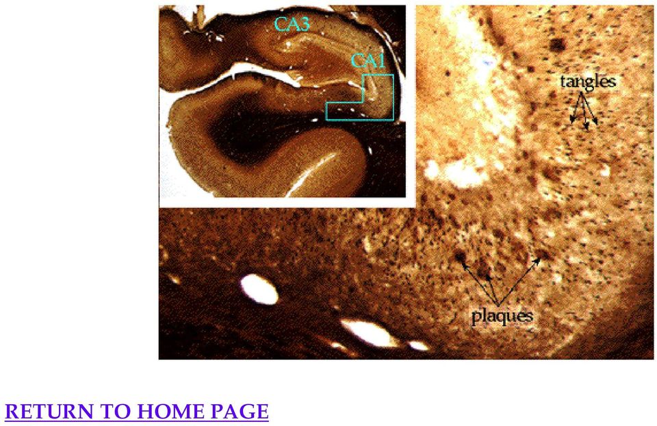
6 called mossy fibers. ("Mossy fibers" is a morphological description for axons with large bulbous terminals, and these are unrelated to those in the cerebellum.) CA3 sends axons called Schaeffer collaterals to CA1, which sends yet another set of fibers to the subiculum. The subiculum is responsible for the output of the hippocampus: it can either send axons directly to the hypothalamus and mammillary bodies via the fornix (remember the fornix?), or it can pass along the information back to entorhinal cortex, which will relay it all back to sensory cortex. It is essentially one continuous pathway that begins in sensory cortex, traverses the hippocampus (loop-the-loop), and returns to sensory cortex. Somewhere in there, memory is born. E. Diseases of the hippocampus: The hippocampus is particularly vulnerable to several disease processes, including ischemia, which is any obstruction of blood flow or oxygen deprivation, Alzheimer's disease, and epilepsy. These diseases selectively attack CA1, which effectively cuts through the hippocampal circuit. Below is a photograph of a normal hippocampus and one which has been deprived of oxygen. You should be able to see the degeneration of CA1 (labeled) and the absence of cell bodies (stained purple). A stroke can have this effect, but there must be bilateral damage of the hippocampi to affect memory. Therefore only situations that deplete blood or oxygen flow to the entire brain will produce a memory deficit. The pathology of severe temporal lobe epilepsy looks very similar to ischemic damage. Alzheimer's disease, although it affects the entire brain, is particularly hard on the CA1 region. Below is a photograph of the hippocampus of an Alzheimer's patient, with the CA1 region magnified. Both extracellular plaques and intracellular tangles are visible - these are the pathological hallmarks of the disease.
, or it can pass along the information back to entorhinal cortex, which will relay it all back to sensory cortex.")
7 RETURN TO HOME PAGE
Brain Structures That are Involved with Memory
 Early Theories of Brain Structures That are Involved with Psychology 372 Sensation Sensory Attention Rehearsal STM Storage Retrieval Physiological Psychology Steven E. Meier, Ph.D. Listen to the audio
Human Neuroanatomy. Grades 9-12. Driving Question: How did the evolution of the human brain impact the structure and function it has today?
 Human Neuroanatomy Grades 9-12 Driving Question: How did the evolution of the human brain impact the structure and function it has today? Objectives: Students will be able to Describe the basic parts and
NEUROANATOMY 6 Limbic System
 NEUROANATOMY 6 Limbic System The Limbic System The part of the brain involved with learning, memory and emotion. It is affected in many neuropsychiatric diseases including schizophrenia, Alzheimer s disease
OLFACTORY PATHWAYS AND LIMBIC SYSTEM
 903 Olfactory and Limbic OLFACTORY PATHWAYS AND LIMBIC SYSTEM I. OLFACTORY PATHWAYS The sense of smell is much less essential than vision, audition or the somatic senses, and will therefore receive less
What is the basic component of the brain and spinal cord communication system?
 EXPLORING PSYCHOLOGY David Myers The Biology of Mind Chapter 2 Neural Communication Neurons How Neurons Communicate How Neurotransmitters Influence Us The Nervous System The Peripheral Nervous System The
Sheep Brain Dissection Picture Guide
 Sheep Brain Dissection Picture Guide Figure 1: Right Hemisphere of Sheep s Brain Figure 2: Underside of Sheep s Brain Figure 3: Saggital cut of Sheep s Brain to reveal subcortical structures Figure 4:
Nervous System Organization. PNS and CNS. Nerves. Peripheral Nervous System. Peripheral Nervous System. Motor Component.
 Nervous System Organization PNS and CNS Chapters 8 and 9 Peripheral Nervous System (PNS) connects CNS to sensory receptors, muscles and glands Central Nervous System (CNS) control/integrating center brain
31.1 The Neuron. BUILD Vocabulary. Lesson Objectives
 Name Class Date 31.1 The Neuron Lesson Objectives Identify the functions of the nervous system. Describe the function of neurons. Describe how a nerve impulse is transmitted. BUILD Vocabulary A. The chart
Sheep Brain Dissection
 Sheep Brain Dissection http://www.carolina.com/product/preserved+organisms/preserved+animals+%28mammal s%29/sheep+organs/preserved+sheep+dissection.do Michigan State University Neuroscience Program Brain
Vision: Receptors. Modes of Perception. Vision: Summary 9/28/2012. How do we perceive our environment? Sensation and Perception Terminology
 How do we perceive our environment? Complex stimuli are broken into individual features, relayed to the CNS, then reassembled as our perception Sensation and Perception Terminology Stimulus: physical agent
Vocabulary & General Concepts of Brain Organization
 Vocabulary & General Concepts of Brain Organization Jeanette J. Norden, Ph.D. Professor Emerita Vanderbilt University School of Medicine Course Outline Lecture 1: Vocabulary & General Concepts of Brain
Nervous System: PNS and CNS
 Nervous System: PNS and CNS Biology 105 Lecture 10 Chapter 8 Outline I. Central Nervous System vs Peripheral Nervous System II. Peripheral Nervous System A. Somatic Nervous System B. Autonomic Nervous
The Brain of a Normal Human
 The Brain of a Normal Human Your Brain Evolved Over Time Human Brain Logic and reasoning Mammalian Brain More complex feelings and reactions Lizard Brain Basic functions The Brain Stem or Hindbrain (The
3. The neuron has many branch-like extensions called that receive input from other neurons. a. glia b. dendrites c. axons d.
 Chapter Test 1. A cell that receives information and transmits it to other cells via an electrochemical process is called a(n) a. neuron b. hormone c. glia d. endorphin Answer: A difficulty: 1 factual
Lab Exercise 9. Nervous Tissue. Brain. Cranial Nerves. Spinal Cord. Spinal Nerves
 Lab Exercise 9 Nervous Tissue Brain Cranial Nerves Spinal Cord Spinal Nerves Textbook Reference: See Chapter 11 for histology of nerve tissue and spinal cord See Chapter 12 for brain and spinal cord anatomy
2 Neurons. 4 The Brain: Cortex
 1 Neuroscience 2 Neurons output integration axon cell body, membrane potential Frontal planning control auditory episodes soma motor Temporal Parietal action language objects space vision Occipital inputs
Student Academic Learning Services Page 1 of 8 Nervous System Quiz
 Student Academic Learning Services Page 1 of 8 Nervous System Quiz 1. The term central nervous system refers to the: A) autonomic and peripheral nervous systems B) brain, spinal cord, and cranial nerves
Chapter 7: The Nervous System
 Chapter 7: The Nervous System I. Organization of the Nervous System Objectives: List the general functions of the nervous system Explain the structural and functional classifications of the nervous system
Figure 2 Figure 3 Figure 1
 The brain is organized into three tiers; a lower tier made up by the brainstem and cerebellum, a middle tier containing the thalamus, basal ganglia and many components of the limbic system and an upper
Lecture One: Brain Basics
 Lecture One: Brain Basics Brain Fractured Femur Bone Spinal Cord 1 How does pain get from here to here 2 How does the brain work? Every cell in your body is wired to send a signal to your brain The brain
Basic Brain Information
 Basic Brain Information Brain facts Your brain weighs about 3lbs, or just under 1.5Kg It has the texture of blancmange Your brain is connected to your spinal cord by the brain stem Behind your brain stem
CENTRAL NERVOUS SYSTEM. Sensory Pathway (PNS) OVERVIEW OF SPINAL CORD ANATOMY OF THE SPINAL CORD FUNCTIONS OF THE SPINAL CORD
 OVERVIEW OF SPINAL CORD ANATOMY OF THE SPINAL CORD FUNCTIONS OF THE SPINAL CORD") CENTRAL NERVOUS SYSTEM Central nervous system (CNS) brain and spinal cord enclosed in bony coverings Functions of the spinal cord spinal cord reflexes integration ti (summation of inhibitory and excitatory)
CENTRAL NERVOUS SYSTEM Central nervous system (CNS) brain and spinal cord enclosed in bony coverings Functions of the spinal cord spinal cord reflexes integration ti (summation of inhibitory and excitatory)
Explore the Neuroscience for Kids Web Site (ANSWERS) Start at: http://faculty.washington.edu/chudler/neurok.html
 Start at: http://faculty.washington.edu/chudler/neurok.html") NAME Explore the Neuroscience for Kids Web Site (ANSWERS) Start at: http://faculty.washington.edu/chudler/neurok.html On the left side, click on Explore, then click on The Neuron, then click on Millions
NAME Explore the Neuroscience for Kids Web Site (ANSWERS) Start at: http://faculty.washington.edu/chudler/neurok.html On the left side, click on Explore, then click on The Neuron, then click on Millions
Name: Teacher: Olsen Hour:
 Name: Teacher: Olsen Hour: The Nervous System: Part 1 Textbook p216-225 41 In all exercises, quizzes and tests in this class, always answer in your own words. That is the only way that you can show that
Chapter Fourteen. Emotion, Reward, Aggression, and Stress
 Chapter Fourteen Emotion, Reward, Aggression, and Stress EMOTIONS! Emotions generally include a Physical component, and a Subjective component or quality, and a Valence Emotions a product of Evolution?
Learning with Your Brain. Teaching With the Brain in Mind
 Learning with Your Brain Should what (and how) we teach be associated with what we know about the brain and the nervous system? Jonathan Karp, Ph.D. Dept of Biology 5/20/2004 Teaching With the Brain in
CSE511 Brain & Memory Modeling. Lect04: Brain & Spine Neuroanatomy
 CSE511 Brain & Memory Modeling CSE511 Brain & Memory Modeling Lect02: BOSS Discrete Event Simulator Lect04: Brain & Spine Neuroanatomy Appendix of Purves et al., 4e Larry Wittie Computer Science, StonyBrook
Slide 4: Forebrain Structures. Slide 5: 4 Lobes of the Cerebral Cortex. Slide 6: The Cerebral Hemispheres (L & R)
") Slide 1: [Film Clip: The Brain #2- Phineas Gage] Integrated Bodily Communications Within Brain (Hemispheres and structures) The remaining Nervous System Endocrine System (Hormonal communication) Our bodies-
Slide 1: [Film Clip: The Brain #2- Phineas Gage] Integrated Bodily Communications Within Brain (Hemispheres and structures) The remaining Nervous System Endocrine System (Hormonal communication) Our bodies-
Brain Development. Genetic make-up... is not the major determiner
 Brain Development Presented by: Linda Alsop SKI-HI Institute Utah State University Genetic make-up... is not the major determiner Early experiences are so powerful that they can completely change the way
Brain Matters: Brain Anatomy
 1 : Brain Anatomy Lesson Overview Students share what they already know about brain structure and function, and then, guided by descriptions of brain regions explore the G2C Online 3-D Brain to learn more
The Brain. What is it? Neurons Glial Cells Connective Tissue Connective Fiber White Matter Grey Matter Cerebro-spinal Fluid
 The Brain What is it? Neurons Glial Cells Connective Tissue Connective Fiber White Matter Grey Matter Cerebro-spinal Fluid A More Realistic View When we look at the brain we see mostly the Cerebral Cortex
Brain Power. Counseling and Mental Health
 Brain Power Counseling and Mental Health TEA COPYRIGHT Copyright Texas Education Agency, 2012. These Materials are copyrighted and trademarked as the property of the Texas Education Agency (TEA) and may
Nervous System. from the Human Body Systems Series. catalog # 2890. Published & Distributed by AGC/UNITED LEARNING
 Nervous System from the Human Body Systems Series catalog # 2890 Published & Distributed by AGC/UNITED LEARNING 1560 Sherman Avenue Suite 100 Evanston, IL 60201 1-800-323-9084 24-Hour Fax No. 847-328-6706
The Hypothalamus. Functions of the Hypothalamus. The Hypothalamus. Medical Neuroscience Dr. Wiegand
 The Medical Neuroscience Dr. Wiegand Neural Influences Neural Influences Hormonal Influences Hormonal Influences The Autonomic Nervous System Limbic System Endocrine System system Endocrine 1 The Diencephalon
BIO130 Chapter 14 The Brain and Cranial Nerves Lecture Outline
 BIO130 Chapter 14 The Brain and Cranial Nerves Lecture Outline Brain structure 1. Cerebrum Hemispheres: left & right Cerebral cortex Gyri Sulci Fissures Longitudinal fissure Corpus callosum Lobes Central
LESSON 4: BRAIN STRUCTURE AND FUNCTION
 LESSON 4: BRAIN STRUCTURE AND FUNCTION THE TRIUNE BRAIN Neo? axon brain stem cerebral hemispheres cortex dendrite limbic system neural plasticity neurons neurotransmitter sensory flooding sensory gating
Biology Slide 1 of 38
 Biology 1 of 38 2 of 38 35-2 The Nervous System What are the functions of the nervous system? 3 of 38 35-2 The Nervous System 1. Nervous system: a. controls and coordinates functions throughout the body
DISSECTION OF THE SHEEP'S BRAIN
 DISSECTION OF THE SHEEP'S BRAIN Introduction The purpose of the sheep brain dissection is to familiarize you with the threedimensional structure of the brain and teach you one of the great methods of studying
Nervous System sensor input integration motor output sensory organs central nervous system
 Nervous System Nervous system performs three overlapping functions of sensor input, integration, and motor output. This process is generally the same even at a very primitive level of nervous system, but
Integration and Coordination of the Human Body. Nervous System
 I. General Info Integration and Coordination of the Human Body A. Both the and system are responsible for maintaining 1. Homeostasis is the process by which organisms keep internal conditions despite changes
The brain structure and function
 The brain structure and function This information is an extract from the booklet Understanding brain tumours. You may find the full booklet helpful. We can send you a copy free see page 5. Contents Introduction
What role does the nucleolus have in cell functioning? Glial cells
 Nervous System Lab The nervous system of vertebrates can be divided into the central nervous system, which consists of the brain and spinal cord, and the peripheral nervous system, which contains nerves,
Functions of the Brain
 Objectives 0 Participants will be able to identify 4 characteristics of a healthy brain. 0 Participants will be able to state the functions of the brain. 0 Participants will be able to identify 3 types
the puzzle packet! Brought to you by
 the puzzle packet! Brought to you by It s Mindboggling! word search The following brain-related words all appear in It s Mindboggling! How many can you find? Words may appear in all directions, including
1. Which of the following is NOT part of the diencephalon? a. Pineal gland b. Tectum c. Interthalamic adhesion d. Hypothalamus e.
 1. Which of the following is NOT part of the diencephalon? a. Pineal gland b. Tectum c. Interthalamic adhesion d. Hypothalamus e. Thalamus 2. The is the primary relay station for sensory information coming
Homework Help Stroke
 The Brain & Strokes Your brain is the most complex organ in your body. It is the command centre for everything you do, think, sense and say! It has over 100 billion special nerve cells called neurons.
Unit 2 - Subcortical systems, neurochemistry and brain function
 Unit 2 - Subcortical systems, neurochemistry and brain function Subcortical anatomy: Most of the five major subdivisions of the brain are subcortical. I. Telencephalon (cortical - part of forebrain) -
BIOLOGY STUDY PACKET THE BRAIN
 BIOLOGY STUDY PACKET THE BRAIN SC.912.L.14.26 AA Spring 2012 The intent of this packet is to supplement regular classroom instruction, not to replace it. This also supposes that the students have access
Mini-atlas of the Marmoset Brain
 Mini-atlas of the Marmoset Brain http://marmoset-brain.org Aya Senoo Tokyo University of Agriculture and Technology Hironobu Tokuno Tokyo Metropolitan Institute of Medical Science Charles Watson Curtin
Chapter 3 The Anatomy of the Nervous System
 Chapter 3 The Anatomy of the Nervous System Systems, Structures, and Cells That Make Up Your Nervous System 1 General Layout of the Nervous System Central Nervous System (CNS) Brain (in the skull) Spinal
Chapter 7: The Nervous System
 Chapter 7: The Nervous System Objectives Discuss the general organization of the nervous system Describe the structure & function of a nerve Draw and label the pathways involved in a withdraw reflex Define
The Visual Cortex 0 http://www.tutis.ca/neuromd/index.htm 20 February 2013
 T he Visual Cortex 0 Chapter contents Contents Chapter 2... 0 T he Visual Cortex... 0 Chapter Contents... 1 Introduction... 2 Optic Chiasm... 2 Where do the eye's ganglion cells project to?... 3 To where
Origin of Electrical Membrane Potential
 Origin of Electrical Membrane Potential parti This book is about the physiological characteristics of nerve and muscle cells. As we shall see, the ability of these cells to generate and conduct electricity
North Bergen School District Benchmarks
 Grade: 10,11, and 12 Subject: Anatomy and Physiology First Marking Period Define anatomy and physiology, and describe various subspecialties of each discipline. Describe the five basic functions of living
Neurophysiology. 2.1 Equilibrium Potential
 2 Neurophysiology 2.1 Equilibrium Potential An understanding of the concepts of electrical and chemical forces that act on ions, electrochemical equilibrium, and equilibrium potential is a powerful tool
Slide 1. Slide 2. Slide 3
 Slide 1 The Role of Experience on the Developing Brain Barb Jackson, Ph.D. Director, Education & Child Development Munroe-Meyer Institute University of Nebraska Medical Center Omaha, NE USA The purpose
Function (& other notes)
") LAB 8. ANATOMY OF THE HUMAN BRAIN In this exercise you each will map the human brain both anatomy and function so that you can develop a more accurate picture of what s going on in your head :-) EXTERNAL
LAB 8. ANATOMY OF THE HUMAN BRAIN In this exercise you each will map the human brain both anatomy and function so that you can develop a more accurate picture of what s going on in your head :-) EXTERNAL
2401 : Anatomy/Physiology
 Dr. Chris Doumen Week 7 2401 : Anatomy/Physiology The Brain Central Nervous System TextBook Readings Pages 431 through 435 and 463-467 Make use of the figures in your textbook ; a picture is worth a thousand
It s All in the Brain!
 It s All in the Brain! Presented by: Mari Hubig, M.Ed. 0-3 Outreach Coordinator Educational Resource Center on Deafness What is the Brain? The brain is a muscle In order to grow and flourish, the brain
Epilepsy and Neuropsychology Dr. Sare Akdag, RPsych
 Epilepsy and Neuropsychology Dr. Sare Akdag, RPsych Most people living with epilepsy do not experience serious problems with their thinking. However, there are aspects of thinking that can be affected
The intermedius nucleus of the medulla: A potential site for the integration of cervical information and the generation of autonomic responses
 The intermedius nucleus of the medulla: A potential site for the integration of cervical information and the generation of autonomic responses 1 Journal of Chemical Neuroanatomy November 2009, 38, pp.
Chapter 9 Nervous System
 Chapter 9 Nervous System Nervous System function: The nervous system is composed of neurons and neuroglia. at the ends of peripheral nerves gather information and convert it into nerve impulses. When sensory
Brains, Ontologies & Virtual Machines
 How Minds Work Brains, Ontologies & Virtual Machines Stan Franklin Computer Science Division & Institute for Intelligent Systems The University of Memphis 1 Question: How do minds work? What would an answer
Chapter 4. The Brain
 Chapter 4 The Brain The Nervous System Central Nervous System (CNS) receives, processes, interprets and stores info (taste, sound, smell, color etc.) Sends information to muscles, glands and internal organs
U N IT 10 NE RVOUS SYS TE M REVIEW 1. Which of the following is controlled by the somatic nervous system? A. rate of heartbeat B.
 U N IT 10 NE RVOUS SYS TE M REVIEW 1. Which of the following is controlled by the somatic nervous system? A. rate of heartbeat B. contraction of skeletal muscles C. increased blood flow to muscle tissue
http://abcnews.go.com/politics/video/obama-says-brain-initiative-will-be-transformative-18861944
 http://abcnews.go.com/politics/video/obama-says-brain-initiative-will-be-transformative-18861944 What are the nervous system s functions? The nervous system organizes and controls an individual s appropriate
How To Understand The Hypothalamus
 883 Hypothalamus HYPOTHALAMUS Introduction The hypothalamus is a very small, but extremely important part of the diencephalon that is involved in the mediation of endocrine, autonomic and behavioral functions.
Sensory Organs (Receptors) Sensory Physiology. Sensory Adaptation. Four Steps to Sensation. Types of Sensors Structural Design
 Sensory Physiology. Sensory Adaptation. Four Steps to Sensation. Types of Sensors Structural Design") Sensory Organs (Receptors) Sensory Physiology Chapter 10 Monitor the internal and external environment Transmit peripheral signals to CNS for processing Critical for homeostasis Types of Sensors Structural
Sensory Organs (Receptors) Sensory Physiology Chapter 10 Monitor the internal and external environment Transmit peripheral signals to CNS for processing Critical for homeostasis Types of Sensors Structural
Emotions, Aggression and Stress
 Emotions, Aggression and Stress Chapter 15 1 Emotion Emotion is a complicated psychological concept that includes wide range of observable behaviors, expressed feelings and changes in bodily state. Due
Adapted from Human Anatomy & Physiology by Marieb and Hoehn (9 th ed.)
") BRAIN ANATOMY Adapted from Human Anatomy & Physiology by Marieb and Hoehn (9 th ed.) The anatomy of the brain is often discussed in terms of either the embryonic scheme or the medical scheme. The embryonic
BRAIN ANATOMY Adapted from Human Anatomy & Physiology by Marieb and Hoehn (9 th ed.) The anatomy of the brain is often discussed in terms of either the embryonic scheme or the medical scheme. The embryonic
Diagram 2(i): Structure of the Neuron
: Structure of the Neuron") Diagram 2(i): Structure of the Neuron Generally speaking, we can divide the nervous system into different parts, according to location and function. So far we have mentioned the central nervous system
Diagram 2(i): Structure of the Neuron Generally speaking, we can divide the nervous system into different parts, according to location and function. So far we have mentioned the central nervous system
CHAPTER 11: NERVOUS SYSTEM II: DIVISIONS OF THE NERVOUS SYSTEM OBJECTIVES: 1. Outline the major divisions of the nervous system.
 CHAPTER 11: NERVOUS II: DIVISIONS OF THE NERVOUS OBJECTIVES: 1. Outline the major divisions of the nervous system. NERVOUS CENTRAL NERVOUS (BRAIN & SPINAL CORD) (INTERNEURONS) PERIPHERAL NERVOUS (CRANIAL
Pierre Gloor (1923 2003): An Appreciation
: An Appreciation") Epilepsia, 45(7):882 886, 2004 Blackwell Publishing, Inc. C 2004 International League Against Epilepsy In Memoriam Pierre Gloor (1923 2003): An Appreciation Massimo Avoli On October 24, 2003, the world
Epilepsia, 45(7):882 886, 2004 Blackwell Publishing, Inc. C 2004 International League Against Epilepsy In Memoriam Pierre Gloor (1923 2003): An Appreciation Massimo Avoli On October 24, 2003, the world
Nerve Cell Communication
 Nerve Cell Communication Core Concept: Nerve cells communicate using electrical and chemical signals. Class time required: Approximately 2 forty minute class periods Teacher Provides: For each student
Brain Tumor 101. Shanna Armstrong, RN Neuro Oncology Nurse Clinician UC Brain Tumor Center
 Brain Tumor 101 Shanna Armstrong, RN Neuro Oncology Nurse Clinician UC Brain Tumor Center Objectives Identify the different parts of the brain Describe how each part of the brain works Connect each part
3) Cerebral Cortex & Functions of the 4 LOBES. 5) Cranial Nerves (Nerves In the Cranium, i.e., Head)
 Cerebral Cortex & Functions of the 4 LOBES. 5) Cranial Nerves (Nerves In the Cranium, i.e., Head)") Lecture 5 (Oct 8 th ): ANATOMY and FUNCTION OF THE NERVOUS SYSTEM Lecture Outline 1) Basic Divisions (CNS vs. PNS, Somatic vs. Autonomic) and Directional Terms 2) The Brain (Hindbrain/ Midbrain/ Forebrain)
Lecture 5 (Oct 8 th ): ANATOMY and FUNCTION OF THE NERVOUS SYSTEM Lecture Outline 1) Basic Divisions (CNS vs. PNS, Somatic vs. Autonomic) and Directional Terms 2) The Brain (Hindbrain/ Midbrain/ Forebrain)
Module 1: The Brain and the Central Nervous System (CNS)
") Module 1: The Brain and the Central Nervous System (CNS) By the end of this unit, the learner will be able to: Describe the anatomy of the brain and the central nervous system Identify regions of the brain
Module 1: The Brain and the Central Nervous System (CNS) By the end of this unit, the learner will be able to: Describe the anatomy of the brain and the central nervous system Identify regions of the brain
BIOL 1108 Vertebrate Anatomy Lab
 BIOL 1108 Vertebrate Anatomy Lab This lab explores major organs associated with the circulatory, excretory, and nervous systems of mammals. Circulatory System Vertebrates are among the organisms that have
Background on Brain Injury
 CHAPTER 1 Background on Brain Injury In this chapter, you will: Read about Alberta s definition of Acquired Brain Injury and how that affects which supports you will be able to access. Learn about the
Class 10 NCERT Science Text Book Chapter 7 Control and Coordination
 Class 10 NCERT Science Text Book Chapter 7 Control and Coordination Question 1: What is the difference between a reflex action and walking? A reflex action is a rapid, automatic response to a stimulus.
Chapter 15 Anatomy and Physiology Lecture
 1 THE AUTONOMIC NERVOUS SYSTEM Chapter 15 Anatomy and Physiology Lecture 2 THE AUTONOMIC NERVOUS SYSTEM Autonomic Nervous System (ANS) regulates the activity of smooth muscles, cardiac muscles, and certain
Chapter 2. The Biological Bases of Psychological Functioning
 Chapter 2 The Biological Bases of Psychological Functioning Outline I. Neurons: The Building Blocks of the Nervous System A. A neuron is a nerve cell that transmits information from one part of the body
How are Parts of the Brain Related to Brain Function?
 How are Parts of the Brain Related to Brain Function? Scientists have found That the basic anatomical components of brain function are related to brain size and shape. The brain is composed of two hemispheres.
Parts of the Brain. Chapter 1
 Chapter 1 Parts of the Brain Living creatures are made up of cells. Groups of cells, similar in appearance and with the same function, form tissue. The brain is a soft mass of supportive tissues and nerve
Name Date Hour. Nerve Histology Microscope Lab
 Name Date Hour Nerve Histology Microscope Lab PRE-LAB: Answer the following questions using your reading and class notes before starting the microscope lab. 1. What is the difference between the functions
Anatomy Review Graphics are used with permission of: adam.com (http://www.adam.com/) Benjamin Cummings Publishing Co (http://www.awl.com/bc).
 Benjamin Cummings Publishing Co (http://www.awl.com/bc).") Page 1. Introduction The structure of neurons reflects their function. One part of the cell receives incoming signals. Another part generates outgoing signals. Anatomy Review Graphics are used with permission
Page 1. Introduction The structure of neurons reflects their function. One part of the cell receives incoming signals. Another part generates outgoing signals. Anatomy Review Graphics are used with permission
D.U.C. Assist. Lec. Faculty of Dentistry General Physiology Ihsan Dhari. The Autonomic Nervous System
 The Autonomic Nervous System The portion of the nervous system that controls most visceral functions of the body is called the autonomic nervous system. This system helps to control arterial pressure,
Lesson # 5A Workshop: Complex Networks and Graphs (Adapted from Teaching Engineering s It s a Connected World )
") Subject area / course / grade level: Science Grade 7-10 Summary: Students are introduced to networks and systems. The students will use this knowledge to create their own model of a neural network (Lesson
Subject area / course / grade level: Science Grade 7-10 Summary: Students are introduced to networks and systems. The students will use this knowledge to create their own model of a neural network (Lesson
Cortical Visual Impairment An introduction
 Cortical Visual Impairment An introduction This is a general introduction to Cortical Visual Impairment intended for families and support workers who may live with or support children and adults with learning
Cerebellum and Basal Ganglia
 Cerebellum and Basal Ganglia 1 Contents Cerebellum and Basal Ganglia... 1 Introduction... 3 A brief review of cerebellar anatomy... 4 Basic Circuit... 4 Parallel and climbing fiber input has a very different
About Brain Injury: A Guide to Brain Anatomy Information from http://www.waiting.com, 1997-2002, Becca, Ltd.
 About Brain Injury: A Guide to Brain Anatomy Information from http://www.waiting.com, 1997-2002, Becca, Ltd. Brain Anatomy Definitions Brainstem: The lower extension of the brain where it connects to the
What Is an Arteriovenous Malformation (AVM)?
?") What Is an Arteriovenous Malformation (AVM)? From the Cerebrovascular Imaging and Intervention Committee of the American Heart Association Cardiovascular Council Randall T. Higashida, M.D., Chair 1 What
What Is an Arteriovenous Malformation (AVM)? From the Cerebrovascular Imaging and Intervention Committee of the American Heart Association Cardiovascular Council Randall T. Higashida, M.D., Chair 1 What
1 PYRAMIDS - CORTICOSPINAL FIBERS
 151 Brain stem Pyramids/Corticospinal Tract 1 PYRAMIDS - CORTICOSPINAL FIBERS The pyramids are two elongated swellings on the ventral aspect of the medulla. Each pyramid contains approximately 1,000,000
THE BRAIN AND CRANIAL NERVES
 THE BRAIN AND CRANIAL NERVES The Brain - made up of a trillion neurons - weighs about 3 lbs - has four principle parts 1. Brain stem - medulla oblongata, pons, midbrain (mesencephalon) 2. Diencephalon
Peripheral Nervous System
 Nervous system consists of: Peripheral Nervous System CNS = brain and spinal cord ~90% (90 Bil) of all neurons in body are in CNS PNS = Cranial nerves and spinal nerves, nerve plexuses & ganglia ~10% (10
Exercise. Rule #1 Exercise boosts brain power.
 Exercise Rule #1 Exercise boosts brain power. Our brains were built for walking 12 miles a day! To improve your thinking skills, move. Exercise gets blood to your brain, bringing it glucose for energy