Postgraduate Course 10 Malignant pleural effusion management
|
|
|
- Logan Eaton
- 10 years ago
- Views:
Transcription
1 ERS International Congress Amsterdam September 2015 Postgraduate Course 10 Malignant pleural effusion management Thank you for viewing this document. We would like to remind you that this material is the property of the author. It is provided to you by the ERS for your personal use only, as submitted by the author by the author Saturday, 26 September :30 13:00 Room 13.2 RAI

2 During the session access the voting questions here: You can access an electronic copy of these educational materials here: To access the educational materials on your tablet or smartphone please find below a list of apps to access, annotate, store and share pdf documents. Apple ios Adobe Reader - FREE - With the Adobe Reader app you can highlight, strikethrough, underline, draw (freehand), comment (sticky notes) and add text to pdf documents using the typewriter tool. It can also be used to fill out forms and electronically sign documents. Mendeley - FREE - Mendeley is a free reference manager and PDF reader with which you can make your own searchable library, read and annotate your PDFs, collaborate with others in private groups, and sync your library across all your devices. Notability Notability uses CloudServices to import and automatically backup your PDF files and allows you to annotate and organise them (incl. special features such as adding a video file). On ipad, you can bookmark pages of a note, filter a PDF by annotated pages, or search your note for a keyword. Android Adobe Reader - FREE - The Android version of Adobe Reader lets you view, annotate, comment, fill out, electronically sign and share documents. It has all of the same features as the ios app like freehand drawing, highlighting, underlining, etc. iannotate PDF - FREE - You can open multiple PDFs using tabs, highlight the text and make comments via handwriting or typewriter tools. iannotate PDF also supports Box OneCloud, which allows you to import and export files directly from/to Box. ezpdf Reader With the ezpdf Reader you can add text in text boxes and sticky notes; highlight, underline, or strikethrough texts or add freehand drawings. Add memo and append images, change colour / thickness, resize and move them around as you like.
, comment (sticky notes) and add text to pdf documents using the typewriter tool.")
3 Postgraduate Course 10 Malignant pleural effusion management AIMS: To understand the new roles that molecular biology and histo-/cytopathology play in the diagnosis and management (treatment and prognosis) of malignant pleural effusions, and how to use these techniques (processing and data interpretation) in clinical practice. TARGET AUDIENCE: Trainees, pulmonary physicians, thoracic surgeons, pathologists, molecular biologists, and scientists. CHAIRS: P. Astoul (Marseille, France), M. Skrzypski (Sopot, Poland) COURSE PROGRAMME PAGE 09:30 Which material in which disease? 5 S. Fernandez Bussy (Santiago, Chile) 10:15 How to send your material to pathology 82 P. Schnabel (Homburg/Saar, Germany) 11:00 Break 11:30 Interpretation of the results in the clinic 83 A. Scherpereel (Lille, France) 12:15 Atypical mesothelial hyperplasia and other pitfalls in MPE diagnosis 102 F. Galateau-Salle (Caen, France) Faculty disclosures 108 Faculty contact information 109
 COURSE PROGRAMME PAGE 09:30 Which material in which disease? 5 S. Fernandez Bussy (Santiago, Chile) 10:15 How to send your material to pathology 82 P.")
4 Regular physical activity improves quality of life and fitness in healthy individuals and people living with lung conditions. In partnership with: Farbe/colour: PANTONE 288 CV ERS/ELF ER Journal 210x280 ad_v2 AW.indd 4 29/07/ :23

5 Malignant Pleural Effusion Dr Sebastian Fernandez Bussy Clinica Alemana-Universidad del Desarrollo Interventional Pulmonology Av Vitacura 5951 Santiago, Región Metropolitana CHILE SUMMARY Metastatic disease to the pleura occur much more frequently than primary pleural neoplasms, causing approximately pleural effusions in the USA each year, as compared to Diffuse Malignant Mesothelioma (DMM), which causes approximately 1500 pleural effusions each year. This is a ratio of over 130 pleural metastases for each primary pleural DMM. Metastases to the pleura exhibit characteristics, including pleural effusions, pleural thickening and pleural-based masses, found with primary pleural neoplasms and reactive pleural processes. Common metastases to the pleura include lymphoma, lung cancer and breast cancer; however, malignancies from any primary may metastasize to the pleura. The pleura can be invaded directly from neighboring structures (lung, chest wall -- including breast in some cases -- diaphragm and mediastinum) but most of the pleural malignancies arise, according to necropsy studies, from tumor emboli to the visceral pleura, with secondary seeding to the parietal pleura. The effusion can develop as a direct consequence of neoplastic pleural involvement, but it can also occur in cases with lymphatic blockade at the mediastinal level. Diagnostic thoracentesis should be performed in every pleural effusion not clearly associated to heart failure, hepathic hydrothorax or chronic renal failure, and also in patients with the above conditions not responding to the appropriate treatment. Malignant transudative effusions can occur in lung cancer with obstruction of the mainstem bronchus, but also in lymphoma and other malignancies. Blockade of lymphatic drainage may be invoked as one of the possible pathogenetic mechanisms, but an underlying cardiac failure or other conditions that are usually associated with transudative effusions should be taken into account also. Many studies have shown a large variation in the diagnostic sensitivity of pleural fluid cytological analysis, ranging from 40 87%2. In particular, a cytomorphologic distinction between reactive mesothelial cells, mesothelioma, and metastatic adenocarcinoma, as well as between lymphomas and reactive lymphocytosis, can often be difficult because of significant overlapping cytologic features. Therefore, other procedures, such as immunohistochemistry (IHC) using monoclonal antibodies against tumor markers and chromosomal analysis, complement cytology in the diagnosis of MPEs. IHC staining can be performed on conventional cytology specimens and cell blocks. There have been many reports on the application of different IHC markers in pleural effusion samples to diagnosis MPE and to identify the primary site of origin. Differential cell counts in pleural fluid are never diagnostic, but can help (more than two-thirds of lymphocytic pleural effusions are the result of malignancy or tuberculosis). Pleural fluid eosinophilia (> 10% eosinophils in the total nucleated cell count) is virtually never diagnostic because it can be due to idiopathic (25%), malignant (20%), infectious (20%) or miscellaneous 5
, which causes approximately 1500 pleural effusions each year. This is a ratio of over 130 pleural metastases for each primary pleural DMM.")
6 benign conditions (35%) [48]. It should be stressed that the higher the percentage of pleural eosinophils (e.g., > 40%), the lower the likelihood of malignancy and the greater the likelihood of an unknown etiology. A negative Adenosin deaminase (ADA) is useful to rule out tuberculous pleuritis in lymphocytepredominant effusions [12]. In addition, extremely high ADA (> 250 U/L) in pleural fluid should raise suspicion of a non-tuberculous effusion, particularly lymphoma or complicated parapneumonic effusion. The yield from sending more than two separate pleural fluid specimens for cytology is very low and should be discouraged: In a study of 603 patients with MPEs, cytological analysis had an overall sensitivity of 50% (95% confidence interval (CI): %) for the first specimen and 57% (95% CI: %) and 58.2% (95% CI: %) for the second and third separate examinations, respectively. For both direct smear/cytospin and cell block preparations, 150 ml or more were recommended, whereas 60 ml were adequate when only direct smear/cytospin was used for the diagnosis of MPE. To separate epithelial mesothelioma from adenocarcinoma, at least two mesothelial (e.g., calretinin, keratin 5/6, WT-1 protein) and two carcinoma (e.g., thyroid transcription factor-1- TTF-1-, CEA, B72.3) markers should be used [56]. In a MPE of unknown origin, a positive TTF- 1 stain strongly suggests a primary non-small-cell lung cancer. Immunocytochemistry in pleural fluid can also help in some therapeutic decisions, like selection of targeted therapy in lung cancer [59], and hormonal therapy. Thus, expression of oestrogen/progestogen receptors or Her- 2-neu may have both diagnostic (breast cancer) and therapeutic implications. Flow cytometry has the advantage of fast and observer independent results and can be very helpful in identifying pleural involvement of hematologic malignancies, and in distinguishing malignant from atypical mesothelial cells. There are an increasing number of MPE patients as the incidences of lung cancer and other malignant cancers have increased. Although cytological or histopathologic changes act as the gold standard of MPE diagnosis, these changes are not observed in the bodies of all patients with MPE. Moreover, many patients are unwilling to undergo a biopsy examination. Many researchers have evaluated multiple assays to find out a necessary to find a highly specific and sensitive MPE marker to improve detection accuracy, reduce patient morbidity and inconvenience, and decrease follow-up financial pressure. A panel of PF tumor markers, including CEA, CA 125, CA 15-3 and CYFRA 21-1, have been tested. Although the measure of these molecular markers can serve as a diagnostic guide, they cannot be used exclusively to confirm a diagnosis. Mesothelin levels have been examined in many series of serum samples; however problems of confounding by kidney function and low sensitivity in presymptomatic individuals have limited its acceptance as a diagnostic marker. In comparison there have been far fewer studies of the role of mesothelin measurements in pleural effusions in a diagnostic setting. Tumor marker measurements in effusions have the potential to be more sensitive than serum measures because proximity of the fluid to the tumor allows continuous sampling of the tumor and because effusions are derived from an already symptomatic group. Mesothelin levels greater than 20 nm in effusions are highly suggestive of malignancy, particularly of MM. With a sensitivity of 67% and specificity of 95% for distinguishing Malignant Mesothelioma (MM) from all other effusions this study confirms that measurement of mesothelin level contributes to the diagnostic 6

7 investigation of patients with pleural effusions. As 33% of MM cases do not have an elevated mesothelin level this test is not sufficient for diagnostic purposes as a stand-alone test. The finding that non-mm mesothelin positive cases are primarily due to other malignancies supports the notion that the value of this test is in differentiating malignant from nonmalignant effusions. One of the early biomarkers, proposed over 30 years ago, for mesothelioma was hyaluronic acid. However, the technically demanding nature of the hyaluronic acid assay limited follow-up studies. Recently, two independent studies have been reported that find combining hyaluronic acid with mesothelin or MPF improves sensitivity for mesothelioma, while maintaining specificity when measured in effusion supernatants. Sensitivity of mesothelin for mesothelioma in a dichotomous differential diagnostic setting has been improved by using additional exclusion markers, such as carcinoembryonic antigen, has been reported. Survivin has been confirmed as a novel member of the inhibitor of apoptosis protein (IAP) family. Reportedly, survivin exerts antiapoptosis effects and regulates cell division in tumors and normal tissues. During tumorigenesis, surviving expression is inversely correlated with apoptosis inhibition but positively correlated with proliferation and angiogenesis. Survivin has been widely studied as a prognostic marker in NSCLC, and it plays a crucial role in regulating cell proliferation, apoptosis, and angiogenesis. Many studies have shown that the positive expression of survivin is significantly related to lymph node metastasis and cancers of late clinical stage, such as MPE. In recent years, surviving has generated much interest as a novel biomarker for MPE diagnosis, and it appears to be a useful marker of MPE. It has been reported that levels of plasma (not serum) fibulin-3 can distinguish healthy persons with exposure to asbestos from patients with mesothelioma, and can further differentiate mesothelioma effusions from other malignant and benign effusions. Although, there is still not enough information about the routine use of fibulin-3. For tissue sample, needle pleural biopsy (US or CT guided) or thoracoscopy, can be used. Pleural malignant mesothelioma (MM) is an aggressive asbestos-induced malignancy. The diagnosis of MM is often difficult and may take several weeks to months to establish. Current guidelines recommend that a diagnosis be based on demonstration of invasion by tumor cells; however, not all patients are able to undergo invasive procedures to obtain a biopsy, and even when tissue is obtained diagnosismay still be difficult. In MM, patients often present with a pleural effusion which can be used for diagnostic purposes. Although there is controversy regarding the cytological analysis of cells from pleural effusions, with sensitivities reported in several studies to be around 30%, the limitation of the technique relates primarily to difficulty in distinguishing MM from benign mesothelial cells. However, a high sensitivity and specificity can be achieved if a standardized approach to cytodiagnosis is used. MPM grows in the pleural cavity diffusely more often than as a localized mass. According to WHO histological classification of tumors of the pleura, diffuse MM is classified into four types; epithelioid, sarcomatoid, desmoplastic, and biphasic mesothelioma. Epithelioid mesothelioma (MPM of epithelial type) is most common, and better prognosis has been reported for patients with MPM of epithelial type than those with MPM of other types. An extrapleural pneumonectomy (EPP), considered to be a curative intent surgery for MPM, is characterized by stripping of the affected parietal pleura from the endothoracic fascia of the chest wall and en bloc removal of the affected parietal pleura and lung. To establish radical cure of MPM, it is therefore indispensable to find earlystage MPM of epithelial type, in which mesothelial proliferation is localized on the serosal surface of parietal pleura. Morphological discrimination between MPM 7

8 of epithelial type and reactive mesothelial hyperplasia (RMH) is difficult, and the most reliable pathological criterion for malignancy is mesothelial proliferation invading deeply into subpleural adipose tissues. When a patient presents with an undiagnosed pleural effusion, thoracentesis is generally the first intervention performed. If no definite cancer cells are apparent in the fluid and no alternative cause for the effusion can be found then thoracoscopy with biopsy will be considered if malignancy is a significant possibility. However, in some patients with significant underlying comorbidities and/or frailty in whom thoracoscopy is considered too high risk, or where thoracoscopy is not available, or declined by the patient, measurement of serum and pleural effusion mesothelin levels is a useful step a positive result dramatically increases the likelihood of there being a pleural malignancy present. Although soluble mesothelin has a useful clinical role in diagnosis because of its high specificity, its sensitivity remains limited. That means that a positive result is informative, but a negative one is not. A better biomarker for pleural malignant mesothelioma has yet to be identified. REFERENCES 1. Lamia H. Shabaan et al. Value of thoracoscopic pleural brush in the diagnosis of exudative pleural effusion. Egyptian Journal of Chest Diseases and Tuberculosis (2012) 61, Georgia Karpathiou et al. Pleural neoplastic pathology. Respiratory Medicine (2015) xx, 1e13 3. Henderson DW, et al. Challenges and controversies in the diagnosis of mesothelioma: Part 1. Cytology-only diagnosis, biopsies, immunohistochemistry, discrimination between mesothelioma and reactive mesothelial hyperplasia, and biomarkers. J Clin Pathol 2013;66: Henderson DW, et al. Challenges and controversies in the diagnosis of malignant mesothelioma: Part 2. Malignant mesothelioma subtypes, pleural synovial sarcoma, molecular and prognostic aspects of mesothelioma, BAP1, aquaporin-1 and microrna. J Clin Pathol 2013;66: Philip T. Cagle et al. Pathology of the pleura: What the pulmonologists need to know Respirology (2011) 16, Tohru Tsujimura et al. Pathological and molecular biological approaches to early mesothelioma. Int J Clin Oncol (2012) 17: Aur_elien de Reynies et al. Molecular Classification of Malignant Pleural Mesothelioma: Identification of a Poor Prognosis Subgroup Linked to the Epithelial-to-Mesenchymal Transition. Clin Cancer Res; 20(5) March 1, P. M. Cury et al. The use of histological and mmunohistochemical markers to distinguish pleural malignant mesothelioma and in situ mesothelioma from reactive mesothelial hyperplasia and reactive pleural fibrosis. J. Pathol. 189: (1999) 9. Mark R. Wick et al. Immunohistochemical differential diagnosis of pleural effusions, with emphasis on malignant mesothelioma. Current Opinion in Pulmonary Medicine 2001, 7: Jenette Creaney et al. Comparison of mesothelin and fibulin-3 in pleural fluid and serum as markers in malignant Mesothelioma. Curr Opin Pulm Med 2015, 21: Shi Chen et al. The diagnostic value of survivin in malignant pleural effusion: A metaanalysis. Clinica Chimica Acta 441 (2015) Jenette Creaney et al. Pleural Fluid Mesothelin as an Adjunct to the Diagnosis of Pleural Malignant Mesothelioma. Disease Markers Volume A.M. Egan et al. Malignant pleural effusion. Q J Med 2014; 107:

9 14. P. Korczynski et al. Diagnostic utility of pleural fluid and serum markers in differentiation between malignant and non-malignant pleural effusions. Eur J Med Res (2009) 14 (Suppl. IV): C. Aleman et al., Differentiating between malignant and idiopathic pleural effusions: the value of diagnostic procedures. Q J Med 2007; 100: EVALUATION 1. Please mark the incorrect answer a. Malignant transudative effusions can occur in lung cancer with obstruction of the mainstem bronchus b. Blockade of lymphatic drainage may be invoked as one of the possible pathogenetic mechanisms of malignant pleural effusion c. Most of the pleural malignancies arise from tumor emboli to the visceral pleura, with secondary seeding to the parietal pleura d. Pleural fluid eosinophilia (> 10% eosinophils in the total nucleated e. cell count) is always diagnostic of malignant pleural effusion 2. Please mark the correct answer a. The yield from sending more than two separate pleural fluid specimens for cytology is very low b. In a MPE of unknown origin, a positive TTF-1 stain strongly suggests a primary non-small-cell lung cancer c. At least two mesothelial markers (e.g., calretinin, keratin 5/6, WT-1 protein) should be used to identified epithelial mesothelioma d. Mesothelin levels greater than 20 nm in effusions are highly suggestive of malignancy, particularly of malignant mesothelioma e. All the above 3. Please mark the correct answer a. Sensitivity of mesothelin for mesothelioma has been improved by using additional exclusion markers, such as carcinoembryonic antigen b. Survivin has been confirmed as a novel member of the inhibitor of apoptosis protein (IAP) family c. During tumorigenesis, Survivin expression is inversely correlated with apoptosis inhibition but positively correlated with proliferation and angiogenesis d. Survivin has been widely studied as a prognostic marker in NSCLC e. All the above 4. Please mark a true statement regarding malignant mesothelioma diagnosis a. Mesothelin pleural level has high sensitivity and specificity b. Low mesothelin rules out a malignant effusion c. Mesothelin level has low sensitivity and high specificity d. All the above 9

10 Malignant Pleural Effusion: which material for what disease Sebastian Fernandez-Bussy, MD 10

11 I have no real or perceived conflicts of interest that relate to this presentation: Affiliation/Financial Interest Grants/research support: Commercial Company Honoraria or consultation fees: Participation in a company sponsored bureau: Stock shareholder: Spouse/partner: Other support/potential conflict of interest: This event is accredited for CME credits by EBAP and EACCME and speakers are required to disclose their potential conflict of interest. The intent of this disclosure is not to prevent a speaker with a conflict of interest (any significant financial relationship a speaker has with manufacturers or providers of any commercial products or services relevant to the talk) from making a presentation, but rather to provide listeners with information on which they can make their own judgments. It remains for audience members to determine whether the speaker s interests, or relationships may influence the presentation. The ERS does not view the existence of these interests or commitments as necessarily implying bias or decreasing the value of the speaker s presentation. Drug or device advertisement is forbidden. 11

12 12

13 Tumor markers seem to be a promising alternative and have been proposed to aid in the differentiation of the PE etiology. These include carcinoembryonic antigen (CEA), cancer antigens: CA- 125, CA 15-3, CA 19-9, CA 72-4, cytokeratin fragments (CYFRA 21-1), neuron specific enolase (NSA), and squamous cell carcinoma antigen (SCCA). However, the clinical value of these markers is still unclear. Eur J Med Res (2009) 14(Suppl. IV):
, neuron specific enolase (NSA), and squamous cell carcinoma antigen (SCCA).")
14 Pleural fluid (mean volume 50 ml) and blood samples for tumor marker measurements were collected during the first thoracentesis. They were centrifuged at 2000 rpm for 10 min and the supernatant was frozen at -20 C until assayed. Concentrations of CEA, CA- 125, CYFRA 21-1, and NSE were measured using electrochemiluminescence immunoassays on Roche Elecsys 1010 analyzer (Roche Diagnostics; Mannheim, Germany). Eur J Med Res (2009) 14(Suppl. IV):
. Eur J Med Res (2009) 14(Suppl.")
15 The diagnostic utility was calculated for: 1) single tumor marker level in the pleural fluid (P) and serum (S); 2) single marker pleural fluid/serum level ratio (R); 3) a combination of four markers in the pleural fluid and serum. In the last case, the result was considered positive when the level of at least two markers was higher than the respective marker cut-off value. Eur J Med Res (2009) 14(Suppl. IV):

16 Eur J Med Res (2009) 14(Suppl. IV):
:")
17 Eur J Med Res (2009) 14(Suppl. IV):
:")
18 The median pleural fluid concentrations of the investigated tumor markers were significantly higher in malignant exudates compared with non-malignant effusions, with the exception of CA-125 which was similar. An analysis of the serum concentrations revealed comparable values of CA-125 and NSE in both groups, while the CEA and CYFRA 21-1 levels were significantly higher in malignant effusions. The pleural fluid/serum concentration ratios were similar for all markers in both investigated groups, with the exception of CEA which was significantly higher in the malignant group. Eur J Med Res (2009) 14(Suppl. IV):

19 Eur J Med Res (2009) 14(Suppl. IV):
:")
20 Eur J Med Res (2009) 14(Suppl. IV):
:")
21 CEA in PE proved to be the marker with the highest diagnostic accuracy. The area under curve (AUC) in the ROC analysis for this parameter was Previous studies which included this marker revealed the following AUC values: 0.97; 0.84, and 0.72 [3, 9, 10]. The diagnostic accuracy for serum measurements in our study was lower and equaled to Eur J Med Res (2009) 14(Suppl. IV):
22 CYFRA 21-1 is another marker with a high diagnostic yield, but contrary to the three other markers the serum, rather than plural fluid, level of CYFRA 21-1 shows a higher overall diagnostic accuracy. CYFRA 21-1, soluble fragments of cytokeratin 19, is expressed by all histological types of lung cancer, especially by squamous lung cancer [12]. Similarly to CEA, increased levels of CYFRA 21-1 in the pleural fluid may result from impaired lymphatic drainage or cancer involvement of the pleura. In our study, the AUC for this marker in serum was Eur J Med Res (2009) 14(Suppl. IV):
23 Studies in which lung cancer was the predominant cause of PE, revealed a higher diagnostic yield of this parameter. Serum CYFRA 21-1 has been found useful for the diagnosis of lung cancer, particularly of the squamous cell type. However, Dejsomritrutai et al [14] found that CYFRA 21-1 is a good marker also in a population of patients with PE caused by adenocarcinoma. These authors reported CYFRA 21-1 sensitivity of 81.5% and 74.1% in serum and the pleural fluid, respectively. The specificity of this marker was also high and reached as much as 97.1% for both serum and PE. Eur J Med Res (2009) 14(Suppl. IV):
24 We found a high sensitivity, but low diagnostic accuracy, for NSE. NSE is a known marker of small cell carcinoma. The sensitivity of pleural fluid NSE concentration was as high as 94.4%, while for the serum measurements it was 80.6%. However, the specificity of this marker was relatively low, thus the AUC reached 0.65 for pleural fluid and 0.62 for serum. Eur J Med Res (2009) 14(Suppl. IV):
25 CA-125 is particularly useful in detecting the recurrence of ovarian cancer [15], although the marker is also observed in malignancies of the endometrium, lung, breast, and gastrointestinal tract. In our patients, the accuracy of CA-125 measurement in PE and serum was 0.64 and 0.60, respectively. Eur J Med Res (2009) 14(Suppl. IV):
26 In the present study, a combination of four markers (CEA, CA-125, CYFRA 21-1, and NSE), with a cut-off for two or more markers, gave sensitivity and specificity of 91.7% and 86.8% for pleural measurements, 86.1% and 71.1% for serum, and 100% and 60.5% for the pleural fluid/ serum ratio, respectively. The AUC calculated from the ROC analysis achieved as much as 0.89, 0.82, and 0.88 for the pleural fluid, serum, and pleural fluid/serum ratio, respectively. It seems that the pleural fluid/serum marker ratio does not add any essential clinical information. A meta-analysis by Liang et al [16] showed similar results with the combinations of different tumor markers, e.g., the AUC for CEA/CA-125 was 0.86 and for CEA/ CYFRA Eur J Med Res (2009) 14(Suppl. IV):
27 27
28 28
29 29
30 30
31 31
32 32
33 33
34 34
35 35
36 36
37 37
38 38

39 Tuberc Respir Dis 2014;76:
40 Tuberc Respir Dis 2014;76:
41 First, measurement of pleural CEA is likely to be a useful diagnostic tool for confirming MPE and is useful for the differential diagnosis between malignant pleural mesothelioma and metastatic lung cancer. A high level of pleural CEA seems to rule out malignant mesothelioma. Second, CA 15-3, CA 19-9, and CYFRA 21-1 are highly specific but insufficiently sensitive to diagnose MPE, and the combination of two or more tumor markers appears to increase the diagnostic sensitivity. Therefore, the results of tumor marker assays should be interpreted in parallel with clinical findings and with the results of conventional tests. Tuberc Respir Dis 2014;76:
42 Vascular endothelial growth factor (VEGF) as a diagnostic biomarker of MPE because of the high levels of VEGF present in MPE14. VEGF is thought to be the key mediator in the formation of MPE via increased vascular permeability and vascular leakage of fluid. A recent meta-analysis based on 1,025 patients in 10 studies concluded that VEGF might play a role in the diagnosis of MPE, while its diagnostic value is not satisfactory (Table 2)15. Mesothelin and fibulin-3 in pleural effusion have also been introduced as potential new biomarkers to detect pleural mesothelioma at an earlier stage. Tuberc Respir Dis 2014;76:

43 43
44 Twenty-one studies with a total of 2,861 cases were included in present meta-analysis. The sensitivity, specificity, positive likelihood ratio (PLR), negative likelihood ratio (NLR) and DOR of CA 15-3 in the diagnosis of MPE were 0.58 [95% confidence interval (CI), ], 0.91 (95% CI, ), 8.93 (95% CI, ), 0.46 (95% CI, ) and (95% CI, ), respectively. In addition, the area under the curve (AUC) was In conclusion, due to the significantly high specificity of pleural CA 15-3 in detecting MPE, it may play a pivotal role in screening to identify patients who may benefit from further invasive pathologic examination, particularly in those presenting clinical manifestations of MPE but with negative cytological findings of the pleural fluid. However, ruling out MPE by testing CA15-3 alone is not recommended due to its limited sensitivity. 44
45 45
46 46
47 The present meta-analysis suggested that the diagnostic value of CA 15-3 for MPE was far from perfection. Combining CA 15-3 with other markers may be an appropriate method for improving the diagnostic accuracy. The study by Romero et al found that the sensitivity and specificity of carcinoembryonic antigen combined with CA 15-3 in pleural fluid were 71% and 96%, respectively, which was better than testing CA 15-3 alone (15). Another study reported that the combination of thymidine kinase with CA 15-3 and procalcitonin appeared to be an optimal combination, nearly enabling differential diagnosing in all types of effusion 47
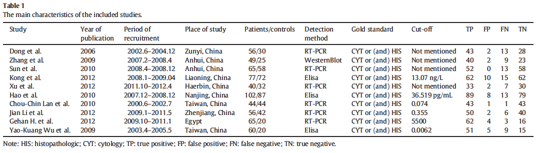
48 The diagnosis of malignant pleural effusion (MPE) is often challenging because differentiating MPE from benign effusion can be difficult using the currently available tools derived from thoracentesis; only approximately 50 70% of patients with MPE can be diagnosed by cytological examination of the pleural fluids [1]. Therefore, various tumor markers found in pleural effusions, including carcinoembryonic antigen (CEA), Cyfra 21-1, CA125, CA19-9, neuron-specific enolase, and squamous cell carcinoma antigen, have been investigated for their ability to differentiate malignant from benign pleural effusions [5 7]. However, the sensitivity of these tests is relatively low, and false-positive results have been described in almost all series. Thus, it is necessary to identify and evaluate more sensitive biological markers for MPE diagnosis [8]. The ideal marker would be office-based, rapid, and inexpensive and would have high sensitivity and specificity in the target population; this might reduce the burden and expense of frequent biopsies of the pleura. 48
49 Background: Survivin in pleural effusion is a promising marker for the diagnosis of malignant pleural effusion. Methods: Based on the principles and methods of Cochrane systematic reviews, PubMed, EMBASE, Web of Knowledge (ISI), the Cochrane Library, and China National Knowledge Infrastructure (CNKI) databases were searched to identify studies that assessed the diagnostic value of survivin in pleural effusion for malignant pleural effusion. Stata 12 and Meta-disc 1.4 software were used to test the heterogeneity and to perform the meta-analysis. Results: Our search returned 167 articles, ofwhich ten fulfilled the inclusion criteria. These studies included a total of 614 patients with malignant pleural effusion and 430 patients with benign pleural effusion as controls. The summary assessments revealed that the pooled sensitivity was 0.86 (95% CI: ) and the pooled specificity was 0.92 (95% CI: ). The positive likelihood ratio was 8.76 (95% CI: ), the negative likelihood ratio was 0.16 (95% CI: ) and the diagnostic odds ratio (DOR) was (95% CI: ). The area under the curve (AUC) for the pleural effusion survivin tests was , and the *Q index estimate for these tests was Conclusions: Survivin in pleural effusion has potential diagnostic value with advanced sensitivity and specificity and it can be used as adjunct tool for noninvasive diagnosis of malignant pleural effusion. 49
50 Survivin is a 12-amino acid protein (16.5 kda) that is located at chromosomal region 17q25. Survivin is an inhibitor of programmed cell death and mediates the suppression of apoptosis by inhibiting caspases 3 and 7, the terminal effectors in apoptotic protease cascades [10]. The survivin promoter is highly active in human tumor cells, but not in normal cells, and is up-regulated by hypoxia in tumors [11]. It is undetectable in normal differentiated tissues but is notably expressed in patients with lung cancer, breast cancer, bladder cancer, multiple myeloma, and lymphoma. 50
51 51
52 52
53 53
54 54
55 55

56 56
57 57

58 58
59 Measurements. Pleural effusion samples were collected prospectively from 1331 consecutive patients. Mesothelin levels were determined by commercial ELISA in effusions and their relationship to concurrent pathology reporting and final clinical diagnosis was determined. Results pleural effusion samples from1331 individuals were analysed.the final clinical diagnosis was 183 MM, 436 non-mm malignancy, and 712 nonmalignant effusions. Effusion mesothelin had a sensitivity of 67% formm at 95% specificity. Mesothelin was elevated in over 47% of MM cases in effusions obtained before definitive diagnosis of MM was established. In the setting of inconclusive effusion cytology, effusion mesothelin had a positive predictive value of 79% for MM and 94% for malignancy. Conclusions. A mesothelin-positive pleural effusion, irrespective of the identification of malignant cells, indicates the likely presence of malignancy and adds weight to the clinical rationale for further investigation to establish a malignant diagnosis. 59
60 60

61 61
62 Purpose of review Malignant mesothelioma is an asbestos-induced, aggressive tumour, which frequently presents with pleural effusion. There are over 60 reported causes that can result in the development of a pleural effusion. Currently, there are no tumour biomarkers in widespread clinical use for the differential diagnosis of mesothelioma from other diseases. With the incidence of mesothelioma expected to continue to increase, it is timely to review the current status of effusion-based biomarkers for mesothelioma diagnosis. Recent findings The majority of recent studies have evaluated soluble mesothelin in effusions in a diagnostic setting for mesothelioma. However, at high specificity, the sensitivity of the assay is limited to approximately 60% at the time of diagnosis. There is considerable research effort directed toward the identification of new markers for mesothelioma through a variety of genomic, proteomic and immunomic based platforms. One of the few new biomarkers to be identified through a biomarker discovery pipeline and evaluated in pleural effusions is fibulin-3. Preliminary results on the diagnostic accuracy of fibulin-3 have been inconsistent. Summary To date, soluble mesothelin remains the best available biomarker for mesothelioma and a positive result is clinically useful in patients with pleural effusions in whom the diagnosis is uncertain. 62
63 63
64 At a histopathologic level, there are essentially four circumstances in which immunophenotypic study plays an important role in the recognition of mesothelioma. (1) Mesothelioma versus adenocarcinoma (2) Sarcomatoid mesothelioma versus primary or metastatic pleural sarcoma versus metastatic sarcomatoid Carcinoma (3) Mesothelioma versus reactive mesothelial hyperplasia (4) Desmoplastic sarcomatoid mesothelioma versus fibrous pleuritis 64
65 Each of the above-cited diagnostic questions requires a different panel of immunohistochemical reagents. In cases of suspected spindle-cell or desmoplastic mesothelioma, these should include antibodies to keratin and calretinin; other markers are necessary only to subtype truly mesenchymal keratin-negative neoplasms. Recently, antibodies to cytokeratins 5/6 have been advocated as contextually specific markers for mesothelioma. Ordoñez found that 40 cases of mesothelioma were positive for keratins 5/6, whereas adenocarcinomas of the lung were negative. Nevertheless, the selected analyses have also found that 11 to 14% of nonpulmonary adenocarcinomas show focal reactivity for cytokeratins 5/6. Cytokeratins 7, 8, 18, and 19 are seen in all mesotheliomas and adenocarcinomas, whereas cytokeratins 5, 6, 14, and 17 are observed in some mesotheliomas but not adenocarcinomas. Unfortunately, the latter markers are absent in sarcomatoid tumors. 65
66 Carcinoembryonic antigen: Virtually all pleural mesotheliomas lack CEA, whereas the vast majority of pulmonary adenocarcinomas express that marker. CD15 has a high level of specificity for adenocarcinoma among all epithelial tumors. Nevertheless, occasional mesotheliomas (usually primary peritoneal lesions) may show focal staining for this marker. B72.3 This antibody recognizes a widely distributed epithelial determinant (tumor-associated glycoprotein-72), which is a cell-membrane glycoprotein. Irrespective of their sites of origin, a majority of adenocarcinomas show reactivity with B72.3. Ber-EP4 is another generic epithelial marker that was originally thought to be specific for adenocarcinomas. It is now apparent that roughly 20% of mesotheliomas demonstrate Ber-EP4 reactivity as well. 66
67 Calretinin is a cytoplasmic and nuclear calcium-binding protein. In contrast to most antibodies used for the evaluation of possible mesotheliomas, labeling for calretinin represents positive support for that diagnosis. This marker is present in more than 85% of mesotheliomas of the epithelial type. The authors own experience is that the majority of sarcomatoid lesions are, in fact, positive for calretinin. 67
68 68
69 69
70 70
71 71
.")
72 Aquaporin-1 (AQP1) is a molecule involved in the growth of MM cells, and yet is a factor reported to correlate with improved survival rates for MM with an epithelioid component, in comparison to AQP1-poor MM, as assessed from AQP1 expression by epithelioid MM cells only (apart from co-expression by stromal endothelial cells in addition to the tumour cells). Prognostic factors for MM include not only the histological subtypes, but other independent variables that include (among others), AQP1 expression by mesothelioma cells, the clinical status of the patient, the serum neutrophil:lymphocyte ratio and blood thrombocytosis 72

73 73
74 74

75 75

76 76
77 Mesothelial Hyperplasia Vs Epitheliod Mesothelioma Husain A., et al., Arch Pathol Lab Med 2013;137:

78 Epitheliod Mesothelioma Vs Lung AdenoCa Husain A., et al., Arch Pathol Lab Med 2013;137:

79 Sarcomatoid Mesothelioma Vs SCC and TCC Scherpereel A, et al. Eur Respir J 2010;35:
80 Fibrous Pleurisy vs Sarcomatoid Mesothelioma Husain A., et al., Arch Pathol Lab Med 2013;137:
81 Benign Vs Malignant 81
82 How to send your material to pathology Prof. Dr Philipp A. Schnabel Instit. Alllgemeine&Spezielle Pathologie University of Saarlandes Homburg/Saar GERMANY 82
83 Interpretation of the results in the clinic Prof. Arnaud Scherpereel Pulmonary and Thoracic Oncology Department Hôpital Calmette University Hospital of Lille (France) INSERM Unit 1019 Pasteur Institute of Lille 2 Avenue Oscar Lambret Lille FRANCE [email protected] 83
84 MALIGNANT PLEURAL EFFUSION MANAGEMENT ERS 2015 Congress Amsterdam (Netherlands) PG 10 Session : Malignant pleural effusion management : From standard cytopathology to molecular biology Saturday, 26 September 2015 Pr Arnaud SCHERPEREEL Pulmonary and Thoracic Oncology Department Hôpital Calmette University Hospital of Lille (France) INSERM Unit 1019 Pasteur Institute of Lille [email protected] 84
85 Financial disclosures I have no conflict of interest for this lecture 85
86 Let s talk about Pathogenesis, Diagnosis and Management of: Malignant pleural effusions (MPE), pleural metastases and malignant pleural mesothelioma (MPM) *Ref: chapter Pleural and chest wall tumours A Scherpereel. ERS Handbook
87 Pleural Malignancies Clinical presentation = usually pleural effusion Malignant pleural effusion (MPE) found in 6% of cancers history; reveals the cancer in half cases But in necropsy series: pleural effusion is only found in half of pleural tumors Dyspnea: most frequent symptom; other NON specific signs: cough, chest pain, asthenia and weight loss, more rarely fever, pneumothorax Light RW & Lee YCG. Textbook of Pleural Disease, 2 nd ed
 - MPM: 95% (unilateral+++) Lung, breast, ovary and gut cancers and lymphoma = 80% of MPE Primitive malignancy: not found in 10% of cases")
88 Malignant pleural effusion (MPE) patients / year in the USA; incidence: - Breast Cancer: 1 /3 - Lung Cancer: 1 /4 (most common etiology >40% of cases) - MPM: 95% (unilateral+++) Lung, breast, ovary and gut cancers and lymphoma = 80% of MPE Primitive malignancy: not found in 10% of cases 88
89 Pathogenesis of malignant pleura Still partly unknown 89
90 Pathogenesis of malignant pleura Pleural tumor involvement may result from different mechanisms: 1. A blood dissemination or more often from tumor emboli to the visceral pleura with secondary seeding to the parietal pleura (main mechanism for lung cancer) 2. direct invasion from adjacent structures (lung, chest wall...) by the tumor (lung or breast cancer) 3. Blood metastases to the parietal pleura from tumors other than lung cancer 4. lymph vessels spreading 5. Effusion may be due to the pleural tumor lesions or to a lymphatic blockade at the mediastinal level Sahn SA. Malignant pleural effusions. Pleural diseases. Philadelphia : WB Saunders, 2004 :
91 Pathogenesis of malignant pleura (2) MPE depend also on interactions between tumor cells and mesothelial cells through growth factors such as VEGF that increase vascular permeability and angiogenesis. Finally, MPE need several pathogenic factors such as: Increase of capillaries permeability Lesions of the lymphatic system draining the pleural space (pores of Wang at the postero-basal parietal pleura) or at the level of the thoracic channel Pericardial involvement 91 Sahn SA. Malignant pleural effusions. Pleural diseases. Philadelphia : WB Saunders, 2004 :
92 Pathogenesis of malignant pleura (3) CAUTION : a pleural effusion is NOT systematically synonym of MPE in the context of tumor because it may be induced by other mechanisms : pneumonia and/or atelectasia due to bronchial obstruction transudate induced by severe denutrition or cardiac failure or even a drug (MTX, CPM ) or radiotherapy-induced effusion (breast cancer+++) Therefore the strategy (diagnosis/ttt) may differ whether the patient has a cancer background or not, but should always rely on cytology or better on histology+++ 92
93 Prognosis of MPE and pleural metastases Poor prognosis (one year MS = 13%) However survival may greatly vary according to the primary cancer: lung cancer << breast cancer or lymphoma New 2009 TNM staging of NSCLC: MPE (or pericardial effusion) or pleural metastases = stage T4 (TNM 1997) NOW STAGE M1a (IV) Survival at 5 years of tumors ct4n0m0 by pleural involvement =2% (similar to M1 patients) vs by other cause = 25% (p = 0,0001) Journal of Thoracic Oncology 2007; 2 :
94 Malignant (Pleural) Mesothelioma Very agressive tumor issued from mesothelial cells mostly of the pleura (MPM : 80% of the cases) Bad prognosis : median survival = 9-12 months 94
95 Diagnostic issues in malignant pleural diseases: a specific clinical profile? Pleural effusion +++ Pleural thickening Pneumothorax (MPM, underlying pulmonary disease ) Non-specific signs: dyspnea, cough, chest pain, asthenia and weight loss 95
96 Management of MPE and MPM MPE: please refer to ERS Handbook MPM: refer to ERS/ESTS Guidelines Scherpereel et al, ERJ 2010 (ongoing update by European Taskforce) 96
97 Clinical and/or radiological diagnosis of unilateral Pleural Effusion: MPE? Previous asbestos exposure? Clinical and/or radiological suspicion of MPM Small effusion, hemostasis disturbancies = relative contra-indications for ultra-sound (US)-guided procedure by trained physicians Thoracocentesis for diagnosis and drainage Proceed to MPM algorythm Exsudate Light s Criteria Transudate: seak and treat cause of transudate Cytology: tumor cells? NO If no argument for tuberculosis, infection YES NO Previous diagnosis of malignancy (lung, breast )? YES Chest CT scan mandatory if no contralateral mediastinal shift on chest Xray bronchoscopy if main bronchus stenosis is suspected: trapped lung? Is pleural histology needed for diagnostic clarity? Otherwise start treatment 97
98 Chest CT scan mandatory if no contralateral mediastinal shift on chest Xray bronchoscopy if main bronchus stenosis is suspected: trapped lung?! Blind pleural biopies if suspicion of tuberculosis? Thoracoscopy Surgery at risk Contra-indications Thoracoscopy Pleural symphysis In the same time if large effusion AND bulky tumor lesions of the pleura Histological proof of pleural metastases CT scan or US -guided transthoracic biopsies Mini-thoracotomy for pleural biopsies Pleurodesis (Talc poudrage ) also if reccurent pleural effusion; alternatives: talc slurry, undwelled catheter Treatment of the primary cancer (chemotherapy and/or hormone therapy and best supportive care (BSC)...) 98
99 Medical Thoracoscopy or VATS: Gold standard diagnostic procedure Sensitivity : 95% Specificity : 100% Complications (non lethal) : < 10% 99
100 MPE aspect in thoracoscopy (Source : Pr A. Scherpereel and Dr Ph. Ramon) 100
101 Thank you for your attention Welcome to join the European Respiratory Society group on Pleural and Mediastinal malignancies (11.2) of the Thoracic Oncology Assembly (N 11) +++! 101
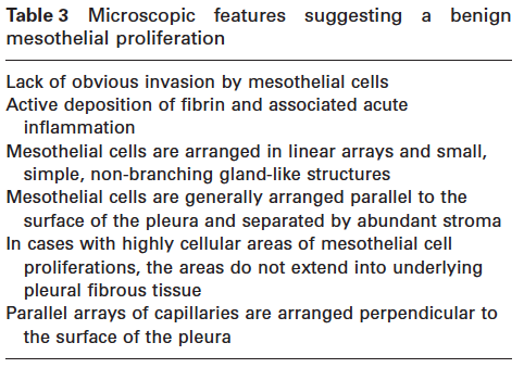
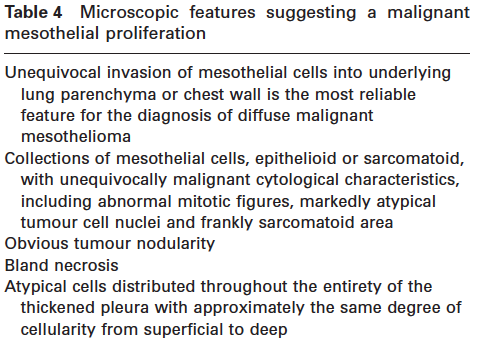
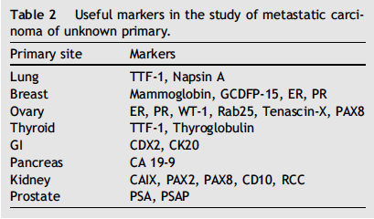
102 SUMMARY Atypical mesothelial hyperplasia and other pitfalls in MPE diagnosis Prof. Françoise Galateau-Salle Centre Leon Bérard Lyon University Hospital Caen Avenue de la Côte de Nacre Cedex 05, Caen FRANCE Atypical mesothelial hyperplasia happens in response to a great variety of benign or malignant processes including infectious pleuritis, pneumothorax, underlying pneumonia, connective tissue diseases (rheumatoid arthritis and systemic lupus erythematosus), pulmonary embolism, drug reactions, and asbestos associated effusions or in association with underlying bronchopulmonary carcinoma. Moreover, exudative pleural effusion in most cases is the initial symptom revealing a mesothelioma or a metastasis as the presenting site of another cancer. According to the literature 50% of patients with a metastatic disease develop pleural effusion and 75% of them are symptomatic. In the majority of the case the primary site is found with in order of frequency from Antunes et al (1, 2): lung, breast, ovarian, lymphoma, GI, GU tract, mesothelioma. In 10% of the cases the primary site remains unknown (2). It also may represent a precancerous step or the early phase of a malignant mesothelioma. Consequently, the separation between benign mesothelial superficial proliferation and a malignant mesothelioma in a patient with a unilateral pleural effusion is crucial for the patient care and is extremely challenging for both clinician and pathologist. On the pathologist side, a first look on cell morphology, raise several questions; is the sample significant enough for an accurate diagnosis???, Is the process malignant??? Then when the process looks malignant, the followed questions; is the lesion a malignant mesothelioma or another tumor mimicking a mesothelioma. Carcinoma may superficially metastases to the surface of the pleura closely mimicking a malignant mesothelioma. It is well known that sarcomatoid mesothelioma and other spindle cell tumors located to the pleura do not peel in the pleural cavity. This course will focus on epithelioid mesothelioma. Indeed, the diagnostic approach in these situations is dependent on the clinical presentation and on the material sent to the pathology lab. 1. The separation between benign mesothelial proliferations on routine biopsies has been largely described by Churg et al(3-6), and new updates were described in the 2015 WHO classification. Two major scenarios can arise for the pathologist: a. The lesion appears on cytology or tissue biopsy specimens of epithelial/epithelioid type the question is this? i. A reactive mesothelial hyperplasia mimicking a malignant process. ii. If malignant; is the lesion a malignant mesothelioma, a metastasis of a carcinoma/ or of a melanoma. iii. Or a so-called well differentiated superficial papillary mesothelioma (rare mésothéliale tumor of uncertain behavior). b. The lesion is morphologically a spindle cells/fibroblastic proliferation (spindle cells do not peel in the serosal cavity) on tissue biopsy is this? i. A reactive cellular diffuse proliferation (chronic fibrous pleuritis) ii. A rare fibromatois desmoid invading the pleura iii. If malignant; is the lesion a sarcomatoid/desmoplastic mesothelioma versus a metastasis of a sarcomatoid carcinoma or a sarcoma? 102
103 2. The lesion looks malignant the issue to reach a diagnosis;iscytology orcore tissue biopsy or open surgical biopsy the best approach?? 3. Facing these situations, 1. the clinical presentation can help the pathologist in separating a metastasis versus a mesothelioma 2. What are the important elements brought by every practitioner for a quality and safe diagnosis? 3. What is the role in 2015 of ancillary techniques to solve the problem of the separation benign from malignant? 4. How can we avoid pitfalls?? Nowadays, even if pathologists better know how to recognize a malignant mesothelioma on morphology or a metastasis in many casesto separate benign versus malignant mesothelial proliferation in a significant proportion of cases may be extremely challenging. The role of this course is to consider the management of pleural effusion from standard cytopathology to molecular biology and to evaluate if the transition, in 2015, is real in routine practice. The diagnostic approach Pathologist side: When confronted with a pleural process for cytological or histological diagnosis it is essential for the pathologist to know as much as possible of the clinical and radiologic information when they are available. For example it is important for the pathologist to know if it is a free floating pleural effusion without thickening and nodularity (could be a WDPM) or if it is a diffuse process (diffuse pleural thickening with or without nodularity) could be a connective tissue disease or a malignant mesothelioma desmoplastic type, or a mass located in the lung or in the thoracic wall (Sarcomatoid carcinoma versus localized sarcomatoid mesothelioma). It is also important to know the age and the sex of the patient, the context of previous history of cancer, due to secondary tumors affecting the serosal cavities that can be great mimickers of mesothelioma and force to check the expression of organ specific markers for the differential diagnosis (ie ER receptors in women to detect breast metastasis). Clinician side: The technique of the pleural effusion aspiration is crucial and determines the quality of the pre-analytical phase, but is mainly dependent on the clinical presentation, the comorbidities, the experience of the clinician. In the majority of the cases pleural aspiration is the initial step to relieve the patient from dyspnea. The sample should be sent as soon as possible to the pathology lab to avoid autolysis. Unfortunately due to undiagnosed exudative pleural effusion pleuralbiopsy is necessary. In 2015, thoracoscopy and CT-guided core biopsy have a quite similar diagnostic yield for the diagnosis of a malignant diseases including mesothelioma with a reported sensitivity of 76-88% and up to 100% specificity depending of the authors series (1, 2). In order to help, the quality of cytopathological examination, if a biopsy is taken or cell blocks made, the sample should be fixed immediately and preserve in formalin 5%. It is also important to avoid acid fixative (AFA and others new fixatives) as it is responsible of absence of signal by FISH analysis. Even though, an open surgical biopsy is a better material for the accurate diagnosis of subtyping mesothelioma 83% versus 74% on VATS and 44 % with CT-guided biopsy (8, 9). On the pathologist side Cytology samples are the initial and major step to approach the diagnosis of a pleural effusion. The cytologist, without major difficulties, recognizes malignant effusions and many studies have shown that malignant effusions are diagnosed in nearly 60 % by cytological examination alone, but drop down to 30% 103
104 for a diagnosis of mesothelioma. Recently some authors showed high specificity for the diagnosis of mesothelioma, but with the limitation depending on the experience of the cytologist. Paintal et al, and Henderson et al (8-10), have reported the update sensitivity of pleural effusion cytology, which is reported from 16% to 75%, and reported lower when lesions are suspicious for malignancy. The major reason to not use cytology alone is even if it is workable in highly cellular pleural effusion for epithelioid type, the deceptively bland appearance of malignant mesothelioma is a cause of misdiagnosis, while reactive mesothelial proliferation may show severe atypia and invasion of adipose tissue on biopsy remains the definitive criteria for malignancy. In 2015 the criteria for distinguishing diffuse malignant mesothelioma from reactive mesothelial proliferations have been further redefined by Churg, and Galateau Salle et al (3-6). From standard cytopathology to molecular biology. Can we improve the diagnosis of malignancy on cytology and tissue biopsy samples? Updates on ancillary techniques have been extensively described in the new 2015 WHO classification. Up to now a great variety of immunohistochemical markers have shown various level of sensitivity and specificity. Among them the most famous are EMA (epithelial membrane antigen, desmin, p53 the wellknown tumor suppressor gene, but largely not mutated in mesotheliomas, IMP3 (the Insulin like growth factor 2 messenger RNA-binding protein 3), GLUT1 (Glucose transporter 1), ki67(3,5). For all of them, the authors claimed that positive staining were not observed on benign reactive mesothelial cells compared to malignant mesothelioma with statistically significant results. From our experience in a large cohort of cases. The studies were statistically valuable but individually not reliable due to staining observed in benign reactions. Two new molecular makers have recently emerged and have demonstrated their usefulness in the separation of benign mesothelial hyperplasia versus diffuse malignant mesothelioma (4-6). 1. p16 is a tumor suppressor gene. p 16 INK4A (p16) have been reported to be deleted in 60 to 80% of diffuse epithelioid malignant mesothelioma (7) and in nearly 100% of sarcomatoid and desmoplastic malignant mesothelioma. Moreover, p16 deletion has been shown to be correlated with a more aggressive clinical course. p16 homozygous deletion is detected by fluorescence in situ hybridization both on cytological and paraffin embedded tissue biopsy samples and has been considered as a robust marker in separating benign from malignant mesothelial proliferation) (5,6,7). p16 homologous deletion by FISH in more than 200 reported benign atypical mesothelial proliferations reported in the literature have never been observed. p16 immunohistochemical stain does not give the same results and are notup to now reliable. From our MESOPATH experience 20 % of AMH suspicious for malignancy with a p16 homozygous deletion by FISH have turn up in a malignant mesothelioma after 5 to 9 years. 2. BAP-1, the BRCA associated protein 1 is a nuclear ubiquitin hydrolase that is expected to function as a tumor suppressor gene. BAP 1 controls DNA repair and cell cycle proliferation. Bott et al, were the first authors to describe the presence of somatic mutations in malignant mesothelioma followed by Testa et al, who showed the presence of germ line mutations in several families. The concept of BAP1 associated cancer syndrome has emerged soon after showing an increased number of mesothelioma with ocular and cutaneous melanoma, renal cell and breast carcinoma and others.bap1 loss is evaluated by immunohistochemistry and is now routinely used on FFPE tissue biopsy samples, and more recently has been reported to be useful on cell-block sections by immunohistochemistry (9). BAP1 somatic mutations are observed in around 30% of mesothelioma, more often in epithelioid type than in biphasic and sarcomatoid mesothelioma. From our MESOPATH and Churg et al (3, 5, 6), experience BAP1 loss has never been reported in reactive mesothelial proliferation. 104
105 Examples of routine practice situations The challenging separation of benign versus malignant lesions. Taking into account the constraints of technical approaches, there are nowadays major advances in the characterization of atypical mesothelial hyperplasia and mimickers of mesothelioma. A- Superficial mesothelial proliferation benign reactive process versus epithelioid malignant mesothelioma. 1. The sample on cytology (highly cellular atypical mesothelial hyperplasia) or on tissue biopsy shows an atypicalmesothelialproliferation, loss of BAP1 by immunohistochemistryand or/a deletion of p16present in the definitive clinical, radiological and thoracoscopic context of a tumor some authors consider that this is a definitive malignant mesothelioma and represents either 1) the superficial pleural extension of a definitive invasive mesothelioma or 2) an early mesothelioma. 2. The atypical mesothelial hyperplasia is associated with a deletion of p16 but there is no definitive clinical, radiological, thoracoscopic context of tumor, a diagnosis of mesothelioma could not be validated. This can represent sampling error and it is recommended if possible to rebiopsy, or it also can represent a precancerous state the mesothelioma can develop 1 to 9 years later. In this situation a close clinical follow up is recommended and if a context of asbestos exposure is present a CT -scan follow up is considered. If the PET scan shows a hyperfixation a new biopsy if possible for the patient is considered and the pathologist should blocked in totality the samples. 3. The samples shows an atypical mesothelial proliferation and no deletion of p16 is observed in the clinical, radiological context of uncertain tumor. A clinical follow up is only recommended. 4. On the other hand, a strong clinical, and radiological context of mesothelioma is present; the absence of p16 deletion did not eliminate a diagnosis of mesothelioma. p16 deletion is present in 60 to 80% of epithelioid malignant mesothelioma and in 80 to 100% of sarcomatoid mesothelioma. In this situation it is recommended to rebiopsy. 5. Up to now loss of BAP1 has not been observed in reactive mesothelial superficial proliferation and benign processes. Loss of BAP1 is a very promising marker with a high specificity (from our experience 0/23) to detect malignancy. 6. The nuclear staining of BAP1 is present this result did not means the lesion is a benign process. BAP1 loss on tissue biopsy is observed according to series in around 30 to 40% of malignant mesothelioma when all histological type are considered. 7. Recommended scheme from Churg et al B- The other challenging situations are the separation between well differentiated papillary mesothelioma and a reactive superficial mesothelial proliferation or with a more challenging conventional papillary mesothelioma with areas of WPM. WDPMis now well-defined and represents a distinct mesothelial tumor of uncertain behavior (ICD /1) (3, 11). Clinically the presentation is a free floating pleural effusions without pleural thickening or nodularity. The tumor is usually not related to asbestos exposure. It may affect all serosal cavities with a predilection for peritoneum more frequently in women. Histologically it is characterized by a superficial spreading of papillary structures with myxoid vascular cores lined by bland mesothelial cells. These lesions have an indolent clinical outcome and when a minimal invasion is observed, in rare patient, they may recur but no fatal outcomes have been observed. p16 deletion and BAP1 have not been extensively studied on this specific entity and they are no guidelines up to now published. Adequate 105
106 sampling of the lesion, and clinical correlation are required to avoid the diagnosis of an undersampled malignant mesothelioma mimicking a WDPM (3, 11). C- The last challenging situation is the separation between metastatic carcinoma and mesothelioma. A huge number of immunohistochemical markers have been published since the last 2004 WHO classification and the 2015 classification has particularly covered this topic giving recommendations for the management of the patient samples. Nevertheless some situations may be challenging due to metastatic adenocarcinoma that are clinically and pathologically great mimickers of mesothelioma. Breast and lung carcinoma the major causes of pleural metastasis are among them. Calretinin the more sensitive and specific marker for mesothelioma may be positive in both tumors. 38% of triple negative breast carcinoma expresses calretinin with a nuclear staining (12). p40 nuclear expression is a marker of squamous differentiation and has never been observed positive in sarcomatoid/pleomorphic mesothelioma (MESOPATH unpublished data). Moreover, TTF1 positivity is a highly specific marker in favor of a lung origin (30% of the cases of pleomorphic/sarcomatoid carcinoma), andmay avoida misdiagnosis of malignant mesothelioma (3). Conclusion On the past decades and more recently in 2015, many advances have been made for the management of atypical mesothelial hyperplasia versus malignant mesothelioma and other pitfalls in MPE that have promising hopes in clinical practice. These include morphological criteria, the use of BAP1 immunohistochemical stains and p16 homozygous deletion by FISH both on cellblocks and FFPE tissue samples. These are important step forwards in the future role of cytopathological diagnosis in effusions of malignant mesothelioma. However, BAP1 and p16 deletion can be observed in metastatic tumor from other organs and are not, on their own diagnostic, of mesothelioma. REFERENCES 1. Bhatnagar R, Maskell N. The modern diagnosis and management of pleura leffusions. BMJ Sep 8; 351:h Roberts ME, Neville E, Berrisford RG, Antunes G, Ali NJ; BTS Pleural DiseaseGuideline Group. Management of a malignant pleural effusion: British ThoracicSociety Pleural Disease Guideline Thorax Aug; 65 Suppl 2:ii Galateau-Salle F, Churg A, Roggli V, et al. Epithelioid mesothelioma. In Travis WD, Brambilla E, Burke AP, Marx A, Nicholson AG eds. WHO Classification of Tumours of the Lung, Pleura, Thymus and Heart. 4 th ed Lyon, France. IARC Press. 2015: Churg A, Galateau-Salle F. The separation of benign and malignant mesothelial proliferations. Arch Pathol Lab Med. 2012:136 (10): Churg A, Sheffield BS, Galateau-Salle F. New Markers for Separating BenignFrom Malignant Mesothelial Proliferations: Are We There Yet? Arch Pathol Lab Med.2015 Aug Sheffield BS, Hwang HC, Lee AF, Thompson K, Rodriguez S, Tse CH, Gown AM,Churg A. BAP1 immunohistochemistry and p16 FISH to separate benign from malignantmesothelial proliferations. Am J Surg Pathol Jul; 39(7): Chung CT, Santos Gda C, Hwang DM, Ludkovski O, Pintilie M, Squire JA, Tsao MS. FISH assay development for the detection of p16/cdkn2a deletion in malignant pleural mesothelioma. J Clin Pathol Jul; 63(7): Paintal A, Raparia K, Zakowski MF, Nayar R. The diagnosis of malignantmesothelioma in effusion cytology: a reappraisal and results of amulti-institution survey. Cancer Cytopathol Dec; 121(12): Henderson DW, Reid G, Kao SC, van Zandwijk N, Klebe S. Challenges and controversies in the diagnosis of malignant mesothelioma: Part 2. Malignant mesothelioma subtypes, pleural synovial 106
107 sarcoma, molecular and prognostic aspectsof mesothelioma, BAP1, aquaporin-1 and microrna. J Clin Pathol. 2013Oct;66(10): HendersonDW, Reid G, Kao SC, van Zandwijk N, Klebe S. Challenges and controversies in the diagnosis of mesothelioma. Part I: cytology-only diagnosis, biopsies, immunohistochemistry. Discrimination between mesothelioma and reactive mesothelial hyperplasia and biomarkers. J Clin Pathol; 2013; 66; (10): Churg A, Allen T, Borczuk AC, Cagle PT, Galateau-Sallé F, Hwang H, Murer B,Murty VV, Ordonez N, Tazelaar HD, Wick M. Well-differentiated papillarymesothelioma with invasive foci. Am J Surg Pathol Jul; 38(7): Ordóñez NG, Sahin AA. Diagnostic utility of immunohistochemistry indistinguishing between epithelioid pleural mesotheliomas and breast carcinomas: a comparative study. Hum Pathol Jul; 45(7): EVALUATION 1. Mesothelioma can accurately be diagnosed on cytology specimens alone without ancillary techniques a. yes b. no 2. The sensitivity of cytological diagnosis on pleural aspiration in mesothelioma is upgraded with immunohistochemistry a. yes b. no 3. BAP1 loss by immunohistochemistry on FFPE cell blocks and tissue biopsy samples is a robust marker of malignant mesothelioma versus benign reactive mesothelial proliferation a. yes b. no 4. p16 homozygous deletion in pleural effusion observed by FISH assay may be encountered in benign reactive superficial mesothelial proliferation a. yes b. no 5. P16 loss by immunohistochemistry on cell blocks and FFPE biopsy tissue samples is a robust marker of malignancy for the diagnosis of mesothelioma a. yes b. no 6. Calretinin positivity by immunohistochemistry on pleural cytoblocks or FFPE tissue biopsy samples can be observed in mesothelioma and other metastatic carcinoma such as metastatic breast carcinoma. a. yes b. no 107
108 Faculty disclosures Prof. Philippe Astoul was leading courses for which the materials or equipment was provided by: Novatech (Material for thoracoscopy course), Wolf (telescope for hands-on session during thoracoscopy course) and Sebac (IPC for practical sessions during course). He is also a Scientific Board Adviser for Sequana. 108
109 Faculty contact information Prof. Philippe Astoul Department of Thoracic Oncology, Pleural Diseases, and Interventional Pulmonology Hôpital Nord Chemin des Bourrely, cedex Marseille FRANCE Dr Sebastian Fernandez Bussy Clinica Alemana-Universidad del Desarrollo Interventional Pulmonology Av Vitacura 5951 Santiago, Región Metropolitana CHILE Prof. Françoise Galateau-Salle Centre Leon Bérard Lyon University Hospital Caen Avenue de la Côte de Nacre Cedex 05, Caen FRANCE Prof. Arnaud Scherpereel Pulmonary and Thoracic Oncology Department Hôpital Calmette University Hospital of Lille (France) INSERM Unit 1019 Pasteur Institute of Lille 2 Avenue Oscar Lambret Lille FRANCE [email protected] Prof. Dr Philipp A. Schnabel Instit. Alllgemeine&Spezielle Pathologie University of Saarlandes Homburg/Saar GERMANY [email protected] Dr Marcin Skrzypski Department of Oncology and Radiotherapy Medical University of Gdansk ul. Debinki Gdansk POLAND [email protected] 109
Update on Mesothelioma
 November 8, 2012 Update on Mesothelioma Intro incidence and nomenclature Update on Classification Diagnostic specimens Morphologic features Epithelioid Histology Biphasic Histology Immunohistochemical
MALIGNANT MESOTHELIOMA UPDATE ON PATHOLOGY AND IMMUNOHISTOCHEMISTRY
 MALIGNANT MESOTHELIOMA UPDATE ON PATHOLOGY AND IMMUNOHISTOCHEMISTRY Sisko Anttila, MD, PhD Jorvi Hospital Laboratory of Pathology Helsinki University Hospital Espoo, Finland 2nd Nordic Conference on Applied
MALIGNANT MESOTHELIOMA UPDATE ON PATHOLOGY AND IMMUNOHISTOCHEMISTRY
 MALIGNANT MESOTHELIOMA CLASSIFICATION MALIGNANT MESOTHELIOMA UPDATE ON PATHOLOGY AND IMMUNOHISTOCHEMISTRY Sisko Anttila, MD, PhD Jorvi Hospital Laboratory of Pathology Helsinki University Hospital Espoo,
Diagnosis of Mesothelioma Pitfalls and Practical Information
 Diagnosis of Mesothelioma Pitfalls and Practical Information Mary Beth Beasley, M.D. Mt Sinai Medical Ctr Dept of Pathology One Gustave L Levy Place New York, NY 10029 (212) 241-5307 [email protected]
بسم هللا الرحمن الرحيم
 بسم هللا الرحمن الرحيم Updates in Mesothelioma By Samieh Amer, MD Professor of Cardiothoracic Surgery Faculty of Medicine, Cairo University History Wagner and his colleagues (1960) 33 cases of mesothelioma
Practical Effusion Cytology
 Practical Effusion Cytology A Community Pathologist s Approach to Immunocytochemistry in Body Fluid Cytology Emily E. Volk, MD William Beaumont Hospital Troy, MI College of American Pathologists 2004.
The develpemental origin of mesothelium
 Mesothelioma Tallinn 14.12.06 Henrik Wolff Finnish Institute of Occupational Health The develpemental origin of mesothelium Mesodermal cavities (pleura, peritoneum and pericardium ) are lined with mesenchymal
Surgeons Role in Symptom Management. A/Prof Cliff K. C. Choong Consultant Thoracic Surgeon Latrobe Regional Hospital GIPPSLAND
 Surgeons Role in Symptom Management A/Prof Cliff K. C. Choong Consultant Thoracic Surgeon Latrobe Regional Hospital GIPPSLAND Conditions PLEURAL Pleural effusion Pneumothorax ENDOBRONCHIAL Haemoptysis
Outline. Workup for metastatic breast cancer. Metastatic breast cancer
 Metastatic breast cancer Immunostain Update: Diagnosis of metastatic breast carcinoma, emphasizing distinction from GYN primary 1/3 of breast cancer patients will show metastasis 1 st presentation or 20-30
Objectives. Mylene T. Truong, MD. Malignant Pleural Mesothelioma Background
 Imaging of Pleural Tumors Mylene T. Truong, MD Imaging of Pleural Tumours Mylene T. Truong, M. D. University of Texas M.D. Anderson Cancer Center, Houston, TX Objectives To review tumors involving the
A 70-year old Man with Pleural Effusion
 Mesothelioma Diagnosis: Pitfalls and Latest Updates S Klebe and DW Henderson Recommendations Indisputable malignant cells on cytomorphological criteria which demonstrate a mesothelial phenotype, which
Diseases. Inflammations Non-inflammatory pleural effusions Pneumothorax Tumours
 Pleura Visceral pleura covers lungs and extends into fissures Parietal pleura limits mediastinum and covers dome of diaphragm and inner aspect of chest wall. Two layers between them (pleural cavity) contains
Notice of Faculty Disclosure
 The Diagnosis of Malignant Mesothelioma Andrew Churg, MD Department of Pathology University of British Columbia Vancouver, BC, Canada [email protected] Notice of Faculty Disclosure In accordance with
Immunohistochemistry on cytology specimens from pleural and peritoneal fluid
 Immunohistochemistry on cytology specimens from pleural and peritoneal fluid Dr Naveena Singh Consultant Pathologist Bart health NHS Trust London United Kingdom Disclosures and Acknowledgements I have
Cytology : first alert of mesothelioma? Professor B. Weynand, UCL Yvoir, Belgium
 Cytology : first alert of mesothelioma? Professor B. Weynand, UCL Yvoir, Belgium Introduction 3 cavities with the same embryologic origin the mesoderme Pleura Exudates Pleura Peritoneum Pericardium 22%
The diagnostic usefulness of tumour markers CEA and CA-125 in pleural effusion
 Malaysian J Path01 2002; 24(1) : 53-58 The diagnostic usefulness of tumour markers CEA and CA-125 in pleural effusion Pavai STHANESHWAR MD, Sook-Fan YAP FRCPath, FRCPA and Gita JAYARAM MDPath, MRCPath
How To Diagnose And Treat A Tumour In An Effusion
 Effusions of the Serous Cavities Annika Dejmek Professor/Consultant in Cytopathology Clinical Pathology; Department of Laboratory Medicine, Malmö, Lund University 5th EFCS Tutorial Trondheim 2012 Pleura
Recommendations for the Reporting of Pleural Mesothelioma
 Recommendations for the Reporting of Pleural Mesothelioma Association of Directors of Anatomic and Surgical Pathology * DOI: 10.1309/6A30YQHBMTHEJTEM It has been evident for decades that pathology reports
Diagnostic Challenge. Department of Pathology,
 Cytology of Pleural Fluid as a Diagnostic Challenge Paavo Pääkkö,, MD, PhD Chief Physician and Head of the Department Department of Pathology, Oulu University Hospital,, Finland Oulu University Hospital
ASBESTOS EXPOSURE AND SARCOMATOID MALIGNANT PLEURAL MESOTHELIOMA Gorantla Sambasivarao 1, Namballa Usharani 2, Tupakula Suresh Babu 3
 ASBESTOS EXPOSURE AND SARCOMATOID MALIGNANT PLEURAL MESOTHELIOMA Gorantla Sambasivarao 1, Namballa Usharani 2, Tupakula Suresh Babu 3 HOW TO CITE THIS ARTICLE: Gorantla Sambasivarao, Namballa Usharani,
Académie internationale de Pathologie - Division arabe XX ème congrès 24-26 novembre 2008 Alger. Immunohistochemistry in malignant mesotheliomas
 Académie internationale de Pathologie - Division arabe XX ème congrès 24-26 novembre 2008 Alger Immunohistochemistry in malignant mesotheliomas Françoise Thivolet-Béjui Groupement Hospitalier Est Lyon-Bron
PATHOLOGY OF THE PLEURA: Mesothelioma and mimickers Necessity of Immunohistochemistry. M. Praet
 PATHOLOGY OF THE PLEURA: Mesothelioma and mimickers Necessity of Immunohistochemistry M. Praet Pathology of the Pleura Normal serosa: visceral and parietal layers Inflammation Neoplasia: Primary: mesothelioma
YOUR LUNG CANCER PATHOLOGY REPORT
 UNDERSTANDING YOUR LUNG CANCER PATHOLOGY REPORT 1-800-298-2436 LungCancerAlliance.org A GUIDE FOR THE PATIENT 1 CONTENTS What is a Pathology Report?...3 The Basics...4 Sections of a Pathology Report...7
Therapy of pleural effusions Modern techniques
 Therapy of pleural effusions Modern techniques Dr. Melanie Toffel Sugery of the chest Pleural effusion Ethiology In the normal pleural space there is a steady state in which there is a roughly equal rate
SMALL CELL LUNG CANCER
 Protocol for Planning and Treatment The process to be followed in the management of: SMALL CELL LUNG CANCER Patient information given at each stage following agreed information pathway 1. DIAGNOSIS New
Case of the. Month October, 2012
 Case of the Month October, 2012 Case The patient is a 47-year-old male with a 3-week history of abdominal pain. A CT scan of the abdomen revealed a suggestion of wall thickening at the tip of the appendix
Immunohistochemical differentiation of metastatic tumours
 Immunohistochemical differentiation of metastatic tumours Dr Abi Wheal ST1. TERA 3/2/14 Key points from a review article written by Daisuke Nonaka Intro Metastatic disease is the initial presentation in
Neoplasms of the LUNG and PLEURA
 Neoplasms of the LUNG and PLEURA 2015-2016 FCDS Educational Webcast Series Steven Peace, BS, CTR September 19, 2015 2015 Focus o Anatomy o SSS 2000 o MPH Rules o AJCC TNM 1 Case 1 Case Vignette HISTORY:
International Journal of Case Reports in Medicine
 International Journal of Case Reports in Medicine Vol. 2013 (2013), Article ID 409830, 15 minipages. DOI:10.5171/2013.409830 www.ibimapublishing.com Copyright 2013 Andrew Thomas Low, Iain Smith and Simon
Seattle. Case Presentations. Case 1. 76 year old female with a history of breast cancer 12 years ago. Now presents with a pleural effusion.
 Seattle Montreal IAP September 2006 Case Presentations Allen M. Gown, M.D. Medical Director and Chief Pathologist PhenoPath Laboratories Clinical Professor of Pathology University of British Columbia Case
Sternotomy and removal of the tumor
 Sternotomy and removal of the tumor All thymomas originate from epithelial thymic cells 4% of them consist of a pure population of epithelial cells Most have mixed populations of lymphoid cells to a
INTERNATIONAL ASSOCIATION FOR THE STUDY OF LUNG CANCER Prospective Mesothelioma Staging Project
 INTERNATIONAL ASSOCIATION FOR THE STUDY OF LUNG CANCER Prospective Mesothelioma Staging Project Data Forms and Fields in CRAB Electronic Data Capture System - Reduced Set - Pivotal data elements for developing
Brigham and Women s Hospital, Boston, MA, USA; 2 Verastem, Inc., Boston, MA, USA
 Determination of Biomarker Response in a Phase II Window of Opportunity Study of Defactinib (VS 6063), a Focal Adhesion Kinase (FAK) Inhibitor, in Patients with Resectable Malignant Pleural Mesothelioma
Changes in Breast Cancer Reports After Second Opinion. Dr. Vicente Marco Department of Pathology Hospital Quiron Barcelona. Spain
 Changes in Breast Cancer Reports After Second Opinion Dr. Vicente Marco Department of Pathology Hospital Quiron Barcelona. Spain Second Opinion in Breast Pathology Usually requested when a patient is referred
Malignant Mesothelioma
 Malignant Malignant mesothelioma is a tumour originating from mesothelial cells. 85 95% of mesotheliomas are caused by asbestos exposure. It occurs much more commonly in the chest (malignant pleural mesothelioma)
Malignant Mesothelioma
 Malignant mesothelioma is a tumour originating from mesothelial cells. 85 95% of mesotheliomas are caused by asbestos exposure. It occurs much more commonly in the chest (malignant pleural mesothelioma)
GRANT APPLICATION FORM: ORGANISING AN ESO EDUCATIONAL EVENT
 GRANT APPLICATION FORM: ORGANISING AN ESO EDUCATIONAL EVENT 1) TOPIC OF THE EVENT MALIGNANT PLEURAL DISEASE 2) SUGGESTED TITLE : Approach to Pleural Cancer: State-of-the-Art 3) TYPE OF EVENT Course Native
Pathology of lung cancer
 Pathology of lung cancer EASO COURSE ON LUNG CANCER AND MESOTHELIOMA DAMASCUS (SYRIA), MAY 3-4, 2007 Gérard ABADJIAN MD Pathologist Associate Professor, Saint Joseph University Pathology Dept. Hôtel-Dieu
Malignant Mesothelioma Diagnosed by Bronchoscopic Biopsy
 CASE REPORT http://dx.doi.org/10.4046/trd.2015.78.3.297 ISSN: 1738-3536(Print)/2005-6184(Online) Tuberc Respir Dis 2015;78:297-301 Malignant Mesothelioma Diagnosed by Bronchoscopic Biopsy Yeon-Hee Park,
Extrapleural Pneumonectomy for Malignant Mesothelioma: Pro. Joon H. Lee 9/17/2012
 Extrapleural Pneumonectomy for Malignant Mesothelioma: Pro Joon H. Lee 9/17/2012 Malignant Pleural Mesothelioma (Epidemiology) Incidence: 7/mil (Japan) to 40/mil (Australia) Attributed secondary to asbestos
Tumour Markers. What are Tumour Markers? How Are Tumour Markers Used?
 Dr. Anthony C.H. YING What are? Tumour markers are substances that can be found in the body when cancer is present. They are usually found in the blood or urine. They can be products of cancer cells or
Disclosures. Learning Objectives. Effusion = Confusion. Diagnosis Of Serous Cavity Effusions - Beware The Mesothelial Cell!
 Disclosures Diagnosis Of Serous Cavity Effusions - Beware The Mesothelial Cell! No Relevant Financial Relationships with Commercial Interests Syed Z. Ali, M.D. Syed Z. Ali, M.D. Associate Professor of
PRIMARY SEROUS CARCINOMA OF PERITONEUM: A CASE REPORT
 PRIMARY SEROUS CARCINOMA OF PERITONEUM: A CASE REPORT Dott. Francesco Pontieri (*) U.O. di Anatomia Patologica P.O. di Rossano (CS) Dott. Gian Franco Zannoni Anatomia Patologica Facoltà di Medicina e Chirurgia
What is Mesothelioma?
 What is Mesothelioma? Mesothelioma is a rare type of cancer that develops in the mesothelial cells found in one s body. These cells form membranous linings that surround and protect the body s organs and
MesoPDT. Photodynamic Therapy for malignant pleural mesothelioma ONCO-THAI. Image Assisted Laser Therapy for Oncology
 MesoPDT Photodynamic Therapy for malignant pleural mesothelioma ONCO-THAI Image Assisted Laser Therapy for Oncology Unité Inserm ONCO-THAI «Image Assisted Laser Therapy for Oncology» Inserm ONCO-THAI "Image
Understanding Pleural Mesothelioma
 Understanding Pleural Mesothelioma UHN Information for patients and families Read this booklet to learn about: What is pleural mesothelioma? What causes it? What are the symptoms? What tests are done to
INFLAMMATORY PLEURAL EFFUSION
 PLEURA- LESIONS LESIONS OF PLEURA Primary Intra pleural bacterial infections Neoplasm (mesothelioma) Secondary A complication of some underlying disease PLEURAL EFFUSION Common manifestation of both primary
Mesothelioma: Questions and Answers
 CANCER FACTS N a t i o n a l C a n c e r I n s t i t u t e N a t i o n a l I n s t i t u t e s o f H e a l t h D e p a r t m e n t o f H e a l t h a n d H u m a n S e r v i c e s Mesothelioma: Questions
How To Test For Cancer
 Diagnosis Of Serous Cavity Effusions - Beware The Mesothelial Cell! Effusion = Confusion Syed Z. Ali, M.D. Professor of Pathology and Radiology The Johns Hopkins Hospital Baltimore, Maryland Diagnostic
Effusions: Mesothelioma and Metastatic Cancers
 Effusions: Mesothelioma and Metastatic Cancers Malignant Mesothelioma Incidence: 2,500 cases/year ~60-80% pts with pleural MM relationship with asbestos exposure Other risk factors: radiation, other carcinogens,
Clinical cases in Malignant Pleural Mesothelioma: Adherence to the ESMO Clinical Practice Guidelines
 Clinical cases in Malignant Pleural Mesothelioma: Adherence to the ESMO Clinical Practice Guidelines Wieneke Buikhuisen The Netherlands Cancer Institute Amsterdam The Netherlands Case (1) Male, 56 year
Ovarian tumors Ancillary methods
 Ovarian tumors Ancillary methods Ovarian tumor course Oslo, 24-25/11/14 Prof. Ben Davidson, MD PhD Department of Pathology, Norwegian Radium Hospital, Oslo University Hospital, Oslo, Norway Division of
DIAPHRAGM. DIAgnostic and Prognostic biomarkers in the Rational Assessment of Mesothelioma
 DIAPHRAGM DIAgnostic and Prognostic biomarkers in the Rational Assessment of Mesothelioma Kevin Blyth Consultant Respiratory Physician, Southern General Hospital NRS Research Fellow & Honorary Clinical
Disease/Illness GUIDE TO ASBESTOS LUNG CANCER. What Is Asbestos Lung Cancer? www.simpsonmillar.co.uk Telephone 0844 858 3200
 GUIDE TO ASBESTOS LUNG CANCER What Is Asbestos Lung Cancer? Like tobacco smoking, exposure to asbestos can result in the development of lung cancer. Similarly, the risk of developing asbestos induced lung
Male. Female. Death rates from lung cancer in USA
 Male Female Death rates from lung cancer in USA Smoking represents an interesting combination of an entrenched industry and a clearly drug-induced cancer Tobacco Use in the US, 1900-2000 5000 100 Per Capita
PRODYNOV. Targeted Photodynamic Therapy of Ovarian Peritoneal Carcinomatosis ONCO-THAI. Image Assisted Laser Therapy for Oncology
 PRODYNOV Targeted Photodynamic Therapy of Ovarian Peritoneal Carcinomatosis ONCO-THAI Image Assisted Laser Therapy for Oncology Inserm ONCO-THAI «Image Assisted Laser Therapy for Oncology» Inserm ONCO-THAI
TUBERCULOSIS PLEURAL EFFUSION - MANAGEMENT
 TUBERCULOSIS PLEURAL EFFUSION - MANAGEMENT Introduction : ETB 15-20% Pleural effusion 20% in non HIV Under reporting because of AFB negative in fluid In HIV patients: EPTB 20% PTB + EPTB 50% Pleural Effusion
Primary -Benign - Malignant Secondary
 TUMOURS OF THE LUNG Primary -Benign - Malignant Secondary The incidence of lung cancer has been increasing almost logarithmically and is now reaching epidemic levels. The overall cure rate is very low
Targeting Specific Cell Signaling Pathways for the Treatment of Malignant Peritoneal Mesothelioma
 The Use of Kinase Inhibitors: Translational Lab Results Targeting Specific Cell Signaling Pathways for the Treatment of Malignant Peritoneal Mesothelioma Sheelu Varghese, Ph.D. H. Richard Alexander, M.D.
Case based applications part III
 Case based applications part III Los Angeles Society Of Pathologists January 25, 2014 Sanja Dacic, MD, PhD University of Pittsburgh Medical Center 1 CASE 1 A 44-year-old woman with multiple lung nodules.
Report series: General cancer information
 Fighting cancer with information Report series: General cancer information Eastern Cancer Registration and Information Centre ECRIC report series: General cancer information Cancer is a general term for
H. Richard Alexander, Jr., M.D. Department of Surgery and The Greenebaum Cancer Center University of Maryland School of Medicine Baltimore, Md
 Major Advances in Cancer Prevention, Diagnosis and Treatment~ Why Mesothelioma Leads the Way H. Richard Alexander, Jr., M.D. Department of Surgery and The Greenebaum Cancer Center University of Maryland
The Value of Thyroid Transcription Factor-1 in Cytologic Preparations as a Marker for Metastatic Adenocarcinoma of Lung Origin
 Anatomic Pathology / TTF-1 IN CYTOLOGY OF BODY FLUIDS The Value of Thyroid Transcription Factor-1 in Cytologic Preparations as a Marker for Metastatic Adenocarcinoma of Lung Origin Jonathan L. Hecht, MD,
Lung Cancer. This reference summary will help you better understand lung cancer and the treatment options that are available.
 Lung Cancer Introduction Lung cancer is the number one cancer killer of men and women. Over 165,000 people die of lung cancer every year in the United States. Most cases of lung cancer are related to cigarette
20 Diagnostic Cytopathology, Vol 36, No 1 ' 2007 WILEY-LISS, INC.
 Utility of WT-1, p63, MOC31, Mesothelin, and Cytokeratin (K903 and CK5/6) Immunostains in Differentiating Adenocarcinoma, Squamous Cell Carcinoma, and Malignant Mesothelioma in Effusions Robert T. Pu,
OBJECTIVES By the end of this segment, the community participant will be able to:
 Cancer 101: Cancer Diagnosis and Staging Linda U. Krebs, RN, PhD, AOCN, FAAN OCEAN Native Navigators and the Cancer Continuum (NNACC) (NCMHD R24MD002811) Cancer 101: Diagnosis & Staging (Watanabe-Galloway
3-F. Pathology of Mesothelioma
 3-F. Pathology of Mesothelioma Kouki Inai Professor of Department of Pathology, Graduate School of Biomedical Science, Hiroshima University Introduction Mesothelioma is a peculiar type of malignancy, which
Avastin: Glossary of key terms
 Avastin: Glossary of key terms Adenocarcinoma Adenoma Adjuvant therapy Angiogenesis Anti-angiogenics Antibody Antigen Avastin (bevacizumab) Benign A form of carcinoma that originates in glandular tissue.
How To Treat Lung Cancer At Cleveland Clinic
 Treatment Guide Lung Cancer Management The Chest Cancer Center at Cleveland Clinic, which includes specialists from the Respiratory Institute, Taussig Cancer Institute and Miller Family Heart & Vascular
Malignant Lymphomas and Plasma Cell Myeloma
 Malignant Lymphomas and Plasma Cell Myeloma Dr. Bruce F. Burns Dept. of Pathology and Lab Medicine Overview definitions - lymphoma lymphoproliferative disorder plasma cell myeloma pathogenesis - translocations
WORKPLACE SAFETY AND INSURANCE APPEALS TRIBUNAL DECISION NO. 1557/14
 WORKPLACE SAFETY AND INSURANCE APPEALS TRIBUNAL DECISION NO. 1557/14 BEFORE: M. Crystal: Vice-Chair HEARING: August 20, 2014 at Toronto Written DATE OF DECISION: December 4, 2014 NEUTRAL CITATION: 2014
Summary of treatment benefits
 Risk Management Plan PEMETREXED Powder for concentrate for Solution for infusion Pemetrexed is also indicated as monotherapy for the maintenance treatment of locally advanced or metastatic non small cell
L Lang-Lazdunski, A Bille, S Marshall, R Lal, D Landau, J Spicer
 Pleurectomy/decortication, hyperthermic pleural lavage with povidone-iodine and systemic chemotherapy in malignant pleural mesothelioma. A 10-year experience. L Lang-Lazdunski, A Bille, S Marshall, R Lal,
7. Prostate cancer in PSA relapse
 7. Prostate cancer in PSA relapse A patient with prostate cancer in PSA relapse is one who, having received a primary treatment with intent to cure, has a raised PSA (prostate-specific antigen) level defined
General Rules SEER Summary Stage 2000. Objectives. What is Staging? 5/8/2014
 General Rules SEER Summary Stage 2000 Linda Mulvihill Public Health Advisor NCRA Annual Meeting May 2014 National Center for Chronic Disease Prevention and Health Promotion Division of Cancer Prevention
Carbohydrate antigen 19 9 (CA 19 9) (serum, plasma)
 (serum, plasma)") Carbohydrate antigen 19 9 (CA 19 9) (serum, plasma) 1 Name and description of analyte 1.1 Name of analyte Carbohydrate antigen 19 9 (CA 19 9) 1.2 Alternative names Cancer antigen 19 9, cancer antigen GI
Carbohydrate antigen 19 9 (CA 19 9) (serum, plasma) 1 Name and description of analyte 1.1 Name of analyte Carbohydrate antigen 19 9 (CA 19 9) 1.2 Alternative names Cancer antigen 19 9, cancer antigen GI
Today s Topics. Tumors of the Peritoneum in Women
 Today s Topics Tumors of the Peritoneum in Women Charles Zaloudek, M.D. Department of Pathology 505 Parnassus Ave., M563 University of California, San Francisco San Francisco, CA USA [email protected]
A Cytokeratin- and Calretinin-negative Staining Sarcomatoid Malignant Mesothelioma
 A Cytokeratin- and Calretinin-negative Staining Sarcomatoid Malignant Mesothelioma MICHAEL G. HURTUK and MICHELE CARBONE Cardinal Bernadin Cancer Center, Cancer Immunology Program, Department of Pathology,
Principal Investigator: Valerie W. Rusch, MD, FACS, Chief, Thoracic Surgery Memorial Sloan-Kettering Cancer Center
 Protocol 1101-1088 Phase I study of intra-pleural administration of GL-ONC1 in patients with malignant pleural effusion: primary, metastases and mesothelioma Principal Investigator: Valerie W. Rusch, MD,
J of Evidence Based Med & Hlthcare, pissn- 2349-2562, eissn- 2349-2570/ Vol. 2/Issue 33/Aug. 17, 2015 Page 5063
 PERITONEAL MALIGNANT MESOTHELIOMA: A RARE S. R. Dhamotharan 1, S. Shanthi Nirmala 2, F. Celine Foustina Mary 3, M. Arul Raj Kumar 4, R. Vinothprabhu 5 HOW TO CITE THIS ARTICLE: S. R. Dhamotharan, S. Shanthi
One out of every two men and one out of every three women will have some type of cancer at some point during their lifetime. 3
 1. What is cancer? 2. What causes cancer?. What causes cancer? 3. Can cancer be prevented? The Facts One out of every two men and one out of every three women will have some type of cancer at some point
Malignant Pleural Diseases Advances Clinicians Should Know F Gleeson
 Malignant Pleural Diseases Advances Clinicians Should Know F Gleeson The following relevant disclosures, conflicts of interest and/ or financial relationships exist related to this presentation: Consultant
Surgery. Wedge resection only part of the lung, not. not a lobe, is removed. Cancer Council NSW
 The treatment you receive will depend on your lung cancer type, for example, whether you have a non-small cell lung cancer Adenocarcinoma or Squamous cell carcinoma, and if this is a sub-type with a mutation.
Small Cell Lung Cancer
 Small Cell Lung Cancer Types of Lung Cancer Non-small cell carcinoma (NSCC) (87%) Adenocarcinoma (38%) Squamous cell (20%) Large cell (5%) Small cell carcinoma (13%) Small cell lung cancer is virtually
This factsheet aims to outline the characteristics of some rare lung cancers, and highlight where each type of lung cancer may be different.
 There are several different kinds of lung cancer, often referred to as lung cancer subtypes. Some of these occur more often than others. In this factsheet we will specifically look at the subtypes of cancers
Survey of Mesothelioma Associated with Asbestos Exposure in Japan
 The research and development and the dissemination projects related to the 13 fields of occupational injuries and illnesses Survey of Mesothelioma Associated with Asbestos Exposure in Japan Clinical characteristics
The Need for Accurate Lung Cancer Staging
 The Need for Accurate Lung Cancer Staging Peter Baik, DO Thoracic Surgery Cancer Treatment Centers of America Oklahoma Osteopathic Association 115th Annual Convention Financial Disclosures: None 2 Objectives
Lung Cancer: Diagnosis, Staging and Treatment
 PATIENT EDUCATION patienteducation.osumc.edu Lung Cancer: Diagnosis, Staging and Treatment Cancer begins in our cells. Cells are the building blocks of our tissues. Tissues make up the organs of the body.
Targeted Therapy What the Surgeon Needs to Know
 Targeted Therapy What the Surgeon Needs to Know AATS Focus in Thoracic Surgery 2014 David R. Jones, M.D. Professor & Chief, Thoracic Surgery Memorial Sloan Kettering Cancer Center I have no disclosures
Malignant Mesothelioma Current Approaches to a Difficult Problem. Raja M Flores, MD Thoracic Surgery Memorial Sloan-Kettering Cancer Center
 Malignant Mesothelioma Current Approaches to a Difficult Problem Raja M Flores, MD Thoracic Surgery Memorial Sloan-Kettering Cancer Center Malignant Pleural Mesothelioma Clinical Presentation Insidious
Lung Carcinomas New 2015 WHO Classification. Spasenija Savic Pathology
 Lung Carcinomas New 2015 WHO Classification Spasenija Savic Pathology ***EXPECTED SPRING 2015*** This authoritative, concise reference book provides an international standard for oncologists and pathologists
Screening, early referral and treatment for asbestos related cancer
 Screening, early referral and treatment for asbestos related cancer Marc de Perrot, MD, MSc, FRCSC Toronto Mesothelioma Research Program University of Toronto Asbestos related diseases Mesothelioma Lung
LYMPHOMA. BACHIR ALOBEID, M.D. HEMATOPATHOLOGY DIVISION PATHOLOGY DEPARTMENT Columbia University/ College of Physicians & Surgeons
 LYMPHOMA BACHIR ALOBEID, M.D. HEMATOPATHOLOGY DIVISION PATHOLOGY DEPARTMENT Columbia University/ College of Physicians & Surgeons Normal development of lymphocytes Lymphocyte proliferation and differentiation: