Contents. Introduction. 1. Doppler ultrasound: principles and practice (Colin Deane)
|
|
|
- Isaac Warren
- 8 years ago
- Views:
Transcription
1 Contents Introduction 1. Doppler ultrasound: principles and practice (Colin Deane) Introduction Basic principles Continuous wave and pulsed wave Ultrasound flow modes Factors affecting the color flow image Spectral or pulsed wave Doppler Blood flow measurements 2. Safety of diagnostic ultrasound in fetal scanning (Colin Deane) Introduction Effects Output regulations, standards and guidelines - who does what? A practical approach to fetal scanning Selected WFUMB statements on the safety of diagnostic ultrasound 3. Methodology of Doppler assessment of the placental and fetal circulations Factors affecting flow velocity waveform Uteroplacental circulation Umbilical artery flow Fetal arterial flow Fetal cardiac flow Fetal venous flow 4. Doppler studies in fetal hypoxemic hypoxia Fetal oxygenation Pathological findings in pre-eclampsia and intrauterine growth restriction Doppler studies 5. Screening for placental insufficiency by uterine artery Doppler Introduction Studies in selected populations Studies in unselected populations Prophylaxis studies Conclusions 6. Doppler studies in red blood cell isoimmunization Pathophysiology Diagnosis and treatment of fetal anemia Doppler studies Conclusions

2 7. Doppler studies in pregnancies with maternal diabetes mellitus Pathophysiology Doppler studies of the umbilical and uterine arteries Doppler studies of the fetal middle cerebral artery and aorta Doppler studies of the fetal heart Conclusions 8. Doppler studies in preterm prelabor amniorrhexis Pathophysiology Doppler studies Conclusions 9. Doppler studies in maternal autoimmune disease Systemic lupus erythematosus Antiphospholipid syndrome Doppler studies Conclusions 10. Doppler studies in post-term pregnancies Post-term pregnancy Doppler studies Conclusions 11. Doppler studies in twin pregnancy Chorionicity in twins Pregnancy complications Doppler studies in twins Doppler studies in twin-to-twin transfusion syndrome Conclusions 12. Color Doppler sonography in the assessment of the fetal heart (Rabih Chaoui) Introduction Examination of the normal heart Examination of the abnormal heart Differential diagnosis of tricuspid regurgitation 13. Color Doppler sonography in the diagnosis of fetal abnormalities (Rabih Chaoui) Introduction Placental and umbilical vessels Renal vessels Intracranial vessels Intrathoracic vessels Intra-abdominal vessels Fetal tumors Visualization of fluid movements Differential diagnosis of oligohydramnios Doppler in Obstetrics Copyright 2002 by The Fetal Medicine Foundation

3 Introduction Doppler assessment of the placental circulation plays an important role in screening for impaired placentation and its complications of pre-eclampsia, intrauterine growth restriction and perinatal death. Assessment of the fetal circulation is essential in the better understanding of the pathophysiology of a wide range of pathological pregnancies and their clinical management. This book provides a comprehensive account of Doppler ultrasound in Obstetrics and will be of value to those involved in antenatal care and fetal medicine. The first chapter explains how the competent use of Doppler ultrasound techniques requires an understanding of the hemodynamics within vessels, the capabilities and limitations of Doppler ultrasound, and the different parameters which contribute to the flow display. Chapter 2 examines how ultrasound can cause thermal and mechanical effects in the body and emphasizes the responsibility of sonographers in ensuring that ultrasound is used safely. Chapter 3 describes the methodology for obtaining and analyzing flow velocity waveforms from the uterine and umbilical arteries and fetal heart, arteries and veins and explains the physiological changes that occur during pregnancy. Chapter 4 reviews the effects of impaired placental perfusion on fetal oxygenation and the hemodynamic responses to fetal hypoxemia. Chapter 5 summarizes the results of screening studies involving assessment of impedance to flow in the uterine arteries in identifying pregnancies at risk of the complications of impaired placentation, and examines the value of prophylactic treatment with low-dose aspirin, vitamins C and E and nitric oxide donors in reducing the risk for subsequent development of pre-eclampsia. The hemodynamic responses to fetal anemia and the value of Doppler ultrasound in the management of red cell isoimmunized pregnancies are described in Chapter 6. Chapter 7 outlines the relation between impedance to flow in the uterine and umbilical arteries and maternal glycemic control or maternal nephropathy and vasculopathy in diabetes mellitus. It also describes the hemodynamic consequences of fetal acidemia and hypertrophic cardiomyopathy. Chapter 8 discusses the potential value of Doppler ultrasound in the management of pregnancies with preterm prelabor amniorrhexis, both in terms of distinction between infected and non-infected cases and in the prediction of pulmonary hypoplasia. The value of uterine and umbilical artery Doppler in identifying pregnancies at risk of pre-eclampsia, intrauterine growth restriction and perinatal death in systemic lupus erythematosus and antiphospholipidsyndrome is summarized in Chapter 9. Chapter 10 reviews the Doppler findings in the placental and fetal circulations in post-term pregnancies and examines the value of Doppler in the prediction of perinatal death. Chapter 11 presents the Doppler findings in twin pregnancies and the hemodynamic changes associated with discordant fetal growth due to placental insufficiency and twin-to-twin transfusion syndrome. Chapters 12 and 13 describe the application of color Doppler in the diagnosis of cardiac and extracardiac abnormalities, respectively. As with the introduction of any new technology into routine clinical practice, it is essential that those undertaking Doppler assessment of the placental and fetal circulations are adequately trained and their results are subjected to rigorous audit. The Fetal Medicine Foundation, under the auspices of the International Society of Ultrasound in Obstetrics and Gynecology, has introduced a process of training and certification to help to establish high standards of scanning on an international basis. The Certificates of Competence in Doppler assessment of the placental and fetal circulations are awarded to those sonographers that can perform these scans to a high standard, can demonstrate a good knowledge of the indications and limitations of Doppler and can interpret the findings in both high-risk and lowrisk pregnancies. Doppler in Obstetrics Copyright 2002 by The Fetal Medicine Foundation

4 Chapter 1 Doppler ultrasound: principles and practice by Colin Deane INTRODUCTION In recent years, the capabilities of ultrasound flow imaging have increased enormously. Color flow imaging is now commonplace and facilities such as power or energy Doppler provide new ways of imaging flow. With such versatility, it is tempting to employ the technique for ever more demanding applications and to try to measure increasingly subtle changes in the maternal and fetal circulations. To avoid misinterpretation of results, however, it is essential for the user of Doppler ultrasound to be aware of the factors that affect the Doppler signal, be it a color flow image or a Doppler sonogram. Competent use of Doppler ultrasound techniques requires an understanding of three key components: (1) The capabilities and limitations of Doppler ultrasound; (2) The different parameters which contribute to the flow display; (3) Blood flow in arteries and veins. This chapter describes how these components contribute to the quality of Doppler ultrasound images. Guidelines are given on how to obtain good images in all flow imaging modes. For further reading on the subject, there are texts available covering Doppler ultrasound and blood flow theory in more detail 1-3. BASIC PRINCIPLES Ultrasound images of flow, whether color flow or spectral Doppler, are essentially obtained from measurements of movement. In ultrasound scanners, a series of pulses is transmitted to detect movement of blood. Echoes from stationary tissue are the same from pulse to pulse. Echoes from moving scatterers exhibit slight differences in the time for the signal to be returned to the receiver (Figure 1). These differences can be measured as a direct time difference or, more usually, in terms of a phase shift from which the Doppler frequency is obtained (Figure 2). They are then processed to produce either a color flow display or a Doppler sonogram.

5 Figure 1: Ultrasound velocity measurement. The diagram shows a scatterer S moving at velocity V with a beam/flow angle θ. The velocity can be calculated by the difference in transmit-to-receive time from the first pulse to the second (t2), as the scatterer moves through the beam. Figure 2: Doppler ultrasound. Doppler ultrasound measures the movement of the scatterers through the beam as a phase change in the received signal. The resulting Doppler frequency can be used to measure velocity if the beam/flow angle is known.

6 As can be seen from Figures 1 and 2, there has to be motion in the direction of the beam; if the flow is perpendicular to the beam, there is no relative motion from pulse to pulse. The size of the Doppler signal is dependent on: (1) Blood velocity: as velocity increases, so does the Doppler frequency; (2) Ultrasound frequency: higher ultrasound frequencies give increased Doppler frequency. As in B-mode, lower ultrasound frequencies have better penetration. (3) The choice of frequency is a compromise between better sensitivity to flow or better penetration; (4) The angle of insonation: the Doppler frequency increases as the Doppler ultrasound beam becomes more aligned to the flow direction (the angle θbetween the beam and the direction of flow becomes smaller). This is of the utmost importance in the use of Doppler ultrasound. The implications are illustrated schematically in Figure 3. Figure 3: Effect of the Doppler angle in the sonogram. (A) higher-frequency Doppler signal is obtained if the beam is aligned more to the direction of flow. In the diagram, beam (A) is more aligned than (B) and produces higher-frequency Doppler signals. The beam/flow angle at (C) is almost 90 and there is a very poor Doppler signal. The flow at (D) is away from the beam and there is a negative signal. All types of Doppler ultrasound equipment employ filters to cut out the high amplitude, low-frequency Doppler signals resulting from tissue movement, for instance due to vessel wall motion. Filter frequency can usually be altered by the user, for example, to exclude frequencies below 50, 100 or 200 Hz. This filter frequency limits the minimum flow velocities that can be measured. CONTINUOUS WAVE AND PULSED WAVE As the name suggests, continuous wave systems use continuous transmission and reception of ultrasound. Doppler signals are obtained from all vessels in the path of the ultrasound beam (until the ultrasound beam becomes sufficiently attenuated due to depth). Continuous wave Doppler ultrasound is unable to determine the specific location of velocities within the beam and cannot be used to produce color flow images. Relatively inexpensive Doppler ultrasound systems are available which employ continuous wave probes to give Doppler output without the addition of B-mode images. Continuous wave Doppler is also used in adult cardiac scanners to investigate the high velocities in the aorta.
 The choice of frequency is a compromise between better sensitivity to flow or better penetration; (4) The angle of insonation: the Doppler frequency increases as the Doppler ultrasound beam")
7 Doppler ultrasound in general and obstetric ultrasound scanners uses pulsed wave ultrasound. This allows measurement of the depth (or range) of the flow site. Additionally, the size of the sample volume (or range gate) can be changed. Pulsed wave ultrasound is used to provide data for Doppler sonograms and color flow images. Aliasing Pulsed wave systems suffer from a fundamental limitation. When pulses are transmitted at a given sampling frequency (known as the pulse repetition frequency), the maximum Doppler frequency ƒd that can be measured unambiguously is half the pulse repetition frequency. If the blood velocity and beam/flow angle being measured combine to give a ƒd value greater than half of the pulse repetition frequency, ambiguity in the Doppler signal occurs. This ambiguity is known as aliasing. A similar effect is seen in films where wagon wheels can appear to be going backwards due to the low frame rate of the film causing misinterpretation of the movement of the wheel spokes.
, the maximum Doppler frequency ƒd that can be measured unambiguously is half the pulse repetition")
8 Figure 4 : Aliasing of color doppler imaging and artefacts of color. Color image shows regions of aliased flow (yellow arrows). Figure 5 : Reduce color gain and increase pulse repetition frequency. Figure 6 (a,b): Example of aliasing and correction of the aliasing. (a) Waveforms with aliasing, with abrupt termination of the peak systolic and display this peaks bellow the baseleinesonogram clear without aliasing. (b) Correction: increased the pulse repetition frequency and adjust baseline (move down) The pulse repetition frequency is itself constrained by the range of the sample volume. The time interval between sampling pulses must be sufficient for a pulse to make the return journey from the transducer to the reflector and back. If a second pulse is sent before the first is received, the receiver cannot discriminate between the reflected signal from both pulses and ambiguity in the range of the sample volume ensues. As the depth of investigation increases, the journey time of the pulse to and from the reflector is increased, reducing the pulse repetition frequency for unambiguous ranging. The result is that the maximum ƒd measurable decreases with depth. Low pulse repetition frequencies are employed to examine low velocities (e.g. venous flow). The longer interval between pulses allows the scanner a better chance of identifying slow flow. Aliasing will occur if low pulse repetition frequencies or velocity scales are used and high velocities are encountered (Figure 4,5 and 6). Conversely, if a high pulse repetition frequency is used to examine high velocities, low velocities may not be identified.
 Correction: increased the pulse repetition frequency and adjust baseline (move down) The pulse repetition frequency is itself constrained by the range of the sample volume.")
9 Figure 7 (a,b): Color flow imaging: effects of pulse repetition frequency or scale. (above) The pulse repetition frequency or scale is set low (yellow arrow). The color image shows ambiguity within the umbilical artery and vein and there is extraneous noise. (b) The pulse repetition frequency or scale is set appropriately for the flow velocities (bottom). The color image shows the arteries and vein clearly and unambiguously.
 The pulse repetition frequency or scale is set appropriately for the flow velocities (bottom).")
10 ULTRASOUND FLOW MODES Since color flow imaging provides a limited amount of information over a large region, and spectral Doppler provides more detailed information about a small region, the two modes are complementary and, in practice, are used as such. Color flow imaging can be used to identify vessels requiring examination, to identify the presence and direction of flow, to highlight gross circulation anomalies, throughout the entire color flow image, and to provide beam/vessel angle correction for velocity measurements. Pulsed wave Doppler is used to provide analysis of the flow at specific sites in the vessel under investigation. When using color flow imaging with pulsed wave Doppler, the color flow/bmode image is frozen while the pulsed wave Doppler is activated. Recently, some manufacturers have produced concurrent color flow imaging and pulsed wave Doppler, sometimes referred to as triplex scanning. When these modes are used simultaneously, the performance of each is decreased. Because transducer elements are employed in three modes (B-mode, color flow and pulsed wave Doppler), the frame rate is decreased, the color flow box is reduced in size and the available pulse repetition frequency is reduced, leading to increased susceptibility to aliasing. Power Doppler is also referred to as energy Doppler, amplitude Doppler and Doppler angiography. The magnitude of the color flow output is displayed rather than the Doppler frequency signal. Power Doppler does not display flow direction or different velocities. It is often used in conjunction with frame averaging to increase sensitivity to low flows and velocities. It complements the other two modes (Table 01). Hybrid color flow modes incorporating power and velocity data are also available from some manufacturers. These can also have improved sensitivity to low flow. A brief summary of factors influencing the displays in each mode is given in the following sections. Most of these factors are set up approximately for a particular mode when the application (e.g. fetal scan) is chosen, although the operator will usually alter many of the controls during the scan to optimize the image. Table 1: Flow imaging modes Spectral Doppler Examines flow at one site Detailed analysis of distribution of flow Good temporal resolution can examine flow waveform Allows calculations of velocity and indices Color flow Overall view of flow in a region Limited flow information Poor temporal resolution/flow dynamics (frame rate can be low when scanning deep) color flow map (diferent color maps) direction information velocity information (high velocity & low velocity) turbulent flows

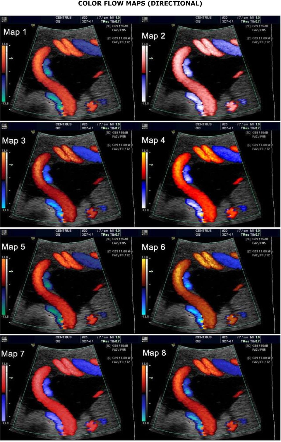
11 COLOR FLOW MAPS (DIRECTIONAL)
12 Power/energy/amplitude flow Sensitive to low flows No directional information in some modes Very poor temporal resolution Susceptible to noise "Color Power Angio" of the Circle of Willis "Color Power Angio" of a submucosus fibroid, note the small vessels inside the tumor.

13 COLOR POWER/ENERGY DOPPLER (AMPLITUDE FLOW)

14 Color flow imaging Color flow Doppler ultrasound produces a color-coded map of Doppler shifts superimposed onto a B-mode ultrasound image (Color Flow Maps). Although color flow imaging uses pulsed wave ultrasound, its processing differs from that used to provide the Doppler sonogram. Color flow imaging may have to produce several thousand color points of flow information for each frame superimposed on the B-mode image. Color flow imaging uses fewer, shorter pulses along each color scan line of the image to give a mean frequency shift and a variance at each small area of measurement. This frequency shift is displayed as a color pixel. The scanner then repeats this for several lines to build up the color image, which is superimposed onto the B-mode image. The transducer elements are switched rapidly between B- mode and color flow imaging to give an impression of a combined simultaneous image. The pulses used for color flow imaging are typically three to four times longer than those for the B-mode image, with a corresponding loss of axial resolution. Assignment of color to frequency shifts is usually based on direction (for example, red for Doppler shifts towards the ultrasound beam and blue for shifts away from it) and magnitude (different color hues or lighter saturation for higher frequency shifts). The color Doppler image is dependent on general Doppler factors, particularly the need for a good beam/flow angle. Curvilinear and phased array transducers have a radiating pattern of ultrasound beams that can produce complex color flow images, depending on the orientation of the arteries and veins. In practice, the experienced operator alters the scanning approach to obtain good insonation angles so as to achieve unambiguous flow images. Table 2: Factors affecting color flow image Main factors Power: transmitted power into tissue* Gain: overall sensitivity to flow signals Frequency: trades penetration for sensitivity and resolution* Pulse repetition frequency (also called scale): low pulse repetition frequency to look at low velocities, high pulse repetition frequency reduces aliasing* Area of investigation: larger area reduces frame rate* Focus: color flow image optimized at focal zone* Other factors Triplex color: pulse repetition frequency and frame rate reduced by need for B-mode/spectral pulses Persistence: high persistence produces smoother image but reduces temporal resolution* Pre-processing: trades resolution against frame rate* Filter: high filter cuts out more noise but also more of flow signal* Post-processing assigns color map/variance* * Settings appropriate for specific examinations assigned by set-up/application keys FACTORS AFFECTING THE COLOR FLOW IMAGE The controls that affect the appearance of the color flow image are summarized in Table 2. The main factors include: (1) Power and gain:color flow uses higher-intensity power than B-mode. Attention should be paid to safety indices. Power and gain should be set to obtain good signal for flow and to minimize the signals from surrounding tissue.

15 Figure 8 : Setting the color gain to minimize the signals (artefacts) from surrondng tissue, on left color gain = 71, then on right decreasing the color gain to 35. (2) Frequency selection: Many scanner/transducer combinations permit changes of frequency. High frequencies give better sensitivity to low flow and have better spatial resolution. Low frequencies have better penetration (Figure 5) and are less susceptible to aliasing at high velocities. (3) Velocity scale/pulse repetition frequency: Low pulse repetition frequencies should be used to examine low velocities but aliasing may occur if high velocities are encountered (Figura 7a,b). (4) Region of interest: Because more pulses are needed to look at flow than for the B-mode image, reducing the width and maximum depth of the color flow area under investigation will usually improve frame rate and may allow a higher color scan line density with improved spatial resolution (Figure 9). (5) Focus: The focus should be at the level of the area of interest. This can make a significant difference to the appearance and accuracy of the image (Figure 7). Figure 9 : Set the focus at the region of interest, and also could use more than one focal zone. In practice, the operator will make many changes to the controls and will try different probe positions to optimize the image. Practical guidelines are given in Table 3.
 Velocity scale/pulse repetition frequency: Low pulse repetition frequencies should be used to examine low velocities but aliasing may occur if high velocities are encountered (Figura 7a,b).")
16 Table 3: Color flow imaging: practical guidelines (1) Select the appropriate applications/set-up key. This optimizes parameters for specific examinations (2) Set power to within fetal study limits. Adjust color gain. Ensure focus is at the region of interest and adjust gain to optimize color signal (3) Use probe positioning/beam steering to obtain satisfactory beam/vessel angle (4) Adjust pulse repetition frequency/scale to suit the flow conditions. Low pulse repetition frequencies are more sensitive to low flows/velocities but may produce aliasing. High pulse repetition frequencies reduce aliasing but are less sensitive to low velocities (5) Set the color flow region to appropriate size. A smaller color flow box may lead to a better frame rate and better color resolution/sensitivity SPECTRAL OR PULSED WAVE DOPPLER Pulsed wave Doppler ultrasound is used to provide a sonogram of the artery or vein under investigation (Figure 12). The sonogram provides a measure of the changing velocity throughout the cardiac cycle and the distribution of velocities in the sample volume (or gate) (Figure 11). If an accurate angle correction is made, then absolute velocities can be measured. The best resolution of the sonogram occurs when the B-mode image and color image are frozen, allowing all the time to be employed for spectral Doppler. If concurrent imaging is used (real-time duplex or triplex imaging), the temporal resolution of the sonogram is compromised. Figure 10 (a): Doppler spectra of uterine artery flow. The color flow image allows beam/flow angle visualization. The sonogram shows high velocities throughout the cardiac cycle, indicating low distal resistance.

17 Figure 10 (b): Doppler spectra of uterine artery flow. The sonogram shows a pulsatile flow waveform with low diastolic velocities. This is indicative of high distal resistance Figure 11: Setting up the sample volume. (b) - direction of the Doppler beam, (g) - gate or sample volume (a) - angle correction Sonogram of the descending aorta. With the angle correction the peak velocities could be measured. FACTORS AFFECTING THE SPECTRAL IMAGE The controls that affect the appearance of the sonogram are summarized in Table 4. The main factors include: (1) Power and gain: Pulsed wave Doppler uses higher intensity power than B-mode. Attention should be paid to safety indices. Power and gain should be set so that clear signals are obtained. (2) Velocity scale/pulse repetition frequency: Low pulse repetition frequencies should be used to look at low velocities but aliasing may occur if high velocities are encountered. (3) Gate size: If flow measurements are being attempted, the whole vessel should be insonated. A large gate may include signals from adjacent vessels (Figure 13).

18 Table 4: Factors affecting the spectral Doppler image Main factors Power: transmitted power into tissue* Gain: overall sensitivity to flow signals Pulse repetition frequency (also called scale): low pulse repetition frequency to look at low velocities, high pulse repetition frequency reduces aliasing* Gate size* Beam steering can allow improved beam/flow angle for better accuracy of velocity calculation* Live duplex/triplex spectral resolution constrained by need for B-mode/color pulses Other factors Gate: sharpness of resolution* Filter: high filter cuts out more noise but more of flow signal* Post-processing: assigns brightness to output* *Settings appropriate for specific examinations assigned by set-up/application keys Figure 12: Umbilical cord displaying umbilical artery (red) and umbilical vein (blue), the gate or sample volume include both signals (left). Sonogram of the umbilical artery and vein (right).
")
19 Figure 13: Influence of gate size. The spectral Doppler gate insonates an artery and vein and the sonogram shows flow from both of these vessels. The calculation of mean velocity (arrow) is meaningless since velocities from one vessel subtract from those of the other Guidelines for a practical approach to obtain good-quality spectral images are given in Table 5. Table 5: Spectral Doppler imaging: practical guidelines (1) Set power to within fetal study limits (2) Position the pulsed wave Doppler cursor on the vessel to be investigated (3) Adjust gain so that the sonogram is clearly visible and free of noise (4) Use probe positioning/beam steering to obtain a satisfactory beam/vessel angle. Angles close to 90 will give ambiguous/unclear values. The beam/vessel angle should be 60 or less if velocity measurements are to be made (5) Adjust the pulse repetition frequency/scale and baseline to suit flow conditions. The sonogram should be clear and not aliased (6) Set the sample volume to correct size. Correct the angle to obtain accurate velocities. Use the B-mode and color flow image of the vessel to make the angle correction BLOOD FLOW MEASUREMENTS Velocity measurement Theoretically, once the beam/flow angle is known, velocities can be calculated from the Doppler spectrum as shown in
 Set power to within fetal study limits (2) Position the pulsed wave Doppler cursor on the vessel to be investigated (3) Adjust gain so that")
20 the Doppler equation. However, errors in the measured velocity may still occur 1,4. Sources of error can be broadly divided into three categories. (1) Errors can arise in the formation of the Doppler spectrum due to: (a) Use of multiple elements in array transducers; (b) Non-uniform insonation of the vessel lumen; (c) Insonation of more than one vessel; (d) Use of filters removing low-velocity components. (2) Errors can arise in the measurement of the ultrasound beam/flow velocity angle. (a) Use of high angles ( θ> 60 ) may give rise to error because of the comparatively large changes in the cosine of the angle which occur with small changes of angle (Figure 14). (b) The velocity vector may not be in the direction of the vessel axis. (3) Errors can arise in the calculation packages provided by the manufacturers for analysis of the Doppler spectrum (for instance, of intensity weighted mean velocity). (a) While efforts can be made to minimize errors, the operator should be aware of their likely range. It is good practice to try to repeat velocity measurements, if possible using a different beam approach, to gain a feel for the variability of measurements in a particular application. However, even repeated measurements may not reveal systematic errors occurring in a particular machine. (b) The effort applied to produce accurate velocity measurements should be balanced against the importance of absolute velocity measurements for an investigation. (c) Changes in velocity and velocity waveform shape are often of more clinical relevance when making a diagnosis. In this and other cases, absolute values of velocity measurement may not be required.
 Use of high angles ( θ> 60 ) may give rise to error because of the comparatively large changes in the cosine of the angle which occur with small changes of angle (Figure 14).")
21 Figure 14: Effect of high vessel/beam angles. (a) and (b) A scan of fetal aortic flow is undertaken at a high beam/vessel angle. Beam/flow angles should be kept to to 60 or less. A huge discrepancy is observed when using angles > 60. If absolute velocities are to be measured, beam/flow angles should be kept to 60 or less.
22 Calculation of absolute flow Total flow measurement using color or duplex Doppler ultrasound is fraught with difficulties, even under ideal conditions 5. Errors that may arise include: (1) Those due to inaccurate measurement of vessel cross-sectional area, for example the cross-sectional area of arteries which pulsate during the cardiac cycle; (2) Those originating in the derivation of velocity (see above). These errors become particularly large when flow calculations are made in small vessels; errors in measurement of diameter are magnified when the diameter is used to derive cross-sectional area. As with velocity measurements, it is prudent to be aware of possible errors and to conduct repeatability tests. Flow waveform analysis Non-dimensional analysis of the flow waveform shape and spectrum has proved to be a useful technique in the investigation of many vascular beds. It has the advantage that derived indices are independent of the beam/flow angle. Changes in flow waveform shape have been used to investigate both proximal disease (e.g. in the adult peripheral arterial circulation) and distal changes (in the fetal circulation and uterine arteries). While the breadth of possible uses shows the technique to be versatile, it also serves as a reminder of the range of factors which cause changes to the local Doppler spectrum. If waveform analysis is to be used to observe changes in one component of the proximal or distal vasculature, consideration must be given to what effects other components may have on the waveform. Flow waveform shape: indices of measurement Many different indices have been used to describe the shape of flow waveforms 1. Techniques range from simple indices of systolic to diastolic flow to feature extraction methods such as principal component analysis. All are designed to describe the waveform in a quantitative way, usually as a guide to some kind of classification. In general, they are a compromise between simplicity and the amount of information obtained. Figure 15: Arterial velocity sonogram (waveform).
23 The relative merits of indices used in uterine arteries have been discussed elsewhere 6,7. Commonly used indices available on most commercial scanners are: (1) Resistance index (RI) (also called resistive index or Pourcelot s index); (2) Systolic/diastolic (S/D) ratio, sometimes called the A/B ratio; (3) Pulsatility index (PI) 8. These indices are all based on the maximum Doppler shift waveform and their calculation is described in Figure 12. The PI takes slightly longer to calculate than the RI or S/D ratio because of the need to measure the mean height of the waveform. It does, however, give a broader range of values, for instance in describing a range of waveform shapes when there is no end-diastolic flow. Figure 16: Flow velocity indices In addition to these indices, the flow waveform may be described or categorized by the presence or absence of a particular feature, for example the absence of end-diastolic flow and the presence of a post-systolic notch. Generally, a low pulsatility waveform is indicative of low distal resistance and high pulsatility waveforms occur in highresistance vascular beds (Figure 8), although the presence of proximal stenosis, vascular steal or arteriovenous fistulas can modify waveform shape. Care should be taken when trying to interpret indices as absolute measurements of either upstream or downstream factors. For example, alterations in heart rate can alter the flow waveform shape and cause significant changes in the value of indices.
24 REFERENCES 1. Evans DH, McDicken WN, Skidmore R, Woodcock JP. Doppler Ultrasound: Physics, Instrumentation, and Clinical Applications. Chichester: Wiley, Powis RL, Schwartz RD. Practical Doppler Ultrasound for the Clinician. Williams and Wilkins, Goldberg BB, Merton DA, Deane CR. An Atlas of Ultrasound Color Flow Imaging. London: Martin Dunitz, Gill RW. Measurement of blood flow by ultrasound: accuracy and sources of error. Ultrasound Med Bio, 1985;7: Rourke C, Hendrickx P, Roth U, Brassel F, Creutzig A, Alexander K. Color and conventional image-directed ultrasonography: accuracy and sources of error in quantitative blood flow measurement. J Clin Ultrasound 1992;20: Thompson RS, Trudinger BJ, Cook CM. A comparison of Doppler ultrasound waveform indices in the umbilical artery. I. Indices derived from the maximum velocity waveform. Ultrasound Med Biol 1986;12: Thompson RS, Trudinger BJ, Cook CM. A comparison of Doppler ultrasound waveform indices in the umbilical artery. II. Indices derived from the mean velocity and first moment waveforms. Ultrasound Med Biol 1986;12: Gosling RG, King DH. Continuous wave ultrasound as an alternative and complement to X-rays in vascular examination. In Reneman RS, ed. Cardiovascular Applications of Ultrasound. Amsterdam: North Holland, 1974: Doppler in Obstetrics Copyright 2002 by The Fetal Medicine Foundation
25 Chapter 2 Safety of diagnostic ultrasound in fetal scanning by Colin Deane INTRODUCTION Diagnostic ultrasound is generally perceived by users and patients as a safe technique with no adverse effects. Since ultrasound is so widely used in pregnancy, it is essential for all practitioners to ensure that its use remains safe. Ultrasound causes thermal and mechanical effects in tissue which are increased as the output power is increased. In the last decade, there has been a general trend towards increased output with the introduction of color flow imaging, more use of pulsed spectral Doppler and higher demands on B-mode imaging 1. In response to these increases, recommendations for the safe use of ultrasound have been issued by several bodies. In addition, recent regulations have changed the emphasis of responsibility so that more onus is now placed on the operator to ensure that ultrasound is used safely. This chapter summarizes the effects and the standards issued and outlines recommendations for safe use in obstetric practice. EFFECTS Ultrasound is a mechanical energy in which a pressure wave travels through tissue. Reflection and scattering back to the transducer are used to form the image. The physical effects of ultrasound are generally categorized as: (1) Thermal effects heating of tissue as ultrasound is absorbed by tissue. Heat is also produced at the transducer surface; (2) Cavitation the formation of gas bubbles at high negative pressure; (3) Other mechanical effects radiation forces leading to streaming in fluids and stress at tissue interfaces. The implications of these effects have been determined by in vitro, animal and human epidemiological studies and are briefly summarized below. Thermal effects As the ultrasound waves are absorbed, their energy is converted into heat. The level of conversion is highest in tissue with a high absorption coefficient, particularly in bone, and is low where there is little absorption (e.g. amniotic fluid). The temperature rise is also dependent on the thermal characteristics of the tissue (conduction of heat and perfusion), the ultrasound intensity and the length of time for which the tissue volume is scanned. The intensity is, in turn, dependent on the power output and the position of the tissue in the beam profile. The intensity at a particular point is altered by many of the operator controls, for example power output, mode (B-mode, color flow, spectral Doppler), scan depth, focus, zoom and area of color flow imaging. With so many variables, it has proved difficult to model temperature rises in tissue. In vitro studies have been used with a worst case model of tissue to predict temperature rises o, for instance in the formation of thermal indices (see below). The transducer face itself can become heated during an examination. Heat is localized to the tissue in contact with the transducer. Cavitation Cavitation is the formation of transient or stable bubbles, described as inertial or non-inertial cavitation. Inertial cavitation has the most potential to damage tissue and occurs when a gas-filled cavity grows, during pressure rarefaction of the ultrasound pulse, and contracts, during the compression phase. Collapse of the bubble can generate
26 local high temperatures and pressures. It has been hypothesized that ultrasonically induced cavitation is the cause of hemorrhage in the lungs and intestines in animal studies 2 6. In these studies, effects have been seen at tissue interfaces with gas. The absence of gas in fetuses means that the threshold for cavitation is high and does not occur at current levels of diagnostic ultrasound. The introduction of contrast agents leads to the formation of microbubbles that potentially provide gas nuclei for cavitation. The use of contrast agents lowers the threshold at which cavitation occurs, but this is not current practice in obstetrics. Other mechanical effects The passage of ultrasound through tissue causes a low-level radiation force on the tissue. This force produces a pressure in the direction of the beam and away from the transducer and should not be confused with the oscillatory pressure of the ultrasound itself. The pressure that results and the pressure gradient across the beam are very low, even for intensities at the higher end of the diagnostic range 7. The effect of the force is manifest in volumes of fluid where streaming can occur with motion within the fluid. The fluid velocities which result are low and are unlikely to cause damage. Effects on fetuses Effects are divided into mechanical and thermal. For mechanical effects, there is no evi-dence that cavitation occurs in fetal scanning. In a study of low-amplitude lithotripsy pulses in mouse fetuses, there has been concern that hemorrhage may be the result of tissue movement caused by radiation forces 8. There is no evidence that this occurs in vivo in fetal scanning. The primary concern in fetal imaging is temperature rise. It is known that hyperthermia is teratogenic. The efforts of investigators have concentrated on defining the temperature increases and exposure times which may give rise to biological effects and on determining the ultrasound levels which might, in turn, lead to those temperature rises. With this information, criteria have been identified for the safe use of diagnostic ultrasound. Temperature rises of 2.5 C have been demonstrated in excised unperfused guinea pig brain tissue after 2 minutes exposure to ultrasound at the high end of pulsed wave Doppler ultrasound intensity levels 9. At the bone surface, temperature increases of up to 5 C were found. In a study on sheep using different intensity criteria 10, the temperature rise in utero was found to be 40% lower than that in the equivalent non-perfused test. While the observed temperature increases occurred in high-intensity modes (typical of pulsed wave Doppler used at maximum power), these levels of intensity are achievable with some current scanner/transducer combinations. The issue of sensitivity of fetal tissue to temperature rise is complex and is not completely understood. Acute and chronic temperature rises have been investigated in animals, but study designs and results are varied. Work carried out in this field is summarized elsewhere 11. The uncertainty over chronic changes is reflected in the WFUMB guidelines 12. These state that ultrasound that produces temperature rises of less than 1.5 C may be used without reservation. They also state that ultrasound exposure causing temperature rises of greater than 4 C for over 5 min should be considered potentially hazardous. This leaves a wide range of temperature increases which are within the capability of diagnostic ultrasound equipment to produce and for which no time limits are recommended. Epidemiology Several studies have examined the development of fetuses receiving different levels of ultrasound investigation. In trials comparing ultrasound screened and non-screened groups, there has generally been no difference in birth weights between groups. There have been no unequivocal data to suggest that there is impaired development of hearing, vision, behavior or neurological function due to ultrasound screening. In a large, randomized trial of over 3200 pregnant women in which half were offered routine ultrasonography at 19 and 32 weeks, there was no evidence of impaired growth or neurological development up to follow-up at 8 9 years. There was a possible association of lefthandedness amongst boys undergoing ultrasonography 13. Scanning of this group was performed with B-mode only. There have been concerns that epidemiological studies to date do not reflect the higher output capabilities of modern scanners.
27 OUTPUT REGULATIONS, STANDARDS AND GUIDELINES WHO DOES WHAT? Regulations governing the output of diagnostic ultrasound have been largely set by the USA s Food and Drug Administration (FDA), although the International Electrotechnical Commission (IEC) is currently in the process of setting internationally agreed standards. The relevant national societies for ultrasound users (e.g. American Institutue of Ultrasound in Medicine (AIUM), British Medical Ultrasound Society (BMUS)) usually have safety committees who offer advice on the safe use of ultrasound. In 1992, the AIUM, in conjunction with the National Electrical Manufacturers Association (NEMA) developed the Output Display Standard (ODS), including the thermal index and mechanical index which have been incorporated in the FDA s new regulations 14,15. Within Europe, the Federation of Societies of Ultrasound in Medicine and Biology (EFSUMB) also addresses safety and has produced safety guidelines (through the European Committee for Ultrasound Radiation Safety). The World Federation (WFUMB) held safety symposia in 1991 (on thermal issues) and 1996 (thermal and non-thermal issues), at which recommendations were proffered. Following review, these were published in 1992 and 1998 as guidelines. Past regulations The initial FDA regulations on ultrasound output were produced in These imposed application-specific limits, based on existing output levels which had demonstrated no adverse effects. Limits were divided into: (1) Ophthalmic applications; (2) Fetal and other (including abdominal, pediatric, small parts); (3) Cardiac; (4) Peripheral vessels. For spatial peak time-averaged intensity (I-SPTA) (the measure most associated with temperature rise), the maximum levels were: Ophthalmic 17 mw/cm 2 Fetal and other 94 mw/cm 2 Cardiac 430 mw/cm 2 Peripheral vessel 720 mw/cm 2 Scanners typically had a key/button which limited output for obstetric applications. Although power and intensity limits could be exceeded in some scanners, especially when using pulsed wave Doppler or color Doppler, this required a deliberate effort on the behalf of the users. Current regulations In revising its regulations in 1993, the FDA 15 altered its approach to ultrasound safety. The new regulations combine an overall limit of I-SPTA of 720 mw/cm 2 for all equipment with a system of output displays to allow users to employ effective and judicious levels of ultrasound appropriate to the examination undertaken. The new regulations allow an eight-fold increase in ultrasound intensity to be used in fetal examinations. They place considerably more responsibility on the user to understand the output measurements and to use them in their scanning. The output display is based on two indices, the mechanical index (MI) and the thermal index (TI).
28 Mechanical index The mechanical index is an estimate of the maximum amplitude of the pressure pulse in tissue. It gives an indication as to the relative risk of mechanical effects (streaming and cavitation). The FDA regulations allow a mechanical index of up to 1.9 to be used for all applications except ophthalmic (maximum 0.23). Thermal index The thermal index is the ratio of the power used to that required to cause a maximum temperature increase of 1 C. A thermal index of 1 indicates a power causing a temperature increase of 1 C. A thermal index of 2 would be twice that power but would not necessarily indicate a peak temperature rise of 2 C. Because temperature rise is dependent on tissue type and is particularly dependent on the presence of bone, the thermal index is subdivided into three indices: (1) TIS: thermal index for soft tissue; (2) TIB: thermal index with bone at/near the focus; (3) TIC: thermal index with bone at the surface (e.g. cranial examination). For fetal scanning, the highest temperature increase would be expected to occur at bone and TIB would give the worst case conditions. The mechanical index and thermal index must be displayed if the ultrasound system is capable of exceeding an index of 1. The displayed indices are based on the manufacturer s experimental and modelled data. These measurements are not infallible; an independent study has demonstrated significant discrepancies over declared I-SPTA output of up to 400% 16. Future IEC standards An IEC standard (Draft IEC 61681) is being drawn up to establish a safety classification for ultrasound equipment based on its ability to produce cavitation or a temperature rise. The standard proposes two classifications of equipment: class A, which has a lower output and for which no output display is required, and class B which has a higher output and for which an output display is required. The draft is currently undergoing review. Guidelines Ultrasound organizations have produced statements on the safe use of ultrasound. These are not regulatory statements but are intended to educate and advise. WFUMB guidelines have been issued in two special issues of Ultrasound in Medicine and Biology Statements and recommendations are given on B-mode scanning, Doppler imaging, transducer heating, thermal effects (see page 33). The AIUM have produced statements on the safety of ultrasound. They are available from the AIUM office and can be obtained from the AIUMwebsite The European Committee for Ultrasound Radiation Safety has published statements 18,19 on the use of pulsed Doppler measurement in fetuses, stating that its use in routine examinations during the period of organogenesis is considered inadvisable at present. A PRACTICAL APPROACH TO SAFE FETAL SCANNING No injurious effects have been identified from ultrasound scanning of the fetus. However, changes in power output, increased use of Doppler ultrasound and a change in regulations governing outputs means that every measure should be taken by users to maintain safe practices. Scanning practice The ALARA ("As Low As Reasonably Achievable") principle should be maintained. Power outputs used should be adequate to conduct the examination. If in doubt, use a low power and increase it as necessary. Application keys for obstetrics should bring in each mode at its lowest output so that the operator is required to increase power if the examination demands it. B-mode generally has the lowest power output and intensity. M-mode, color flow and spectral Doppler have higher outputs which can cause more heating at the site of examination. The examination should begin with B-mode and use color and spectral Doppler only when necessary.
29 The intensity (and temperature rise) is highly dependent on scanner settings. For example, the intensity changes in response to changes in: (a) Power Output, (b) Depth of examination, (c) Mode used (color flow, spectral Doppler), (d) Transmitted frequency used, (e) Color pulse repetition frequency (scale), (f) Region of color flow interest, (g) Focus. If the display for the scanner/transducer combination shows thermal and mechanical indices, the indices should be readily visible. Of the thermal indices, TIB is most relevant to heating in the second and third trimesters. The operator should be aware of changes to the indices in response to changes in control settings. Special care should be taken in febrile patients, since ultrasound heating will cause additional heating to the fetus. The WFUMB recommends that ultrasound causing a temperature rise of no more than 1.5 C may be used without reservation on thermal grounds. Thermal indices exceeding 1.5 should not be used routinely and, if required for specific diagnostic information, should be used for the minimum time necessary. The influence of higher intensity levels can be moderated by moving the transducer so that specific areas of tissue are not subjected to long periods of higher intensity investigation. Do not scan for longer than is necessary to obtain the diagnostic information. SELECTED WFUMB STATEMENTS ON THE SAFETY OF DIAGNOSTIC ULTRASOUND B-mode imaging (issued 1992) Known diagnostic ultrasound equipment as used today for simple B-mode imaging operates at acoustic outputs that are not capable of producing harmful temperature rises. Its use in medicine is therefore not contraindicated on thermal grounds. This includes endoscopic, transvaginal and transcutaneous applications. Doppler (1992) It has been demonstrated in experiments with unperfused tissue that some Doppler diagnostic equipment has the potential to produce biologically significant temperature rises, specifically at bone/soft tissue interfaces. The effects of elevated temperatures may be minimized by keeping the time during which the beam passes through any one point in tissue as short as possible. Where output power can be controlled, the lowest available power level consistent with obtaining the desired diagnostic information should be used. Although the data on humans are sparse, it is clear from animal studies that exposures resulting in temperatures less than 38.5 C can be used without reservation on thermal grounds. This includes obstetric applications. Transducer heating (1992) A substantial source of heating may be the transducer itself. Tissue heating from this source is localized to the volume in contact with the transducer.
Doppler Ultrasound in the Management of Fetal Growth Restriction Chukwuma I. Onyeije, M.D. Atlanta Perinatal Associates
 Doppler Ultrasound in the Management of Fetal Growth Restriction Chukwuma I. Onyeije, M.D. Atlanta Perinatal Associates 1 For your convenience a copy of this lecture is available for review and download
Doppler. Doppler. Doppler shift. Doppler Frequency. Doppler shift. Doppler shift. Chapter 19
 Doppler Doppler Chapter 19 A moving train with a trumpet player holding the same tone for a very long time travels from your left to your right. The tone changes relative the motion of you (receiver) and
Applications of Doppler Ultrasound in Fetal Growth Assessment. David Cole
 Applications of Doppler Ultrasound in Fetal Growth Assessment David Cole Aims The aim of this presentation is to consider the use of Doppler ultrasound to investigate and monitor those pregnancies at risk
BIOMEDICAL ULTRASOUND
 BIOMEDICAL ULTRASOUND Goals: To become familiar with: Ultrasound wave Wave propagation and Scattering Mechanisms of Tissue Damage Biomedical Ultrasound Transducers Biomedical Ultrasound Imaging Ultrasonic
VFI Technology to Change the Way You Work
 Technology to Change the Way You Work Vascular Ultrasound Made Easier Vector Flow Imaging VFI VFI is a ground-breaking technology that can revolutionize the workflow for many Doppler ultrasound applications.
Principles of Medical Ultrasound. Pai-Chi Li Department of Electrical Engineering National Taiwan University
 Principles of Medical Ultrasound Pai-Chi Li Department of Electrical Engineering National Taiwan University What is Medical Ultrasound? Prevention: actions taken to avoid diseases. Diagnosis: the process
TISSUE MIMICKING GEL QUALITY LE PHANTOM SERIES DESIGN. performance the ultrasound labs ofand. icking material has the same attenuation mim-
 QUALITY Tissue Benefits Mimicking of s RMI recognized RMI as the ultrasound standard phantoms for quality are performance the ultrasound labs ofand hospitals, manufacturers. clinics Sophisticated and ultrasound
Ny teknologi: Fagdagene ved St. Olavs Hospital Lasse Løvstakken Dept. Circulation and Medical Imaging 11.06.2010
 1 Ny teknologi: Ultralyd måler m blodstrøm Fagdagene ved St. Olavs Hospital Lasse Løvstakken Dept. Circulation and Medical Imaging 11.06.2010 2 Conventional imaging methods of blood flow using ultrasound
Umbilical Artery Doppler Waveform Indices in Normal Pregnancies
 Thai Journal of Obstetrics and Gynaecology June 2000, Vol. 12, pp. 103-107 OBSTETRICS Umbilical Artery Doppler Waveform Indices in Normal Pregnancies Pharuhas Chanprapaph MD, Chanane Wanapirak MD, Theera
Ultrasound. Sound waves
 Ultrasound Basic Idea Send waves into body which are reflected at the interfaces between tissue Return time of the waves tells us of the depth of the reflecting surface History First practical application,
School of Diagnostic Medical Sonography
 Semester 1 Orientation - 101 This class is an introduction to sonography which includes a basic anatomy review, introduction to sonographic scanning techniques and physical principles. This curriculum
Spectral Doppler Sonography: Waveform Analysis and Hemodynamic Interpretation
 Chapter 4 Spectral Doppler Sonography: Waveform Analysis and Hemodynamic Interpretation Dev Maulik The spectral Doppler power waveform contains an immense amount of hemodynamic information from the sampled
Assessment of umbilical arterial and venous flow using color Doppler
 Ultrasound Obstet Gynecol 1999;14:250 255 Assessment of umbilical arterial and venous flow using color C. Lees, G. Albaiges, C. Deane*, M. Parra and K. H. Nicolaides Harris Birthright Research Centre and
Duplication Images in Vascular Sonography
 Article Duplication Images in Vascular Sonography Jonathan M. Rubin, MD, PhD, Jing Gao, MD, Keith Hetel, MD, Robert Min, MD Objective. The purpose of this study was to determine the characteristics and
EFSUMB EUROPEAN FEDERATION OF SOCIETIES FOR ULTRASOUND IN MEDICINE AND BIOLOGY Building a European Ultrasound Community
 MINIMUM TRAINING REQUIREMENTS FOR THE PRACTICE OF MEDICAL ULTRASOUND IN EUROPE Appendix 8: Vascular Ultrasound Level 1 Training and Practice Practical training should involve at least two half day ultrasound
Preparation iagnostic Medical Sonographer Overview"
 Diagnostic Medical Sonographer Overview The Field - Preparation - Specialty Areas - Day in the Life - Earnings - Employment - Career Path Forecast - Professional Organizations The Field Diagnostic imaging
Subclavian Steal Syndrome By Marta Thorup
 Subclavian Steal Syndrome By Marta Thorup Definition Subclavian steal syndrome (SSS), is a constellation of signs and symptoms that arise from retrograde flow of blood in the vertebral artery, due to proximal
How can I be sure it will be performed to a high standard?
 FREQUENTLY ASKED QUESTIONS (About ultrasound scans at Derriford) What is an Ultrasound Scan? How does it work? What are the dangers? What are the advantages? Who will perform my scan? How can I be sure
Vascular Laboratory Fundamentals. Robert Mitchell MD, RPVI Duke University Medical Center 5/2/08
 Vascular Laboratory Fundamentals Robert Mitchell MD, RPVI Duke University Medical Center 5/2/08 Goals of Lecture Understand basic ultrasound principles Understand normal and abnormal arterial hemodynamics
Monitoring of Cerebral Blood Flow. Transcranial Doppler Laser Doppler Flowmetry Thermal dilution method (Hemedex)
") Monitoring of Cerebral Blood Flow Transcranial Doppler Laser Doppler Flowmetry Thermal dilution method (Hemedex) Ultrasound in Tissue Some Facts: blood cell tissue probe ultrasound travels at a constant
Monitoring of Cerebral Blood Flow Transcranial Doppler Laser Doppler Flowmetry Thermal dilution method (Hemedex) Ultrasound in Tissue Some Facts: blood cell tissue probe ultrasound travels at a constant
Antonio Rampoldi Struttura Complessa di Radiologia Interventistica Azienda Ospedale Niguarda Ca Granda, Milano. Terminology
 Biophysical principles and clinical applications of MRgFUS Alberto Torresin Struttura Complessa di Fisica Sanitaria Azienda, Milano Università degli Studi di Milano Dip. di Fisica Antonio Rampoldi Struttura
Company profile. Quote from the analyst of. Company Milestone. Feel the Difference. Tel: +86-755-26722860 Fax: +86-755-26722850
 Company profile Since the establishment, SonoScape has committed to provide high standard medical equipments for healthcare of human beings. SonoScape specializes in the development and production of diagnostic
Hunting Bats. Diagnostic Ultrasound. Ultrasound Real-time modality
 Diagnostik Ultrasound Basic physics, image reconstruction and signal processing Per Åke Olofsson Dpt of Biomedical Engineering, Malmö University Hospital, Sweden Ultrasound Real-time modality 17-WEEK FETAL
Assessment of Fetal Growth
 Assessment of Fetal Growth Unit / Trust: 1. INTRODUCTION The aim of this guideline template is to outline the methods used to assess fetal growth and the referral pathways utilising customised antenatal
School of Diagnostic Medical Sonography Course Catalog
 School of Diagnostic Medical Sonography Course Catalog 2 School of Diagnostic Medical Sonography Course Schedule Our program provides a broad base of education and performance- based clinical experience
TCD in Intensive Care
 TCD in Intensive Care Background: Transcranial Doppler (TCD) ultrasonography is a technique that uses a hand-held Doppler transducer (placed on the surface of the cranial skin) to measure the velocity
Signal Extraction Technology
 Signal Extraction Technology Technical bulletin Introduction Masimo SET pulse oximetry is a new and fundamentally distinct method of acquiring, processing and reporting arterial oxygen saturation and pulse
CORD BLOOD COLLECTION / ANALYSIS- AT BIRTH
 WOMEN AND NEWBORN HEALTH SERVICE King Edward Memorial Hospital CLINICAL GUIDELINES OBSTETRICS AND MIDWIFERY King Edward Memorial Hospital WOMEN AND NEWBORN HEALTH SERVICE INTRAPARTUM CARE SPECIMEN COLLECTION
Section Two: Arterial Pressure Monitoring
 Section Two: Arterial Pressure Monitoring Indications An arterial line is indicated for blood pressure monitoring for the patient with any medical or surgical condition that compromises cardiac output,
History of the American Institute of Ultrasound in Medicine s Efforts to Keep Ultrasound Safe
 Commemoration History of the American Institute of Ultrasound in Medicine s Efforts to Keep Ultrasound Safe Wesley L. Nyborg, PhD Physics Department University of Vermont Burlington, Vermont USA Abbreviations
Reliable versatility. Philips HD5 ultrasound system
 Reliable versatility Philips HD5 ultrasound system Affordability and features in one Designed to perform as you need it Every day, your patients come to you for high-quality care. Now, there s an ultrasound
Diagnostic Medical Sonography
 Diagnostic Medical Sonography 130 Diagnostic Medical Sonography Location: Trenholm Campus - Bldg. H Program Information Diagnostic Medical Sonography (DMS) is a diagnostic procedure that uses high frequency
Basics of Ultrasound Imaging
 2 Basics of Ultrasound Imaging Vincent Chan and Anahi Perlas Introduction... Basic Principles of B-Mode US... Generation of Ultrasound Pulses... Ultrasound Wavelength and Frequency... Ultrasound Tissue
STATISTICAL ANALYSIS OF ULTRASOUND ECHO FOR SKIN LESIONS CLASSIFICATION HANNA PIOTRZKOWSKA, JERZY LITNIEWSKI, ELŻBIETA SZYMAŃSKA *, ANDRZEJ NOWICKI
 STATISTICAL ANALYSIS OF ULTRASOUND ECHO FOR SKIN LESIONS CLASSIFICATION HANNA PIOTRZKOWSKA, JERZY LITNIEWSKI, ELŻBIETA SZYMAŃSKA *, ANDRZEJ NOWICKI Institute of Fundamental Technological Research, Department
Ultrasound - Vascular
 Scan for mobile link. Ultrasound - Vascular Vascular ultrasound uses sound waves to evaluate the body s circulatory system and help identify blockages and detect blood clots. A Doppler ultrasound study
School of Diagnostic Medical Sonography Course Catalog
 School of Diagnostic Medical Sonography Course Catalog 2 School of Diagnostic Medical Sonography Course Schedule Our program provides a broad base of education and performance- based clinical experience
Samsung Ultrasound H60
 Samsung Medison is a global leading medical devices company. Founded in 1985, the company now sells cutting-edge medical devices including diagnostic ultrasound, digital X-ray and blood analyzer around
Ultrasonic Wave Propagation Review
 Ultrasonic Wave Propagation Review Presented by: Sami El-Ali 1 1. Introduction Ultrasonic refers to any study or application of sound waves that are higher frequency than the human audible range. Ultrasonic
X-ray (Radiography) - Chest
 - Chest") Scan for mobile link. X-ray (Radiography) - Chest What is a Chest X-ray (Chest Radiography)? The chest x-ray is the most commonly performed diagnostic x-ray examination. A chest x-ray produces images of
Scan for mobile link. X-ray (Radiography) - Chest What is a Chest X-ray (Chest Radiography)? The chest x-ray is the most commonly performed diagnostic x-ray examination. A chest x-ray produces images of
An abdominal ultrasound produces a picture of the organs and other structures in the upper abdomen.
 Scan for mobile link. Ultrasound - Abdomen Ultrasound imaging of the abdomen uses sound waves to produce pictures of the structures within the upper abdomen. It is used to help diagnose pain or distention
Hemodynamic Monitoring: Principles to Practice M. L. Cheatham, MD, FACS, FCCM
 SUMMARY HEMODYNAMIC MONITORING: FROM PRINCIPLES TO PRACTICE Michael L. Cheatham, MD, FACS, FCCM Director, Surgical Intensive Care Units Orlando Regional Medical Center Orlando, Florida Fluid-filled catheters
her Admissions Information Diploma 122 2,425 Hours: program. sonography too specializes in general sonography field. good health of the
 1 Diagnostic Medical Sonograp her Award: Quarter Credits: Hours: Weeks: Diploma 122 2,425 80 total instructional weeks Educational Objective The Diagnostic Medical Sonographer program is designed to provide
High-quality/High-resolution Digital Ultrasound Diagnostic Scanner
 Hitachi Review Vol. 52 (2003), No. 4 207 High-quality/High-resolution Digital Ultrasound Diagnostic Scanner Tsuyoshi Otake Toshihiko Kawano Takashi Sugiyama Tsuyoshi Mitake Shinichiro Umemura, Ph. D. OVERVIEW:
FXA 2008. UNIT G485 Module 4 5.4.3 Ultrasound. Candidates should be able to :
 1 Candidates should be able to : ULTRASOUND Describe the properties of ultrasound. ULTRASOUND is any sound wave having a frequency greater than the upper frequency limit of human hearing (20 khz). Describe
Ultrasound Technologies. PD1v. vascular pocket doppler. Operating Instructions
 Ultrasound Technologies PD1v vascular pocket doppler Operating Instructions Before using your Pocket Doppler for the first time, please read these operating instructions carefully. 1 INTRODUCTION The UltraTec
Vascular Technology (VT) Content Outline Anatomy & physiology 20% Cerebrovascular Cerebrovascular normal anatomy Evaluate the cerebrovascular vessels
 Content Outline Anatomy & physiology 20% Cerebrovascular Cerebrovascular normal anatomy Evaluate the cerebrovascular vessels") Vascular Technology (VT) Content Outline Anatomy & physiology 20% normal anatomy Evaluate the cerebrovascular vessels hemodynamics Evaluate the cerebrovascular vessels for normal perfusion normal anatomy
Vascular Technology (VT) Content Outline Anatomy & physiology 20% normal anatomy Evaluate the cerebrovascular vessels hemodynamics Evaluate the cerebrovascular vessels for normal perfusion normal anatomy
Reavis High School Anatomy and Physiology Curriculum Snapshot
 Reavis High School Anatomy and Physiology Curriculum Snapshot Unit 1: Introduction to the Human Body 10 days As part of this unit, students will define anatomy, physiology, and pathology. They will identify
Routine Ultrasound Equipment Tests for Quality Assurance. Contents
 Routine Ultrasound Equipment Tests for Quality Assurance James A. Zagzebski, Ph.D. Nick Rupert, M.S. Ryan DeWall, M.S. Tomy Varghese, Ph.D. Dept. of Medical Physics University of Wisconsin, Madison, WI,
Vivid S6. Cardiovascular ultrasound system. imagination at work. GE Healthcare
 GE Healthcare Vivid S6 Cardiovascular ultrasound system imagination at work Taking care of everyone. Strong performance and innovative miniaturization make the Vivid S6 a highly desirable system for a
Universal Fetal Cardiac Ultrasound At the Heart of Newborn Well-being
 Universal Fetal Cardiac Ultrasound At the Heart of Newborn Well-being Optimizes detection of congenital heart disease (chd) in the general low risk obstetrical population Daniel J. Cohen, M.D. danjcohen@optonline.net
Ischemia and Infarction
 Harvard-MIT Division of Health Sciences and Technology HST.035: Principle and Practice of Human Pathology Dr. Badizadegan Ischemia and Infarction HST.035 Spring 2003 In the US: ~50% of deaths are due to
ASSOCIATE OF APPLIED SCIENCE IN DIAGNOSTIC MEDICAL SONOGRAPHY (DMS AAS) 130 quarter credit units / 2,160 clock hours / 84 weeks (20-32 hours per
 130 quarter credit units / 2,160 clock hours / 84 weeks (20-32 hours per") ASSOCIATE OF APPLIED SCIENCE IN DIAGNOSTIC MEDICAL SONOGRAPHY (DMS AAS) 130 quarter credit units / 2,160 clock hours / 84 weeks (20-32 hours per week) Educational Objective: The Associate of Applied Science
ASSOCIATE OF APPLIED SCIENCE IN DIAGNOSTIC MEDICAL SONOGRAPHY (DMS AAS) 130 quarter credit units / 2,160 clock hours / 84 weeks (20-32 hours per week) Educational Objective: The Associate of Applied Science
Brain Injury during Fetal-Neonatal Transition
 Brain Injury during Fetal-Neonatal Transition Adre du Plessis, MBChB Fetal and Transitional Medicine Children s National Medical Center Washington, DC Brain injury during fetal-neonatal transition Injury
Transmission Line and Back Loaded Horn Physics
 Introduction By Martin J. King, 3/29/3 Copyright 23 by Martin J. King. All Rights Reserved. In order to differentiate between a transmission line and a back loaded horn, it is really important to understand
What Does Pregnancy Have to Do With Blood Clots in a Woman s Legs?
 Patient s Guide to Prevention of Blood Clots During Pregnancy: Use of Blood-Thinning A Patient s Guide to Prevention of Blood Clots During Pregnancy: Use of Blood-Thinning Drugs to Prevent Abnormal Blood
How To Use An Ultrasound For Medical Research
 Scan for mobile link. General Ultrasound What is General Ultrasound Imaging? Ultrasound is safe and painless, and produces pictures of the inside of the body using sound waves. Ultrasound imaging, also
Fetal Thermal Effects of Diagnostic Ultrasound
 Fetal Thermal Effects of Diagnostic Ultrasound Abbreviations ALARA, as low as reasonably achievable; dga, days gestational age; FDA, Food and Drug Administration; HSP, heat shock protein; I SPTA, spatial-peak
CHAPTER 4 QUALITY ASSURANCE AND TEST VALIDATION
 CHAPTER 4 QUALITY ASSURANCE AND TEST VALIDATION CINDY WEILAND AND SANDRA L. KATANICK Continued innovations in noninvasive testing equipment provide skilled sonographers and physicians with the technology
Magnetic Resonance Imaging
 Magnetic Resonance Imaging What are the uses of MRI? To begin, not only are there a variety of scanning methodologies available, but there are also a variety of MRI methodologies available which provide
Section Four: Pulmonary Artery Waveform Interpretation
 Section Four: Pulmonary Artery Waveform Interpretation All hemodynamic pressures and waveforms are generated by pressure changes in the heart caused by myocardial contraction (systole) and relaxation/filling
Laminar and Turbulent flow. Flow Sensors. Reynolds Number. Thermal flow Sensor. Flow and Flow rate. R = Mass Flow controllers
 Flow and Flow rate. Laminar and Turbulent flow Laminar flow: smooth, orderly and regular Mechanical sensors have inertia, which can integrate out small variations due to turbulence Turbulent flow: chaotic
Robot Perception Continued
 Robot Perception Continued 1 Visual Perception Visual Odometry Reconstruction Recognition CS 685 11 Range Sensing strategies Active range sensors Ultrasound Laser range sensor Slides adopted from Siegwart
Ultrasound Billing CPT Codes Summary and Notes
 Ultrasound Billing CPT Codes Summary and Notes CPT codes for ultrasound examinations are considered to be complete studies unless specified as limited studies in their code definitions. A limited study
OHTAC Recommendation
 OHTAC Recommendation Multiple Sclerosis and Chronic Cerebrospinal Venous Insufficiency Presented to the Ontario Health Technology Advisory Committee in May 2010 May 2010 Issue Background A review on the
X-ray (Radiography), Chest
, Chest") X-ray (Radiography), Chest What is a Chest X-ray (Chest Radiography)? The chest x-ray is the most commonly performed diagnostic x-ray examination. A chest x-ray makes images of the heart, lungs, airways,
X-ray (Radiography), Chest What is a Chest X-ray (Chest Radiography)? The chest x-ray is the most commonly performed diagnostic x-ray examination. A chest x-ray makes images of the heart, lungs, airways,
Application of Ultrasound, MRI and PET in Cardiac Disease Diagnosis
 Application of Ultrasound, MRI and PET in Cardiac Disease Diagnosis Zarina Che Amin Ultrasound is defined as sound above the hearing range of normal human and similar to audible sound. Ultrasound provides
Note: The left and right sides of the heart must pump exactly the same volume of blood when averaged over a period of time
 page 1 HEART AS A PUMP A. Functional Anatomy of the Heart 1. Two pumps, arranged in series a. right heart: receives blood from the systemic circulation (via the great veins and vena cava) and pumps blood
Sound Power Measurement
 Sound Power Measurement A sound source will radiate different sound powers in different environments, especially at low frequencies when the wavelength is comparable to the size of the room 1. Fortunately
A PHOTOGRAMMETRIC APPRAOCH FOR AUTOMATIC TRAFFIC ASSESSMENT USING CONVENTIONAL CCTV CAMERA
 A PHOTOGRAMMETRIC APPRAOCH FOR AUTOMATIC TRAFFIC ASSESSMENT USING CONVENTIONAL CCTV CAMERA N. Zarrinpanjeh a, F. Dadrassjavan b, H. Fattahi c * a Islamic Azad University of Qazvin - nzarrin@qiau.ac.ir
Sonography. 1. Introduction. 2. Documentation of Compliance. 3. Didactic Competency Requirements. 4. Clinical Competency Requirements
 PRIMARY CERTIFICATION Sonography 1. Introduction Candidates for certification and registration are required to meet the Professional Education Requirements specified in the ARRT Rules and Regulations.
Advances in scmos Camera Technology Benefit Bio Research
 Advances in scmos Camera Technology Benefit Bio Research scmos camera technology is gaining in popularity - Why? In recent years, cell biology has emphasized live cell dynamics, mechanisms and electrochemical
PRINCIPLES OF DOPPLER ECHOCARDIOGRAPHY AND THE DOPPLER EXAMINATION #1
 PRINCIPLES OF DOPPLER ECHOCARDIOGRAPHY AND THE DOPPLER EXAMINATION #1 Joseph A. Kisslo, MD David B. Adams, RDCS INTRODUCTION Doppler echocardiography is a method for detecting the direction and velocity
Ultrasound in Vascular Surgery. Torbjørn Dahl
 Ultrasound in Vascular Surgery Torbjørn Dahl 1 The field of vascular surgery Veins dilatation and obstruction (varicose veins and valve dysfunction) Arteries dilatation and narrowing (aneurysms and atherosclerosis)
Human Exposure Limits
 Human Exposure Limits Session 3 0 Version December 2014 Learning objectives In this session we will: Learn about the international exposure limits for workers and the public Learn about methods for assessing
Diagnosis Codes for Pregnancy and Complications of Pregnancy
 This list is for informational purposes only and is not a binding or definitive list of covered conditions. It is not a guarantee of coverage; coverage depends on the available benefits and eligibility
Placenta, Cord, & Fluid
 , Cord, & Fluid Abruption Accreta/Increta/Percreta Chorioangioma Complete Partial Not generally Relevant to U/S Gestational Age (Weeks) Distance from 16-23.9 24 to Internal Os >20 mm No No 11-20 mm 0-10
Advancements in High Frequency, High Resolution Acoustic Micro Imaging for Thin Silicon Applications
 Advancements in High Frequency, High Resolution Acoustic Micro Imaging for Thin Silicon Applications Janet E. Semmens Sonoscan, Inc. 2149 E. Pratt Boulevard Elk Grove Village, IL 60007 USA Phone: (847)
Upper Extremity Vein Mapping for Placement of a Dialysis Access
 VASCULAR TECHNOLOGY PROFESSIONAL PERFORMANCE GUIDELINES Upper Extremity Vein Mapping for Placement of a Dialysis Access This Guideline was prepared by the Professional Guidelines Subcommittee of the Society
Contents. HD11 XE Quick Guide 4535 612 62661
 Contents Using the HD11 XE Help................................................3 Using the HD11 XE Ultrasound System....................................4 Imaging Modes........................................................7
ASSESSMENT REPORT. Program: AAS Diagnostic Medical Sonography Cardiac Vascular Tract
 ASSESSMENT REPORT Date Submitted: February 9, 2015 School: Engelstad School of Health Sciences Program: AAS Diagnostic Medical Sonography Cardiac Vascular Tract Submitted By: Tracy Lopez, Program Director
CHLAMYDIA SCREENING IN WOMEN
 CHLAMYDIA SCREENING IN WOMEN APPLICATIONS OBJECTIVE Purpose of Measure: ELIGIBLE POPULATION Which members are included? STANDARD OF CARE What screening should be done? NCQA ACCEPTED CODES DOCUMENTATION
Physical Science Study Guide Unit 7 Wave properties and behaviors, electromagnetic spectrum, Doppler Effect
 Objectives: PS-7.1 Physical Science Study Guide Unit 7 Wave properties and behaviors, electromagnetic spectrum, Doppler Effect Illustrate ways that the energy of waves is transferred by interaction with
Index-Velocity Rating Development for Rapidly Changing Flows in an Irrigation Canal Using Broadband StreamPro ADCP and ChannelMaster H-ADCP
 Index-Velocity Rating Development for Rapidly Changing Flows in an Irrigation Canal Using Broadband StreamPro ADCP and ChannelMaster H-ADCP HENING HUANG, RD Instruments, 9855 Businesspark Avenue, San Diego,
Blood Pressure Management and Your Pregnancy
 Patient Education Blood Pressure Management and Your Pregnancy This handout explains: How your blood pressure is checked during pregnancy. What preeclampsia is, including risk factors, treatments, and
Screening for chromosomal abnormalities at 10 14 weeks: the role of ductus venosus blood flow
 Ultrasound Obstet Gynecol 1998;12:380 384 Screening for chromosomal abnormalities at 10 14 weeks: the role of ductus venosus blood flow A. Matias*, C. Gomes*, N. Flack*, N. Montenegro and K. H. Nicolaides*
Why is prematurity a concern?
 Prematurity What is prematurity? A baby born before 37 weeks of pregnancy is considered premature. Approximately 12% of all babies are born prematurely. Terms that refer to premature babies are preterm
The process components and related data characteristics addressed in this document are:
 TM Tech Notes Certainty 3D November 1, 2012 To: General Release From: Ted Knaak Certainty 3D, LLC Re: Structural Wall Monitoring (#1017) rev: A Introduction TopoDOT offers several tools designed specifically
Scanning Acoustic Microscopy Training
 Scanning Acoustic Microscopy Training This presentation and images are copyrighted by Sonix, Inc. They may not be copied, reproduced, modified, published, uploaded, posted, transmitted, or distributed
AgoraLink Agora for Life Science Technologies Linköpings Universitet Kurs i Fysiologisk mätteknik Biofluidflöden
 AgoraLink Agora for Life Science Technologies Linköpings Universitet Kurs i Fysiologisk mätteknik Biofluidflöden Fysiologisk mätteknik Anatomy of the heart The complex myocardium structure right ventricle
Normal Intracardiac Pressures. Lancashire & South Cumbria Cardiac Network
 Normal Intracardiac Pressures Lancashire & South Cumbria Cardiac Network Principle Pressures recorded from catheter tip Electrical transducer - wheatstone bridge mechanical to electrical waveform display
Doppler Effect Plug-in in Music Production and Engineering
 , pp.287-292 http://dx.doi.org/10.14257/ijmue.2014.9.8.26 Doppler Effect Plug-in in Music Production and Engineering Yoemun Yun Department of Applied Music, Chungwoon University San 29, Namjang-ri, Hongseong,
Convincing Deans that Ultrasound should be in the Medical Student Curriculum. Richard Hoppmann Dean University of South Carolina School of Medicine
 Convincing Deans that Ultrasound should be in the Medical Student Curriculum Richard Hoppmann Dean University of South Carolina School of Medicine Convincing Deans that Ultrasound should be in the Medical
Phased-Array ROWA-SPA: High-performance testing machine for combined, 100-percent automated testing of square and round bars
 11th European Conference on Non-Destructive Testing (ECNDT 2014), October 6-10, 2014, Prague, Czech Republic Phased-Array ROWA-SPA: High-performance testing machine for combined, 100-percent automated
Leading the New Standards
 Samsung Medison is a global leading medical devices company. Founded in 1985, the company now sells cutting-edge medical devices including diagnostic ultrasound, digital X-ray and blood analyzer, in 110
Overview. What is EMR? Electromagnetic Radiation (EMR) LA502 Special Studies Remote Sensing
 LA502 Special Studies Remote Sensing") LA502 Special Studies Remote Sensing Electromagnetic Radiation (EMR) Dr. Ragab Khalil Department of Landscape Architecture Faculty of Environmental Design King AbdulAziz University Room 103 Overview What
LA502 Special Studies Remote Sensing Electromagnetic Radiation (EMR) Dr. Ragab Khalil Department of Landscape Architecture Faculty of Environmental Design King AbdulAziz University Room 103 Overview What
Normal & Abnormal Intracardiac. Lancashire & South Cumbria Cardiac Network
 Normal & Abnormal Intracardiac Pressures Lancashire & South Cumbria Cardiac Network Principle Pressures recorded from catheter tip Electrical transducer - wheatstone bridge mechanical to electrical waveform
BASIC PHYSICAL PRINCIPLES OF ULTRASOUND
 BASIC PHYSICAL PRINCIPLES OF ULTRASOUND BASIC PHYSICAL PRINCIPLES OF ULTRASOUND INTRODUCTION C ompetent performance of an ultrasound examination of any type is reliant on an understanding of the basic
Inferior Vena Cava filter and removal
 Inferior Vena Cava filter and removal What is Inferior Vena Cava Filter Placement and Removal? An inferior vena cava filter placement procedure involves an interventional radiologist (a specialist doctor)
Contents. Physical and Technical Principles. 1 Physical and Technical Principles of Color Doppler Sonography
 viii Contents Physical and Technical Principles 1 Physical and Technical Principles of Color Doppler Sonography R. Kubale and G. Hetzel Historical Development... 2 B-Mode Sonography... 2 Physical Principles
Epidemiology 521. Epidemiology of Maternal and Child Health Problems. Winter / Spring, 2010
 Extended MPH Degree Program School of Public Health Department of Epidemiology University of Washington Epidemiology 521 Epidemiology of Maternal and Child Health Problems Winter / Spring, 2010 Instructor: