High Resolution Chest CT (HRCT): Protocol, Indications, and Pathologies
|
|
|
- Marilynn Norton
- 9 years ago
- Views:
Transcription
1 High Resolution Chest CT (HRCT): Protocol, Indications, and Pathologies David Levi, MD Atlantic Medical Imaging
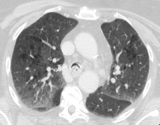
2 What is HRCT ACR defines HRCT as the use of thin section CT images (0.625 mm - 2 mm slice thickness) often with a high-spatial-frequency reconstruction algorithm Requesting physicians sometimes don t understand the definition of HRCT and may order it improperly HRCT does not have to, but also often includes expiratory and prone imaging
3 Benefits of HRCT Gold standard for evaluation of lung parenchyma and airways HRCT can distinguish between the causes of the mosaic attenuation pattern HRCT allows for dynamic evaluation of the airways HRCT findings in interstitial lung disease may have survival implications
4 HRCT Protocol 1) Standard 2.5 mm chest without contrast at full inspiration 1.25 mm images will also be reconstructed using bone algorithm and both sets of images will be sent to PACS 2) Supine expiratory images performed at 1.25 mm with 20 mm gaps, using bone algorithm 3) Prone inspiratory images performed at 1.25 mm with 20 mm gaps, using bone algorithm
5 HRCT Protocol Thin section inspiratory Fine detail of lung parenchyma and airways Volumetric images can be constructed Thin section expiratory Mosaic attenuation pattern Tracheobronchomalacia Thin section prone Atelectasis vs. interstitial lung disease
6 HRCT indications Indications Small airways vs. small vessel disease (mosaic attenuation pattern) Large airways Tracheobronchomalacia Bronchiectasis Restrictive lung diseases Idiopathic interstitial pneumonias Secondary diffuse lung disease

7 Mosaic Attenuation Variable areas of lung attenuation in lobular or multi-lobular distribution Mosaic pattern of fairly well defined areas of low and high attenuation lung is a result of disease demarcated by Secondary Pulmonary Lobule
8 Causes of Mosaic Attenuation in the Lung Small Airway Disease Vascular Disease Primary Parenchymal Disease Reversible (ex asthma) Thromboembolic disease Infectious Fixed (exobliterative bronchiolitis) Pulmonary arterial hypertension Non-infectious Neoplastic


9 Small Airway Disease AIR TRAPPING Abnormal Lung: Lower in attenuation. Cause: Air trapping and decreased blood flow, (combination of hypoxic vasoconstriction and mechanical pressure on vessels from air trapping). When process at lobular or multi-lobular level, mosaic pattern of attenuation results.
10 Small Airway Disease Differential Diagnosis: Reversible Asthma Fixed Bronchiolitis Obliterans (including bronchiectasis associated small airway disease) Example: Swyer-James Syndrome Cystic Fibrosis Allergic Bronchopulmonary Aspergillosis


11 Small Airway Disease CT Findings Expiratory CT scan shows alternating patchy areas of low and high attenuation consistent with air trapping in an individual with Bronchiolitis Obliterans Abnormal Lung Normal Lung
12 Small Airway Disease CT Findings Expiratory CT: Lower attenuation regions of lung: remain lucent show no or minimal change in volume due to air trapping May be necessary for detection of air trapping. Can be used to accentuate attenuation difference. Ancillary Findings: Bronchiectasis, mucoid impaction, tree-in-bud opacities
13 Inspiratory Expiratory Patient with Bronchiolitis Obliterans - Mosaic attenuation and air trapping only seen on expiratory views.
14 Causes of Mosaic Attenuation in the Lung Small Airway Disease Vascular Disease Primary Parenchymal Disease Reversible (ex asthma) Thromboembolic disease Infectious Fixed (exobliterative bronchiolitis) Pulmonary arterial hypertension Non-infectious Neoplastic

15 Vascular Lung Disease DIFFERENTIAL PERFUSION Abnormal Lung: Lower in attenuation. Cause: Decreased perfusion. Regions of oligemia adjacent to normal/hyperemic lung creates mosaic pattern. Mosaic perfusion or Mosaic oligemia are terms also used referring to this particular etiology.

16 Vascular Lung Disease CT Findings Patchy areas of high and low attenuation in a patient with Chronic Thromboembolic Disease Normal Lung Abnormal Lung
17 Vascular Lung Disease CT Findings Expiratory CT: Attenuation of both low and high attenuation lung increases in similar fashion. Volume of normal/abnormal lung decreases similarly. Ancillary Findings: Eccentric filling defects in pulmonary artery and its branches Arterial webs Pruning and/or stenoses Enlargement of main pulmonary artery
 lung.")
18 Elderly female with pulmonary artery hypertension secondary to chronic thromboembolic disease - Vessel number and caliber decreased in lower attenuation (oligemic) lung. - Mediastinal windows demonstrate eccentric filling defect consistent with chronic embolus and enlargement of main pulmonary artery.
19 Small Airways Disease vs. Vascular Disease Expiratory CT Inspiratory Expiratory Cystic Fibrosis Expiratory Image: - Lucent areas more pronounced suggesting air trapping. Inspiratory Expiratory Chronic Thromboembolic Disease Expiratory Image: - Opaque areas remain white while lucent areas remain dark. - Opaque and lucent areas of lungs decrease in size uniformly.
20 Causes of Mosaic Attenuation in the Lung Small Airway Disease Vascular Disease Primary Parenchymal Disease Reversible (ex asthma) Thromboembolic disease Infectious Fixed (exobliterative bronchiolitis) Pulmonary arterial hypertension Non-infectious Neoplastic

21 Primary Parenchymal Lung Disease PATCHY RETICULAR /AIRSPACE DISEASE Abnormal lung: Higher in attenuation. Cause: Partial filling of airspaces/interstitium with fluid, cells, fibrosis. Normal lung adjacent to diseased lung creates mosaic appearance.

22 Primary Parenchymal Lung Disease Filling of airspaces with fluid in patient with Pulmonary Edema creates mosaic appearance CT Findings Normal Lung Abnormal Lung
23 Primary Parenchymal Lung Differential Diagnosis: Infectious: Disease Pneumocystis carinii pneumonia, pyogenic pneumonia Noninfectious: Chronic eosinophilic pneumonia, hypersensitivity pneumonitis, cryptogenic organizing pneumonia, sarcoidosis, alveolar proteinosis, pulmonary edema Neoplastic: Bronchioloalveolar carcinoma, lymphoma

24 Primary Parenchymal Lung Disease Hypersensitivity Pneumonitis Pulmonary Hemorrhage Pulmonary Edema Bronchioloalveolar carcioma
25 Mosaic Attenuation Prominent CT Findings Normal Lung Abnormal Lung Vessel Number and Caliber in Low Attenuation Lung Air trapping on expiratory CT? Small Airways Disease Higher attenuation Lower attenuation decreased yes Vascular Disease Higher attenuation Lower attenuation decreased no Primary Parenchymal Disease Lower attenuation Higher attenuation No difference no

26 Large airways Tracheobronchomalacia is a condition caused by excessive collapsibility of the trachea/bronchi due to weakness of the airway walls or supporting cartilage Patients get chronic inflammation of their downstream small airways due to inability to clear secretions and improper coughing mechanism End-expiratory HRCT can evaluate for tracheobronchomalacia, although dynamic forced expiratory HRCT is the imaging gold standard

27 Tracheomalacia

28 Bronchomalacia Javidan-Nejad C, Radiol Clin North Am Jan;48(1):

29 Bronchiectasis Dipth

30 Diffuse lung disease (DLD) Idiopathic interstitial pneumonia UIP NSIP Secondary DLD Scleroderma Asbestosis Prone HRCT series helps for evaluation of basilar DLD

31 Prone Imaging Silva, C. I. S. et al. Am. J. Roentgenol. 2007;188:

32 UIP

33 NSIP Lynch D A et al. Radiology 2005;236:10-21
 Evaluation for interstitial lung disease, especially those with a basilar predominance Any reasonable request from an")
34 Conclusion At AMI, indications for HRCT include: Evaluation for small airway/small vessel disease (mosaic attenuation) Evaluation for large airway disease (tracheobronchomalacia, bronchiectasis) Evaluation for interstitial lung disease, especially those with a basilar predominance Any reasonable request from an ordering physician

35 Conclusion Mosaic attenuation pattern: air trapping vs. no air-trapping, with additional ancillary findings Tracheobronchomalacia < 50% AP change is normal 50-75% is a grey area > 75% is definitely abnormal Interstitial lung disease Reticulation at the base which resolves on prone imaging = atelectasis
36 References 1. Arakawa et al. Air Trapping on CT of Patients with Pulmonary Embolism. AJR 2002;178: Arakawa et al. Inhomogeneous Lung Attenuation at Thin- Section CT: Diagnostic Value of Expiratory Scans. Radiology 1998;206(1): Guckel et al. Mechanism of Mosaic Attenuation of the lungs on computed tomography in induced bronchospasm. Journal of Applied Physiology 1999:86(2); Hansell D. Small-Vessel Disease of the Lung: CT-Pathologic Correlates. Radiology 2002;225(3): Stern et al. CT Mosaic Pattern of Lung Attenuation: Distinguishing Different Causes. AJR 1995;165: Stern EJ and Frank MS. Small airways disease of the lungs: findings at expiratory CT. AJR 1994;163:37-41
Cystic Lung Diseases. Melissa Price Gillian Lieberman, MD Advanced Radiology Clerkship Beth Israel Deaconess Medical Center November, 2008
 Cystic Lung Diseases Melissa Price Gillian Lieberman, MD Advanced Radiology Clerkship Beth Israel Deaconess Medical Center November, 2008 How do we define a cyst of the lung? Hansell DM, Bankier AA, MacMahon
Radiological Findings in BO
 Radiological Findings in BO BO-Meeting 2016 Schloss Johannisberg Geisenheim - Rheingau Germany Dr. Simon Martin Department of Diagnostic and Interventional Radiology University Hospital Frankfurt Bronchiolitis
Expiratory CT: Correlation with Pulmonary Function Tests and Value for Discriminating Lung Diseases
 Original Article Expiratory CT: Correlation with Pulmonary Function Tests and Value for Discriminating Lung Diseases Ertuğrul Mavili 1, Hakan Büyükoğlan 2, Nurdan Bulut Çomu 1, Mustafa Güleç 1 Erciyes
Respiratory Concerns in Children with Down Syndrome
 Respiratory Concerns in Children with Down Syndrome Paul E. Moore, M.D. Associate Professor of Pediatrics and Pharmacology Director, Pediatric Allergy, Immunology, and Pulmonary Medicine Vanderbilt University
Role of HRCT in Diagnosis of Asbestos Related Pleuro Pulmonary Disease
 Cairo University Role of HRCT in Diagnosis of Asbestos Related Pleuro Pulmonary Disease ESSAY Submitted for partial fulfillment of Master degree in Radio-diagnosis By Rania Adel Abd ElRahem Elghetany (M.B.B.CH.,
Pulmonary Patterns VMA 976
 Pulmonary Patterns VMA 976 PULMONARY PATTERNS Which pulmonary patterns are commonly described in veterinary medicine? PULMONARY PATTERNS Normal Alveolar Interstitial Structured/Nodular Unstructured Bronchial
Pulmonary interstitium. Interstitial Lung Disease. Interstitial lung disease. Interstitial lung disease. Causes.
 Pulmonary interstitium Interstitial Lung Disease Alveolar lining cells (types 1 and 2) Thin elastin-rich connective component containing capillary blood vessels Interstitial lung disease Increase in interstitial
Defending the Rest Basics on Lung Cancer, Other Cancers and Asbestosis: Review of the B-Read and Pulmonary Function Testing
 Defending the Rest Basics on Lung Cancer, Other Cancers and Asbestosis: Review of the B-Read and Pulmonary Function Testing ASBESTOSIS November 2013 Bruce T. Bishop Lucy L. Brandon Willcox & Savage 440
Influenza (Flu) Influenza is a viral infection that may affect both the upper and lower respiratory tracts. There are three types of flu virus:
 Influenza is a viral infection that may affect both the upper and lower respiratory tracts. There are three types of flu virus:") Respiratory Disorders Bio 375 Pathophysiology General Manifestations of Respiratory Disease Sneezing is a reflex response to irritation in the upper respiratory tract and is associated with inflammation
Respiratory Disorders Bio 375 Pathophysiology General Manifestations of Respiratory Disease Sneezing is a reflex response to irritation in the upper respiratory tract and is associated with inflammation
Rheumatoid Arthritis related Lung Diseases: CT Findings 1
 Nobuyuki Tanaka, MD Jeung Sook Kim, MD John D. Newell, MD Kevin K. Brown, MD Carlyne D. Cool, MD Richard Meehan, MD Takuya Emoto, MD Tsuneo Matsumoto, MD David A. Lynch, MB Index terms: Arthritis, rheumatoid,
Recurrent or Persistent Pneumonia
 Recurrent or Persistent Pneumonia Lower Respiratory Tract Dr T Avenant Recurrent or Persistent Pneumonia Definitions Recurrent pneumonia more than two episodes of pneumonia in 18 months Persistent pneumonia
Is CT screening for asbestos-related diseases rational?
 Is CT screening for asbestos-related diseases rational? Narufumi Suganuma, M.D. 1 Yukinori Kusaka, M.D. 1 Harumi Itoh, M.D. 2 1 Division of Environmental Health, Department of International, Social and
Restrictive lung diseases
 Restrictive lung diseases Characterized by reduced compliance of the lung. Prominent changes in the interstitium (interstitial lung disease). Important signs and symptoms: - Dyspnea. - Hypoxia. - With
General Thoracic Surgery ICD9 to ICD10 Crosswalks. C34.11 Malignant neoplasm of upper lobe, right bronchus or lung
 ICD-9 Code ICD-9 Description ICD-10 Code ICD-10 Description 150.3 Malignant neoplasm of upper third of esophagus C15.3 Malignant neoplasm of upper third of esophagus 150.4 Malignant neoplasm of middle
Normal CT scan of the chest
 Normal CT scan of the chest Heart with left and right ventricle showing up lighter (contrast dye) Breast tissue Breast bone (sternum) Breast tissue Left lung (dark area) Right lung (dark area) Rib Main
The Floppy Airway- A Review of Tracheobronchomalacia in Adults
 Jay Pahade, MS IV Gillian Lieberman, MD The Floppy Airway- A Review of Tracheobronchomalacia in Adults Jay Pahade, MS IV University at Buffalo, School of Medicine Gillian Lieberman, MD Harvard School of
Primary -Benign - Malignant Secondary
 TUMOURS OF THE LUNG Primary -Benign - Malignant Secondary The incidence of lung cancer has been increasing almost logarithmically and is now reaching epidemic levels. The overall cure rate is very low
Tests. Pulmonary Functions
 Pulmonary Functions Tests Static lung functions volumes Dynamic lung functions volume and velocity Dynamic Tests Velocity dependent on Airway resistance Resistance of lung tissue to change in shape Dynamic
MECHINICAL VENTILATION S. Kache, MD
 MECHINICAL VENTILATION S. Kache, MD Spontaneous respiration vs. Mechanical ventilation Natural spontaneous ventilation occurs when the respiratory muscles, diaphragm and intercostal muscles pull on the
Restrictive vs. Obstructive Disease (Dedicated to my good friend Joe Walsh)
") Restrictive vs. Obstructive Disease (Dedicated to my good friend Joe Walsh) The field of medicine has a long history of describing or classifying disease. Pulmonary disease is no different. Although there
Restrictive vs. Obstructive Disease (Dedicated to my good friend Joe Walsh) The field of medicine has a long history of describing or classifying disease. Pulmonary disease is no different. Although there
Abuse of inhaled or intravenously injected illicit drugs
 PICTORIAL ESSAY Pulmonary Complications of Illicit Drug Use Differential Diagnosis Based on CT Findings Elsie T. Nguyen, MD, C. Isabela S. Silva, MD, PhD, Carolina A. Souza, MD, and Nestor L. Müller, MD,
Chest 1: Pulmonary Nodule Follow-up: Low-Dose Helical CT (Unenhanced) (Non-metastatic) Gantry Rotation Time. mas (Reg-Lg) 40-80
 (Non-metastatic) Gantry Rotation Time. mas (Reg-Lg) 40-80") Revisions Effective January 2012 Chest 1: Pulmonary Nodule Follow-up: Low-Dose Helical CT (Unenhanced) (Non-metastatic) Technologist Instructions Patient must cough several times prior to scan to clear
Revisions Effective January 2012 Chest 1: Pulmonary Nodule Follow-up: Low-Dose Helical CT (Unenhanced) (Non-metastatic) Technologist Instructions Patient must cough several times prior to scan to clear
Congestive Heart Failure
 William Herring, M.D. 2002 Congestive Heart Failure In Slide Show mode, to advance slides, press spacebar or click left mouse button Congestive Heart Failure Causes of Coronary artery disease Hypertension
The Fatal Pulmonary Artery Involvement in Behçet s Disease
 The Fatal Pulmonary Artery Involvement in Behçet s Disease Dr. Vedat Hamuryudan Div. Rheumatology, Dept. Internal Medicine Cerrahpasa Medical Faculty, University of Istanbul 33 years old man Sept 2011:
RESPIRATORY VENTILATION Page 1
 Page 1 VENTILATION PARAMETERS A. Lung Volumes 1. Basic volumes: elements a. Tidal Volume (V T, TV): volume of gas exchanged each breath; can change as ventilation pattern changes b. Inspiratory Reserve
How To Understand Aetiology Of Pulmonary Fibrosis
 Usual interstitial pneumonia-pattern fibrosis in surgical lung biopsies. Clinical, radiological and histopathological clues to aetiology Maxwell Smith, 1 Mercedes Dalurzo, 2 Prasad Panse, 3 James Parish,
PULMONARY PHYSIOLOGY
 I. Lung volumes PULMONARY PHYSIOLOGY American College of Surgeons SCC Review Course Christopher P. Michetti, MD, FACS and Forrest O. Moore, MD, FACS A. Tidal volume (TV) is the volume of air entering and
ACR STR PRACTICE PARAMETER FOR THE PERFORMANCE OF HIGH- RESOLUTION COMPUTED TOMOGRAPHY (HRCT) OF THE LUNGS IN ADULTS
 OF THE LUNGS IN ADULTS") The American College of Radiology, with more than 30,000 members, is the principal organization of radiologists, radiation oncologists, and clinical medical physicists in the United States. The College
The American College of Radiology, with more than 30,000 members, is the principal organization of radiologists, radiation oncologists, and clinical medical physicists in the United States. The College
Milano, 9-12 Luglio 2012 Hotel Enterprise
 Milano, 9-12 Luglio 2012 Hotel Enterprise Organized by: Endorsed by: BACKGROUND The HERMES National Summer School is an educational project organized by SIMeR and established with the goal to develop standards
Pathophysiology of hypercapnic and hypoxic respiratory failure and V/Q relationships. Dr.Alok Nath Department of Pulmonary Medicine PGIMER Chandigarh
 Pathophysiology of hypercapnic and hypoxic respiratory failure and V/Q relationships Dr.Alok Nath Department of Pulmonary Medicine PGIMER Chandigarh Jan 2006 Respiratory Failure inadequate blood oxygenation
CPT codes are for information only; consult your payer organization for reimbursement information.
 CPT codes are for information only; consult your payer organization for reimbursement information. Coverage for Spirometry/Oximetry Spirometry is a component of pulmonary function testing (PFTs). PFTs
Occupational Lung Disease. SS Visser Internal Medicine UP
 Occupational Lung Disease SS Visser Internal Medicine UP Classification Anorganic ( mineral ) dust/pneumoconiosis Fibrogenic - silica, asbestos, talc, silicates Non-fibrogenic - Fe, barium, tin Immunologic/Pharmcologic
Evaluation and treatment of emphysema in a preterm infant
 ISPUB.COM The Internet Journal of Pediatrics and Neonatology Volume 11 Number 1 Evaluation and treatment of emphysema in a preterm infant T Saad, P Chess, W Pegoli, P Katzman Citation T Saad, P Chess,
INTERSTITIAL LUNG DISEASE Paul F. Simonelli, MD, Ph.D.
 INTERSTITIAL LUNG DISEASE Paul F. Simonelli, MD, Ph.D. Definition: Interstitial lung disease (ILD) refers to a broad range of conditions that have common clinical, physiological, and radiological features.
Juvenile Dermatomyositis Joseph Junewick, MD FACR
 Juvenile Dermatomyositis Joseph Junewick, MD FACR 10/11/2015 History Child with several month history of weakness, arthralgias and palpable abnormalities at the knee Diagnosis Juvenile Dermatomyositis
Department of Surgery
 What is emphysema? 2004 Regents of the University of Michigan Emphysema is a chronic disease of the lungs characterized by thinning and overexpansion of the lung-like blisters (bullae) in the lung tissue.
The Global Alliance against Chronic Respiratory Diseases
 The Global Alliance against Chronic Respiratory Diseases Pulmonary hypertension Dr Marc Humbert What is the burden of pulmonary hypertension? The true burden of pulmonary hypertension is currently unknown
How To Pay For Respiratory Therapy Rehabilitation
 LCD ID Number L32748 LCD Title Respiratory Therapy Rehabilitation Contractor s Determination Number L32748 AMA CPT/ADA CDT Copyright Statement CPT only copyright 2002-2011 American Medical Association.
Understanding Hypoventilation and Its Treatment by Susan Agrawal
 www.complexchild.com Understanding Hypoventilation and Its Treatment by Susan Agrawal Most of us have a general understanding of what the term hyperventilation means, since hyperventilation, also called
2.06 Understand the functions and disorders of the respiratory system
 2.06 Understand the functions and disorders of the respiratory system 2.06 Understand the functions and disorders of the respiratory system Essential questions What are the functions of the respiratory
Pediatric Respiratory System: Basic Anatomy & Physiology. Jihad Zahraa Pediatric Intensivist Head of PICU, King Fahad Medical City
 Pediatric Respiratory System: Basic Anatomy & Physiology Jihad Zahraa Pediatric Intensivist Head of PICU, King Fahad Medical City Outline Introduction Developmental Anatomy Developmental Mechanics of Breathing
April 2015 CALGARY ZONE CLINICAL REFERENCE PULMONARY CENTRAL ACCESS & TRIAGE
 April 2015 CALGARY ZONE CLINICAL REFERENCE CENTRAL ACCESS & TRIAGE Introduction Pulmonary consulting services are organized through the Calgary Zone Pulmonary Central Access and Triage (PCAT). Working
Idiopathic Pulmonary Fibrosis (IPF) Research
 Research") Idiopathic Pulmonary Fibrosis (IPF): Why Early Referral is Critical Even if Your Patient is Not Eligible for a Clinical Trial Idiopathic Pulmonary Fibrosis (IPF) Research Management of IPF requires a confident
Idiopathic Pulmonary Fibrosis (IPF): Why Early Referral is Critical Even if Your Patient is Not Eligible for a Clinical Trial Idiopathic Pulmonary Fibrosis (IPF) Research Management of IPF requires a confident
Interstitial Lung Diseases ACOI Board Review 2013
 Interstitial Lung Diseases ACOI Board Review 2013 Thomas F. Morley, DO, FACOI, FCCP, FAASM Professor of Medicine Director of the Division of Pulmonary, Critical Care and Sleep Medicine UMDNJ-SOM Restrictive
Imaging of Acute Stroke. Noam Eshkar, M.D New Jersey Neuroscience Institute JFK Medical Center Edison Radiology Group
 Imaging of Acute Stroke Noam Eshkar, M.D New Jersey Neuroscience Institute JFK Medical Center Edison Radiology Group Modalities Non Contrast CT (NCCT) Contrast CT Angiography MRI MR Angiography Perfusion
Radiation-Induced Lung Injury
 May 2001 Radiation-Induced Lung Injury Warren Phipps, Harvard Medical School Year III Our Patient D.C. is a 50 year-old woman with a 30-pack year history of smoking who presented to the ED because she
Ventilation Perfusion Relationships
 Ventilation Perfusion Relationships VENTILATION PERFUSION RATIO Ideally, each alveolus in the lungs would receive the same amount of ventilation and pulmonary capillary blood flow (perfusion). In reality,
Francine Lortie-Monette, MD, MSc, CSPQ, MBA Department of Epidemiology and Biostatistics University of Western Ontario 2003
 ASBESTOS Francine Lortie-Monette, MD, MSc, CSPQ, MBA Department of Epidemiology and Biostatistics University of Western Ontario 2003 Asbestosis Asbestosis is a model for other dust diseases as well as
UKRC 2015 Dr Michael Sproule Glasgow
 UKRC 2015 Dr Michael Sproule Glasgow Radiology of Asbestos Related Lung Disease General term given to a group of fibrous minerals containing silica and a variety of other elements. Asbestos: Derived
Occupational Lung Disease. David Perlman, MD
 Occupational Lung Disease David Perlman, MD What causes occupational lung diseases? Breathing bad stuff into your lung Mechanism of particle deposition Large particles (>0.5μM) Impaction Gravitational
IV FOCAL AND MULTIFOCAL LUNG DISEASE
 IV FOCAL AND MULTIFOCAL LUNG DISEASE Roland H. Ingram, Jr., m.d. Chest physicians, when consulted, most often begin their consultation by examining standard posteroanterior and lateral chest radiographs.
The abnormal chest X-ray when to refer to a specialis t
 The abnormal chest X-ray when to refer to a specialis t An abnormal chest X-ray must be followed up. OLGA MZILENI, MB ChB, MMed (Int Med) Professor and Head of Internal Medicine and Pulmonology, University
Sunday. Cases of the Day. Track 1 Grand C. Track 2 Grand D. 7:30 8:00 AM Coffee and Pastries Grand Assembly
 April 8, 2001 General Session Grand I & J 7:30 8:00 AM Coffee and Pastries Grand Assembly Cases of the Day Moderator: Melissa L. Rosado de Christenson, MD 7:30 7:45 AM Case of the Day Kyung Soo Lee, MD
These factors increase your chance of developing emphysema. Tell your doctor if you have any of these risk factors:
 Emphysema Pronounced: em-fiss-see-mah by Debra Wood, RN En Español (Spanish Version) Definition Emphysema is a chronic obstructive disease of the lungs. The lungs contain millions of tiny air sacs called
CT scans and IV contrast (radiographic iodinated contrast) utilization in adults
 utilization in adults") CT scans and IV contrast (radiographic iodinated contrast) utilization in adults At United Radiology Group, a majority of CT exams are performed either with IV contrast or without while just a few exams
CT scans and IV contrast (radiographic iodinated contrast) utilization in adults At United Radiology Group, a majority of CT exams are performed either with IV contrast or without while just a few exams
Pulmonary manifestations of connective tissue diseases (SLE,RA, SS, PM/DM)
") Pulmonary manifestations of connective tissue diseases (SLE,RA, SS, PM/DM) Spectrum of pleuro pulmonary involvement in CTD Upper airway involvement cricoarytenoid joint arthritis aspiration pneumonia Smaller
Pulmonary manifestations of connective tissue diseases (SLE,RA, SS, PM/DM) Spectrum of pleuro pulmonary involvement in CTD Upper airway involvement cricoarytenoid joint arthritis aspiration pneumonia Smaller
PARTICLE SIZE AND CHEMISTRY:
 Pneumoconioses LW/Please note: This information is additional to Davidson s Principles and Practice of Medicine. /Hierdie inligting is aanvullend tot Davidson s Principles and Practice of Medicine. Pneumoconioses
Success and Survival in Pulmonary Rehab
 Success and Survival in Pulmonary Rehab 35 Years and Still Growing Valerie McLeod, RRT Manager, Pulmonary Rehabilitation McLaren Flint, MI Disclosure Information I have no disclosures. While some brands
Pulmonary Disorders. Chronic Obstructive Pulmonary Disease (COPD) Chronic Obstructive Pulmonary Disease (COPD)
 Chronic Obstructive Pulmonary Disease (COPD)") RCS 6080 Medical and Psychosocial Aspects of Rehabilitation Counseling Pulmonary Disorders Chronic Obstructive Pulmonary Disease (COPD) Characterized by decreased expiratory airflow Reduction in expiratory
RCS 6080 Medical and Psychosocial Aspects of Rehabilitation Counseling Pulmonary Disorders Chronic Obstructive Pulmonary Disease (COPD) Characterized by decreased expiratory airflow Reduction in expiratory
Diseases. Inflammations Non-inflammatory pleural effusions Pneumothorax Tumours
 Pleura Visceral pleura covers lungs and extends into fissures Parietal pleura limits mediastinum and covers dome of diaphragm and inner aspect of chest wall. Two layers between them (pleural cavity) contains
Interstitial lung disease
 Interstitial lung disease William D Travis Abstract Idiopathic interstitial pneumonias represent an important group of interstitial lung diseases, encompassing seven entities: (1) usual interstitial pneumonia
Oxford Centre for Respiratory Medicine Bronchial-Artery Embolisation Information for patients
 Oxford Centre for Respiratory Medicine Bronchial-Artery Embolisation Information for patients This leaflet tells you about the bronchial-artery embolisation procedure. It explains what is involved and
ACUTE RESPIRATORY DISTRESS SYNDROME (ARDS) S. Agarwal, MD, S. Kache MD
 S. Agarwal, MD, S. Kache MD") ACUTE RESPIRATORY DISTRESS SYNDROME (ARDS) S. Agarwal, MD, S. Kache MD Definition ARDS is a clinical syndrome of lung injury with hypoxic respiratory failure caused by intense pulmonary inflammation that
ACUTE RESPIRATORY DISTRESS SYNDROME (ARDS) S. Agarwal, MD, S. Kache MD Definition ARDS is a clinical syndrome of lung injury with hypoxic respiratory failure caused by intense pulmonary inflammation that
Linfoma maligno pulmonar tratado com Nerium oleander. http://www.drozel.org/eng/diagnosis_malignant_mg.htm CASE REPORT
 Linfoma maligno pulmonar tratado com Nerium oleander http://www.drozel.org/eng/diagnosis_malignant_mg.htm CASE REPORT Diagnosis: Malignant lymphoma, lung cancer A 60-year-old woman experienced pain in
Disease/Illness GUIDE TO ASBESTOS LUNG CANCER. What Is Asbestos Lung Cancer? www.simpsonmillar.co.uk Telephone 0844 858 3200
 GUIDE TO ASBESTOS LUNG CANCER What Is Asbestos Lung Cancer? Like tobacco smoking, exposure to asbestos can result in the development of lung cancer. Similarly, the risk of developing asbestos induced lung
LYMPHOMA IN DOGS. Diagnosis/Initial evaluation. Treatment and Prognosis
 LYMPHOMA IN DOGS Lymphoma is a relatively common cancer in dogs. It is a cancer of lymphocytes (a type of white blood cell) and lymphoid tissues. Lymphoid tissue is normally present in many places in the
IN THE WORKERS COMPENSATION COURT OF THE STATE OF MONTANA 2008 MTWCC 51. WCC No. 2008-2056 LUCILE KILGORE. Petitioner. vs.
 IN THE WORKERS COMPENSATION COURT OF THE STATE OF MONTANA 2008 MTWCC 51 WCC No. 2008-2056 LUCILE KILGORE Petitioner vs. TRANSPORTATION INSURANCE COMPANY Respondent/Insurer. FINDINGS OF FACT, CONCLUSIONS
Idiopathic Pulmonary Fibrosis
 Idiopathic Pulmonary Fibrosis What is Idiopathic Pulmonary Fibrosis? Idiopathic pulmonary fibrosis (IPF) is a condition that causes persistent and progressive scarring of the tiny air sacs (alveoli) in
by Lee S. Newman, M.D., and Cecile S. Rose, M.D., M.P.H.
 OCCUPATIONAL ASBESTOSIS AND RELATED DISEASES by Lee S. Newman, M.D., and Cecile S. Rose, M.D., M.P.H. A 63-year-old man consulted an internist complaining of dyspnea on exertion. He reported the following:
Computed Tomography (CT) - Chest
 - Chest") Scan for mobile link. Computed Tomography (CT) - Chest Computed tomography (CT) of the chest uses special x-ray equipment to examine abnormalities found in other imaging tests and to help diagnose the
Scan for mobile link. Computed Tomography (CT) - Chest Computed tomography (CT) of the chest uses special x-ray equipment to examine abnormalities found in other imaging tests and to help diagnose the
Respiratory failure and Oxygen Therapy
 Respiratory failure and Oxygen Therapy A patient with Hb 15 G % will carry 3X more O2 in his blood than someone with Hb 5G % Give Controlled O2 treatment in acute pulmonary oedema to avoid CO2 retention
Medicare C/D Medical Coverage Policy
 Nebulizer Medications Origination: June 17, 2009 Review Date: October 21, 2015 Next Review: October, 2017 Medicare C/D Medical Coverage Policy DESCRIPTION Nebulizer medications are used to prevent and
Tina Mosaferi, Harvard Medical School Year III Gillian Lieberman, MD
 July 2014 Tina Mosaferi, Harvard Medical School Year III 1. Our Patient-Introduction 2. Asbestos Basics 3. Pulmonary Findings Manifestations demonstrated by companion patients 4. Our patient-conclusion
X-ray (Radiography) - Chest
 - Chest") Scan for mobile link. X-ray (Radiography) - Chest What is a Chest X-ray (Chest Radiography)? The chest x-ray is the most commonly performed diagnostic x-ray examination. A chest x-ray produces images of
Scan for mobile link. X-ray (Radiography) - Chest What is a Chest X-ray (Chest Radiography)? The chest x-ray is the most commonly performed diagnostic x-ray examination. A chest x-ray produces images of
2011 Pulmonary Pathology Society Biennial Meeting Program
 2011 Pulmonary Pathology Society Biennial Meeting Program Thursday, August 18, 2011 7:00 AM Registration and Continental Breakfast 7:45 AM Welcome: Donald Guinee / Elisabeth Brambilla 8:00 AM Interstitial
Cardiac Masses and Tumors
 Cardiac Masses and Tumors Question: What is the diagnosis? A. Aortic valve myxoma B. Papillary fibroelastoma C. Vegetation from Infective endocarditis D. Thrombus in transit E. None of the above Answer:
Multi-slice Helical CT Scanning of the Chest
 Multi-slice Helical CT Scanning of the Chest Comparison of different low-dose acquisitions Lung cancer is the main cause of deaths due to cancer in human males and the incidence is constantly increasing.
A912: Kidney, Renal cell carcinoma
 A912: Kidney, Renal cell carcinoma General facts of kidney cancer Renal cell carcinoma, a form of kidney cancer that involves cancerous changes in the cells of the renal tubule, is the most common type
Pulmonary infections are an important cause of morbidity and
 Diagn Interv Radiol 2008; 14:75-82 Turkish Society of Radiology 2008 CHEST IMAGING ORIGINAL ARTICLE CT findings in immunocompromised patients with pulmonary infections Figen Başaran Demirkazık, Aylin Akın,
Interpretation of Pulmonary Function Tests
 Interpretation of Pulmonary Function Tests Dr. Sally Osborne Cellular & Physiological Sciences University of British Columbia Room 3602, D.H Copp building 604 822-3421 [email protected] www.sallyosborne.com
A Diagnostic Chest XRay: Multiple Myeloma
 Daniela Marinho Tridente, VI FCMSCSP October 2013 A Diagnostic Chest XRay: Multiple Myeloma Daniela Marinho Tridente, VI FCMSCSP Our Learning Agenda Introduction of our patient His imaging data and findings
UNIVERSITA' DEGLI STUDI DI ROMA TOR VERGATA
 SYSTEMATIC PATHOLOGY I IIIYear Scientific Field DISCIPLINE TUTOR Systematic Pathology I MED/21 MED/10 Thoracic Surgery Respiratory Diseases Tommaso Claudio Mineo Paola Rogliani MED/10 Respiratory Diseases
Radiologic Evaluation of Renal Cysts
 Eugene K. Cha, HMS III November 2004 Radiologic Evaluation of Renal Cysts Eugene K. Cha, Harvard Medical School III Renal Anatomy Netter FH. Atlas of Human Anatomy, Second Edition. 2001, p 313. 2 HPI:
Low-dose CT for Pulmonary Embolism
 Low-dose CT for Pulmonary Embolism Gautham Gautham P. P. Reddy, Reddy, MD, MD, MPH MPH University University of of Washington Washington Introduction Introduction CT CT accounts accounts for for > 50%
Suspected pulmonary embolism (PE) in pregnant women
 in pregnant women") Suspected pulmonary embolism (PE) in pregnant women What is a pulmonary embolus? A deep vein thrombosis (DVT) is a blood clot that forms in one of the deep veins of the leg. If the clot moves to the lung,
Suspected pulmonary embolism (PE) in pregnant women What is a pulmonary embolus? A deep vein thrombosis (DVT) is a blood clot that forms in one of the deep veins of the leg. If the clot moves to the lung,
Review Article Imaging Diagnosis of Interstitial Pneumonia with Emphysema (Combined Pulmonary Fibrosis and Emphysema)
") Volume 2012, Article ID 816541, 9 pages doi:10.1155/2012/816541 Review Article Imaging Diagnosis of Interstitial Pneumonia with Emphysema (Combined Pulmonary Fibrosis and Emphysema) Fumikazu Sakai, 1 Junya
Volume 2012, Article ID 816541, 9 pages doi:10.1155/2012/816541 Review Article Imaging Diagnosis of Interstitial Pneumonia with Emphysema (Combined Pulmonary Fibrosis and Emphysema) Fumikazu Sakai, 1 Junya
Leanne M Poulos Patricia K Correll Brett G Toelle Helen K Reddel Guy B Marks Woolcock Institute of Medical Research, University of Sydney, NSW
 Lung disease in Australia Leanne M Poulos Patricia K Correll Brett G Toelle Helen K Reddel Guy B Marks Woolcock Institute of Medical Research, University of Sydney, NSW Prepared for Lung Foundation Australia