Imaging of Pulmonary Nodules. Bradley R. Trotter, MD, DABR, DABNM Department of Radiology
|
|
|
- Dorothy Green
- 7 years ago
- Views:
Transcription
1 Imaging of Pulmonary Nodules Bradley R. Trotter, MD, DABR, DABNM Department of Radiology

2 Imaging of Pulmonary Nodules: Objectives Review imaging modalities and their use Describe imaging features that either decrease or increase the likelihood of malignancy Present a framework for integration of imaging and clinical information to assist in the management of pulmonary nodules on an individual patient basis

3 Imaging of Pulmonary Nodules: Overview Background: From Nodules to Lung Cancer Review of Imaging Modalities: Tools of the Trade Review of Imaging Features: To benign or not to benign, that is the question. Likelihood of Malignancy: damned lies, and statistics Recommendations: Putting it together

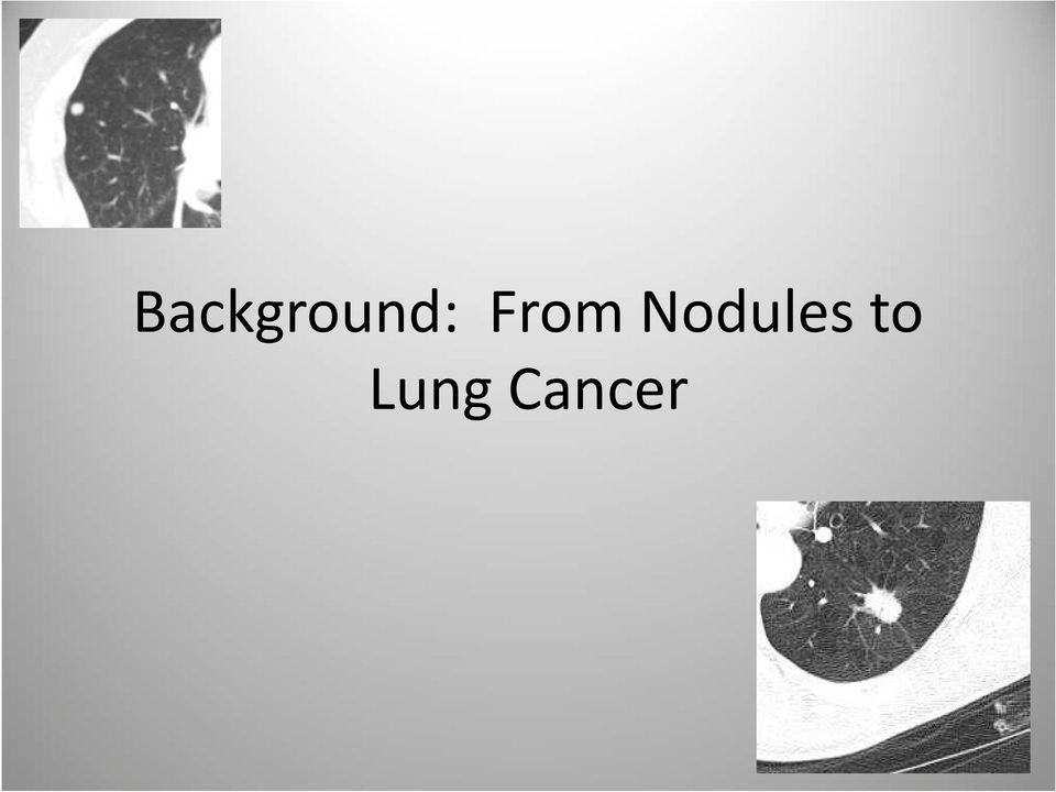
4 Background: From Nodules to Lung Cancer
5 Background: Pulmonary Nodules A round opacity, at least moderately well marginated, and no greater than 3 cm in maximum diameter Round meaning roughly circular or oval shaped but also spherical in its 3- dimensional nature (not flat or plaque-like) Completely surrounded by lung parenchyma and not associated with adenopathy, atelectasis, or pneumonia

6 Background: Pulmonary Nodules May be solitary (SPN) or multiple, which affects likelihood of various differential diagnostic considerations When multiple, imaging features of each nodule identified must be considered, in addition to consideration as a whole Our discussion primarily relates to solitary (or clearly dominant) pulmonary nodules, but is also useful in the setting of multiple nodules
 pulmonary nodules, but is also useful in the setting of")
7 Background: Pulmonary Nodules Very common imaging finding, particularly since advent of helical and multidetector CT Studies report prevalence of one or more nodules from 8% to 69% on CT Vast majority (95%-98% in most studies) are benign
 are")
8 Background: Lung Cancer Our discussion of pulmonary nodules ultimately falls within the context of lung cancer, which directly influences our management decisions regarding nodules A few points are worth noting for our discussion

9 Background: Lung Cancer It is, overall, an aggressive disease: More people in the United States die from lung cancer than from any other type of cancer. This is true for both men and women.

10 Background: Lung Cancer It is a heterogeneous disease: Non-small cell lung cancer (NSCLC) 80%-85% of lung cancer Can be detected at earlier stages with CT possible role for screening with CT NSCLC is our primary focus when discussing imaging of pulmonary nodules SCLC Detection of limited-stage disease unlikely due to its aggressive nature Not the focus of our discussion of nodules

11 Background: Lung Cancer There are known risk factors: Smoking: Accounts for 90% of lung cancers in the U. S. 15x to 30x more likely to develop or die from lung cancer than nonsmokers Risk increases with degree and duration (pack years) Currently estimated 94 million current or former smokers in the U. S. at increased risk
 Currently estimated 94 million")
12 Background: Lung Cancer Other known risk factors include: Second hand smoke Asbestos, radon, or uranium exposure Radiation therapy to the thorax, such as with lymphoma or breast cancer Family history of lung cancer in 1 st degree relative Age: risk increases with age; rare under age 35 Chronic lung diseases Personal history of prior lung cancer

13 Background: Lung Cancer It is a costly disease: Estimated impact on U. S. economy is over $300 billion annually As much as possible, management decisions should include consideration of cost effectiveness that is based on proven clinical outcomes research

14 Review of Imaging Modalities: Tools of the Trade

15 Review of Imaging Modalities: Chest radiograph (CXR) Computed Tomography (CT) Positron Emission Tomography (PET) PET-CT (no, it s not the same thing) (Flouroscopy, MRI, US)
")
16 Low cost Review of Imaging Modalities: Chest Radiograph Low radiation exposure Low utility as primary imaging modality of pulmonary nodules No utility for lung cancer screening

17 Review of Imaging Modalities: Chest Radiograph However, CXR is a common entry point into the evaluation of pulmonary nodules when detected incidentally Prevalence of nodules on CXR is about 0.2% to 9% depending on population

18 Review of Imaging Modalities: Computed Tomography Tomographs (slices) eliminate the problem of superimposed structures on radiographs Volumetric data acquisition on modern scanners allows slice reconstruction in any plane Highly sensitive for detection of pulmonary nodules as small as 1-2 mm Unfortunately limited specificity Can be performed with or without IV contrast enhancement

19 Review of Imaging Modalities: Computed Tomography High spatial and contrast resolution allow determination of important morphologic features of nodules Thin-sections (1-3 mm slice thickness) should be utilized for evaluation of nodule morphology (IV contrast unnecessary)

20 Review of Imaging Modalities: Computed Tomography IV contrast-enhanced densitometry can be performed to assess nodule enhancement characteristics More accurate determination of important ancillary findings: adenopathy, bronchial involvement, pleural involvement, etc. Low-dose (radiation) imaging may be used for any needed follow-up exams (or screening)
 imaging may be used for any needed follow-up exams (or")
21 Review of Imaging Modalities: Positron Emission Tomography IV administration of 18F-fluorodeoxyglucose (FDG) Degree of tissue uptake reflects its relative metabolic activity (glucose) Many malignancies demonstrate significantly greater uptake of FDG compared with normal tissues and appear hot on PET images >90% sensitivity for nodules 1-3 cm; lower specificity of about 80%
22 Review of Imaging Modalities: PET-CT It s like Reese s Peanut Butter Cups It s a PET scan with an anatomic contrast agent It s a CT scan with a metabolic contrast agent Either way, combines benefits of both modalities with higher sensitivity and specificity than for either PET or CT alone
23 Review of Imaging Modalities: Flouroscopy: Biopsy guidance Other Can be used as an inexpensive problem solving tool MRI has little role in imaging of pulmonary nodules but has utility in the evaluation of thoracic malignancies Ultrasound has little role in imaging of pulmonary nodules although may occasionally be used for biopsy localization
24 Review of Imaging Features: To benign or not to benign, that is the question.
25 Review of Imaging Features: To benign or not to benign There are a few imaging features that allow for a confidant benign diagnosis None are reliably diagnostic of malignancy
26 Idealistic: Review of Imaging Features: Goals of Imaging Early and definitive identification of all malignant nodules, and thereby improve patient outcomes Definitively identify all benign nodules, and thereby avoid the morbidity and cost of invasive procedures or further imaging that provide no true benefit Disappointingly, this remains elusive despite extensive experience and research
27 Realistic: Review of Imaging Features: Goals of Imaging Determine which nodules are benign and need no further evaluation Determine which are suspicious for malignancy and refer for definitive resolution For nodules that remain indeterminate: Determine which require biopsy Determine which require follow-up
28 Review of Imaging Features: Benign Benign pattern of calcification Stability of size Presence of fat (Need to keep in mind the definition of a pulmonary nodule)
29 Review of Imaging Features: Benign Benign patterns of calcification in a smoothly marginated nodule (or smoothly lobulated if hamartoma): Diffuse (granuloma) Central (granuloma) Laminated or concentric (granuloma) Popcorn (hamartoma)
30 Review of Imaging Features: Benign Any other pattern of calcification is indeterminate Caveat : patients with history of bone malignancy may have calcified nodules that resemble benign granulomas
31 Review of Imaging Features: Stability of size: Benign Comparison with older exams is essential in evaluation of pulmonary nodules (retrospect?) Measurement accuracy is critical: For spherical structures, a diameter increase of only 26% is a doubling of volume: Equates to a mere 1 mm diameter increase for a 4mm nodule!
32 Review of Imaging Features: Stability of size: Benign Especially for smaller nodules, visual comparison has been shown inaccurate, and physical measurement should be performed Measurement is most accurate on soft-copy images with electronic calipers Automated volumetric analysis software has promise to allow for more precise determination of nodule growth
33 Review of Imaging Features: Stability of size: Benign Any enlargement compared with baseline measurement considered suspicious and should prompt further evaluation Caution: cancers may occasionally demonstrate temporary decrease of overall size a single follow-up study demonstrating decrease of size may not be adequate to confirm a benign etiology
34 Review of Imaging Features: Benign Stability of size: 2 years of stability widely accepted as consistent with benign etiology based on studies of volume doubling-time of benign versus malignant lesions: days typical of malignant nodules with median about days However, reported range is actually quite broad Caveat: subsolid (ground-glass opacity) nodules that are malignant are more likely to have significantly longer doubling time and should undergo longer surveillance (3+ years)
35 Review of Imaging Features: Benign CT 2004 CT 2012
36 Review of Imaging Features: Benign Presence of fat: Within a smooth or smoothly lobulated SPN essentially diagnostic of hamartoma Present in approximately 60% Caution: be certain the lesion contains fat as artifact from volume averaging may mimic Caveat : metastases from liposarcoma and renal cell carcinoma may occasionally contain fat
37 Review of Imaging Features: Benign
38 Review of Imaging Features: Probably Benign Other probably benign imaging findings: FDG PET standardized uptake value (SUV) <2.5 Clustered nodules Very small size (<4 mm <<1%) Concave margins Follow-up CT imaging
39 Review of Imaging Features: Mimics Some benign pulmonary imaging findings may mimic a nodule (experience or cojones): Arteriovenous malformation feeding and draining vessels Mucocele tubular branching mucus filled dilated bronchi Rounded atelectasis comet tail appearance and associated with pleural thickening Flat lesions scarring that may be appreciated with multiplanar imaging
40 Review of Imaging Features: Mimics Rounded atelectasis Flat lesion/scar
41 Review of Imaging Features: Mimics Mucocele Mucocele
42 Review of Imaging Features: Suspicious for Malignancy Larger size: as size approaches 3 cm, likelihood of malignancy approaches 90% Spiculated margin: approximately 90% predictive value for cancer Lobulated margin more suspicious than smooth
43 Review of Imaging Features: Suspicious for Malignancy Subsolid attenuation with ground-glass opacity (GGO) in part or entirety: 34%-43% of GGO nodules malignant 40%-50% of mixed GGO/solid nodules <1.5 cm cancer and likelihood increases with size Cavitation with thick walls: 84%-95% cancer if wall thickness >16 mm 95% benign when wall thickness <4 mm
44 Review of Imaging Features: Suspicious for Malignancy Air containing: air bronchograms, air bronchiolograms, and air filled cystic spaces occur more commonly in malignant than benign nodules Upper lobe location: 70% of cancers in upper lobes; also 1.5x more likely in right lung
45 Review of Imaging Features: Suspicious for Malignancy These findings prompt further evaluation
46 Review of Imaging Features: FDG PET-CT Further Imaging Evaluation Usable for nodules >8-10 mm diameter Caveat: Because volume averaging from motion can artificially decrease apparent FDG activity, PET may be less useful for nodules located near diaphragm or heart, especially if small
47 Review of Imaging Features: FDG PET-CT Further Imaging Evaluation Test result determined by degree of activity in nodule relative to other body structures: Subjective visual evaluation Quantitative evaluation (SUV 2.5) Much more reliable for solid than GGO nodules: >90% sensitivity for cancer in solid nodules (with exception of carcinoid tumor probably 50%) Probably <50% sensitive for purely GGO cancers such as adenocarcinoma in situ (formerly BAC)
48 Review of Imaging Features: Further Imaging Evaluation Solid nodule GGO nodule
49 Review of Imaging Features: FDG PET-CT Further Imaging Evaluation Excellent test when used appropriately but probably a tendency for overutilization Questions to consider: Is it uncertain whether to simply watch and wait or to proceed with biopsy/resection? If yes then Will the PET result determine whether or not to proceed with biopsy/resection? If yes then Proceed with PET imaging
50 Review of Imaging Features: FDG PET-CT Further Imaging Evaluation Most useful and cost effective when: Low to moderate (5%-60%) pre-test probability of malignancy Clinical risk assessment and nodule morphologic characteristics are discordant Indeterminate nodule in a high risk patient
51 Review of Imaging Features: FDG PET-CT Further Imaging Evaluation May be useful in other situations but can be an unnecessary and added expense be judicious: If very low likelihood of malignancy (<5%), PET is not needed to justify observation (watch and wait) with follow-up CTs If high likelihood of malignancy (>60%), PET is not needed to justify biopsy/resection
52 Review of Imaging Features: FDG PET-CT Further Imaging Evaluation All PET negative nodules should be observed with follow-up CTs to confirm stability for at least 2 years: If grow then biopsy
53 Review of Imaging Features: Further Imaging Evaluation Contrast Enhanced CT Densitometry: Usable for nodules at least 10 mm diameter without cavitation or central necrosis Compare baseline unenhanced attenuation with peak contrast-enhanced attenuation <15 Hounsfield units (HU) enhancement is essentially diagnostic of benign etiology (99%) >15 HU is nonspecific Ask your doctor if this test is right for you
54 Likelihood of Malignancy: damned lies, and statistics
55 Likelihood of Malignancy Determining the statistical probability of malignancy for a given nodule is essential to proper management, including decisions regarding use of imaging studies: Qualitatively by an experienced clinician there is probably a tendency to overestimate the probability of malignancy in low risk patients Quantitatively using mathematical model
56 Likelihood of Malignancy: Logistic Regression Model Mayo clinic study using multiple logistic regression analysis identified 6 independent predictors: 3 clinical: Age, smoking, and history of prior extrathoracic cancer more than 5 years earlier 3 Imaging related: Nodule diameter, spiculated margin, and upper lobe location
57 Likelihood of Malignancy: Logistic Regression Model Probability of Malignancy = eˣ/(1+eˣ) Where x = ( x Years of age) + ( x Diameter in mm) if smoker if prior extra-thoracic cancer >5 yrs ago if spiculated margin if upper lobe
58 Likelihood of Malignancy: Logistic Regression Model 40 year old, nonsmoker, no prior malignancy, with a 5 mm smoothly marginated nodule in a lower lobe: Probability of malignancy = 0.9% 65 year old, smoker, no prior malignancy, with a 15 mm spiculated nodule in an upper lobe: Probability of malignancy = 55% 70 year old, smoker, no prior malignancy, with a 25 mm spiculated nodule in an upper lobe: Probability of malignancy = 87%
59 Likelihood of Malignancy: Bayesian Analysis Uses validated likelihood ratios for various independent clinical and imaging variables to estimate the probability of malignancy Based on Bayes Theorem: New odds = Prior odds x Likelihood Ratio Likelihood ratios >1 increase the probability of malignancy while ratios <1 lower it
60 Likelihood of Malignancy: Bayesian Analysis Clinical variables increasing probability: Age >50 years Smoking history 30 pack years Hemoptysis History of prior malignancy
61 Likelihood of Malignancy: Bayesian Analysis Imaging variables increasing probability: Diameter >2.0 cm Upper or middle lobe location Spiculated margin Thick walled cavitation Absence of calcification FDG PET SUV >2.5 CT densitometry enhancement >15 HU
62 Likelihood of Malignancy: SPN Calculator An easily accessible and useable SPN Calculator with both the Bayesian analysis and logistic regression models can be found on the web at: Extremely cool so check it out
63
64 Recommendations: Putting It Together
65 Recommendations: Clinical management of an imaging finding Distill the clinical and imaging variables and formulate a plan of action that is broadly applicable and adheres to the standard of care
66 Recommendations: First things first: Comparison with old imaging studies cannot be overemphasized! (Turn on the retrospectoscope) May obviate need for any further expensive and potentially harmful evaluation. Team effort of clinician, radiologist, and patient. In general, clearly growing nodules should move to tissue diagnosis if not contraindicated If suspected infectious etiology, further diagnostic intervention, therapy, and short-term follow-up imaging may be best initial management
67 Recommendations: Three management categories: Management of small solid nodules 8-10 mm Management of subsolid nodules Management of larger solid nodules >8-10 mm
68 Small Solid Nodules 8-10 mm
69 Recommendations: Small Solid Nodules 8-10 mm 2005 Fleischner Society guidelines for management of small nodules have been widely adopted Apply to: Incidentally detected solid nodules on CT Patients 35 years of age and older Do not apply to: Patient with known or suspected malignancy Patient with unexplained fever
70 Recommendations: Small Solid Nodules 8-10 mm MacMahon H, et al. Guidelines for management of small pulmonary nodules detected on CT scans: a statement from the Fleischner Society. Radiology 2005; 237:
71 Recommendations: Small Solid Nodules 8-10 mm A few key rationales for Fleischner Society Guidelines (FSG): Follow-up intervals for low and high risk categories differ because malignant nodules generally grow faster in smokers Even in smokers, <1% of nodules <4 mm will become lethal cancers, but this increases to 1020% for nodules about 8 mm
72 Recommendations: Small Solid Nodules 8-10 mm A few points regarding FSG: Are for managing nodules 8 mm and not meant to define management of larger nodules FSG clearly indicate flexibility in this category 2005 FSG do not distinguish between single and multiple nodules although primarily focused on solitary solid nodules Size is average of length and width Different management of subsolid nodules
73 Recommendations: Small Solid Nodules 8-10 mm Other patient groups: Patients <35 years old: In general, follow-up CT imaging should be avoided Consider a 6-12 month follow-up CT if have known malignancy Patients with known or suspected malignancy fall outside of the 2005 FSG (not part of the high risk group) and should be managed according to the specific clinical situation or protocol
74 Recommendations: Small Solid Nodules 8-10 mm Additional considerations: For nodules detected incidentally on a CT exam that did not include the entire thorax and future imaging follow-up is planned, consider first obtaining a dedicated CT thorax to assess for other nodules (no consensus) Consider limited coverage on follow-up CTs to reduce radiation exposure
75 Subsolid Nodules
76 Recommendations: Subsolid Nodules Complementary report to Fleischner Society Guidelines regarding management of subsolid nodules published January 2013 Apply to purely GG nodules and part-solid GG nodules and management differs for each Necessarily more varied due to the greater complexity of these lesions
77 Recommendations: Subsolid Nodules Unlike 2005 FSG for small solid nodules: Management does not differentiate smokers and non-smokers Management differs for solitary versus multiple nodules Known or suspected extra-thoracic malignancy does not preclude application No age distinction
78 Recommendations: Subsolid Nodules Recommendations for the Management of Subsolid Pulmonary Nodules Detected at CT: A Statement from the Fleischner Society (Naidich DP, et al. Radiology 2013; 266(1): ) Note. These guidelines assume meticulous evaluation, optimally with contiguous thin sections (1 mm) reconstructed with narrow and/or mediastinal windows to evaluate the solid component and wide and/or lung windows to evaluate the nonsolid component of nodules, if indicated. When electronic calipers are used, bidimensional measurements of both the solid and ground-glass components of lesions should be obtained as necessary. The use of a consistent low-dose technique is recommended, especially in cases for which prolonged follow-up is recommended, particularly in younger patients. With serial scans, always compare with the original baseline study to detect subtle indolent growth.
79 Recommendations: Subsolid Nodules A few key rationales for 2013 FSG: Adenocarcinomas (often subsolid) occur more frequently in younger and nonsmoking individuals Presence of multiple subsolid nodules is frequently encountered Purely GGO nodules are rarely metastatic cancer Subsolid nodules may resolve at short term follow-up Accurate nonsurgical diagnosis is problematic
80 Recommendations: Subsolid Nodules A few key rationales for 2013 FSG: Evidence that delay in surgical resection of slowgrowing pure GGNs does not alter outcome Part-solid nodules that persist are considered malignant until proved otherwise Larger solid components increase likelihood of malignancy and invasive disease (5 mm threshold ) Multiple subsolid nodules that are neoplastic are more likely synchronous primary lesions
81 Recommendations: Subsolid Nodules A few points regarding 2013 FSG: Work in progress, many controversial variables Meticulous evaluation, best with 1 mm slices Consistent CT and measurement technique between studies Size is average of long and short axis dimensions GGO measured on lung window Solid parts measured on mediastinal window Throughout follow-up, sizes should always be compared with the baseline CT
82 Larger Solid Nodules >8-10 mm
83 Recommendations: Larger Solid Nodules >8-10 mm Key imaging step is morphologic characterization with thin-section ( 3 mm) CT: Clearly benign features? If not, detailed accounting of morphology Key clinical step is determining probability of malignancy: Accounting of relevant clinical history Estimate or calculate the probability
84 Recommendations: Larger Solid Nodules >8-10 mm More diagnostic options become feasible with larger nodules: Advanced imaging with FDG PET or contrast enhanced CT densitometry Percutaneous needle biopsy or bronchoscopy Many management algorithms in the literature most are very similar Best integrate clinical and imaging variables
85 A management algorithm for patients with SPNs >8 mm and <30 mm in diameter. Adapted from: Evaluation of patients with pulmonary nodules: when is it lung cancer?, ACCP evidence-based clinical practice guidelines (2nd ed.). Chest 2007; 132: 108S-130S
86 Recommendations: Larger Solid Nodules >8-10 mm Ask your doctor if this test is right for you Details of application depend on available local resources, expertise, and the wishes of the fully informed patient
87 Thank you for your attention
88 Resources cdc.gov Chestx-ray.com Gould MK, et al. Evaluation of patients with pulmonary nodules: when is it lung cancer?, ACCP evidence-based clinical practice guidelines (2nd ed.). Chest 2007; 132: 108S-130S MacMahon H, et al. Guidelines for management of small pulmonary nodules detected on CT scans: a statement from the Fleischner Society. Radiology 2005; 237: NLST Research Team. Reduced lung cancer mortality with low-dose computed tomographic screening. N Engl J Med 2011; 365: Winer-Muram HT. The solitary pulmonary nodule. Radiology 2006; 239: Nicholas E, et al. Evaluation of the solitary pulmonary nodule: A practical approach. Applied Radiology Vol 40, No 12: 6-15 Swenson SJ, et al. The probability of malignancy in solitary pulmonary nodules: application to small radiologically indeterminate nodules. Arch Intern Med 1997; 157: Naidich DP, et al. Recommendations for the management of subsolid pulmonary nodules detected at CT: a statement from the Fleischner Society. Radiology 2013; 266(1):
89 Once upon a time An example of the intersection of imaging and clinical management
90 Once upon a time 67 year old former smoker who reports a past history of pneumonia and secondary basilar scarring CXR 11/2005
91 Once upon a time CT 11/2005: LLL 2.4 x 1.4 cm irregular nodule worrisome for neoplasm Moderate to high risk of malignancy Bronchoscopy done: no malignant cells
92 Once upon a time PET 12/2005: No hypermetabolic activity decreases likelihood of malignancy but not excluded, and short term follow-up CT imaging recommended CT bx planned but cancelled after noting possible decreased size
93 Once upon a time Follow-up CT 2/2006: 2.1 x 1.6 cm stable to slightly decreased size Life goes on
94 Once upon a time Nodule is intermittently seen on abdominal CTs obtained for other reasons, with fluctuating size measurements: 1.8 cm (follow-up recommended) 1.3 x 2.2 cm ( smaller from 2005 ) 1.9 x 2.4 cm (fluctuating size,? atelectasis, chronic infection, or malignancy)
95 Once upon a time CT 2005 CT 2008
96 Once upon a time CT 1/2011: 2.7 x 2.9 cm malignancy diagnosis of exclusion CT bx 2/2011: path not definitive but papillary architecture suggests neoplasm and lowgrade adenocarcinoma not excluded; surgical excision may be needed for definitive dx
97 Once upon a time 3/2011 Whole Body PET-CT: hypermetabolic and highly suspicious of malignancy No PET evidence of local or distant metastatic disease
98 Once upon a time Mediastinoscopy and VATS 4/2011 Partial lobectomy path: 5.0 x 2.5 x 2.5 cm adenocarcinoma mucinous, partially papillary Mediastinal lymph nodes path: Negative T2 N0 M0, Stage IB NSCLC
99 Once upon a time Surveillance CT 4/2012: 1.0 cm nodule at postoperative site PET-CT 4/2012: No hypermetabolism, but small size and basilar location with respiratory motion may reduce reliability of PET evaluation for the nodule CT bx 5/2012: Negative Follow-up CT 8/2012: Stable postoperative changes without evidence of recurrence
Low-dose CT Imaging. Edgar Fearnow, M.D. Section Chief, Computed Tomography, Lancaster General Hospital
 Lung Cancer Screening with Low-dose CT Imaging Edgar Fearnow, M.D. Section Chief, Computed Tomography, Lancaster General Hospital Despite recent declines in the incidence of lung cancer related to the
OPTIMIZING PATIENT EXPOSURE TO IONIZING RADIATION (OPEIR) MEASURES GROUP OVERVIEW 2015 PQRS OPTIONS FOR MEASURES GROUPS:
 MEASURES GROUP OVERVIEW 2015 PQRS OPTIONS FOR MEASURES GROUPS:") OPTIMIZING PATIENT EXPOSURE TO IONIZING RADIATION (OPEIR) MEASURES GROUP OVERVIEW 2015 PQRS OPTIONS F MEASURES GROUPS: 2015 PQRS MEASURES IN OPTIMIZING PATIENT EXPOSURE TO IONIZING RADIATION (OPEIR) MEASURES
OPTIMIZING PATIENT EXPOSURE TO IONIZING RADIATION (OPEIR) MEASURES GROUP OVERVIEW 2015 PQRS OPTIONS F MEASURES GROUPS: 2015 PQRS MEASURES IN OPTIMIZING PATIENT EXPOSURE TO IONIZING RADIATION (OPEIR) MEASURES
Patient sample criteria for the OPEIR Measures Group are all patients regardless of age, that have a specific CT procedure performed:
 OPTIMIZING PATIENT EXPOSURE TO IONIZING RADIATION (OPEIR) MEASURES GROUP OVERVIEW 2016 PQRS OPTIONS F MEASURES GROUPS: 2016 PQRS MEASURES IN OPTIMIZING PATIENT EXPOSURE TO IONIZING RADIATION (OPEIR) MEASURES
UNDERSTANDING SERIES LUNG NODULES. 1-800-298-2436 LungCancerAlliance.org
 UNDERSTANDING SERIES LUNG NODULES 1-800-298-2436 LungCancerAlliance.org CONTENTS What is a Nodule?...2 Finding Nodules...3 If a Nodule is Found... 4 What Happens Next?...6 Questions to Ask about Your Results...7
LUNG CANCER SCREENING: UNDERSTANDING LUNG NODULES. 1-800-298-2436 LungCancerAlliance.org
 LUNG CANCER SCREENING: UNDERSTANDING LUNG NODULES 1-800-298-2436 LungCancerAlliance.org 1 1 CONTENTS What is a Nodule?...3 Finding Nodules...4 If a Nodule Is Found...5 What Happens Next?...7 Questions
Disease/Illness GUIDE TO ASBESTOS LUNG CANCER. What Is Asbestos Lung Cancer? www.simpsonmillar.co.uk Telephone 0844 858 3200
 GUIDE TO ASBESTOS LUNG CANCER What Is Asbestos Lung Cancer? Like tobacco smoking, exposure to asbestos can result in the development of lung cancer. Similarly, the risk of developing asbestos induced lung
Interview with David Djang, MD On PET Scan in Oncology: Principles and Practice
 Interview with David Djang, MD On PET Scan in Oncology: Principles and Practice By Howard (Jack) West, MD May, 2009 Hello and welcome to the GRACE audio podcast on PET scanning. This one is with Dr. David
Objectives. Mylene T. Truong, MD. Malignant Pleural Mesothelioma Background
 Imaging of Pleural Tumors Mylene T. Truong, MD Imaging of Pleural Tumours Mylene T. Truong, M. D. University of Texas M.D. Anderson Cancer Center, Houston, TX Objectives To review tumors involving the
Measure #405: Appropriate Follow-up Imaging for Incidental Abdominal Lesions National Quality Strategy Domain: Effective Clinical Care
 Measure #405: Appropriate Follow-up Imaging for Incidental Abdominal Lesions National Quality Strategy Domain: Effective Clinical Care 2016 PQRS OPTIONS FOR INDIVIDUAL MEASURES: CLAIMS, REGISTRY DESCRIPTION:
Primary -Benign - Malignant Secondary
 TUMOURS OF THE LUNG Primary -Benign - Malignant Secondary The incidence of lung cancer has been increasing almost logarithmically and is now reaching epidemic levels. The overall cure rate is very low
The Need for Accurate Lung Cancer Staging
 The Need for Accurate Lung Cancer Staging Peter Baik, DO Thoracic Surgery Cancer Treatment Centers of America Oklahoma Osteopathic Association 115th Annual Convention Financial Disclosures: None 2 Objectives
Lung cancer forms in tissues of the lung, usually in the cells lining air passages.
 Scan for mobile link. Lung Cancer Lung cancer usually forms in the tissue cells lining the air passages within the lungs. The two main types are small-cell lung cancer (usually found in cigarette smokers)
Lung Cancer: Diagnosis, Staging and Treatment
 PATIENT EDUCATION patienteducation.osumc.edu Lung Cancer: Diagnosis, Staging and Treatment Cancer begins in our cells. Cells are the building blocks of our tissues. Tissues make up the organs of the body.
Lung Cancer Screening
 Lung Cancer Screening Middlesex Hospital Total Lung Care Center Megin Iaccarino RN, BSN Lung Pathway Coordinator and Lung Nurse Navigator Middlesex Hospital Cancer Center and Surgical Alliance Lung Screening
Kidney Cancer OVERVIEW
 Kidney Cancer OVERVIEW Kidney cancer is the third most common genitourinary cancer in adults. There are approximately 54,000 new cancer cases each year in the United States, and the incidence of kidney
What If I Have a Spot on My Lung? Do I Have Cancer? Patient Education Guide
 What If I Have a Spot on My Lung? Do I Have Cancer? Patient Education Guide A M E R I C A N C O L L E G E O F C H E S T P H Y S I C I A N S Lung cancer is one of the most common cancers. About 170,000
Recommendations for cross-sectional imaging in cancer management, Second edition
 www.rcr.ac.uk Recommendations for cross-sectional imaging in cancer management, Second edition Breast cancer Faculty of Clinical Radiology www.rcr.ac.uk Contents Breast cancer 2 Clinical background 2 Who
Thoracic 18F-FDG PETCT
 Thoracic 18F-FDG PETCT RAD Magazine, 41, 482, 13-16 Dr Allanah arker Specialist registrar radiology Dr Nagmi Qureshi Consultant cardiothoracic radiologist Papworth Hospital, Cambridge email: allanahbarker@nhs.net
PET/CT in Lung Cancer
 PET/CT in Lung Cancer Rodolfo Núñez Miller, M.D. Nuclear Medicine and Diagnostic Imaging Section Division of Human Health International Atomic Energy Agency Vienna, Austria GLOBOCAN 2012 #1 #3 FDG-PET/CT
An Update on Lung Cancer Diagnosis
 An Update on Lung Cancer Diagnosis Dr Michael Fanning MBBS FRACGP FRACP RESPIRATORY AND SLEEP PHYSICIAN Mater Medical Centre Outline Risk factors for lung cancer Screening for lung cancer Radiologic follow-up
PET/CT: Basic Principles, Applications in Oncology
 PET/CT: Basic Principles, Applications in Oncology Mabel Djang, HMS III Overview PET Basics and Limitations PET/CT - Advantages and Limitations Applications of PET/CT in oncology Summary 2 Principles of
Lung Cancer Surveillance using low Dose CT scanning Where are We Now?
 Lung Cancer urveillance using low Dose CT scanning Where are We Now? cott wanson Professor Thoracic urgery Brigham and Women s Hospital and Harvard Medical chool Disclosures These slides were kindly provided
Lung Cancer. This reference summary will help you better understand lung cancer and the treatment options that are available.
 Lung Cancer Introduction Lung cancer is the number one cancer killer of men and women. Over 165,000 people die of lung cancer every year in the United States. Most cases of lung cancer are related to cigarette
D. FREQUENTLY ASKED QUESTIONS
 ACR BI-RADS ATLAS D. FREQUENTLY ASKED QUESTIONS 1. Under MQSA, is it necessary to include a numeric assessment code (i.e., 0, 1, 2, 3, 4, 5, or 6) in addition to the assessment category in all mammography
Guideline for the Imaging of Patients Presenting with Breast Symptoms incorporating the guideline for the use of MRI in breast cancer
 Guideline for the Imaging of Patients Presenting with Breast Symptoms incorporating the guideline for the use of MRI in breast cancer Version History Version Date Summary of Change/Process 0.1 09.01.11
Cystic Lung Diseases. Melissa Price Gillian Lieberman, MD Advanced Radiology Clerkship Beth Israel Deaconess Medical Center November, 2008
 Cystic Lung Diseases Melissa Price Gillian Lieberman, MD Advanced Radiology Clerkship Beth Israel Deaconess Medical Center November, 2008 How do we define a cyst of the lung? Hansell DM, Bankier AA, MacMahon
GUIDELINES FOR THE MANAGEMENT OF LUNG CANCER
 GUIDELINES FOR THE MANAGEMENT OF LUNG CANCER BY Ali Shamseddine, MD (Coordinator); as04@aub.edu.lb Fady Geara, MD Bassem Shabb, MD Ghassan Jamaleddine, MD CLINICAL PRACTICE GUIDELINES FOR THE TREATMENT
Lung Cancer Treatment Guidelines
 Updated June 2014 Derived and updated by consensus of members of the Providence Thoracic Oncology Program with the aid of evidence-based National Comprehensive Cancer Network (NCCN) national guidelines,
Radiology Workload and Follow-up Considerations
 Radiology Workload and Follow-up Considerations William C. Black, MD Department of Radiology Norris Cotton Cancer Center Dartmouth-Hitchcock Medical Center william.c.black@hitchcock.org No financial disclosures
Neoplasms of the LUNG and PLEURA
 Neoplasms of the LUNG and PLEURA 2015-2016 FCDS Educational Webcast Series Steven Peace, BS, CTR September 19, 2015 2015 Focus o Anatomy o SSS 2000 o MPH Rules o AJCC TNM 1 Case 1 Case Vignette HISTORY:
Sternotomy and removal of the tumor
 Sternotomy and removal of the tumor All thymomas originate from epithelial thymic cells 4% of them consist of a pure population of epithelial cells Most have mixed populations of lymphoid cells to a
Non-Small Cell Lung Cancer
 Non-Small Cell Lung Cancer About Your Lungs and Lung Cancer How do your lungs work? To understand lung cancer it is helpful to understand your lungs. Your lungs put oxygen into the blood, which the heart
OBJECTIVES By the end of this segment, the community participant will be able to:
 Cancer 101: Cancer Diagnosis and Staging Linda U. Krebs, RN, PhD, AOCN, FAAN OCEAN Native Navigators and the Cancer Continuum (NNACC) (NCMHD R24MD002811) Cancer 101: Diagnosis & Staging (Watanabe-Galloway
Male. Female. Death rates from lung cancer in USA
 Male Female Death rates from lung cancer in USA Smoking represents an interesting combination of an entrenched industry and a clearly drug-induced cancer Tobacco Use in the US, 1900-2000 5000 100 Per Capita
Mesothelioma. 1995-2013, The Patient Education Institute, Inc. www.x-plain.com ocft0101 Last reviewed: 03/21/2013 1
 Mesothelioma Introduction Mesothelioma is a type of cancer. It starts in the tissue that lines your lungs, stomach, heart, and other organs. This tissue is called mesothelium. Most people who get this
CHEST. Lung cancer causes as many deaths as the next four. Supplement. Executive Summary
 CHEST Supplement DIAGNOSIS AND MANAGEMENT OF LUNG CANCER, 3RD ED: ACCP GUIDELINES Diagnosis and Management of Lung Cancer, 3rd ed: American College of Chest Physicians Evidence-Based Clinical Practice
Nicole Kounalakis, MD
 Breast Disease: Diagnosis and Management Nicole Kounalakis, MD Assistant Professor of Surgery Goal of Breast Evaluation The goal of breast evaluation is to classify findings as: normal physiologic variations
A Practical Guide to Advances in Staging and Treatment of NSCLC
 A Practical Guide to Advances in Staging and Treatment of NSCLC Robert J. Korst, M.D. Director, Thoracic Surgery Medical Director, The Blumenthal Cancer Center The Valley Hospital Objectives Revised staging
SPOTS AND DOTS. What Is That In My Lung? Elsira M. Pina, DO FCCP University of Cincinnati Medical Center Pulmonary-Critical Care Medicine
 SPOTS AND DOTS What Is That In My Lung? Elsira M. Pina, DO FCCP University of Cincinnati Medical Center Pulmonary-Critical Care Medicine DISCLOSURE ANTI-TOBACCO PRO-LUNG FORCE TODAY S PRESENTATION Discuss
Moving Beyond RECIST
 Moving Beyond RECIST Ihab R. Kamel, M.D., Ph.D. ikamel@jhmi.edu Associate Professor Clinical Director, MRI Department of Radiology The Johns Hopkins University School of Medicine Outline Standard measures
GENERAL CODING. When you review old cases that were coded to unknown, make corrections based on guidelines in effect at the time of diagnosis.
 GENERAL CODING When you review old cases that were coded to unknown, make corrections based on guidelines in effect at the time of diagnosis. Exception: You must review and revise EOD coding for prostate
PET POSITIVE PLEURAL PLAQUES DECADES AFTER PLEURODESIS: MESOLTHELIOMA? Ellen A. Middleton 1. Jonathan C. Daniel 2. Kenneth S.
 PET POSITIVE PLEURAL PLAQUES DECADES AFTER PLEURODESIS: MESOLTHELIOMA? Ellen A. Middleton 1 Jonathan C. Daniel 2 Kenneth S. Knox 1 Kathleen Williams 1 Departments of Medicine 1 and Surgery 2, University
In Practice Whole Body MR for Visualizing Metastatic Prostate Cancer
 In Practice Whole Body MR for Visualizing Metastatic Prostate Cancer Prostate cancer is the second most common cancer in men worldwide, accounting for 15% of all new cancer cases. 1 Great strides have
Multi-slice Helical CT Scanning of the Chest
 Multi-slice Helical CT Scanning of the Chest Comparison of different low-dose acquisitions Lung cancer is the main cause of deaths due to cancer in human males and the incidence is constantly increasing.
Malignant Pleural Diseases Advances Clinicians Should Know F Gleeson
 Malignant Pleural Diseases Advances Clinicians Should Know F Gleeson The following relevant disclosures, conflicts of interest and/ or financial relationships exist related to this presentation: Consultant
Renal Cysts What should I do now?
 Renal Cysts What should I do now? Dr Edmund Chiong Asst. Professor & Consultant Department of Urology National University Hospital What are renal cysts? Fluid-filled structures in the kidney that are not
Cystic Neoplasms of the Pancreas: A multidisciplinary approach to the prevention and early detection of invasive pancreatic cancer.
 This lecture is drawn from the continuing medical education program Finding Hope: Prevention, Early Detection and Treatment of Pancreatic Cancer, Nov, 2011. Robert P. Jury, MD Cystic Neoplasms of the Pancreas:
2011 Radiology Diagnosis Coding Update Questions and Answers
 2011 Radiology Diagnosis Coding Update Questions and Answers How can we subscribe to the Coding Clinic for ICD-9 guidelines and updates? The American Hospital Association publishes this quarterly newsletter.
General Information About Non-Small Cell Lung Cancer
 General Information About Non-Small Cell Lung Cancer Non-small cell lung cancer is a disease in which malignant (cancer) cells form in the tissues of the lung. The lungs are a pair of cone-shaped breathing
Post-PET Restaging Cancer Form National Oncologic PET Registry
 Post-PET Restaging Cancer Form National Oncologic PET Registry Facility ID #: Registry Case Number: Patient Name: Your patient had a PET scan on: mm/dd/yyyy. The PET scan was done for restaging of (cancer
How To Treat Lung Cancer At Cleveland Clinic
 Treatment Guide Lung Cancer Management The Chest Cancer Center at Cleveland Clinic, which includes specialists from the Respiratory Institute, Taussig Cancer Institute and Miller Family Heart & Vascular
PSA Screening for Prostate Cancer Information for Care Providers
 All men should know they are having a PSA test and be informed of the implications prior to testing. This booklet was created to help primary care providers offer men information about the risks and benefits
In the last hundred years, lung cancer has risen from a
 CONTINUING EDUCATION PET Evaluation of Lung Cancer* Tira Bunyaviroch, MD 1 ; and R. Edward Coleman, MD 2 1 Boston University School of Medicine and Boston Medical Center, Boston, Massachusetts; and 2 Duke
False positive PET in lymphoma
 False positive PET in lymphoma Thomas Krause Introduction and conclusion 2 3 Introduction 4 FDG-PET in staging of lymphoma 34 studies with 2227 Patients CT FDG-PET Sensitivity 63 % 89 % (58%-100%) (63%-100%)
Surgeons Role in Symptom Management. A/Prof Cliff K. C. Choong Consultant Thoracic Surgeon Latrobe Regional Hospital GIPPSLAND
 Surgeons Role in Symptom Management A/Prof Cliff K. C. Choong Consultant Thoracic Surgeon Latrobe Regional Hospital GIPPSLAND Conditions PLEURAL Pleural effusion Pneumothorax ENDOBRONCHIAL Haemoptysis
Infrared Thermography Not a Useful Breast Cancer Screening Tool
 Contact: Jeanne-Marie Phillips Sharon Grutman HealthFlash Marketing The American Society of Breast Surgeons 203-977-3333 877-992-5470 Infrared Thermography Not a Useful Breast Cancer Screening Tool Mammography
PET. Can we afford PET-CT. Positron annihilation. PET-CT scanner. PET detection
 PET-CT Can we afford PET-CT John Buscombe New technology Combines functional information-pet anatomical information-ct Machine able to perform both studies in single imaging episode PET imaging depends
Detection and staging of recurrent prostate cancer is still one of the important clinical problems in prostate cancer. A rise in PSA or biochemical
 Summary. 111 Detection and staging of recurrent prostate cancer is still one of the important clinical problems in prostate cancer. A rise in PSA or biochemical recurrence (BCR) is the first sign of recurrent
Lung Cancer Center: How to Achieve JCI
 07/30/55 1 Lung Cancer Center: How to Achieve JCI Prof. Emeritus Sawang Saenghirunvattana M.D Copyright 2012 07/30/55 2 Technology TECHNOLOGY ROADMAP Emphasis valve EBUS GS AUTOFLUORESCENSE Virtual Bronchoscopy
MANCHESTER Lung Cancer Screening Program Dartmouth-Hitchcock Manchester 100 Hitchcock Way Manchester, NH 03104 (603) 695-2850
 695-2850") LEBANON Lung Cancer Screening Program One Medical Center Drive Lebanon, NH 03756 (603) 650-4400 (866) 966-1601 Toll-free cancer.dartmouth.edu/lungscreening MANCHESTER Lung Cancer Screening Program Dartmouth-Hitchcock
LEBANON Lung Cancer Screening Program One Medical Center Drive Lebanon, NH 03756 (603) 650-4400 (866) 966-1601 Toll-free cancer.dartmouth.edu/lungscreening MANCHESTER Lung Cancer Screening Program Dartmouth-Hitchcock
Patterns of nodal spread in thoracic malignancies
 Patterns of nodal spread in thoracic malignancies Poster No.: C-0977 Congress: ECR 2010 Type: Educational Exhibit Topic: Chest Authors: R. dos Santos, M. Duarte, J. Alpendre, J. Castaño, Z. Seabra, Â.
WA Asbestos Review Program
 WA Asbestos Review Program Dr Fraser Brims Consultant Respiratory Physician, SCGH, Head of Occupational and Respiratory Health Unit, LIWA Asbestos awareness week seminar, 2014 Introduction Asbestos and
ATLAS OF HEAD AND NECK PATHOLOGY THYROID PAPILLARY CARCINOMA
 Papillary carcinoma is the most common of thyroid malignancies and occurs in all age groups but particularly in women under 45 years of age. There is a high rate of cervical metastatic disease and yet
WHAT S WRONG WITH MY GALL BLADDER? GALL BLADDER POLYPS
 WHAT S WRONG WITH MY GALL BLADDER? GALL BLADDER POLYPS This is a patient information booklet providing specific practical information about gall bladder polyps in brief. Its aim is to provide the patient
Mammography Education, Inc.
 Mammography Education, Inc. 2011 LÁSZLÓ TABÁR, M.D.,F.A.C.R (Hon) 3D image of a milk duct MULTIMODALITY DETECTION and DIAGNOSIS of BREAST DISEASES PRAGUE, Czech Republic Crown Plaza, Prague June 29 - July
Diagnosis and Prognosis of Pancreatic Cancer
 Main Page Risk Factors Reducing Your Risk Screening Symptoms Diagnosis Treatment Overview Chemotherapy Radiation Therapy Surgical Procedures Lifestyle Changes Managing Side Effects Talking to Your Doctor
Incidence of Incidental Thyroid Nodules on Computed Tomography (CT) Scan of the Chest Performed for Reasons Other than Thyroid Disease
 Scan of the Chest Performed for Reasons Other than Thyroid Disease") International Journal of Clinical Medicine, 2011, 2, 264-268 doi:10.4236/ijcm.2011.23042 Published Online July 2011 (http://www.scirp.org/journal/ijcm) Incidence of Incidental Thyroid Nodules on Computed
International Journal of Clinical Medicine, 2011, 2, 264-268 doi:10.4236/ijcm.2011.23042 Published Online July 2011 (http://www.scirp.org/journal/ijcm) Incidence of Incidental Thyroid Nodules on Computed
Corporate Medical Policy Electromagnetic Navigation Bronchoscopy
 Corporate Medical Policy Electromagnetic Navigation Bronchoscopy File Name: Origination: Last CAP Review: Next CAP Review: Last Review: electromagnetic_navigation_bronchoscopy 1/2010 3/2016 3/2017 3/2016
Response Criteria for Malignant Lymphoma 2007. Cheson Criteria. Quick Reference Guide
 Response Criteria for Malignant Lymphoma 2007 Cheson Criteria Quick Reference Guide Table of Contents Summary of Assessments...3 Baseline Lesion Burden...4 What isameasurable Lesion?...5 Choosing Target
1. What is the prostate-specific antigen (PSA) test?
 test?") 1. What is the prostate-specific antigen (PSA) test? Prostate-specific antigen (PSA) is a protein produced by the cells of the prostate gland. The PSA test measures the level of PSA in the blood. The doctor
1. What is the prostate-specific antigen (PSA) test? Prostate-specific antigen (PSA) is a protein produced by the cells of the prostate gland. The PSA test measures the level of PSA in the blood. The doctor
Epidemiology, Staging and Treatment of Lung Cancer. Mark A. Socinski, MD
 Epidemiology, Staging and Treatment of Lung Cancer Mark A. Socinski, MD Associate Professor of Medicine Multidisciplinary Thoracic Oncology Program Lineberger Comprehensive Cancer Center University of
Introduction Breast cancer is cancer that starts in the cells of the breast. Breast cancer happens mainly in women. But men can get it too.
 Male Breast Cancer Introduction Breast cancer is cancer that starts in the cells of the breast. Breast cancer happens mainly in women. But men can get it too. Many people do not know that men can get breast
The lungs What is lung cancer? How common is it? Risks & symptoms Diagnosis & treatment options
 Why We re Here The lungs What is lung cancer? How common is it? Risks & symptoms Diagnosis & treatment options What Are Lungs? What Do They Do? 1 Located in the chest Allow you to breathe Provide oxygen
Lung cancer is not just one disease. There are two main types of lung cancer:
 1. What is lung cancer? 2. How common is lung cancer? 3. What are the risk factors for lung cancer? 4. What are the signs and symptoms of lung cancer? 5. How is lung cancer diagnosed? 6. What are the available
SUNY DOWNSTATE MEDICAL CENTER SURGERY GRAND ROUNDS February 28, 2013 VERENA LIU, MD ROSEANNA LEE, MD
 SUNY DOWNSTATE MEDICAL CENTER SURGERY GRAND ROUNDS February 28, 2013 VERENA LIU, MD ROSEANNA LEE, MD Case Presentation 35 year old male referred from PMD with an asymptomatic palpable right neck mass PMH/PSH:
Metastatic Renal Cell Carcinoma: Staging and Prognosis of Three Separate Cases.
 Metastatic Renal Cell Carcinoma: Staging and Prognosis of Three Separate Cases. Abstract This paper describes the staging, imaging, treatment, and prognosis of renal cell carcinoma. Three case studies
Lungenkrebs. Lungenkrebs Häufigkeit
 Lungenkrebs Prof. Dr. E.W. Russi Pneumologie 1.9.2008 Lungenkrebs Häufigkeit The Principles and Practice of Medicine William Osler New York D. Appleton and Company 1892 Section IV DISEASE OF THE RESPIRATORY
Elements of PET/CT Reporting
 Elements of PET/CT Reporting 1. Clinical History a. Indication i. tumor type ii. abnormality to be evaluated iii. specific clinical question b. Relevent history i. biopsy results ii. chemotherapy iii.
LYMPHOMA IN DOGS. Diagnosis/Initial evaluation. Treatment and Prognosis
 LYMPHOMA IN DOGS Lymphoma is a relatively common cancer in dogs. It is a cancer of lymphocytes (a type of white blood cell) and lymphoid tissues. Lymphoid tissue is normally present in many places in the
This factsheet aims to outline the characteristics of some rare lung cancers, and highlight where each type of lung cancer may be different.
 There are several different kinds of lung cancer, often referred to as lung cancer subtypes. Some of these occur more often than others. In this factsheet we will specifically look at the subtypes of cancers
YOUR LUNG CANCER PATHOLOGY REPORT
 UNDERSTANDING YOUR LUNG CANCER PATHOLOGY REPORT 1-800-298-2436 LungCancerAlliance.org A GUIDE FOR THE PATIENT 1 CONTENTS What is a Pathology Report?...3 The Basics...4 Sections of a Pathology Report...7
Lung Cancer. Ossama Tawfik, MD, PhD Professor, Vice Chairman Director of Anatomic &Surgical Pathology University of Kansas School of Medicine
 Lung Cancer Ossama Tawfik, MD, PhD Professor, Vice Chairman Director of Anatomic &Surgical Pathology University of Kansas School of Medicine Alexandria, Egypt July 1-1 3, 2008 OBJECTIVES Describe and
NEOPLASMS C00 D49. Presented by Jan Halloran CCS
 NEOPLASMS C00 D49 Presented by Jan Halloran CCS 1 INTRODUCTION A neoplasm is a new or abnormal growth. In the ICD-10-CM classification system, neoplastic disease is classified in categories C00 through
Case Report: Whole-body Oncologic Imaging with syngo TimCT
 Case Report: Whole-body Oncologic Imaging with syngo TimCT Eric Hatfield, M.D. 1 ; Agus Priatna, Ph.D. 2 ; John Kotyk, Ph.D. 1 ; Benjamin Tan, M.D. 1 ; Alto Stemmer 3 ; Stephan Kannengiesser, Ph.D. 3 ;
THYROID CANCER. I. Introduction
 THYROID CANCER I. Introduction There are over 11,000 new cases of thyroid cancer each year in the US. Females are more likely to have thyroid cancer than men by a ratio of 3:1, and it is more common in
Polyps. Hyperplasias. CAP 2011: Course AP104. The High Risk Benign Endometrium. Mutter and Nucci 1
 Course AP104 Endometrial Hyperplasia A morphologic Definition Hyperplasias Hormonal Effect or Precancer? George L. Mutter, MD Harvard Medical School and Brigham and Women s Hospital Boston, MA Endometrial
Guidelines for Management of Small Pulmonary Nodules Detected on CT Scans: A Statement from the Fleischner Society 1
 Editorials Radiology Heber MacMahon, MB, BCh, BAO John H. M. Austin, MD Gordon Gamsu, MD Christian J. Herold, MD James R. Jett, MD David P. Naidich, MD Edward F. Patz, Jr, MD Stephen J. Swensen, MD Published
Rotation Specific Goals & Objectives: University Health Network-Princess Margaret Hospital/ Sunnybrook Breast/Melanoma
 Rotation Specific Goals & Objectives: University Health Network-Princess Margaret Hospital/ Sunnybrook Breast/Melanoma Medical Expert: Breast Rotation Specific Competencies/Objectives 1.0 Medical History
NICE Pathways bring together all NICE guidance, quality standards and other NICE information on a specific topic.
 bring together all NICE guidance, quality standards and other NICE information on a specific topic. are interactive and designed to be used online. They are updated regularly as new NICE guidance is published.
Evaluation and Management of the Breast Mass. Gary Dunnington,, M.D. Department of Surgery Internal Medicine Ambulatory Conference December 4, 2003
 Evaluation and Management of the Breast Mass Gary Dunnington,, M.D. Department of Surgery Internal Medicine Ambulatory Conference December 4, 2003 Common Presentations of Breast Disease Breast Mass Abnormal
Practice Guidelines in Oncology. A summary of the recommendations and practice guidelines of professional groups. April 2013 EXECUTIVE SUMMARY
 18 F-fluorodeoxyglucose (FDG) PET and PET/CT Practice Guidelines in Oncology A summary of the recommendations and practice guidelines of professional groups April 2013 The recommendations and practice
SMALL CELL LUNG CANCER
 Protocol for Planning and Treatment The process to be followed in the management of: SMALL CELL LUNG CANCER Patient information given at each stage following agreed information pathway 1. DIAGNOSIS New
General Rules SEER Summary Stage 2000. Objectives. What is Staging? 5/8/2014
 General Rules SEER Summary Stage 2000 Linda Mulvihill Public Health Advisor NCRA Annual Meeting May 2014 National Center for Chronic Disease Prevention and Health Promotion Division of Cancer Prevention
Examples of good screening tests include: mammography for breast cancer screening and Pap smears for cervical cancer screening.
 CANCER SCREENING Dr. Tracy Sexton (updated July 2010) What is screening? Screening is the identification of asymptomatic disease or risk factors by history taking, physical examination, laboratory tests
By Anne C. Travis, M.D., M.Sc. and John R. Saltzman, M.D., FACG Brigham and Women's Hospital Harvard Medical School Boston, MA
 SMALL BOWEL BLEEDING: CAUSES, DIAGNOSIS AND TREATMENT By Anne C. Travis, M.D., M.Sc. and John R. Saltzman, M.D., FACG Brigham and Women's Hospital Harvard Medical School Boston, MA 1. What is the small
Lung Cancer Screening
 NCCN Clinical Practice Guidelines in Oncology (NCCN Guidelines ) Lung Cancer Screening Version 1.2012 NCCN.org Continue Version 1.2012, 10/26/11 National Comprehensive Cancer Network, Inc. 2011, All rights
Stephen R. Veach, M.D.
 Stephen R. Veach, M.D. Memorial Sloan-Kettering Cancer Center International Oncology Programs 160 E. 53 rd Street New York, NY 10022 212-610 610-08780878 - tel 212-308 308-7063 - fax veachs@mskcc.org SCREENING
Advances in Differentiated Thyroid Cancer
 Advances in Differentiated Thyroid Cancer Steven A. De Jong, M.D., FACS, FACE Professor and Vice Chair Clinical Affairs Department of Surgery Loyola University Medical Center Thyroid Cancer classification