Radiology Board Review Session. Nancy E. Love DVM, Dip ACVR
|
|
|
- Aubrey Watts
- 9 years ago
- Views:
Transcription


1 Radiology Board Review Session Nancy E. Love DVM, Dip ACVR
2 Introduction Review of COMMON radiographic abnormalities Test will probably have common easy to identify abnormalities seen on a single view Gravid uterus & megaesophagus on sample questions Can t learn everything in 90 minutes!
3 Introduction Small Animal Thorax Abdomen Musculoskeletal Equine Musculoskeletal
4 Small Animal - Thorax Pleural Space Pleural effusion Pneumothorax Mediastinum Cranial Mediastinal Mass Pneumomediastinum Megaesophagus Lungs Alveolar pattern Structured interstitial pattern Heart Left Atrial Enlargement Patent Ductus Arteriosus Heartworm Disease Hypertrophic Cardiomyopathy
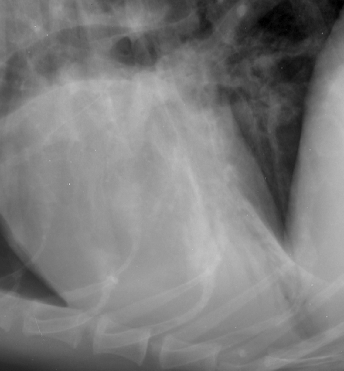
5 Small Animal -Thorax Pleural Effusion Radiographic Findings Increased soft tissue opacity in pleural space Scalloping and apparent elevation of the lung lobe margins Pleural fissure lines, +/- visualization of individual lung lobes Blunting of the costophrenic angles Decreased visualization of the cardiac silhouette &/or diaphragm Often associated with: Rib tumors Lung lobe torsion


6 Pleural Effusion Severe - Inability to see heart & diaphragm - Increased pleural ST opacity - Retraction of lungs from wall Mild - Pleural fissure lines - Increased ST opacity


7 Pleural Effusion - Inability to see heart & diaphragm - Pleural fissure lines - Scalloped lung lobes
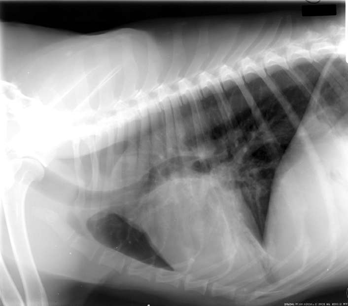
8 Small Animal - Thorax Pneumothorax Radiographic Findings Elevation of the cardiac silhouette from the sternum with a gas opacity ventral to the heart Tenting of diaphragm Retraction of the lung lobes from the body wall No lung markings in the gas opacity/lucency surrounding the retracted lung lobe margins Possible increased pulmonary pattern unstructured interstitial alveolar Collapsed lung lobes Mediastinal shift

9 Pneumothorax - Elevation of heart from sternum - Collapsed lung lobes - No lung markings

10 Pneumothorax - Collapsed lung lobes - No lung markings - Mediastinal shift - Tenting of diaphragm
11 Small Animal - Thorax Cranial Mediastinal Mass Radiographic Findings Widening of the cranial mediastinum Caudal displacement of the cardiac silhouette Tracheal elevation Possible visualization of a circumscribed structure Caudal &/or lateral displacement of the cranial lung lobes


12 Cranial Mediastinal Mass - Widening of cranial mediastinum - Circumscribed structure in cranial mediastinum - Lateral displacement of cranial lung lobes
13 Small Animal - Thorax Pneumomediastinum Radiographic Findings Increased visualization of cranial mediastinal structures
14 Pneumomediastinum
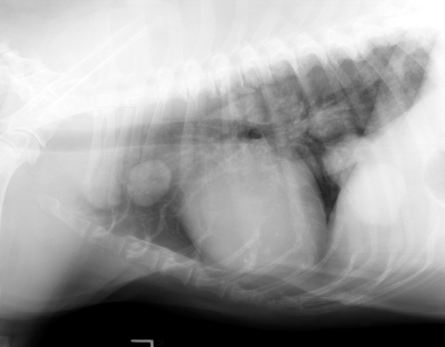
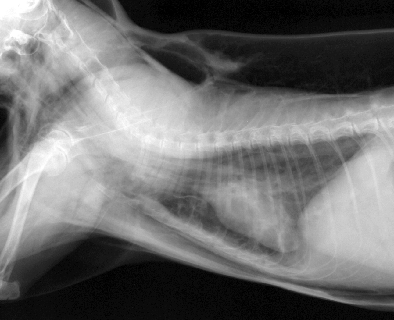
15 Small Animal - Thorax Megaesophagus Radiographic Findings Tubular structure dorsal to the trachea Some portions may be ventral ( draping ) Ventral (& rightward) displacement of the trachea May be fluid, gas or food filled Always check for associated aspiration pneumonia Tracheal stripe sign Often Associated with: Aspiration pneumonia R middle lobe most commonly affected

16 Megaesophagus Tracheal stripe -Tubular gas structure dorsal to trachea - Ventral displacement of trachea - Tracheal stripe sign
17 Megaesophagus Alveolar pattern
18 Small Animal - Thorax Alveolar Pattern Radiographic Findings Lack of visualization of the pulmonary vasculature in the region of the alveolar pattern (due to silhouette effect). Increased soft tissue opacity may be mild, moderate, severe. May be patchy to homogeneous. May have summation effect if alveolar pattern is superimposed over the cardiac silhouette. May have a lobar sign if the disease is along the periphery of a lung lobe. May have an air bronchogram
19 Small Animal - Thorax Alveolar Pattern Differential Diagnosis Bacterial pneumonia Aspiration pneumonia Pulmonary edema Cardiogenic Non-cardiogenic Pulmonary hemorrhage Collapse Lung lobe torsion Inflammatory Neoplasia
20 - Air bronchograms Alveolar pattern

21 Alveolar Pattern
22 Small Animal - Thorax Structured Interstitial Pattern Radiographic Findings Non-cavitary Circumscribed soft tissue, mineral or mixed nodule /mass in the interstitium of the lung May be single or multiple Cavitary Circumscribed nodule /mass containing a gas opacity in the interstitium of the lung May be single or multiple
23 Small Animal - Thorax Structured Interstitial Pattern Differential Diagnosis Neoplasia Fungal mycosis Abscess Eosinophilic granuloma Parasitic pneumonia

24 Non-cavitary Structured Interstitial Pattern Cavitary
25 Small Animal - Thorax L Atrial Enlargement Radiographic Findings Elevation of the caudal lobar bronchi Caudodorsal bulge of the L atrium Increased soft tissue opacity in the region of the L atrium (DV/VD) summation effect Bowlegged cowboy sign separation of the caudal lobar bronchi Important Need to determine if heart failure is present


26 Left Atrial Enlargement - Elevation of bronchi - Increased ST opacity in region of L atrium - Large L auricle
27 Small Animal - Thorax Hypertropic Cardiomyopathy Radiographic Findings May see bulge in the region of the L atrium Possible valentine shaped heart Important Need to determine if heart failure is present


28 Hypertropic Cardiomyopathy - Valentine shaped heart - Caudodorsal bulge in region of L atrium - L heart failure in this example - unstructured interstitial & alveolar lung patterns
29 Small Animal - Thorax Patent Ductus Arteriosus Radiographic Findings 3 bumps Main pulmonary artery enlargement Aortic knob L auricular enlargement Overcirculation L sided heart enlargement
30 Patent Ductus Arteriosus MPA Aortic knob L Auricle L Atrium Overcirculation


31 Patent Ductus Arteriosus
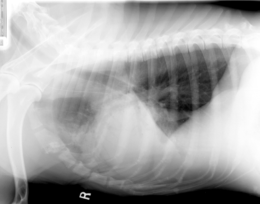
32 Small Animal - Thorax Heartworm Disease Radiographic findings Right heart enlargement Main pulmonary artery enlargement Large, tortuous, blunted arteries Possible pulmonary infiltrates


33 Heartworm Disease R heart enlarge MPA - R sided heart enlargement - Lg, tortuous pulmonary arteries - MPA enlargement

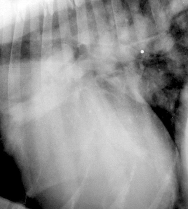
34 Heartworm Disease
Pulmonary Patterns VMA 976
 Pulmonary Patterns VMA 976 PULMONARY PATTERNS Which pulmonary patterns are commonly described in veterinary medicine? PULMONARY PATTERNS Normal Alveolar Interstitial Structured/Nodular Unstructured Bronchial
Radiography provides a rapid, noninvasive
 P ro c e d u re s P ro I M G I N G Peer Reviewed Clifford R. erry, DVM, Diplomate CVR University of Florida Interpreting Small nimal Thoracic Radiographs Radiography provides a rapid, noninvasive mechanism
Administrative. Patient name Date compare with previous Position markers R-L, upright, supine Technical quality
 CHEST X-RAY Administrative Patient name Date compare with previous Position markers R-L, upright, supine Technical quality AP or PA ( with x-ray beam entering from back of patient, taken at 6 feet) Good
Radiology of the Equine Lungs and Thorax (2-Mar-2004)
") In: Equine Respiratory Diseases, P. Lekeux (Ed.) Publisher: International Veterinary Information Service (www.ivis.org), Ithaca, New York, USA. Radiology of the Equine Lungs and Thorax (2-Mar-2004) R.
In: Equine Respiratory Diseases, P. Lekeux (Ed.) Publisher: International Veterinary Information Service (www.ivis.org), Ithaca, New York, USA. Radiology of the Equine Lungs and Thorax (2-Mar-2004) R.
The silhouette sign. Dr Etienne Leroy-Terquem Centre hospitalier de Meulan les Mureaux. France French-cambodian association for pneumology (OFCP)
") The silhouette sign Dr Etienne Leroy-Terquem Centre hospitalier de Meulan les Mureaux. France French-cambodian association for pneumology (OFCP) The silhouette sign When 2 opacities of the same density
The silhouette sign Dr Etienne Leroy-Terquem Centre hospitalier de Meulan les Mureaux. France French-cambodian association for pneumology (OFCP) The silhouette sign When 2 opacities of the same density
Congestive Heart Failure
 William Herring, M.D. 2002 Congestive Heart Failure In Slide Show mode, to advance slides, press spacebar or click left mouse button Congestive Heart Failure Causes of Coronary artery disease Hypertension
Ultrasound of the Thorax (Noncardiac)
") Ultrasound of the Thorax (Noncardiac) Martha Moon Larson, DVM, MS KEYWORDS Ultrasound Thorax Pleural effusion Mediastinum Lung Ultrasound of the noncardiac thorax is an important supplemental imaging modality
Ultrasound of the Thorax (Noncardiac) Martha Moon Larson, DVM, MS KEYWORDS Ultrasound Thorax Pleural effusion Mediastinum Lung Ultrasound of the noncardiac thorax is an important supplemental imaging modality
Radiation-Induced Lung Injury
 May 2001 Radiation-Induced Lung Injury Warren Phipps, Harvard Medical School Year III Our Patient D.C. is a 50 year-old woman with a 30-pack year history of smoking who presented to the ED because she
Chest X-ray STUDY GUIDE 2014-2015
 Internal Medicine Clerkship Chest X-ray STUDY GUIDE 2014-2015 10 Steps/ABC s for Reading Chest X-Rays & Table of Contents to be used with CXR CD-ROM #1 10 Steps / ABC s for Reading Chest X-Rays 0 Normal
Pericardium. Pericardial Diseases. Function of Pericardium 10/1/2012
 NO LASIX, PLEASE! PERICARDIAL DISEASE IN THE DOG Pericardium Michael Luethy, DVM Diplomate ACVIM Cardiology September 13 th, 2012 Tough, outer, parietal pericardium Delicate, serous, visceral pericardium
Chapter 2 Cardiac Interpretation of Pediatric Chest X-Ray
 Chapter 2 Cardiac Interpretation of Pediatric Chest X-Ray Ra-id Abdulla and Douglas M. Luxenberg Key Facts The cardiac silhouette occupies 50 55% of the chest width on an anterior posterior chest X-ray
CHAPTER 1: THE LUNGS AND RESPIRATORY SYSTEM
 CHAPTER 1: THE LUNGS AND RESPIRATORY SYSTEM INTRODUCTION Lung cancer affects a life-sustaining system of the body, the respiratory system. The respiratory system is responsible for one of the essential
Diseases. Inflammations Non-inflammatory pleural effusions Pneumothorax Tumours
 Pleura Visceral pleura covers lungs and extends into fissures Parietal pleura limits mediastinum and covers dome of diaphragm and inner aspect of chest wall. Two layers between them (pleural cavity) contains
General Thoracic Surgery ICD9 to ICD10 Crosswalks. C34.11 Malignant neoplasm of upper lobe, right bronchus or lung
 ICD-9 Code ICD-9 Description ICD-10 Code ICD-10 Description 150.3 Malignant neoplasm of upper third of esophagus C15.3 Malignant neoplasm of upper third of esophagus 150.4 Malignant neoplasm of middle
The abnormal chest X-ray when to refer to a specialis t
 The abnormal chest X-ray when to refer to a specialis t An abnormal chest X-ray must be followed up. OLGA MZILENI, MB ChB, MMed (Int Med) Professor and Head of Internal Medicine and Pulmonology, University
Pulmonary interstitium. Interstitial Lung Disease. Interstitial lung disease. Interstitial lung disease. Causes.
 Pulmonary interstitium Interstitial Lung Disease Alveolar lining cells (types 1 and 2) Thin elastin-rich connective component containing capillary blood vessels Interstitial lung disease Increase in interstitial
Chapter 2 - Anatomy & Physiology of the Respiratory System
 Chapter 2 - Anatomy & Physiology of the Respiratory System Written by - AH Kendrick & C Newall 2.1 Introduction 2.2 Gross Anatomy of the Lungs, 2.3 Anatomy of the Thorax, 2.4 Anatomy and Histology of the
NORMAL CHEST RADIOGRAPHY. Front and lateral view
 NORMAL CHEST RADIOGRAPHY Front and lateral view Dr Etienne Leroy-Terquem Centre hospitalier de Meulan les Mureaux. France French-cambodian association for pneumology (OFCP) OFCP How to obtain a good quality
FELINE PLEURAL DISEASE Diagnosis and Treatment
 FELINE PLEURAL DISEASE Diagnosis and Treatment Philip Padrid, DVM Southwest Regional Medical Director Veterinary Centers of America Associate Professor of Medicine University of Chicago (adjunct) The Ohio
Linfoma maligno pulmonar tratado com Nerium oleander. http://www.drozel.org/eng/diagnosis_malignant_mg.htm CASE REPORT
 Linfoma maligno pulmonar tratado com Nerium oleander http://www.drozel.org/eng/diagnosis_malignant_mg.htm CASE REPORT Diagnosis: Malignant lymphoma, lung cancer A 60-year-old woman experienced pain in
Normal CT scan of the chest
 Normal CT scan of the chest Heart with left and right ventricle showing up lighter (contrast dye) Breast tissue Breast bone (sternum) Breast tissue Left lung (dark area) Right lung (dark area) Rib Main
MEDICAL REPORT MEDICAL HISTORY QUESTIONS
 MEDICAL HISTORY QUESTIONS PAGE 1 OF 7 IF YOUR ANSWER IS YES TO ANY OF THE FOLLOWING QUESTIONS, PLEASE PROVIDE ADDITIONAL INFORMATION INCLUDING: DIAGNOSIS, DATE, AND TREATMENT (INCLUDING MEDICATIONS AND/OR
The WHO manual of diagnostic imaging. Radiographic Anatomy and Interpretation of the chest and the pulmonary System
 The WHO manual of diagnostic imaging Radiographic Anatomy and Interpretation of the chest and the pulmonary System The WHO manual of diagnostic imaging Radiographic Anatomy and Interpretation of the Chest
Thoracic Cavity. Photo: This normal canine lung collapsed when the thorax was opened and the negative pressure was lost in the thorax.
 Thoracic Cavity There are significant anatomical differences in the mediastinum of domestic animals. For instance, bovines, like humans, have well-developed mediastinal separation between the left and
Proceedings of the 34th World Small Animal Veterinary Congress WSAVA 2009
 www.ivis.org Proceedings of the 34th World Small Animal Veterinary Congress WSAVA 2009 São Paulo, Brazil - 2009 Next WSAVA Congress : Reprinted in IVIS with the permission of the Congress Organizers THORACIC
Cardiac Masses and Tumors
 Cardiac Masses and Tumors Question: What is the diagnosis? A. Aortic valve myxoma B. Papillary fibroelastoma C. Vegetation from Infective endocarditis D. Thrombus in transit E. None of the above Answer:
Handbook of radiographic positions and projections in the dog
 THE VETERINARY PUBLISHING COMPANY COMPANION ANIMALS Handbook of radiographic positions and projections in the dog Aimed at veterinary surgeons, students, teachers and other professionals in the veterinary
A. function: supplies body with oxygen and removes carbon dioxide. a. O2 diffuses from air into pulmonary capillary blood
 A. function: supplies body with oxygen and removes carbon dioxide 1. ventilation = movement of air into and out of lungs 2. diffusion: B. organization a. O2 diffuses from air into pulmonary capillary blood
Imaging of the chest. Katarzyna Wypych Zbigniew Serafin
 Imaging of the chest Katarzyna Wypych Zbigniew Serafin Bronchial carcinoma Lung cancer (or frequently if somewhat incorrectly known as bronchogenic carcinoma) is the most common cause of cancer in men,
Best Practice Model for Imaging of Community Acquired Pneumonia. Karen E. Conner, MD
 Best Practice Model for Imaging of Community Acquired Pneumonia Karen E. Conner, MD Chest Radiology and CT Section Chief, Intermountain Healthcare; Chair, QA Committee, Mountain Medical Physician Specialists
NEEDLE THORACENTESIS Pneumothorax / Hemothorax
 NEEDLE THORACENTESIS Pneumothorax / Hemothorax By: Steven Jones, NREMT-P Pneumothorax Pneumothorax is a collection of air or gas in the pleural space of the lung, causing the lung to collapse. Pneumothorax
Ultrasonography of the Adrenal Glands CVM 6105 Kari L. Anderson, DVM, Diplomate ACVR Associate Clinical Professor of Veterinary Radiology
 1: US of adrenal glands, KLA Ultrasonography of the Adrenal Glands CVM 6105 Kari L. Anderson, DVM, Diplomate ACVR Associate Clinical Professor of Veterinary Radiology Ultrasound has quickly become an important
6. Histopathology of Alveoli 7. Surfactant 8. Blood supply of lungs 9. Lymphatics of Lungs 10. Nerve supply of Lungs 11. Pleura 12.
 ANATOMY OF LUNGS - 1. Gross Anatomy of Lungs 2. Surfaces and Borders of Lungs 3. Hilum and Root of Lungs 4. Fissures and Lobes of Lungs 5. Bronchopulmonary segments 6. Histopathology of Alveoli 7. Surfactant
Radiological Findings in BO
 Radiological Findings in BO BO-Meeting 2016 Schloss Johannisberg Geisenheim - Rheingau Germany Dr. Simon Martin Department of Diagnostic and Interventional Radiology University Hospital Frankfurt Bronchiolitis
How To Teach An Integrated Ultrasound
 University of South Carolina School of Medicine Integrated Ultrasound Curriculum iusc Richard Hoppmann The Integrated Ultrasound Curriculum Initiated 2006 First (M1) and Second (M2) Year Medical Students
Primary -Benign - Malignant Secondary
 TUMOURS OF THE LUNG Primary -Benign - Malignant Secondary The incidence of lung cancer has been increasing almost logarithmically and is now reaching epidemic levels. The overall cure rate is very low
UKRC 2015 Dr Michael Sproule Glasgow
 UKRC 2015 Dr Michael Sproule Glasgow Radiology of Asbestos Related Lung Disease General term given to a group of fibrous minerals containing silica and a variety of other elements. Asbestos: Derived
Common types of congenital heart defects
 Common types of congenital heart defects Congenital heart defects are abnormalities that develop before birth. They can occur in the heart's chambers, valves or blood vessels. A baby may be born with only
Abdomen X-Ray (AXR) Collimation is ideally from diaphragms to lower border of the symphysis pubis and the lateral skin margins.
 Collimation is ideally from diaphragms to lower border of the symphysis pubis and the lateral skin margins.") Abdomen X-Ray (AXR) Collimation is ideally from diaphragms to lower border of the symphysis pubis and the lateral skin margins. LMP of child-bearing age female patients should be checked. 1. Acute abdomen
Abdomen X-Ray (AXR) Collimation is ideally from diaphragms to lower border of the symphysis pubis and the lateral skin margins. LMP of child-bearing age female patients should be checked. 1. Acute abdomen
Patient Possible differentials Recommended diagnostics Puppy or kitten with a soft systolic murmur
 Cardiac Auscultation 101 Terri DeFrancesco, DVM, DACVIM (Cardiology), DACVECC Associate Professor in Cardiology and Critical Care NC State University College of Veterinary Medicine Email: [email protected]
Lung Carcinoid Tumor
 Lung Carcinoid Tumor What are lung carcinoid tumors? Lung carcinoid tumors (also known as lung carcinoids) are a type of lung cancer, which is a cancer that starts in the lungs. Cancer starts when cells
Chest Pain. Acute Myocardial Infarction: Differential Diagnosis and Patient Management. Common complaint in ED. Wide range of etiologies
 Acute Myocardial Infarction: Differential Diagnosis and Patient Management Presented By: Barbara Furry, RN-BC, MS, CCRN, FAHA Director The Center of Excellence in Education Director of HERO Chest Pain
Lung & Thorax Exams. Charlie Goldberg, M.D. Professor of Medicine, UCSD SOM [email protected]
 Lung & Thorax Exams Charlie Goldberg, M.D. Professor of Medicine, UCSD SOM [email protected] Lung Exam Includes Vital Signs & Cardiac Exam 4 Elements (cardiac & abdominal too) Observation Palpation Percussion
Dr. Weyrich G04: Anterior Thoracic Wall, Breast and Lymphatic System
 Dr. Weyrich G04: Anterior Thoracic Wall, Breast and Lymphatic System Reading: 1. Gray s Anatomy for Students, Chapter 3 2. Dissection Guide for Human Anatomy, Lab 4 Objectives: 1. Osteocartilaginous thoracic
Recurrent or Persistent Pneumonia
 Recurrent or Persistent Pneumonia Lower Respiratory Tract Dr T Avenant Recurrent or Persistent Pneumonia Definitions Recurrent pneumonia more than two episodes of pneumonia in 18 months Persistent pneumonia
Chest radiography is the most
 ... Chest Radiography for Radiologic Technologists DAN L. HOBBS, M.S.R.S., R.T.(R)(CT)(MR) The chest exam is performed more frequently than any other exam in the imaging department. It is important for
Sample Learning Objectives for a Medical School Radiology Curriculum: Listed by Subjects
 Sample Learning Objectives for a Medical School Radiology Curriculum: Listed by Subjects This document lists sample learning objectives by subject matter The numerical ranking in parenthesis following
INFLAMMATORY PLEURAL EFFUSION
 PLEURA- LESIONS LESIONS OF PLEURA Primary Intra pleural bacterial infections Neoplasm (mesothelioma) Secondary A complication of some underlying disease PLEURAL EFFUSION Common manifestation of both primary
The Lewin Group undertook the following steps to identify the guidelines relevant to the 11 targeted procedures:
 Guidelines The following is a list of proposed medical specialty guidelines that have been found for the 11 targeted procedures to be included in the Medicare Imaging Demonstration. The list includes only
Neoplasms of the LUNG and PLEURA
 Neoplasms of the LUNG and PLEURA 2015-2016 FCDS Educational Webcast Series Steven Peace, BS, CTR September 19, 2015 2015 Focus o Anatomy o SSS 2000 o MPH Rules o AJCC TNM 1 Case 1 Case Vignette HISTORY:
Pulmonary Ventilation
 Pulmonary Ventilation Graphics are used with permission of: Pearson Education Inc., publishing as Benjamin Cummings (http://www.aw-bc.com) Page 1. Introduction Pulmonary ventilation, or breathing, is the
LYMPHOMA IN DOGS. Diagnosis/Initial evaluation. Treatment and Prognosis
 LYMPHOMA IN DOGS Lymphoma is a relatively common cancer in dogs. It is a cancer of lymphocytes (a type of white blood cell) and lymphoid tissues. Lymphoid tissue is normally present in many places in the
Sir William Osler: Listen to the patient; the patient tells you everything.
 Sir William Osler: Listen to the patient; the patient tells you everything. Jean-Martin Charcot: The patient is a liar. Epidemiology of Mesothelioma Jeffrey H. Mandel, MD, MPH Division of Environmental
Restrictive lung diseases
 Restrictive lung diseases Characterized by reduced compliance of the lung. Prominent changes in the interstitium (interstitial lung disease). Important signs and symptoms: - Dyspnea. - Hypoxia. - With
Cystic Lung Diseases. Melissa Price Gillian Lieberman, MD Advanced Radiology Clerkship Beth Israel Deaconess Medical Center November, 2008
 Cystic Lung Diseases Melissa Price Gillian Lieberman, MD Advanced Radiology Clerkship Beth Israel Deaconess Medical Center November, 2008 How do we define a cyst of the lung? Hansell DM, Bankier AA, MacMahon
Radiology of Asbestos-related Diseases
 8/25/03 9/19/03 Radiology of Asbestos-related Diseases Joan S. Hu,, Harvard Medical School Year III Gillian Lieberman, M.D. Asbestos-related conditions I. Benign Pleural Disease II. III. IV. A. Pleural
Abuse of inhaled or intravenously injected illicit drugs
 PICTORIAL ESSAY Pulmonary Complications of Illicit Drug Use Differential Diagnosis Based on CT Findings Elsie T. Nguyen, MD, C. Isabela S. Silva, MD, PhD, Carolina A. Souza, MD, and Nestor L. Müller, MD,
Influenza (Flu) Influenza is a viral infection that may affect both the upper and lower respiratory tracts. There are three types of flu virus:
 Influenza is a viral infection that may affect both the upper and lower respiratory tracts. There are three types of flu virus:") Respiratory Disorders Bio 375 Pathophysiology General Manifestations of Respiratory Disease Sneezing is a reflex response to irritation in the upper respiratory tract and is associated with inflammation
Respiratory Disorders Bio 375 Pathophysiology General Manifestations of Respiratory Disease Sneezing is a reflex response to irritation in the upper respiratory tract and is associated with inflammation
CT scans and IV contrast (radiographic iodinated contrast) utilization in adults
 utilization in adults") CT scans and IV contrast (radiographic iodinated contrast) utilization in adults At United Radiology Group, a majority of CT exams are performed either with IV contrast or without while just a few exams
CT scans and IV contrast (radiographic iodinated contrast) utilization in adults At United Radiology Group, a majority of CT exams are performed either with IV contrast or without while just a few exams
Evaluation and treatment of emphysema in a preterm infant
 ISPUB.COM The Internet Journal of Pediatrics and Neonatology Volume 11 Number 1 Evaluation and treatment of emphysema in a preterm infant T Saad, P Chess, W Pegoli, P Katzman Citation T Saad, P Chess,
Management of Chest Tubes and Air Leaks after Lung Resection
 Management of Chest Tubes and Air Leaks after Lung Resection Emily Kluck PA-C The Johns Hopkins Hospital Baltimore, MD AATS 2014, Toronto, CAN April 2014 Management of Chest Tubes 1 Overview Review the
CT findings in Differential Diagnosis between Tuberculous Pleurisy and Malignant Effusion
 CT findings in Differential Diagnosis between Tuberculous Pleurisy and Malignant Effusion Poster No.: E-0084 Congress: ESTI 2012 Type: Scientific Exhibit Authors: S. S. Shim, Y. Kim; Seoul/KR Keywords:
Objectives. Mylene T. Truong, MD. Malignant Pleural Mesothelioma Background
 Imaging of Pleural Tumors Mylene T. Truong, MD Imaging of Pleural Tumours Mylene T. Truong, M. D. University of Texas M.D. Anderson Cancer Center, Houston, TX Objectives To review tumors involving the
ERCIYES UNIVERSITY MEDICAL FACULTY CARDIOVASCULAR SURGERY
 ERCIYES UNIVERSITY MEDICAL FACULTY Code - Title Stage of study MED511 THORACIC AND CARDIOVASCULAR SURGERY 5 th semester LOCAL CREDIT: 2 ECTS CREDITS: 3 Coordinating Lecturer ALL CONTACTS CONCERNİNG SOCRATE
PNEUMOMEDIASTINUM: A PATIENT PRESENTATION
 November 2002 PNEUMOMEDIASTINUM: A PATIENT PRESENTATION Alden Chip McDonald, III Harvard Medical School, Year III AGENDA I. Patient Presentation II. Diagnosis of Pneumomediastinum III. Causes of Pneumomediastinum
MECHINICAL VENTILATION S. Kache, MD
 MECHINICAL VENTILATION S. Kache, MD Spontaneous respiration vs. Mechanical ventilation Natural spontaneous ventilation occurs when the respiratory muscles, diaphragm and intercostal muscles pull on the
CHEST TUBES AND CHEST DRAINAGE SYSTEMS
 CHEST TUBES AND CHEST DRAINAGE SYSTEMS Central Nursing Orientation April 2008 Revised September 2011 OBJECTIVES Describe common tubes and indications for use at LHSC Review indications and contraindications,
Emergency care of the patient with acute respiratory distress
 Emergency care of the patient with acute respiratory distress Lesley King, MVB, Dipl. ACVECC, Dipl. ACVIM, Dipl. ECVIM (CA) Philadelphia School of Veterinary Medicine, University of Pennsylvania, Philadelphia,
6/30/15. ! Atrioventricular valve insufficiency (AVVI) is the most common cardiac disease in the dog
 is the most common cardiac disease in the dog") Jeremy Orr DVM, DVSc, DACVIM (Cardiology) Colorado Veterinary Medical Association Conference 2015 September 18, 2015! Review atrioventricular valve insufficiency! Presenting complaints! Key physical examination
Jeremy Orr DVM, DVSc, DACVIM (Cardiology) Colorado Veterinary Medical Association Conference 2015 September 18, 2015! Review atrioventricular valve insufficiency! Presenting complaints! Key physical examination
Expiratory CT: Correlation with Pulmonary Function Tests and Value for Discriminating Lung Diseases
 Original Article Expiratory CT: Correlation with Pulmonary Function Tests and Value for Discriminating Lung Diseases Ertuğrul Mavili 1, Hakan Büyükoğlan 2, Nurdan Bulut Çomu 1, Mustafa Güleç 1 Erciyes
New Cardiothoracic Surgery CPT Codes for 2013
 New Cardiothoracic Surgery CPT Codes for 2013 There were several changes to the cardiothoracic surgery CPT codes for 2013. There are five new codes in the general thoracic surgery section, with one revised
Non-Small Cell Lung Cancer
 Non-Small Cell Lung Cancer About Your Lungs and Lung Cancer How do your lungs work? To understand lung cancer it is helpful to understand your lungs. Your lungs put oxygen into the blood, which the heart
General Information About Non-Small Cell Lung Cancer
 General Information About Non-Small Cell Lung Cancer Non-small cell lung cancer is a disease in which malignant (cancer) cells form in the tissues of the lung. The lungs are a pair of cone-shaped breathing
April 2015 CALGARY ZONE CLINICAL REFERENCE PULMONARY CENTRAL ACCESS & TRIAGE
 April 2015 CALGARY ZONE CLINICAL REFERENCE CENTRAL ACCESS & TRIAGE Introduction Pulmonary consulting services are organized through the Calgary Zone Pulmonary Central Access and Triage (PCAT). Working
An Introduction to Anatomy and Physiology
 An Introduction to Anatomy and Physiology Objectives Define anatomy and physiology Identify the levels of organization in organisms from simplest to most complex Identify the organ systems of the human
Occupational Lung Disease. David Perlman, MD
 Occupational Lung Disease David Perlman, MD What causes occupational lung diseases? Breathing bad stuff into your lung Mechanism of particle deposition Large particles (>0.5μM) Impaction Gravitational
CARDIAC TAMPONADE SECONDARY TO INTRAPERICARDIAL TUMOR IN A DOG. CASE REPORT
 Scientific Works. Series C. Veterinary Medicine. Vol. LIX (3) ISSN 2065-1295, ISSN Online 2067-3663, ISSN-L 2065-1295 bstract CRDIC TMONDE SECONDRY TO INTRERICRDIL TUMOR IN DOG. CSE REORT ndrei ISN 1,
Acute heart failure may be de novo or it may be a decompensation of chronic heart failure.
 Management of Acute Left Ventricular Failure Acute left ventricular failure presents as pulmonary oedema due to increased pressure in the pulmonary capillaries. It is important to realise though that left
The Newborn With a Congenital Disorder. Chapter 14. Copyright 2008 Wolters Kluwer Health Lippincott Williams & Wilkins
 The Newborn With a Congenital Disorder Chapter 14 Congenital Anomalies or Malformations May be caused by genetic or environmental factors Approximately 2% to 3% of all infants born have a major malformation
Cardiac and Pulmonary Issues in the Elite Athlete. Keep Your Edge Hockey Sports Medicine 2015 Toronto August 28-30 2015
 Cardiac and Pulmonary Issues in the Elite Athlete Keep Your Edge Hockey Sports Medicine 2015 Toronto August 28-30 2015 THORACIC EMERGENCIES Hockey related thoracic trauma is blunt Injury to the boney
Preoperative Laboratory and Diagnostic Studies
 Preoperative Laboratory and Diagnostic Studies Preoperative Labratorey and Diagnostic Studies The concept of standardized testing in all presurgical patients regardless of age or medical condition is no
Lung Cancer: Diagnosis, Staging and Treatment
 PATIENT EDUCATION patienteducation.osumc.edu Lung Cancer: Diagnosis, Staging and Treatment Cancer begins in our cells. Cells are the building blocks of our tissues. Tissues make up the organs of the body.
Mesothelioma. 1995-2013, The Patient Education Institute, Inc. www.x-plain.com ocft0101 Last reviewed: 03/21/2013 1
 Mesothelioma Introduction Mesothelioma is a type of cancer. It starts in the tissue that lines your lungs, stomach, heart, and other organs. This tissue is called mesothelium. Most people who get this
Pericardial Effusions Diagnosis and Treatment
 The Vet Education International Online Veterinary Conference 2013 Pericardial Effusions Diagnosis and Treatment With Dr Richard Woolley Specialist in Cardio-Respiratory Medicine July2013 Vet Education
Anatomy Pleura Visceral Layer outer surface of lung Separates lobes of lung from each other Parietal Layer inner surface chest wall Pleural linings Co
 Pleural diseases Dr. JM Nel Department of Pulmonology Pleural diseases Anatomy Empyema Physiology Pleurisy Pleural effusion Spontaneous Pneumothorax Asbestos- related pleural disease 1 Anatomy Pleura Visceral
Multi-slice Helical CT Scanning of the Chest
 Multi-slice Helical CT Scanning of the Chest Comparison of different low-dose acquisitions Lung cancer is the main cause of deaths due to cancer in human males and the incidence is constantly increasing.
A Practical Guide to Advances in Staging and Treatment of NSCLC
 A Practical Guide to Advances in Staging and Treatment of NSCLC Robert J. Korst, M.D. Director, Thoracic Surgery Medical Director, The Blumenthal Cancer Center The Valley Hospital Objectives Revised staging
Disease/Illness GUIDE TO ASBESTOS LUNG CANCER. What Is Asbestos Lung Cancer? www.simpsonmillar.co.uk Telephone 0844 858 3200
 GUIDE TO ASBESTOS LUNG CANCER What Is Asbestos Lung Cancer? Like tobacco smoking, exposure to asbestos can result in the development of lung cancer. Similarly, the risk of developing asbestos induced lung
Radiologic Diagnosis of Spinal Metastases
 September 2002 Radiologic Diagnosis of Spinal Metastases Natalie J. M. Dailey, Harvard Medical Student Year III Our Patient s Presenting Story 70 year old male Presents to the hospital for laparascopic
Metastatic Cervical Cancer s/p Radiation Therapy, Radical Hysterectomy and Attempted Modified Internal Hemipelvectomy
 Metastatic Cervical Cancer s/p Radiation Therapy, Radical Hysterectomy and Attempted Modified Internal Hemipelvectomy Sarah Hutto,, MSIV Marc Underhill, M.D. January 27, 2009 Past History 45 yo female