Understanding Digital Breast Tomosynthesis
|
|
|
- Basil Harold Daniel
- 7 years ago
- Views:
Transcription
1 Understanding Digital Breast Tomosynthesis Sharon Walenga, B.S. RT(R)(M) Clinical Manager of Breast Health and Radiation Oncology Advocate Lutheran General Hospital, Park Ridge, IL Jean Paquelet, M.D., FACR Director of Breast Imaging McKee Medical Center, Loveland, CO & Harmony Breast Diagnostic Center, Fort Collins, CO R. Edward Hendrick, Ph.D., FACR Clinical Professor, Department of Radiology, University of Colorado Denver, School of Medicine, Aurora, CO

2 Objectives: Upon completion, participant will be able to: 1. Understand the design and performance of different manufacturers' digital breast tomosynthesis systems 2. Describe the clinical application and performance differences between digital breast tomosynthesis and digital mammography 3. Understand the quality control tests technologists should be able to perform on digital breast tomosynthesis systems
3 Tomosynthesis Acquisition Tube motion X-ray Tube Compression Plate Breast Digital Detector X-ray tube moves in an arc around the breast Series of low dose images are acquired at different angles Total dose similar to standard breast exam
4 Tomosynthesis Acquisition Tube motion X-ray Tube Reconstructed planes Compression Plate Breast Digital Detector X-ray tube moves in an arc around the breast Series of low dose images are acquired at different angles Collected data permits reconstruction of parallel planes, each plane in-focus, with out-of-plane tissues blurred
5 Digital Breast Tomosynthesis Acquisition Each DBT acquisition consists of 9-25 separate projections that permit reconstruction of multiple planes in the breast, each plane in focus Overlapping out-of-plane tissues are blurred Yields clearer lesion margins than 2D in nonfatty breasts

6 DBT Acquisition and Reconstruction Reconstruction at at Height height B Reconstruction at height Height A A Low-dose X-Ray sweep Height A Height B Projection views 5 Projection Views Plane B Plane A
7 Effect of Sweep Angle 15 o Sweep 50 o Sweep 2D Narrower Sweep Wider Sweep Wider sweep angle give more complete blurring of tissues outside the focal plane Narrower sweep angle makes lesion margins appear sharper
8 Reconstructed DBT Images Can Be Reconstructed as Planes or Slabs Single Plane 11 cm Slab

9 3 DBT Systems Are FDA Approved for Clinical Use in the U.S. Hologic Dimensions GE SenoClaire Siemens Inspiration

10 FDA Approval of DBT Hologic Dimensions received FDA approval Feb 2011 Hologic s original approach: CC and MLO DBT + 2D DM Hologic s new approach: CC and MLO DBT + Synthetic 2D (C-view) GE SenoClaire received FDA approval August 2014 GE approach: 3D MLO DBT + 2D CC view Siemens Inspiration DBT received FDA approval in April 2015 Siemens approach: CC and MLO DBT + 2D CC and MLO
11 Differences Among FDA-approved DBT Systems Manufacturer: Hologic Dimensions GE SenoClaire Siemens Inspritation Detector motion rotating static static Detector pixel size (μm) 70 (140 DBT) Tube motion continuous step-and-shoot continuous Angular range (degrees) Number of projections Scan time(seconds) 4 < 10 s 25 Grid NO YES NO Reconstruction algorithm FBP iterative iterative
12 Step-and-shoot vs. Continuous Manufacturer: GEH Hologic Siemens Tube motion: step-and-shoot continuous continuous Step-and-shoot Continuous
13
14
15

16 Example #1: Multifocal Cancer MX (LCC) MX (LMLO) DBT (LMLO) DETECTION OF MULTIPLE LESIONS: DBT > MX plane 27 of 84 Images courtesy of Dr. Gisella Gennaro
17 Example #2: Invasive Ductal Cancer MX (RCC) MX (RMLO) DBT (RMLO) LESION DETECTION: DBT ONLY plane 25 of 69 Images courtesy of Dr. Gisella Gennaro
18 Radiation Doses in DBT Each individual DBT projection is very low dose - Hologic approach of acquiring DBT + 2D in both CC & MLO projections has a total dose that is about x the dose of 2-view DM - Newer Hologic approach of acquiring only DBT views and reconstructing synthetic 2D views (C-view) has a total dose that is 1x-1.5x times that of DM - For GE, dose for a DBT view ~ dose for a 2D view - For Siemens, single-view DBT dose is x higher than single-view DM dose, depending on breast thickness (bigger difference for thinner breasts)
19 Digital Breast Tomosynthesis (DBT) Radiologist s Perspective Jean Paquelet, MD, FACR Director of Breast Imaging McKee Medical Center Loveland Colorado and Harmony Breast Diagnostic Center Fort Collins Colorado 4/22/
20 DBT: A Much Better Mammogram Decreased Recall Rates (improved specificity) compared to 2D mammography Recall rates (currently 7-10 patients per 100 2D screening exams) for DBT reduced 10-42% Decreased recalls primarily due to elimination of superimposed structures (summation densities) The reduction in recall rates was most pronounced for patients undergoing their first mammogram and for patients with scattered fibroglandular densities and for heterogeneously dense breasts
21 DBT: A Much Better Mammogram Compared to conventional 2D digital mammography, DBT detects more breast cancers (increased sensitivity) Sharpe et al reported a 54.3% increase in breast cancer detection rate with DBT compared to 2D mammography. In a screening population their cancer detection rate rose from 3.5 cancers per thousand women screened to 5.4 cancers per thousand The additional cancers detected with DBT are almost all invasive cancers Most noninvasive cancers (DCIS: Ductal carcinoma in situ) manifest as calcifications. Detection of calcification is not improved with DBT compared to 2D technique
22 DBT: A Much Better Mammogram How will your facility use it? Will all mammography units be DBT or just some? How will you triage patients? Randomly? By breast density? By patient preference or insurance coverage? By exam type? For screening? Diagnostic? Both? Your workstation may dictate this choice
23 Invasive Ductal Cancer RMLO tomo slice RCC tomo slice DBT better detects invasive cancer due to elimination of overlapping 4/22/ structures and to better lesion border depiction
24 DBT: Interpretation For an average 55 mm thick breast, the radiologist will be viewing about 250 images 2D or synthesized (composite) views: 4 images DBT slices: 55 one mm thick slices for each CC and MLO view: 220 images DBT slabs or thick slices: 6 one centimeter thick images for each CC and MLO view : 24 images. Viewing DBT images with thicker slices helps the radiologist appreciate calcification clusters
25 DBT: Interpretation Currently, most DBT images are viewed on proprietary dedicated workstations. All work stations are not created equal. Some are multimodality? Interpretation from PACS Interpretation time for DBT slightly more twice that of 2D mammograms Increased interpretation time for screening exams may be partially offset by fewer recalls for DX In my practice we have increased FTE for radiologists by 33%
26 RMLO tomo cine RMLO single tomo slice 4/22/ Invasive Ductal Cancer
27 RCC tomo cine RCC single tomo slice 4/22/ Invasive Ductal Cancer
28 DBT: Changing Work up of Screen Detected Findings Many masses seen at screening DBT do not require recall for additional views. The screening DBT images often define borders and triangulate the lesion well enough to proceed directly to US and avoid additional mammographic views Even when a finding is seen on a single projection, DBT does provide more information for triangulation However, most facilities report performing more ultrasound than was necessary prior to introduction of DBT. We have increased US staffing by 20%
29 Simple Cyst(tomo slices) 4/22/
30 Tomo Slice High Grade DCIS 2D Mag view DBT: Calcifications 4/22/
31 Tomo Slice High Grade DCIS 2D Mag view DBT: Calcifications 4/22/ Work-up of calcifications unchanged: Mag views, 90degree lateral
32 Features that look different on DBT vs 2D LCC tomo slice LMLO tomo slice Lumpectomy Scar Scars are often much more impressive on DBT than on 2D imaging Scar markers/diagrams of scars/history particularly important 4/22/
33 Zipper Artifact from Marking Clip 4/22/ Tomo cine LCC 2D LCC
34 LCC tomo slice LMLO tomo slice 4/22/ Skin calcifications & skin lesions easily recognized on DBT
35 Key Points DBT detects more cancers and has fewer recalls than 2D mammograms Additional cancers detected are nearly all invasive Even with DBT, some cancers are missed. Typically the missed cancers are non-calcified, non-spiculated lesions in dense breasts Introduction of DBT may require additional personnel due to increased US volume and radiologist s increased interpretation time.
36 Sharon Walenga, BS RT(R)(M) Clinical Manager of Breast Health and Radiation Oncology TOMOSYNTHESIS FOR TECHNOLOGISTS
37 Advocate Lutheran General Hospital Caldwell Breast Center
38 Caldwell Breast Center Breast Imaging Center of Excellence (BICOE) from American College of Radiology NAPBC- National Accreditation Program for Breast Centers Perform mammography, breast ultrasound and breast MRI examinations with biopsy capability in all modalities Consistent 99% patient satisfaction First in the Midwest to perform Tomosynthesis
39 Mammography- Through the Years Xerography Analog Digital Tomosynthesis
40 Contrary to Popular Opinion Digital Breast Tomosynthesis is not quite 3D.
41 The Path of Tomosynthesis Entered Hologic Pivotal Multicenter Tomosynthesis Trial with PI Betty Rafferty, MD at Massachusetts General Hospital, Boston, MA: A Multicenter, Controlled Trail to Evaluate the Hologic Tomosynthesis Mammography System At LGH- 5 technologists and 4 Physicians trained for 8 hours each to consent, perform and interpret examinations
42 Beta Tomosynthesis Study at LGH Installed beta unit April 2010 Total of 22 sites across country participating Approximately 3,200 total subjects enrolled Imaged 120 patients at ALGH Involved in 2 arms of the study: Screening arm No prior surgical procedures No biopsy clips Female Biopsy arm Same as screening arm exclusion criteria Recommended for biopsy based on recent diagnostic examination
43 Overall Experience Technologist experience Switch to new machine - very user friendly Faster QC Patient experience Little to no difference Some patients concerned about additional radiation Less painful Radiologist experience Longer to interpret screening Helpful in diagnostic setting
44 Transition to Commercial Use DBT was FDA approved in February 2011 LGH was able to secure donated funds to purchase our Beta Unit Started imaging in June 13, 2011 Purchased additional units in 2012, 2014 and 2016
45 Outcomes Oslo Breast Cancer Screening Trail % increase in the detection of invasive breast cancers 27% increase in the detection of all cancers (invasive and in situ cancers combined) 15% decrease in false- positive rates Journal of American Medical Association- Breast Cancer Screening Using Tomosynthesis in Combination With Digital Mammography % increase in the detection of invasive breast cancers 29% increase in the detection of all cancers 15% decrease in the recall rates
46 Implementation
47 Your Team Project Manager Lead Interpreting Radiologist, Manager Sales person, Field Engineer, Connectively specialist from your chosen vendor Maintenance Pacs Administrator/ Clinical Engineer Finance Charge master/ris Medical Physicist
48 Considerations Which patients to image? How many images should you take? Reimbursement/charging patients Do you need an order? Educating referring physicians Should you consent the patients?
49 Which Patients to Image? FDA approval for both screening and diagnostic imaging Who is the Focus? Screening or Diagnostic patients? Do we designate patients who might be better Tomosynthesis candidates? We have limited number of machines!
50 What we did Diagnostic patients (specifically call backs and patients who present with a problem) priority Patients requesting Tomosynthesis from the screening populationscheduled. After more requests for DBT screening and obtaining a 2 nd unitwent to all Tomo after 3:00pm Monday-Friday and all day Saturday. Exception is request Diagnostic either by Radiologist or Referring Physician. With 4 th room purchase in % of screenings are performed with DBT
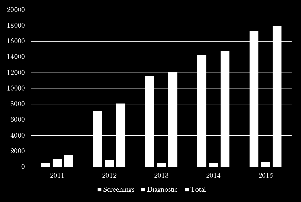
51 Volumes
52 Imaging Screening Standard CC and MLO Implant- ID only Mosaic- Largest part Diagnostic Architectural distortion- spot Skin Calcifications and palpable abnormalities- omits tangential
53 Choices Hologic Combo- 3D then 2D Tomo- 3D only Combo HD- 3D, 2D and C-view GE 2D- CC and MLO 3D only in MLO Premium view
54 Replacement of 2D Synthesized 3D image to eventually replace 2D Can only be used on Standard imaging Cannot be used on spots, mags or tangentials Great for highlighting faint calcifications
55 Reimbursement CPT code for screening Medicare declared as standard of care Most private insurance companies are paying 2D + 3D plus physician component but very inconsistent.
56 Exploding Charge The charge for the 3-D mammography will be set up so you will bill the regular mammography charge plus the add-on charge (they are still looking into the CAD component). The charge for the add-on will be about $X. Generally, insurance companies follow Medicare in their billing and payment methodologies. However, we have some insurance companies that pay us a flat rate for a mammography, whether it is analog, digital, 3D or whatever. We have also had an issue in the past where one insurance company would not pay for the CAD component which they considered experimental although all other insurance companies would. This has since been resolved. We do not anticipate that the insurance companies will not cover this if ordered by a physician, but you never know for sure until we start billing it, and at that time we will work with them so it is covered. Beth Hickey, Director of Finance
57 Charge Master Contact Charge Master to create codes FFDM Screening / Diagnostic Implants Unilateral CAD/ No CAD
58 Do you need an Order? Currently, no additional orders are necessary for Tomosynthesis examinations, though many physican s offices are requesting.
59 Education Referring Physicians Important to educate referring physicians Send a letter to hospital staff via and regular mailboxes Visit offices to answer questions Inform marketing team with basics
60 Is a Consent Needed? No written consent is necessary since the examination is not invasive May want to consider discussing: Examination a few seconds longer under the same compression Machine moves on an arc with Tomosynthesis Advantage of seeing through tissue and potentially increasing detection of breast cancer and deceasing additional imaging
61 Patient Radiation dose Tungsten target instead of Molybdenum 20% less dose on 2D Average glandular dose needs to be less than 300mrad 2D Selenia = 193mrad 2D Dimensions = 134mrad 3D Portion = 156mrad Combo 2D + 3D = 291mrad Data based off phantom image- not patient dose
62 Upgrade needed- Manager 3 Hologic Selenia / 4 Dimensions plus 2 R2 checkers and 1 Cenova Anticipate all machines to be converted to Dimensions and therefore upgrade is needed for speed and viewing ease All SecurView software upgraded and Manager Hardware replaced (Power Edge T610) Purchased SecureXchange to pull priors when patient enters system (RIS)
63 Upgrade needed- Manager Dell PowerEdge 2900 Server- Old Tower Chassis (2) Quad-Core Processors 4GB RAM Memory 10/100/1000 BaseT Ethernet Interface Dell PowerEdge T610 Server- New Windows Server 2003 Dual Quad-Core Processors at 2.5 GHz Hard drives are RAID TB This unit can handle 80 patients per hour assuming 2 prior studies for each patient. It can connect to up to 10 SVDX Client workstations.
64 PACS Memory 2D imaging Screening (4 images) 8x10 uses 17 MB per image = 68 MB 10x12 uses 27 MB per image = 108 MB 3D combo- 2D + 3D (4 view screening) 200 MB compressed file Currently the 3D images are not able to be visualized on Pacs though they are archived. In future, there will be a fully compatible diacom but will require 400 MB.
65 Cost Mammography unit 2D Dimensions 3D Option + License CAD- Cenova + License Options SecureView Reading Station Secure Xchange Localization kit Paddle holder Electrical requirements ACR
66 We got the Power! Selenia- 220 line (208), 35 amp Dimensions 220 line (208), 40 amp
67 Money AND Time? Installation- 4 to 5 days Remove old unit- 1 day Installation of machine- 2-3 days Physicist testing- 1 day
68 Education Technologist application training on site 8hrs required Radiologist Webinar or Conference- 8 hrs required
69 2D Accreditation - ACR New Unit addendum If more than 13 months left on Accreditation $850 per unit now $1000 Transfers of current expiration date Intial/ Early Renewal If less than 13 months left on Accreditation $1475 for first unit now $1700 $1300 for additional units now $1500 Good for 3 Years
70 2D Accreditation - ACR Submit to ACR via website Physicist forms MQSA Requirements for Mammography Equipment checklist Medical Physicist s QC Test Summary form Withdrawn Unit Memo New Unit Addendum Once confirmation is received, start 2D
71 FDA for 3D EmittingProducts/MammographyQualityStandardsActandProgram/ FacilityCertificationandInspection/ucm htm Mammography Evaluation Survey Phantom Image (3D mode) Certificate extension requirements form
72 No, tomos performed until The Food and Drug Administration (FDA) has approved your FullField Digital Mammography (FFDM) system. You may begin using your Hologic Selenia Dimensions Digital Breast Tomosynthesis (DBT) System unit for clinical use on patients. Please see attached approval letter. If you have any question, please contact me at Denise Robinson FDA/MQSA Program Phone:
73 Positioning/ Workflow Benefits Fast paddle- better compression Automatic MLO positioning both gantry and paddle Fingerprint sign on Error fixes Drawbacks Precise Positioning Faceshield Motion Not able to be viewed in PACs
74 Quality Control Geometry Calibration Bi-annual- QC for individual tomo slices Compression Test Bi-annual Compression Thickness Indicator Bi-weekly
75 Quality Control- Weekly Flat field Artifact Evaluation Filter check Rhodium- 2D Silver- 2D Aluminum- 3D Gain Callibration Focal Spots Tomo- resets digital pictures Rh/Lg- Rhodium Af/Lg- Silver Rh/Sm- Rhodium done with mag stand Af/Sm- Silver done with mag stand
76 Quality Control- Weekly Phantom Combo 2D 3D- scroll until brightest specks (slice 15) CNR/SNR No more calculations!
77 Quality Control Repeat Analysis Can be done through website via IP address Through unit For any upgrades- make sure you check release notes- possible technique changes
78 GE - checklist
Role of the Medical Physicist in Clinical Implementation of Breast Tomosynthesis
 Role of the Medical Physicist in Clinical Implementation of Breast Tomosynthesis Bob Liu, Ph.D. Department of Radiology Massachusetts General Hospital And Harvard Medical School Digital Breast Tomosynthesis
Hologic Selenia Dimensions C-View Software Module. October 24, 2012
 Hologic Selenia Dimensions C-View Software Module October 24, 2012 Introduction and Agenda Peter Soltani, Ph.D. Senior VP & GM, Breast Health Hologic, Inc. Agenda Technology Overview Clinical Overview
Mammography. What is Mammography?
 Scan for mobile link. Mammography Mammography is a specific type of breast imaging that uses low-dose x-rays to detect cancer early before women experience symptoms when it is most treatable. Tell your
How To Compare Mammography To A 3D System
 Radiology Advisory Panel Meeting Hologic Selenia Dimensions 3D System with C-View Software Module FDA Review Robert Ochs, PhD Branch Chief Mammography, Ultrasound, and Imaging Software Branch Division
Digital Breast Tomosynthesis QC Requirements
 Digital Breast Tomosynthesis QC Requirements AAPM Spring Clinical Meeting March 8, 2015 Michael S Glaser, MS, DABR Alliance Medical Physics, LLC Learning Objectives 1. GE SenoClaire - Physicist & Technologist
MQSA and ACR Digital Breast Tomosynthesis Mammography Accreditation
 US FFDM Mammography Facilities and Units MQSA and ACR Digital Breast Tomosynthesis Mammography Accreditation Pamela L. Platt, BSRT(R)(M)(CV) FDA Liaison, ACR Breast Imaging Accreditation Program In 2000
Surveying and QC of Stereotactic Breast Biopsy Units for ACR Accreditation
 Surveying and QC of Stereotactic Breast Biopsy Units for ACR Accreditation LORAD Stereotactic Breast Biopsy System AAPM Spring Clinical Meeting Phoenix, AZ March 17, 2013 Melissa C. Martin, M.S., FACR,
American College of Radiology Mammography Accreditation Program
 American College of Radiology Mammography Accreditation Program Objectives Overview of the ACR Mammography Accreditation Program Interrelationship between mammography accreditation, FDA certification and
Mammography. For 35+ years, screenfilm has been the gold standard for breast cancer detection
 Digital Mammography Upright Stereo Mammography For 35+ years, screenfilm has been the gold standard for breast cancer detection IN THE BEGINNING Mammography technology has come a long way since the first
A Guide to Breast Imaging: The Latest Technology for Screening and Detecting Breast Cancer
 A Guide to Breast Imaging: The Latest Technology for Screening and Detecting Breast Cancer Sally Herschorn, MD Associate Professor of Radiology University of Vermont College of Medicine Medical Director
Update on MQSA and ACR Mammography Accreditation
 MQSA - Who s Who Update on MQSA and ACR Mammography Accreditation The Law: Mammography Quality Standards Act (MQSA) The Regulator: US Food and Drug Administration (FDA) Pamela L. Platt, BSRT(R)(M)(CV)
IAC Ch 41, p.1. Procedure means a stereotactically guided breast biopsy performed on a patient for diagnostic purposes.
 IAC Ch 41, p.1 641 41.7 (136C) X-ray machines used for stereotactically guided breast biopsy. 41.7(1) Definitions. In addition to the definitions provided in rules 641 38.2(136C), 641 40.2(136C), and 641
Quality Control of Full Field Digital Mammography Units
 Quality Control of Full Field Digital Mammography Units Melissa C. Martin, M.S., FACMP, FACR, FAAPM Melissa@TherapyPhysics.com 310-612-8127 ACMP Annual Meeting Virginia Beach, VA May 2, 2009 History of
BREAST IMAGING SOLUTIONS. Integrated Breast Imaging Workflow Solutions
 BREAST IMAGING SOLUTIONS Integrated Breast Imaging Workflow Solutions Seamless Workflow Solutions Customized to Your Needs Hologic understands that a well structured connectivity plan benefits your whole
Infrared Thermography Not a Useful Breast Cancer Screening Tool
 Contact: Jeanne-Marie Phillips Sharon Grutman HealthFlash Marketing The American Society of Breast Surgeons 203-977-3333 877-992-5470 Infrared Thermography Not a Useful Breast Cancer Screening Tool Mammography
Update on ACR Digital Mammography QC Manual
 Update on ACR Digital Mammography QC Manual Priscilla F. Butler, M.S. Medical Physicist and Senior Director, ACR, Reston, VA (with thanks to Eric Berns, Ph.D.) Overview Phantom Specifications QC Manual
Digital Mammography Update: Design and Characteristics of Current Systems
 : Design and Characteristics of Current Systems Kalpana M. Kanal, Ph.D., DABR Assistant Professor Department of Radiology University of Washington Seattle, Washington AAPM Annual Meeting 2009 Anaheim,
Breast Density Legislation: Implications for primary care providers
 Breast Density Legislation: Implications for primary care providers Deborah J. Rhodes MD Associate Professor of Medicine 2012 MFMER slide-1 Disclosure Relevant financial relationship(s) None Off-label
Breast Imaging Made Brief and Simple. Jane Clayton MD Associate Professor Department of Radiology LSUHSC New Orleans, LA
 Breast Imaging Made Brief and Simple Jane Clayton MD Associate Professor Department of Radiology LSUHSC New Orleans, LA What women are referred for breast imaging? Two groups of women are referred for
2014 Meeting of Mid-Atlantic States Radiation Control Programs March 25, 2014
 2014 Meeting of Mid-Atlantic States Radiation Control Programs March 25, 2014 Interpreting Physicians Final Requirements for Interpreting Physicians Meet Initial Qualifications License Certification OR
Q: What differentiates a diagnostic from a screening mammography procedure?
 The following Q&As address Medicare guidelines on the reporting of breast imaging procedures. Private payer guidelines may vary from Medicare guidelines and from payer to payer; therefore, please be sure
MAMMOGRAPHY GOALS AND OBJECTIVES
 MAMMOGRAPHY GOALS AND OBJECTIVES GOALS: After completion of the mammography rotations, the resident will be able to: 1. Demonstrate learning of the knowledge-based objectives-(practice Base Learning) 2.
Dose Measurement in Mammography; What are we measuring? David E. Hintenlang, Ph.D. DABR University of Florida
 Dose Measurement in Mammography; What are we measuring? David E. Hintenlang, Ph.D. DABR University of Florida Average Glandular Dose Required measurement performed by medical physicist as part of Mammography
Scan Time Reduction and X-ray Scatter Rejection in Dual Modality Breast Tomosynthesis. Tushita Patel 4/2/13
 Scan Time Reduction and X-ray Scatter Rejection in Dual Modality Breast Tomosynthesis Tushita Patel 4/2/13 Breast Cancer Statistics Second most common cancer after skin cancer Second leading cause of cancer
Imaging Viewing Imaging Viewing Mammography Mammography
 INTUITIVE PC-BASED DICOM VIEWING AND ROUTING SOLUTIONS that provide unmatched functionality, flexibility and superior data management even in the most demanding production environments. Enterprise Grade
Personalized Breast Screening Service
 Frequently Asked Questions WHAT IS BREAST DENSITY? Breasts are made up of a mixture of fibrous, glandular and fatty tissue. Your breasts are considered if you have predominantly fibrous or glandular tissue
D. FREQUENTLY ASKED QUESTIONS
 ACR BI-RADS ATLAS D. FREQUENTLY ASKED QUESTIONS 1. Under MQSA, is it necessary to include a numeric assessment code (i.e., 0, 1, 2, 3, 4, 5, or 6) in addition to the assessment category in all mammography
MQSA, FDA Regulations, and Inspections
 The American College of Radiology Mammography Accreditation Program: Frequently Asked Questions (Updated: December 15, 2015) Table of Contents MQSA, FDA Regulations, and Inspections... 2 ACR Mammography
Introduction. Digital Mammography Detector QC FFDM. What is FFDM QC and why is it. important? What to know before you start. Eric A. Berns, Ph.D.
 Slide 1 Digital Mammography Detector QC Slide 2 Introduction What is FFDM QC and why is it important? Eric A. Berns, Ph.D. What to know before you start eric.berns@gmail.com Overview and compare QC tests
How To Write A Draft Report On Breast Cancer Screening
 Health Technology Assessment Appropriate Imaging for Breast Cancer Screening in Special Populations Draft Evidence Report: & December 10, 2014 Health Technology Assessment Program (HTA) Washington State
The American College of Radiology Stereotactic Breast Biopsy Accreditation Program: Frequently Asked Questions (Updated: August 24, 2015)
") The American College of Radiology Stereotactic Breast Biopsy Accreditation Program: Frequently Asked Questions (Updated: August 24, 2015) Table of Contents Stereotactic Breast Biopsy Accreditation Program
The American College of Radiology Stereotactic Breast Biopsy Accreditation Program: Frequently Asked Questions (Updated: August 24, 2015) Table of Contents Stereotactic Breast Biopsy Accreditation Program
Medicare Part B. Mammograms - Updated Billing Guide for Screening and Diagnostic Tests
 Mammograms - Updated Billing Guide for Screening and Diagnostic Tests This article from Medicare B News Issue 223 dated October 21, 2005 is being updated and reprinted to ensure that the Noridian Administrative
BREAST IMAGING. Developed by the Ad Hoc Committee on Resident and Fellow Education of the Society of Breast Imaging
 BREAST IMAGING Developed by the Ad Hoc Committee on Resident and Fellow Education of the Society of Breast Imaging Stephen A. Feig, M.D., Chair Ferris Hall, M.D. Debra Ikeda, M.D. Ellen Mendelson, M.D.
ACR AAPM SIIM PRACTICE PARAMETER FOR DETERMINANTS OF IMAGE QUALITY IN DIGITAL MAMMOGRAPHY
 The American College of Radiology, with more than 30,000 members, is the principal organization of radiologists, radiation oncologists, and clinical medical physicists in the United States. The College
Q.A. Collectible. Sponsored by CRCPD s Committee on Quality Assurance in Diagnostic X-Ray (H-7)
") Q.A. Collectible Sponsored by CRCPD s Committee on Quality Assurance in Diagnostic X-Ray (H-7) Mammography Phantom Image Quality Evaluation (from the American College of Radiology 1999 Mammography Quality
Q.A. Collectible Sponsored by CRCPD s Committee on Quality Assurance in Diagnostic X-Ray (H-7) Mammography Phantom Image Quality Evaluation (from the American College of Radiology 1999 Mammography Quality
Supports screening, diagnostic, and multimodality workflows
 GE Healthcare Universal Viewer Breast Imaging Product Data Sheet Supports screening, diagnostic, and multimodality workflows Introduction Centricity PACS with Universal Viewer 1 puts clinical insight within
Breast Sonography general goal. Optimizing Breast Sonography. BUS indications -- all. Breast Sonography specific goals.
 Optimizing general goal Cindy Rapp BS, RDMS, FAIUM, FSDMS University of Colorado Hospital Denver, Colorado to make a more specific diagnosis than can be made with clinical and mammographic findings alone
CT RADIATION DOSE REPORT FROM DICOM. Frank Dong, PhD, DABR Diagnostic Physicist Imaging Institute Cleveland Clinic Foundation Cleveland, OH
 CT RADIATION DOSE REPORT FROM DICOM Frank Dong, PhD, DABR Diagnostic Physicist Imaging Institute Cleveland Clinic Foundation Cleveland, OH CT Patient comes out... Patient goes in... Big Black Box Radiology
BREAST CHARACTERISTICS AND DOSIMETRIC DATA IN X RAY MAMMOGRAPHY - A LARGE SAMPLE WORLDWIDE SURVEY
 BREAST CHARACTERISTICS AND DOSIMETRIC DATA IN X RAY MAMMOGRAPHY - A LARGE SAMPLE WORLDWIDE SURVEY N. GEERAERT a,b,c, R. KLAUSZ a, S. MULLER a, I. BLOCH c, H. BOSMANS b a GE Healthcare, Buc, France b Department
UNITED STATES OF AMERICA FOOD AND DRUG ADMINISTRATION + + + + + + RADIOLOGICAL DEVICES PANEL + + + October 24, 2012 8:00 a.m.
 1 1 UNITED STATES OF AMERICA DEPARTMENT OF HEALTH AND HUMAN SERVICES FOOD AND DRUG ADMINISTRATION + + + CENTER FOR DEVICES AND RADIOLOGICAL HEALTH MEDICAL DEVICES ADVISORY COMMITTEE + + + RADIOLOGICAL
Measuring Size and Distance in Digital Mammography
 On the matter of size and distance measurements in digital mammography David A. Clunie 2007/03/18 Radiologists reading mammograms have a need to measure size and distance in order to estimate interval
VI. FREQUENTLY ASKED QUESTIONS CONCERNING BREAST IMAGING AUDITS
 ACR BI-RADS ATLAS VI. FREQUENTLY ASKED QUESTIONS CONCERNING BREAST IMAGING AUDITS American College of Radiology 55 ACR BI-RADS ATLAS A. All Breast Imaging Modalities 1. According to the BI-RADS Atlas,
COST EFFECTIVE FLAT PANEL DIGITAL RADIOGRAPHY UPGRADE SOLUTIONS
 COST EFFECTIVE FLAT PANEL DIGITAL RADIOGRAPHY UPGRADE SOLUTIONS DRive is a digital imaging DR hardware & Software solution designed for General Radiography of anatomy. It is intended to replace film/screen
Coronis 5MP Mammo. The standard of care for digital mammography
 Coronis 5MP Mammo The standard of care for digital mammography The standard of care For thousands of women every day, details make all the difference. This understanding, along with many years of commitment
Breast Ultrasound: Benign vs. Malignant Lesions
 October 25-November 19, 2004 Breast Ultrasound: Benign vs. Malignant Lesions Jill Steinkeler,, Tufts University School of Medicine IV Breast Anatomy Case Presentation-Patient 1 62 year old woman with a
Breast MRI Quality Control
 Donna M. Reeve, MS, DABR, DABMP Department of Imaging Physics Educational Objectives Discuss the importance of breast MRI quality control (QC). Provide an overview of the new ACR Breast MRI Accreditation
Sustaining a High-Quality Breast MRI Practice
 Sustaining a High-Quality Breast MRI Practice Christoph Lee, MD, MSHS Associate Professor of Radiology Adjunct Associate Professor, Health Services University of Washington September 11, 2015 Overview
Concepts for High-Resolution Low-Dose CT of the Breast
 RSNA 2012 Refresher Course 721B, Chicago, Nov. 30, 2012 Concepts for High-Resolution Low-Dose CT of the Breast Disclosures WAK is founder, shareholder and CEO of CT Imaging GmbH, Erlangen, Germany. Willi
Great Strides MARKEY COMPREHENSIVE BREAST CARE CENTER: SETTING A NEW STANDARD OF CARE
 Great Strides MARKEY COMPREHENSIVE BREAST CARE CENTER: SETTING A NEW STANDARD OF CARE Markey Cancer Center s Comprehensive Breast Care Center is the first health care provider in Kentucky to offer new
Patient Prep Information
 Stereotactic Breast Biopsy Patient Prep Information Imaging Services Cannon Memorial Hospital Watauga Medical Center Table Weight Limits for each facility Cannon Memorial Hospital Watauga Medical Center
How To Improve Your Ct Image Quality
 Translating Protocols Between Scanner Manufacturer and Model Cynthia H. McCollough, PhD, FACR, FAAPM Professor of Radiologic Physics Director, CT Clinical Innovation Center Department of Radiology Mayo
Stereotactic Breast Biopsy
 Scan for mobile link. Stereotactic Breast Biopsy Stereotactic breast biopsy uses mammography a specific type of breast imaging that uses low-dose x-rays to help locate a breast lump or abnormality and
DATE NAME: DOB: / / AGE: EMAIL ADDRESS: CELL PHONE NUMBER/BEST CONTACT NUMBER: REFERRING PHYSICIAN: EMERGENCY CONTACT NAME, PHONE NUMBER
 Mammosafe 5300 North Braeswood Boulevard Suite 4-400 Houston, Texas 77096 832.975.7824 BREAST HISTORY FORM DATE NAME: DOB: / / AGE: EMAIL ADDRESS: CELL PHONE NUMBER/BEST CONTACT NUMBER: REFERRING PHYSICIAN:
Moving Forward What does this mean for the Medical Physicist and the Imaging Community?
 Moving Forward What does this mean for the Medical Physicist and the Imaging Community? John M. Boone, Ph.D., FAAPM, FACR Professor and Vice Chairman of Radiology University of California Davis Medical
Purchasing a cardiac CT scanner: What the radiologist needs to know
 Purchasing a cardiac CT scanner: What the radiologist needs to know Maria Lewis ImPACT St George s Hospital, London maria.lewis@stgeorges.nhs.uk CT scanner development Slice wars 1998 Increased z-coverage
MDCT Technology. Kalpana M. Kanal, Ph.D., DABR Assistant Professor Department of Radiology University of Washington Seattle, Washington
 MDCT Technology Kalpana M. Kanal, Ph.D., DABR Assistant Professor Department of Radiology University of Washington Seattle, Washington ACMP Annual Meeting 2008 - Seattle, WA Educational Objectives Historical
Cone Beam Reconstruction Jiang Hsieh, Ph.D.
 Cone Beam Reconstruction Jiang Hsieh, Ph.D. Applied Science Laboratory, GE Healthcare Technologies 1 Image Generation Reconstruction of images from projections. textbook reconstruction advanced acquisition
Mammography Education, Inc.
 Mammography Education, Inc. 2011 LÁSZLÓ TABÁR, M.D.,F.A.C.R (Hon) 3D image of a milk duct MULTIMODALITY DETECTION and DIAGNOSIS of BREAST DISEASES PRAGUE, Czech Republic Crown Plaza, Prague June 29 - July
Clinic. ED Trauma Trauma Stroke. OR Neuro/Spine. Critical Care. Neuro ENT. Diagnostic. Pediatric. Radiology. Plastics Thoracic. Neuro.
 www.neurologica.com Critical Care Clinic Operating Room Radiology ED Trauma OR Neuro/Spine Pediatric Plastics Thoracic General Interventional Radiology ENT ED Trauma Trauma Stroke Critical Care Neuro Pediatric
LA Hardesty MD, D Glueck PhD, K Lind MPH, JR Lambert, DJ Knapp BS RT. Recommended Frequency of Screening Mammography Continues to be Debated
 Improving Compliance with Screening Mammography Guidelines in an Insured Population by Initiating a Mobile Mammography Program to Increase Access in a Metropolitan Area LA Hardesty MD, D Glueck PhD, K
INTERDISCIPLINARY CONFERENCE. Florence/Firenze, Italy Nov 27-29, 2012 Centro Congressi al Duomo, Firenze BREAST SEMINAR SERIES
 Since breast cancer is not a systemic disease from inception, when the imagers find in situ and 1-14 mm invasive breast cancer, it is the surgeon, specialized in the treatment of breast diseases, who should
Breast cancer close to the nipple: Does this carry a higher risk ofaxillary node metastasesupon diagnosis?
 Breast cancer close to the nipple: Does this carry a higher risk ofaxillary node metastasesupon diagnosis? Erin I. Lewis, BUSM 2010 Cheri Nguyen, BUSM 2008 Priscilla Slanetz, M.D., MPH Al Ozonoff, Ph.d.
Allegheny Health Network. Breast Care Center
 Allegheny Health Network Breast Care Center Breast health is a top priority of every woman. At Allegheny Health Network, you ll find a full array of breast care services designed with your needs in mind.
Insight HealthCare Information Systems OmniCare 7.3 Update Overview
 New Software Release: OmniCare Version 7.3: Overview The staff at Insight have been hard at work, developing the latest version of OmniCare, Version 7.3. We re proud of this new release and are confident
The American College of Radiology Breast Ultrasound Accreditation Program: Frequently Asked Questions
 The American College of Radiology Breast Ultrasound Accreditation Program: Frequently Asked Questions Table of Contents APPLICATION - GENERAL... 1 MOVED FACILITIES AND UNITS... 3 EQUIPMENT... 4 PERSONNEL...
Evaluation and Management of the Breast Mass. Gary Dunnington,, M.D. Department of Surgery Internal Medicine Ambulatory Conference December 4, 2003
 Evaluation and Management of the Breast Mass Gary Dunnington,, M.D. Department of Surgery Internal Medicine Ambulatory Conference December 4, 2003 Common Presentations of Breast Disease Breast Mass Abnormal
Breast Cancer. CSC Cancer Experience Registry Member, breast cancer
 ESSENTIALS Breast Cancer Take things one step at a time. Try not to be overwhelmed by the tidal wave of technical information coming your way. Finally you know your body best; you have to be your own advocate.
Harlem Hospital Center Integrated Radiology Residency Program Mammography Educational goals and objectives
 Harlem Hospital Center Integrated Radiology Residency Program Mammography Educational goals and objectives Rotation 1 (Radiology year 1/2) Knowledge Based Objectives: At the end of the rotation, the resident
A Breakthrough in Breast Specimen Imaging
 BreAST HEALTH SOLUTIONS A Breakthrough in Breast Specimen Imaging Trident Specimen Radiography System Superb Image Quality for Streamlined Workflow The Trident system s sharp, high quality images allow
HIGH PERFORMANCE MOBILE SURGICAL C-ARM KMC-950
 HIGH PERFORMANCE MOBILE SURGICAL C-ARM 1K x 1k CCD Digital Camera System H.F. GENERATOR & ROTATING ANODE TUBE WITH DIGITAL WORKSTATION DESCRIPTION: Mobile Surgical C-arm systems are integrated with a triple
Guideline for the Imaging of Patients Presenting with Breast Symptoms incorporating the guideline for the use of MRI in breast cancer
 Guideline for the Imaging of Patients Presenting with Breast Symptoms incorporating the guideline for the use of MRI in breast cancer Version History Version Date Summary of Change/Process 0.1 09.01.11
Mammomat Inspiration Mammomat Inspiration Prime
 s Data Sheet Mammomat Inspiration Mammomat Inspiration Prime Digital Mammography Platform for Screening, Diagnostics, Biopsy and Tomosynthesis www.siemens.com/inspiration Mammomat Inspiration Digital Mammography
R/F. Efforts to Reduce Exposure Dose in Chest Tomosynthesis Targeting Lung Cancer Screening. 3. Utility of Chest Tomosynthesis. 1.
 R/F Efforts to Reduce Exposure Dose in Chest Tomosynthesis Targeting Lung Cancer Screening Department of Radiology, National Cancer Center Hospital East Kaoru Shimizu Ms. Kaoru Shimizu 1. Introduction
Annual MQSA Inspection Questions NMQAAC April 19, 2004
 1 1 1 1 1 1 1 1 0 1 0 1 0 1 0 1 Annual MQSA Inspection Questions NMQAAC April 1, 00 1.0 Inspection Information 1.1 Name and Address 1. Equipment Registration.0 Facility Inspection Download.0 Facility Inspections.0
National Medical Policy
 National Medical Policy Subject: Breast Tomosynthesis (Digital), 3-D Mammography Policy Number: NMP 526 Effective Date: December 2013 Update: April 2016 This National Medical Policy is subject to the terms
Goals and Objectives: Breast Cancer Service Department of Radiation Oncology
 Goals and Objectives: Breast Cancer Service Department of Radiation Oncology The breast cancer service provides training in the diagnosis, management, treatment, and follow-up of breast malignancies, including
An Overview of Digital Imaging Systems for Radiography and Fluoroscopy
 An Overview of Digital Imaging Systems for Radiography and Fluoroscopy Michael Yester, Ph.D. University of Alabama at Birmingham Outline Introduction Imaging Considerations Receptor Properties General
Breast Cancer: from bedside and grossing room to diagnoses and beyond. Adriana Corben, M.D.
 Breast Cancer: from bedside and grossing room to diagnoses and beyond Adriana Corben, M.D. About breast anatomy Breasts are special organs that develop in women during puberty when female hormones are
SITE IMAGING MANUAL ACRIN 6698
 SITE IMAGING MANUAL ACRIN 6698 Diffusion Weighted MR Imaging Biomarkers for Assessment of Breast Cancer Response to Neoadjuvant Treatment: A sub-study of the I-SPY 2 TRIAL Version: 1.0 Date: May 28, 2012
Digital Mammogram National Database
 Digital Mammogram National Database Professor Michael Brady FRS FREng Medical Vision Laboratory Oxford University Chairman: Mirada Solutions Ltd PharmaGrid 2/7/03 ediamond aims construct a federated database
Mammography as you ve never seen it before
 CARESTREAM Mammography Workstation and PACS Mammography as you ve never seen it before Whether you re planning a move to digital mammography or you ve already transitioned to digital, it s important to
A new innovation to protect, share, and distribute healthcare data
 A new innovation to protect, share, and distribute healthcare data ehealth Managed Services ehealth Managed Services from Carestream Health CARESTREAM ehealth Managed Services (ems) is a specialized healthcare
Rotation Specific Goals & Objectives: University Health Network-Princess Margaret Hospital/ Sunnybrook Breast/Melanoma
 Rotation Specific Goals & Objectives: University Health Network-Princess Margaret Hospital/ Sunnybrook Breast/Melanoma Medical Expert: Breast Rotation Specific Competencies/Objectives 1.0 Medical History
Segmentation of Breast Tumor from Mammographic Images Using Histogram Peak Slicing Threshold
 International Journal of Computer Sciences and Engineering Open Access Research Paper Volume-4, Special Issue-1 E-ISSN: 2347-2693 Segmentation of Breast Tumor from Mammographic Images Using Histogram Peak
Quality Control and Maintenance Programs
 Quality Control and Maintenance Programs Cari Borrás, D.Sc., FACR, FAAPM Visiting Professor DOIN-DEN / UFPE Recife, Pernambuco, Brazil Co-Chair, IUPESM Health Technology Task Group 1 Medical Imaging Equipment
Registration of X ray mammograms and 3 dimensional speed of sound images of the female breast
 Diploma Thesis 2008 Registration of X ray mammograms and 3 dimensional speed of sound images of the female breast by Marie Holzapfel - Student number: 164122 - - Course: TIT05ANS - Company: Forschungszentrum
System Requirements for Microsoft Dynamics SL 2015
 System for Microsoft Dynamics SL 2015 Overview This document contains the minimum client hardware requirements, server recommendations and Terminal Server minimum hardware requirements supported by the
DR Retrofit Kit 1417 WL Configuration Integrated PACS
 DR Retrofit Kit 1417 WL Configuration Integrated PACS Description: The DR Retrofit Kit 1417WL - Configuration Integrated PACS makes the use of CR Cassettes/Imaging Plates or the use of analogue x-ray films,
Mammography Applications for MAMMOMAT 1000/3000 Nova
 Mammography Applications for MAMMOMAT 1000/3000 Nova Introduction This booklet is intended as an application handbook for use with Siemens MAMMOMAT 1000 and 3000 Nova. The booklet contains advice on positioning,
The Principles of Contrast Mammography
 The Principles of Contrast Mammography Andrew Smith, Ph.D. Vice President Imaging Science, Hologic Introduction Multiple clinical studies have demonstrated that the use of screening mammography reduces
Provincial Quality Management Programs for Mammography, Colonoscopy and Pathology in Ontario
 Provincial Quality Management Programs for Mammography, Colonoscopy and Pathology in Ontario Quality Management Partnership Consultation Materials: Mammograp hy October 20, 2014 Table of Contents 1.0 Background
Redesigning Health Insurance Benefits, Payment and Performance Improvement Programs. Pay for Performance Subcommittee Committee Meeting #3
 Redesigning Health Insurance Benefits, Payment and Performance Improvement Programs Pay for Performance Subcommittee Committee Meeting #3 Statement of Harvey L. Neiman., MD, FACR Executive Director American
A d v a n c e d 1 2 8
 Advanced 128 SCENARIA Advanced 128 Hitachi Innovation at Work for You. Continuing its commitment to the SCENARIA scalable CT platform, Hitachi introduces cutting-edge 128-slice technology with the SCENARIA
Chapter 15. Creating the Digital Image. Objectives. Key Terms
 Chapter 15 Creating the Digital Image Objectives Describe how a digital image is created and processed. Sequence the steps of image production and evaluation of the digital image. Identify the components
Accelerated Partial Breast Irradiation (APBI) for Breast Cancer [Pre-authorization Required]
![Accelerated Partial Breast Irradiation (APBI) for Breast Cancer [Pre-authorization Required]](/thumbs/33/16383859.jpg "Accelerated Partial Breast Irradiation (APBI) for Breast Cancer [Pre-authorization Required]") Accelerated Partial Breast Irradiation (APBI) for Breast Cancer [Pre-authorization Required] Medical Policy: MP-SU-01-11 Original Effective Date: February 24, 2011 Reviewed: February 24, 2012 Revised:
Accelerated Partial Breast Irradiation (APBI) for Breast Cancer [Pre-authorization Required] Medical Policy: MP-SU-01-11 Original Effective Date: February 24, 2011 Reviewed: February 24, 2012 Revised:
BREAST IMAGING FOR SCREENING AND DIAGNOSING CANCER
 MEDICAL POLICY BREAST IMAGING FOR SCREENING AND DIAGNOSING CANCER Policy Number: 2015T0375N Effective Date June 1, 2015 Table of Contents BENEFIT CONSIDERATIONS COVERAGE RATIONALE APPLICABLE CODES.. DESCRIPTION
Low-dose CT Imaging. Edgar Fearnow, M.D. Section Chief, Computed Tomography, Lancaster General Hospital
 Lung Cancer Screening with Low-dose CT Imaging Edgar Fearnow, M.D. Section Chief, Computed Tomography, Lancaster General Hospital Despite recent declines in the incidence of lung cancer related to the
GE Healthcare. pet/ct for simulation. Precision in motion.
 GE Healthcare pet/ct for simulation. Precision in motion. Patient profile H: 5 10 W: 370 lbs BMI: 53 Each cancer patient is just as unique as his or her cancer. Approaches to cancer treatment are changing
Accelerated Partial Breast Irradiation (APBI) for Breast Cancer [Preauthorization Required]
![Accelerated Partial Breast Irradiation (APBI) for Breast Cancer [Preauthorization Required]](/thumbs/33/16383903.jpg "Accelerated Partial Breast Irradiation (APBI) for Breast Cancer [Preauthorization Required]") Accelerated Partial Breast Irradiation (APBI) for Breast Cancer [Preauthorization Required] Medical Policy: MP-SU-01-11 Original Effective Date: February 24, 2011 Reviewed: Revised: This policy applies
Accelerated Partial Breast Irradiation (APBI) for Breast Cancer [Preauthorization Required] Medical Policy: MP-SU-01-11 Original Effective Date: February 24, 2011 Reviewed: Revised: This policy applies
(1) Interpreting physicians. All physicians interpreting mammograms shall meet the following qualifications:
 Interpreting physicians. All physicians interpreting mammograms shall meet the following qualifications:") DEPARTMENT OF HEALTH AND HUMAN SERVICES Food and Drug Administration 21 CFR Part 900 Quality Mammography Standards Subpart B Quality Standards and Certification Sec. 900.12 Quality standards. (a) Personnel.
DEPARTMENT OF HEALTH AND HUMAN SERVICES Food and Drug Administration 21 CFR Part 900 Quality Mammography Standards Subpart B Quality Standards and Certification Sec. 900.12 Quality standards. (a) Personnel.