Anterior Approach in Supine Position. Surgical Technique
|
|
|
- Dayna Hudson
- 7 years ago
- Views:
Transcription
1 Anterior Approach in Supine Position Surgical Technique

2
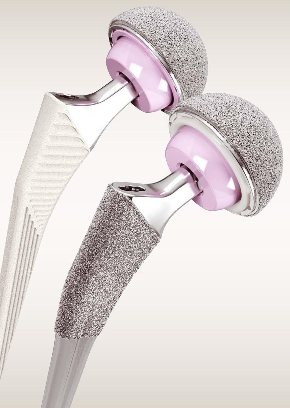
3 Introduction This brochure presents the Anterior Approach in the supine position for hip arthroplasty. The approach uses the internervous plane between the femoral nerve on the medial, superior and inferior gluteal nerve on the lateral side. There is no resection of any muscular attachment making this a truly tissue sparing approach. Hueter in1882 first described the Anterior Approach making this the oldest approach to the hip joint. The approach gained popularity in France in the early fifties with LeTournel and the Judet brothers, who recognised the importance of preserving the muscles surrounding the hip joint. They advocated the use of an orthopaedic traction table to facilitate access to femur and acetabulum, however this has certain disadvantages. In this brochure we describe the Anterior Approach, without the need for an orthopaedic traction table. The supine position gives optimal control of acetabular cup placement, stability and leg length. The procedure can be done with standard Operating Room equipment, but specific instruments as described in this brochure help facilitate the operation. Fluoroscopy can also be used to improve accuracy of cup and stem placement. This approach allows placement of both cemented and uncemented stems and may be considered for all patients regardless of Body Mass Index. There is however a learning curve with the Anterior Approach and it is advised to follow a dedicated learning journey and begin with selected simple cases. Hans-Erik Henkus Tom Hogervorst John Van Overschelde Kristoff Corten Bone Preserving Hip Systems The DePuy Synthes portfolio of hip products offers bone preserving options for patients undergoing the Anterior Approach. When evaluating the Anterior Approach, an overall plan to maximise soft tissue preservation and bone preservation should be considered. The Anterior Approach, when combined with a bone preserving femoral stem, achieves both soft tissue preservation and bone preservation for the patient. DePuy offers two bone preserving stems, which complement the Anterior Approach; the CORAIL Hip System, which has 25 years of clinical success and the TRI-LOCK Bone Preservation Stem. Both stems provide options to treat a spectrum of patient anatomies. The PINNACLE Acetabular Cup System also offers an ideal solution for tissue sparing surgery, with flexible bearing options and specialised instrumentation. 1

4 Preoperative Planning The primary goal of total hip arthroplasty is the anatomic reconstruction of the hip joint, resulting in favourable prosthetic joint load and function. Mechanically, the goals are to create a stable articulation with optimised range of motion, restored biomechanics for muscular efficiency and equalised limb lengths. Meeting these goals begins with a thorough analysis of the hip with comparison to the contralateral side in anterior/ posterior (A/P) and lateral projections. Figure 1 Please see the surgical techniques for the TRI-LOCK Bone Preservation Stem ( ) and the CORAIL Hip System ( ) for further information on preoperative planning. Patient Positioning This approach may be considered for all patients. The patient lies in a supine position, preferably on a radiolucent table that can extend/break at the position of the anterior superior iliac spine (ASIS). The arm on the operative side can be positioned as shown in Figure 2a or positioned over the patient's body as shown in Figure 2b. It is important to drape both legs individually so they are mobile throughout the procedure. As shown in Figure 2c. Figure 2a. Figure 2b. Figure 2c. 2
 and the CORAIL Hip System (9066-35-025) for further information on preoperative planning.")
5 Initial Incision Planning Three landmarks need to be identified (Figure 3): 1. The anterior superior iliac spine (ASIS). 2. The tip of the greater trochanter. 3. The lateral side of the patella. The interval between the tensor fascia lata (TFL) laterally and the sartorius muscle medially can be palpated following a line between the ASIS and the lateral side of the patella (Figure 4) The incision starts 2-3 cm posterior and 1-2 cm distal to the anterior superior iliac spine and extends in a distal direction 2-3 cm below the greater trochanter. The incision should be lateral to the interval between the tensor and sartorius muscle avoiding the lateral femoral cutaneous nerve that runs variably in the fascia over the sartorius muscle (Figure 5). Figure 3 Sartorius Generally the incision should run centrally along the muscle belly of the TFL. However, ensure the fascia incision is somewhat anterior to avoid damage to the perforating artery supplying the skin dorsal to the incision. Tensor Facia Lata Figure 4 Figure 5 3

6 Initial Skin Incision Make the initial skin incision and divide the subcutaneous tissue in line with the incision. When the fascia and tensor muscle is identified the surgeon can palpate the ASIS. Start the fascia incision two finger breaths from the ASIS. A longitudinal incision is made through the translucent facia lata in line with the fibres over the belly of the muscle. (Figure 6). The fascia on the medial side is lifted from the muscle itself and the intermuscular interval between the tensor laterally and the sartorius muscle medially is entered using your finger, making sure to stay within the TFL sheath. (Figure 7). Facia Lata Figure 6 Sartorius Tensor Facia Lata Figure 7 4

7 Capsule Exposure There is a thin fascia overlying the capsule which has to be opened first, to expose the pericapsular fat, underneath which is the joint capsule itself. The capsule of the hip on the lateral side and the anterior part of the greater trochanter can now be palpated. A retractor is positioned extracapsularly underneath the gluteus miniumus and lateral to the neck, retracting the tensor muscle. During the whole procedure care should be taken to protect the tensor muscle from tearing. The lateral femoral circumflex vessels cross the intermuscular interval just distally of the intertrochanteric line (Figure 8). These vessels must be clamped or cauterised; If you can t find them, they will find you!. Rectus Femoris Circumflex Vessels Vastus Intermedius Tensor Facia Lata Capsule The vessels indicate the level of a plane that is just superficial of the fat pad, over the anterior capsule. Once these vessels have been cauterised you can identify the fat pad under the rectus femoris muscle, removal of the fat pad will identify the anterior capsule. This will allow identification the reflected femoral head of the rectus femoris muscle, which follows the anterior acetabular rim and can be elevated from the anteromedial part of the capsule using a rasparatory. A second retractor can now be placed extracapsularly on the medial side proximal to the trochanter minor retracting the rectus femoris muscle medially exposing the whole anterior part of the capsule (Figure 9). Figure 8 Vastus Lateralis Capsule Vastus Intermedius Tensor Facia Lata Figure 9 5

8 Capsule Incision Vastus Lateralis Capsule The capsule can be incised in an inverted T or H shape or excised at surgeons will. The intertrochanteric line forms the inferior border and is identified distally by the junction of the capsule and the origin of the vastus lateralis muscle. Two retractors can now be placed intracapsularly on both sides of the femoral neck. Tip: On the lateral side a weight can be used on the retractor to retract the tensor muscle. Vastus Intermedius Tensor Facia Lata Figure 10 6

9 Femoral Neck Resection The intertrochanteric line, which can also be seen on the X-ray can be used as a reference for the osteotomy of the femoral neck. Reposition the laterally placed retractor to protect the greater trochanter. Use a long narrow saw blade to cut the femoral neck at a level that is appropriate for the chosen femoral stem (Figure 11). Vastus Lateralis Rectus Femoris After completing the osteotomy a chisel can be used to flip the femoral neck towards the front. This will allow the introduction of a corkscrew into the femoral head. Vastus Intermedius Before the femoral head can be removed, it may be necessary to release the posterior capsule attached to the femoral head. The corkscrew can be used as a joystick. Twist the head several times before removing it. In a case where the hip is stiff or there are acetabular osteophytes hampering the removal of the head, a slice of bone from the femoral neck, can be taken out first; creating space for the femoral head to be removed (Figure 12). Tensor Facia Lata Figure 11 Caution: Try to prevent levering on the tensor muscle with the corkscrew to avoid damaging the anterior fibers. In a case of difficulty in retrieving the head you can cut the labrum and remove more antero-superior capsule. Alternatively you can place the corkscrew in the head/neck junction anteriorly and pull in the direction of the socket. Figure 12 7

10 Acetabular Preparation and Reaming The preparation of the cup is similar to any other approach. Insert a retractor just over the anterior acetabular rim retracting the reflected head of the rectus muscle. A second retractor is placed underneath the posterior rim of the acetabulum. A third retractor may be placed under the transverse ligament to hold down the femur. In cases where the reflected head of the rectus muscle obstructs good visualisation of the acetabulum an additional retractor can be placed over the anterior inferior acetabular rim. The reflected head of the rectus muscle can be sharply detached from the capsule. Caution: be aware of the femoral nerve about 2 cm medial of the anterior acetabular rim and the Psoas tendon that can be damaged if the retractor is wrongly placed. Figure 13 Remove the acetabular labrum and remaining soft tissues obstructing good visualisation before acetabular reaming. Reaming can be done with either a straight or angled reamer. The transverse acetabular ligament and antero-lateral acetabular rim can be used as landmarks. In difficult cases an image intensifier can be used. Straight Handled Acetabular Reamer Curved Handled Acetabular Reamer Figure 14 8

11 Acetabular Trial and Implantation To check the quality of the acetabular reaming a Pinnacle trial shell can be impacted before the definitive PINNACLE cup is placed. The orientation of the cup can be checked using the anatomical landmarks of the antero-lateral acetabular rim and the transverse acetabular ligament. Be certain the PINNACLE cup is well seated in the acetabulum and does not overhang anteriorly causing impingement of the Psoas tendon. An image intensifier can be used easily in the supine position. Straight PINNACLE Cup Inserter Caution: be aware that in this approach the femur can force the cup into too much inclination and anteversion. Please refer to the PINNACLE Surgical technique for full details. Curved PINNACLE Cup Inserter Figure 15 Motion to adjust inclination Motion to adjust anteversion Figure 16 9

12 Cup Positioning Peer reviewed publications highlight the importance of acetabular component positioning in relation to short and long term outcomes during total hip arthroplasty for all types of bearing materials. 1-8 Cup positioning should be varied to optimise fixation, range of motion and dislocation resistance and minimise the likelihood of subluxation, impingement and edge loading. This may be assessed during pre-operative planning, acetabular preparation and cup trialling. Suboptimal component positioning may lead to edge loading, dislocation, increased wear, elevated metal ion release, ceramic squeaking and polyethylene fracture 1-8 The target cup inclination (as measured on radiographs) should be taking into account local soft tissue and anatomic landmarks. The target cup anteversion (as measured on radiographs) should be taking into account local soft tissue and anatomic landmarks. An alignment guide is provided to assist with cup positioning; however, cup orientation in the patient depends on patient position. The alignment guide does not allow for variation in patient position with respect to the operating table and it should be noted that patient orientation can vary throughout the procedure. 10

13 Leg Positioning For the femur, the critical step is the release of the capsule at the postero-superior neck remnant and fossa Piriformis. This can be accomplished by pulling the femur forward using a bone hook and electrocautery to release the capsule. Care should be taken not to release the Piriformis tendon. The insertion of the Piriformis tendon can be seen medially of the neck-trochanter junction when the leg is in (90 ) external rotation. Make some vertical cuts of the posterior capsule, till the femur is coming up Pull the femur upwards and laterally judging the effect of the release as you cut. The proximal femur is presented within the wound by putting the leg in a lazy figure of four position, extending / breaking the operating table by 20 30, elevating the femur with a bone hook and stabilising this position with a double bent retractor behind the greater trochanter (Figure 18). Figure 17 A second retractor is placed on the medial side of the calcar pushing the femur laterally. Tip: An inadequate release of the posterior capsule exposing the proximal femur can be hampered when the greater trochanter is stuck behind the pelvis. This can be solved by pulling the proximal femur lateral and then forward while releasing the capsule. When the exposure is not good, look at positioning in terms of adduction. Figure 18 Figure 19 11

14 CORAIL Hip System Femoral Canal Preparation Figure 20 Use the CORAIL broaches to create the femoral cavity by compacting the cancellous bone. To avoid varus positioning, it is important to enter the femoral canal as laterally as needed. Similarly, to avoid three point fixation in the lateral plane, insert the broach handle along the posterior wall of the proximal femur. This also allows the broach to follow the correct anteversion. Note that the posterior wall is located medially in the wound when the femur is externally rotated. Use a chisel or a canal finder to check the correct entry alignment (Figure 20). Begin with the smallest broach attached to the broach handle and increase the size of broaches one at a time. Stop broaching when axial and rotational stability are achieved in order to preserve cancellous bone and encourage osteointegration (Figure 21). The anteversion is automatically set by the anatomy of the femur. Please refer to the CORAIL Surgical Technique for full details (Cat. No.: ). The CORAIL system offers additional offset handles for patients where the iliac crest tends to obstruct a straight entrance to the femoral shaft (Figure 22). Figure 21 Caution: be aware that the lazy figure of four can put the patient's femur into internal rotation which can influence stem anteversion. Anteversion can be checked by referring to the epiphyseal line between the distal femur and the patella. Curved Lateral, Anterior Lateral, Direct Anterior Dual Offset Direct Anterior Extra Curved Direct Anterior Figure 22 12
.")
15 CORAIL Hip System Trial Reduction & Implantation With the last broach in situ, attach the selected neck and femoral head trials (Figure 23) and reduce the hip. When desired stability is achieved, remove the broach, the femoral head and neck trials. Do not irrigate or dry the femoral canal. This will help to preserve the quality of the compacted cancellous bone that encourages osteointegration. First insert the definitive liner into the cup. Insert the CORAIL stem and impact it for the last centimetres until stability is achieved (Figure 24). Clean and dry the taper thoroughly to ensure it is completely free of debris. Place the appropriate femoral head onto the taper. Using the head impactor, engage the head with moderate blows. Clean the bearing surfaces, and reduce the hip. Please refer to the CORAIL surgical technique for full details (Cat. No.: ). The supine position gives you optimal control of stability and leg length. The reduction should be effortless. However, the hip should remain stable throughout a full range of motion including extension, external rotation and maximum flexion and internal rotation. Figure 23 Leg length can be assessed by either looking at the knees, the malleoli or the heels. Figure 24 13
. Clean and dry the taper thoroughly to ensure it is completely free of debris.")
16 TRI-LOCK Bone Preservation Stem Femoral Canal Preparation Utilise the modular box osteotome to enter the femoral canal and to establish version. If needed the box osteotome can be used to clear bone laterally. In some cases, particularly in hard bone,the femoral canal can be developed using a small instrument such as a Canal Finder (Cat. No ). Approach enabling broach handle options The TRI-LOCK Bone Preservation Stem offers several broach handles that enable the many surgical approaches for hip replacement. The Curved and Dual Offset broach handles are available for the anterior approach technique. Figure 25 Curved Dual Offset Begin using a broach at least two sizes smaller than the preoperatively templated stem size. The starter broach can be used when needed for small femoral geometries, or for clearing bone laterally. While taking care to maintain proper alignment and version, sequentially advance the broaches down the femoral canal. Continue to increase broach size until intimate contact is made between the broach and the medial and lateral cortices. The final size is achieved when the broach maintains axial and rotational stability, and is at a seating level that recreates proper leg length. Calcar planing is optional, as the TRI-LOCK Bone Preservation Stem is a collarless design. With the final broach fully seated, place the planer over the broach stud. Apply power prior to engaging the calcar to prevent the planer from binding. Mill the calcar to the level of the broach face. Caution: be aware that the "lazy figure of four" can put the patient's femur into internal rotation that can influence stem anteversion. Anteversion can be checked by referring to the epiphyseal line between the distal femur and the patella. Figure 26 14

17 TRI-LOCK Bone Preservation Stem Trial Reduction & Implantation Trial neck segments and trial heads are available to assess proper component position, joint stability, range-of-motion and leg length. Standard and high offset options are available for each stem size. Offset increases 6-8 mm (depending on stem size) from the standard to the high offset option, via direct lateralisation. With the final broach in-situ, attach the appropriate trial neck and trial head. First insert the definitive liner into the cup. Reduce the hip and assess what adjustments, if any, are required to ensure stability through a full range of motion. When stability is achieved, note the broach size and head/neck offset. Stem inserters are available with various geometries, this enables each surgeon to decide according to their surgical preference. The retaining stem inserter can be used if a positive connection between the implant and instrument is required. Select the stem size that corresponds to the final broach. In the area of GRIPTION coating, the implant is oversized by 0.25 mm per side relative to the broach. Introduce the implant into the femoral canal by hand. Take care to orient the implant with proper alignment and version. Using moderate mallet blows, advance the stem into position. The implant is fully seated when the top of the GRIPTION coating reaches the level where the face of the broach previously sat and the implant is stable. Excessive force should not be needed to seat the stem. Figure 27 Approach enabling stem inserter options Threaded Straight Modular Curved Modular Offset Modular Following the final trial reduction, clean and dry the taper thoroughly to ensure it is completely free of debris. Place the appropriate femoral head onto the taper. Using the head impactor, engage the head with light taps. Clean the bearing surfaces, and reduce the hip. Figure 28 Bullet-tip Modular Please refer to the TRI-LOCK Bone Preservation Stem surgical technique for full details (Cat. No ). The supine position gives you optimal control of stability and leg length. The reduction should be effortless. However, the hip should remain stable throughout a full range of motion including extension, external rotation and maximum flexion and internal rotation. Leg length can be assessed by either looking at the knees, the malleoli or the heels. Figure 29 15

18 Final Closure Capsular closure can be performed if desired. The fascia lata is closed with a running suture, followed by subcutaneous and skin sutures. A wound drain can be used at will. Figure 30 Figure 31 16

19 Surgical Tips Hans-Erik Henkus During the whole procedure care should be taken to protect the tensor muscle from tearing. Make use of an image intensifier in difficult cases when you can not rely on the patient's anatomy. Beside an inadequate release of the posterior capsule exposing the proximal femur can be hampered when the greater trochanter is stuck behind the pelvis. This can be solved by pulling the proximal femur lateral and then forward while releasing the capsule. When broaching the femoral canal, The direct anterior approach tends to force you in a varus position. Enter the femoral canal as lateral as needed and use the canal finder. Do not start broaching until you are absolutely certain about the position of the proximal femur. You can extend the incision proximally towards the anterior superior iliac spine and then bending laterally following the iliac crest doing a classic Smith-Peterson approach. You can extend the incision distally going laterally reaching the femur behind the vastus lateralis muscle. This approach can be done in almost every patient. However attention should be given to the very obese, especially when there is overhanging paninculus hampering the entrance of the femoral canal. The hardest cases are muscular males and cases with a short femoral neck or extreme coxa vara. John Van Overschelde Before draping, assess patient positioning on table and look for leg length discrepancy. Make the incision of the facia over the purple muscle belly, avoid the white region medial and lateral of it otherwise you will end up in the wrong plane. While resecting the capsule clean the fossa digitalis completely so you can hook the tip of the greater trochanter with your index finger. Protect the tensor facia lata with a retractor during osteotomy of the femoral neck and be careful when taking out the head not to tear the tensor with some sharp ends of the neck. Use a curved reamer and impactor to avoid steep positioning of the cup. When lifting the femur push the retractor in the direction of the fibres of the facia lata muscle so you avoid tearing the muscle. Resect enough lateral cortical bone before broaching to avoid varus positioning of the stem. At the end of the intervention take a second look at the circumflex vessels and coagulate them if necessary. 17

20 References 1. Brodner W, Grübl A, Jankovsky R, Meisinger V, Lehr S, Gottsauner-Wolf FJ. Cup inclination and serum concentration of cobalt and chromium after metal-on-metal total hip arthroplasty. J Arthroplasty. 2004;19(8 Suppl 3): Williams S, Leslie I, Isaac G, Jin Z, Ingham E, Fisher J. Tribology and wear of metal-on-metal hip prostheses: influence of cup angle and head position. J Bone Joint Surg. 2008;90A Suppl 3: Udomkiat P, Dorr LD, Wan Z. Cementless hemispheric porous-coated sockets implanted with press-fit technique without screws: average ten-year follow-up. J Bone Joint Surg. 2002;84A: Schmalzried TP, Guttmann D, Grecula M, Amstutz H. The relationship between the design, position, and articular wear of acetabular components inserted without cement and the development of pelvic osteolysis. J Bone Joint Surg. 1994;76A: Kennedy JG, Rogers WB, Soffee KE, et al. Effect of acetabular component orientation on recurrent dislocation, pelvic osteolysis, polyethylene wear and component migration. J Arthroplasty 1998;13: Prudhommeaux F, Hamadouche M, Nevelos J, et al. Wear of alumina-on-alumina total hip arthroplasty at a mean 11-year followup. Clin Orthop Relat Res. 2000; 397: Walter WL, O Toole GC, Walter WK, Ellis A, Zicat BA. Squeaking in ceramic-on ceramic hips: the importance of acetabular component orientation. J Arthroplasty. 2007;22: Tower SS, Currier JH, Currier BH, Lyford KA, Van Citters DW, Mayor MB. Rim cracking of the cross-linked longevity polyethylene acetabular liner after total hip arthroplasty. J Bone Joint Surg Am Oct;89(10): This publication is not intended for distribution in the USA. DePuy Orthopaedics EMEA is a trading division of DePuy International Limited. Registered Office: St. Anthony s Road, Leeds LS11 8DT, England Registered in England No DePuy Orthopaedics, Inc. 700 Orthopaedic Drive Warsaw, IN USA Tel: +1 (800) Fax: +1 (574) depuysynthes.com DePuy International Ltd St Anthony s Road Leeds LS11 8DT England Tel: +44 (0) Fax: +44 (0) DePuy International Ltd. and DePuy Orthopaedics, Inc All rights reserved version 2 Issued: 04/13 CA#DPEM/ORT/1212/0407a

BONE PRESERVATION STEM
 TRI-LOCK BONE PRESERVATION STEM Featuring GRIPTION Technology SURGICAL TECHNIQUE IMPLANT GEOMETRY Extending the TRI-LOCK Stem heritage The original TRI-LOCK Stem was introduced in 1981. This implant was
MIS Direct Anterior Approach Surgical Protocol
 Joint Replacements MIS Direct Anterior Approach Surgical Protocol The Right Procedure and the Right Implant for the Right Patient Scientific advice and text: Franz Rachbauer, M.D., M.A.S., M.Sc. Associate
Total hip arthroplasty (THA) through the anterior approach
 through the anterior approach") Primary Total Hip Arthroplasty with a Minimally Invasive Anterior Approach Jonathan G. Yerasimides, MD, and Joel M. Matta, MD Total hip arthroplasty (THA) through the anterior approach is a minimally invasive
Primary Total Hip Arthroplasty with a Minimally Invasive Anterior Approach Jonathan G. Yerasimides, MD, and Joel M. Matta, MD Total hip arthroplasty (THA) through the anterior approach is a minimally invasive
Surgical Approaches to Total Hip Arthroplasty
 Surgical Approaches to Total Hip Arthroplasty Daniel Kelmanovich, 1 Michael L. Parks, MD, 2 Raj Sinha, MD, PhD, 3 and William Macaulay, MD 4 Surgical exposure of the hip for trauma, infection, or reconstruction
Minimally Invasive Hip Replacement through the Direct Lateral Approach
 Surgical Technique INNOVATIONS IN MINIMALLY INVASIVE JOINT SURGERY Minimally Invasive Hip Replacement through the Direct Lateral Approach *smith&nephew Introduction Prosthetic replacement of the hip joint
VERSYS HERITAGE CDH HIP PROSTHESIS. Surgical Technique for CDH Hip Arthroplasty
 VERSYS HERITAGE CDH HIP PROSTHESIS Surgical Technique for CDH Hip Arthroplasty SURGICAL TECHNIQUE FOR VERSYS HERITAGE CDH HIP PROSTHESIS CONTENTS ANATOMICAL CONSIDERATIONS....... 2 PREOPERATIVE PLANNING............
Zimmer M/L Taper Hip Prosthesis. Surgical Technique
 Zimmer M/L Taper Hip Prosthesis Surgical Technique Zimmer M/L Taper Hip Prosthesis 1 Zimmer M/L Taper Hip Prosthesis Surgical Technique Table of Contents Preoperative Planning 2 Determination of Leg Length
P REPLACEMENT SURGERY
 P REPLACEMENT SURGERY DIRECT ANTERIOR APPROACH M I N I M I Z I N G R E C O V E R Y. M A X I M I Z I N G R E S U L T S. CENTER FOR MINIMAL INVASIVE JOINT SURGERY 2301 25TH STREET SOUTH FARGO ND 58103 701-241-9300
The Anterior Approach for Total Hip Replacement. Background and Operative Technique
 The Anterior Approach for Total Hip Replacement Background and Operative Technique Joel M. Matta, MD Hip and Pelvis Institute at Saint John s Health Center, Director Santa Monica, California RECOVERY FUNCTION
Zimmer M/L Taper Hip Prosthesis with Kinectiv Technology. Surgical Technique
 Zimmer M/L Taper Hip Prosthesis with Kinectiv Technology Surgical Technique Zimmer M/L Taper Hip Prosthesis with Kinectiv Technology 1 Zimmer M/L Taper Hip Prosthesis with Kinectiv Technology Surgical
Metasul LDH Large Diameter Head
 Metasul LDH Large Diameter Head Surgical Technique Metasul LDH Large Diameter Head Surgical Technique Enhancing Stability and Increasing Range of Motion Metasul LDH Large Diameter Head Surgical Technique
PINNACLE. Polyethylene Surgical Technique. Surgical Technique
 PINNACLE Hip Solutions Polyethylene Surgical Technique Surgical Technique Table of Contents Surgical technique Introduction 4 Templating and Pre-Operative Planning 6 Surgical Approach Anterolateral 8
Versa-Fx II Femoral Fixation System Surgical Techniques
 Versa-Fx II Femoral Fixation System Surgical Techniques Versa-Fx II Femoral Fixation System Surgical Techniques 1 Surgical Technique For Fixation Of Intertrochanteric and Supracondylar Fractures of the
MET: Posterior (backward) Rotation of the Innominate Bone.
 Rotation of the Innominate Bone.") MET: Posterior (backward) Rotation of the Innominate Bone. Purpose: To reduce an anterior rotation of the innominate bone at the SI joint. To increase posterior (backward) rotation of the SI joint. Precautions:
MET: Posterior (backward) Rotation of the Innominate Bone. Purpose: To reduce an anterior rotation of the innominate bone at the SI joint. To increase posterior (backward) rotation of the SI joint. Precautions:
ACCOLADE II. Orthopaedics. Femoral Hip System. Surgical Technique
 ACCOLADE II Femoral Hip System Orthopaedics Surgical Technique Indications The indications for use of the total hip replacement prostheses include: Noninflammatory degenerative joint disease, including
Exeter X3 RimFit. Acetabular Cup Surgical Protocol
 Exeter X3 RimFit Acetabular Cup Surgical Protocol 2 Exeter X3 RimFit Surgical Protocol Table of Contents Indications and Contraindications... 4 Introduction... 5 Surgical Protocol... 6 Step 1 - Pre-Operative
Chapter 9 The Hip Joint and Pelvic Girdle
 Copyright The McGraw-Hill Companies, Inc. Reprinted by permission. The Hip Joint and Pelvic Girdle Chapter 9 The Hip Joint and Pelvic Girdle Structural Kinesiology R.T. Floyd, Ed.D, ATC, CSCS Hip joint
Aesculap Orthopaedics Metha
 Aesculap Orthopaedics Metha Short Hip Stem Evolving the State of Arthroplasty 2 Evolving the State of Arthroplasty Contents Short Hip Stem System 4 Short Stem Anchoring Concept 6 Variable Implant Options
The Right Choice. Exeter. total hip system
 The Right Choice Exeter total hip system Exeter The Right Choice Anatomic Reconstruction Offset The objectives of total hip replacement are to: relieve pain increase mobility and function Achieving a correct
Why an Exactech Hip is Right for You
 Why an Exactech Hip is Right for You Why do I need a total hip replacement? Which surgical approach is best for me? How long will it last? Which implant is right for me? Founded in 1985 by an orthopaedic
Surgical technique. Sagitta EVL
 Surgical technique Sagitta EVL Sagitta EVL The range Offset 7 mm The collar neck increases by mm from the size. 1 Size L (mm) Standard Offset DP+ 1. / 0 10 6 L 0+ 1 6.. 1 16 6.6.6 10 7 1 7.. 160 0 7 16
Product overview and surgical technique
 Product overview and surgical technique Overview Every patient moves differently 1 and their total hip replacement should be optimised to account for this. The orientation of the acetabular cup is one
Restoration Modular Cone Body/Conical Distal Stem Femoral Components Using the Restoration Modular Instrument System Surgical Technique
 Restoration Modular Cone Body/Conical Distal Stem Femoral Components Using the Restoration Modular Instrument System Surgical Technique CONE CONICAL BROACHED CALCAR MILLED CONE MT3 FLUTED PLASMA CONICAL
Design surgeon list. Smith & Nephew thanks the following surgeons for their participation as part of the R3 system design team:
 Surgical Technique Design surgeon list Smith & Nephew thanks the following surgeons for their participation as part of the R3 system design team: Robert Barrack, MD St. Louis, Missouri Robert Bourne, MD
Wagner Cone Prosthesis Hip Stem. Surgical Technique
 Wagner Cone Prosthesis Hip Stem Surgical Technique Wagner Cone Prosthesis Hip Stem Surgical Technique 3 Surgical Technique Wagner Cone Prosthesis Table of Contents History 4 Design Features 4 Wagner Cone
Introduction to the Bertram Hip Spacer
 Introduction to the Bertram Hip Spacer Approximately 200,000 hip replacement surgeries are done every year in United States. Fortunately the incidence of infection is routinely 0.5 percent-1 percent. However
How To Use A Phoenix Retrograde Femoral Nail
 Phoenix Retrograde Femoral Nail System Featuring CoreLock Technology Surgical Technique Contents Introduction... Page 1 Indications... Page 2 Design Features... Page 3 Surgical Technique... Page 6 Product
Femoral head and neck ostectomy (FHO) is a commonly
 is a commonly") P ro c e d u re s P ro O R T H O P E D I C S Peer Reviewed Laura E. Peycke, DVM, MS, Diplomate CVS Texas &M University Femoral Head & Neck Ostectomy Femoral head and neck ostectomy (FHO) is a commonly
P ro c e d u re s P ro O R T H O P E D I C S Peer Reviewed Laura E. Peycke, DVM, MS, Diplomate CVS Texas &M University Femoral Head & Neck Ostectomy Femoral head and neck ostectomy (FHO) is a commonly
ReCap Product Rationale
 FLH 155 01/09 Biomet UK Ltd Waterton Industrial Estate Bridgend, South Wales CF31 3XA, United Kingdom Tel. +44 (0)1656 655221 Fax. +44 (0)1656 645454 ReCap Product Rationale A complete resurfacing system
Musculoskeletal Ultrasound Technical Guidelines. IV. Hip
 European Society of MusculoSkeletal Radiology Musculoskeletal Ultrasound Technical Guidelines IV. Ian Beggs, UK Stefano Bianchi, Switzerland Angel Bueno, Spain Michel Cohen, France Michel Court-Payen,
Rejuvenate Total Hip System Surgical Protocol. Rejuvenate Modular Hip Stem Enhanced Stability 1 Proven Modularity 2 Intraoperative Flexibility
 Rejuvenate Total Hip System Surgical Protocol Rejuvenate Modular Hip Stem Enhanced Stability 1 Proven Modularity 2 Intraoperative Flexibility Rejuvenate Total Hip System Surgical Protocol Table of Contents
Accolade. System. Brochure. Achieving Perfect Balance. Cemented and Cementless Femoral Hip System
 Accolade System Brochure Achieving Perfect Balance Cemented and Cementless Femoral Hip System Accolade Cemented Hip System Potential for Perfect Balance The Accolade C Femoral Component: A perfect balance
DUAL INCISION HIP REPLACEMENT ST. CLOUD HOSPITAL, ST. CLOUD, MINNESOTA Broadcast November 16, 2004
 DUAL INCISION HIP REPLACEMENT ST. CLOUD HOSPITAL, ST. CLOUD, MINNESOTA Broadcast November 16, 2004 NARRATOR A new technique in dual incision MIS hip replacement surgery is redefining the way surgeries
Zimmer M/L Taper Hip Prosthesis with Kinectiv Technology
 Zimmer M/L Taper Hip Prosthesis with Kinectiv Technology Hips designed to fit the unique anatomies of men and women Independent control for a natural fit Simple, practical solutions for optimal restoration
CPT 12/14 Hip System. Primary Hip Athroplasty Surgical Technique
 1 CPT 12/14 Hip System Primary Hip Athroplasty Surgical Technique 2 CPT 12/14 Hip System Surgical Technique CPT 12/14 Hip System Surgical Technique 3 PRIMARY SURGICAL TECHNIQUE FOR THE CPT 12/14 COLLARLESS
Anatomy and Pathomechanics of the Sacrum and Pelvis. Charles R. Thompson Head Athletic Trainer Princeton University
 Anatomy and Pathomechanics of the Sacrum and Pelvis Charles R. Thompson Head Athletic Trainer Princeton University Simplify Everything There are actually only three bones: Two innominates, one sacrum.
DRAFT. Triathlon TS Knee System. Surgical Protocol. Version 1
 DRAFT Version 1 Triathlon TS Knee System Table of Contents Acknowledgments..........................................................2 Tibial Canal Preparation....................................................4
Posterior Referencing. Surgical Technique
 Posterior Referencing Surgical Technique Posterior Referencing Surgical Technique INTRO Introduction Instrumentation Successful total knee arthroplasty depends in part on re-establishment of normal lower
Anterior Lumbar Interbody Fusion (ALIF). Instrument set supports placement of ALIF spacers using anterior or anterolateral approach.
. Instrument set supports placement of ALIF spacers using anterior or anterolateral approach.") Anterior Lumbar Interbody Fusion (ALIF). Instrument set supports placement of ALIF spacers using anterior or anterolateral approach. Technique Guide Instruments and implants approved by the AO Foundation
Anterior Lumbar Interbody Fusion (ALIF). Instrument set supports placement of ALIF spacers using anterior or anterolateral approach. Technique Guide Instruments and implants approved by the AO Foundation
Malleolar fractures Anna Ekman, Lena Brauer
 Malleolar fractures Anna Ekman, Lena Brauer How to use this handout? The left column is the information as given during the lecture. The column at the right gives you space to make personal notes. Learning
Zimmer Natural Nail System. Cephalomedullary Nail Surgical Technique STANDARD
 Zimmer Natural Nail System Cephalomedullary Nail Surgical Technique STANDARD Zimmer Natural Nail System Cephalomedullary Nail Surgical Technique - Standard 1 Zimmer Natural Nail System Cephalomedullary
The information contained in this document is intended for healthcare professionals only.
 The information contained in this document is intended for healthcare professionals only. Dall-Miles Cabling System Dall-Miles Recon and Trauma Cable System Trochanteric Reattachment Using the Trochanteric
Converge CSTi Porous Acetabular Cup System. Surgical Technique
 Converge CSTi Porous Acetabular Cup System Surgical Technique 2 Converge CSTi Porous Acetabular Cup System Surgical Technique Converge CSTi Porous Acetabular Cup System Surgical Technique 3 CONVERGE CSTI
Y O U R S U R G E O N S. choice of. implants F O R Y O U R S U R G E R Y
 Y O U R S U R G E O N S choice of implants F O R Y O U R S U R G E R Y Y O U R S U R G E O N S choice of implants F O R Y O U R S U R G E R Y Your Surgeon Has Chosen the C 2 a-taper Acetabular System The
An overview of the anatomy of the canine hindlimb
 An overview of the anatomy of the canine hindlimb Darren Kelly Artwork by Paddy Lennon Original photos courtesy of Mary Ferguson Students at University College Dublin, School of Veterinary Medicine. Video
Restoration Modular. Revision Hip System Surgical Protocol
 Orthopaedics Restoration Modular Revision Hip System Restoration Modular Calcar Body/Conical Distal Stem Femoral Components Using the Restoration Modular Instrument System Revision Hip System Orthopaedics
Anterior Hip Replacement
 Disclaimer This movie is an educational resource only and should not be used to manage Orthopaedic health. All decisions about the management of hip replacement and arthritis management must be made in
Spinal Arthrodesis Group Exercises
 Spinal Arthrodesis Group Exercises 1. Two surgeons work together to perform an arthrodesis. Dr. Bonet, a general surgeon, makes the anterior incision to gain access to the spine for the arthrodesis procedure.
Zimmer M/L Taper Hip Prosthesis with Modular Neck Kinectiv Technology March 12, 2009 7:00 PM EDT
 Zimmer M/L Taper Hip Prosthesis with Modular Neck Kinectiv Technology March 12, 2009 7:00 PM EDT Welcome to this "OR Live" Webcast presentation brought to you by Zimmer. During the program, it s easy for
*smith&nephew SLR-PLUS
 Surgical Technique *smith&nephew SLR-PLUS Cementless Revision Stem SLR-PLUS Table of Contents Comments from the Author s Clinic... 3 Indications... 4 Contraindications... 5 Preoperative Planning... 5
Surgical Art. Formulaic Drawing Method. DRAWING WORKSHOP Learning to sketch for patient notes
 DRAWING WORKSHOP Learning to sketch for patient notes Surgical Art Formulaic Drawing Method Formulaic figure drawing systems involve using abstract rhythms and interlocking shapes to construct the human
Zimmer Periarticular Proximal Tibial Locking Plate
 Zimmer Periarticular Proximal Tibial Locking Plate Surgical Technique The Science of the Landscape Zimmer Periarticular Proximal Tibial Locking Plate 1 Table of Contents Introduction 2 Locking Screw Technology
Surgical Technique. Struan H Coleman MD, PhD
 Surgical Technique Guide Struan H Coleman MD, PhD the Author Struan H. Coleman MD, PhD, specializes in Sports Medicine at Hospital for Special Surgery where he treats orthopedic conditions of the shoulder,
How To Fix A Radial Head Plate
 Mayo Clinic CoNGRUENT RADIAL HEAD PLATE Since 1988 Acumed has been designing solutions to the demanding situations facing orthopedic surgeons, hospitals and their patients. Our strategy has been to know
ANTERIOR CERVICAL DISCECTOMY AND FUSION. Basic Anatomical Landmarks: Anterior Cervical Spine
 Anterior In the human anatomy, referring to the front surface of the body or position of one structure relative to another Cervical Relating to the neck, in the spine relating to the first seven vertebrae
Zimmer MOST Options System
 Zimmer MOST Options System Surgical Technique Modular Options for Severe Bone Loss and Trauma Zimmer MOST Options System Surgical Technique 1 MOST Options System Surgical Technique Table of Contents Introduction
PERIPROSTHETIC IMPLANTS
 PERIPROSTHETIC IMPLANTS PRODUCT OVERVIEW CLINICAL SOLUTIONS Periprosthetic fractures present unique challenges, such as how to gain fixation when the medullary canal is occupied. Special techniques and
Primary V40 Surgical Protocol. Exeter. total hip system
 Primary V40 Surgical Protocol Exeter total hip system V40 SURGICAL PROTOCOL HIGHLIGHTS 44mm Head diameter mm 22 26 28 30 32 36 1. Pre-operative Planning: Pre-operative templating is important to select
In this brochure you will: Keep in mind:
 Hip Implant Options Bearing Basics Today s hip implant patients are fortunate. Doctors can now choose from advanced implant materials. One choice doctors make when selecting the implant for you is called
Anatomic Percutaneous Ankle Reconstruction of Lateral Ligaments (A Percutaneous Anti ROLL)
") Anatomic Percutaneous Ankle Reconstruction of Lateral Ligaments (A Percutaneous Anti ROLL) Mark Glazebrook James Stone Masato Takao Stephane Guillo Introduction Ankle stabilization is required when a patient
Anatomic Percutaneous Ankle Reconstruction of Lateral Ligaments (A Percutaneous Anti ROLL) Mark Glazebrook James Stone Masato Takao Stephane Guillo Introduction Ankle stabilization is required when a patient
Hip Arthroscopy Principles and Application J.W. Thomas Byrd, M.D.
 Hip Series Technique Guide Hip Arthroscopy Principles and Application J.W. Thomas Byrd, M.D. As described by: J.W. Thomas Byrd, M.D. Southern Sports Medicine and Orthopaedic Center Nashville, TN Introduction
Lower Extremity Orthopedic Surgery in Cerebral Palsy. Hank Chambers, MD Rady Children s Hospital - San Diego
 Lower Extremity Orthopedic Surgery in Cerebral Palsy Hank Chambers, MD Rady Children s Hospital - San Diego Indications Fixed contracture Joint dislocations Shoe wear problems Pain Perineal hygiene problems
Structure and Function of the Hip
 Structure and Function of the Hip Objectives Identify the bones and bony landmarks of the hip and pelvis Identify and describe the supporting structures of the hip joint Describe the kinematics of the
How To Perform An Elbow Operation
 Coonrad/Morrey Total Elbow Surgical Technique Interchangeability, Anterior Flange, Clinical History Coonrad/Morrey Total Elbow Surgical Technique 1 Coonrad/Morrey Total Elbow Surgical Technique Table
NETWORK FITNESS FACTS THE HIP
 NETWORK FITNESS FACTS THE HIP The Hip Joint ANATOMY OF THE HIP The hip bones are divided into 5 areas, which are: Image: www.health.com/health/static/hw/media/medical/hw/ hwkb17_042.jpg The hip joint is
Achilles Tendon Repair, Operative Technique
 *smith&nephew ANKLE TECHNIQUE GUIDE Achilles Tendon Repair, Operative Technique Prepared in Consultation with: C. Niek van Dijk, MD, PhD KNEE HIP SHOULDER EXTREMITIES Achilles Tendon Repair, Operative
Orthopedic Foot Instruments. Dedicated instruments for reconstructive foot surgery.
 Orthopedic Foot Instruments. Dedicated instruments for reconstructive foot surgery. Surgical Technique This publication is not intended for distribution in the USA. Instruments and implants approved by
CuttingEdge Hip Instrumentation Femoral Surgical Technique for Primary Press-Fit and Cemented Hip Implants.
 CuttingEdge Hip Instrumentation Femoral Surgical Technique for Primary Press-Fit and Cemented Hip Implants. Surgical Protocol Table of Contents INTRODUCTION....2 FEMORAL SURGICAL TECHNIQUE Surgical Approach
Integra. Subtalar MBA and bioblock Implant SURGICAL TECHNIQUE
 Integra Subtalar MBA and bioblock Implant SURGICAL TECHNIQUE Table of contents Introduction Description... 2 Indications... 2 Contraindications... 2 Surgical Technique Step 1: Incision and Dissection...3
Wagner SL Revision Hip Stem
 Wagner SL Revision Hip Stem Surgical Technique Femoral Revision Prosthesis for Extensive Bone Loss Disclaimer This document is intended exclusively for experts in the field, i.e. physicians in particular,
Exeter. Surgical Technique. V40 Stem Cement-in-Cement. Orthopaedics
 Exeter Orthopaedics V40 Stem Cement-in-Cement Surgical Technique Exeter V40 Stem Cement-in-Cement Surgical Technique Table of Contents Indications and Contraindications...2 Warnings and Precautions...2
Technique Guide. Orthopaedic Cable System. Cerclage solutions for general surgery.
 Technique Guide Orthopaedic Cable System. Cerclage solutions for general surgery. Table of Contents Introduction The Orthopaedic Cable System 2 Indications 4 Contraindications 4 Surgical Technique Cerclage
Zimmer FuZion Instruments. Surgical Technique (Beta Version)
") Zimmer FuZion Surgical Technique (Beta Version) INTRO Surgical Technique Introduction Surgical goals during total knee arthroplasty (TKA) include establishment of normal leg alignment, secure implant fixation,
Zimmer FuZion Surgical Technique (Beta Version) INTRO Surgical Technique Introduction Surgical goals during total knee arthroplasty (TKA) include establishment of normal leg alignment, secure implant fixation,
Total Hip Replacement
 Please contactmethroughthegoldcoasthospitaswityouhaveanyproblemsafteryoursurgery. Dr. Benjamin Hewitt Orthopaedic Surgeon Total Hip Replacement The hip joint is a ball and socket joint that connects the
Anterior Superior Iliac Spine. Anterior Inferior Iliac Spine. head neck greater trochanter intertrochanteric line lesser trochanter
 Ilium Bones The Skeleton Ischium Pubis Sacro-iliac Joint Iliac Crest Anterior Superior Superior Pubic Ramus Anterior Inferior Acetabulum Obturator Foramen Ischio-pubic ramus Ischial tuberosity Pubic Crest
TOTAL HIP REPLACEMENT: MODERN SURGERY FOR SEVERE ARTHRITIS OF THE HIP
 TOTAL HIP REPLACEMENT: MODERN SURGERY FOR SEVERE ARTHRITIS OF THE HIP John T. Dearborn, M.D. and Alexander P. Sah, M.D. The Center for Joint Replacement Please read this pamphlet before you see me so that
M/DN Femoral Interlocking & Recon Nail Intramedullary Fixation Surgical Technique
 M/DN Femoral Interlocking & Recon Nail Intramedullary Fixation Surgical Technique M/DN Femoral Interlocking & Recon Nail Intramedullary Fixation 1 Surgical Techniques for Fixation of Femoral Fractures
Patellofemoral Joint: Superior Glide of the Patella
 Patellofemoral Joint: Superior Glide of the Patella Purpose: To increase knee extension. Precautions: Do not compress the patella against the femoral condyles. Do not force the knee into hyperextension
When is Hip Arthroscopy recommended?
 HIP ARTHROSCOPY Hip arthroscopy is a minimally invasive surgical procedure that uses a camera inserted through very small incisions to examine and treat problems in the hip joint. The camera displays pictures
The Pilates Studio of Los Angeles / PilatesCertificationOnline.com
 Anatomy Review Part I Anatomical Terminology and Review Questions (through pg. 80) Define the following: 1. Sagittal Plane 2. Frontal or Coronal Plane 3. Horizontal Plane 4. Superior 5. Inferior 6. Anterior
Technique Guide. DHS/DCS Dynamic Hip and Condylar Screw System. Designed to provide stable internal fixation.
 Technique Guide DHS/DCS Dynamic Hip and Condylar Screw System. Designed to provide stable internal fixation. Table of Contents Introduction Dynamic Hip Screw (DHS) 2 Dynamic Condylar Screw (DCS) 3 Indications
Aesculap Orthopaedics BiCONTACT
 Aesculap Orthopaedics BiCONTACT Hip Endoprosthesis System Bone Preservation. For Years to Come. BiCONTACT Hip Endoprosthesis System Bone Preservation. For Years to Come. 2 Table of contents Objective:
MINIMALLY INVASIVE TOTAL HIP REPLACEMENT VANDERBILT UNIVERSITY MEDICAL CENTER, NASHVILLE, TN Broadcast April 13 2005
 NARRATOR MINIMALLY INVASIVE TOTAL HIP REPLACEMENT VANDERBILT UNIVERSITY MEDICAL CENTER, NASHVILLE, TN Broadcast April 13 2005 For the more than 200,000 people who undergo hip replacement surgery every
13 Adductor Muscle Group Excision
 Malawer Chapter 13 21/02/2001 15:36 Page 243 13 Adductor Muscle Group Excision Martin Malawer and Paul Sugarbaker OVERVIEW The adductor muscle group is the second most common site for high- and low-grade
Knee Kinematics and Kinetics
 Knee Kinematics and Kinetics Definitions: Kinematics is the study of movement without reference to forces http://www.cogsci.princeton.edu/cgi-bin/webwn2.0?stage=1&word=kinematics Kinetics is the study
Arthroscopic Shoulder Instability Repair Using the SUTUREFIX ULTRA Suture Anchor and SUTUREFIX ULTRA Instrumentation System
 *smith&nephew SHOULDER TECHNIQUE GUIDE Arthroscopic Shoulder Instability Repair Using the SUTUREFIX ULTRA Suture Anchor and SUTUREFIX ULTRA Instrumentation System KNEE HIP SHOULDER EXTREMITIES Arthroscopic
Hip Resurfacing 2011 ORIGINAL ARTICLE. James W. Pritchett MD. Introduction. Abstract
 1 ORIGINAL ARTICLE Hip Resurfacing 2011 James W. Pritchett MD Abstract In 1938 Marion Smith-Peterson placed a cobalt chromium cup on a reshaped femoral head to perform the first hip resurfacing. 15 Also,
MDM. Surgical Technique. Modular Dual Mobility Acetabular System. Orthopaedics
 MDM X3 Modular Dual Mobility Acetabular System Orthopaedics Surgical Technique Indications The indications for use of total hip replacement prostheses include: Non-inflammatory degenerative joint disease
THE BENJAMIN INSTITUTE PRESENTS. Excerpt from Listen To Your Pain. Assessment & Treatment of. Low Back Pain. Ben E. Benjamin, Ph.D.
 THE BENJAMIN INSTITUTE PRESENTS Excerpt from Listen To Your Pain Assessment & Treatment of Low Back Pain A B E N J A M I N I N S T I T U T E E B O O K Ben E. Benjamin, Ph.D. 2 THERAPIST/CLIENT MANUAL The
MDMTM. Surgical Technique. Modular Dual Mobility TM Acetabular System. Orthopaedics
 MDMTM X3 Modular Dual Mobility TM Acetabular System Orthopaedics Surgical Technique Indications The indications for use of total hip replacement prostheses include: Non-inflammatory degenerative joint
High-Flex Solutions for the MIS Era. Zimmer Unicompartmental High Flex Knee System
 High-Flex Solutions for the MIS Era Zimmer Unicompartmental High Flex Knee System Zimmer Unicompartmental High Flex Knee Built On Success In today s health care environment, meeting patient demands means
Zimmer Small Fragment Universal Locking System. Surgical Technique
 Zimmer Small Fragment Universal Locking System Surgical Technique Zimmer Small Fragment Universal Locking System 1 Zimmer Small Fragment Universal Locking System Surgical Technique Table of Contents Introduction
S-ROM NOILES. This publication is not intended for distribution in the USA. SURGICAL TECHNIQUE
 S-ROM NOILES This publication is not intended for distribution in the USA. SURGICAL TECHNIQUE CONTENTS INTRODUCTION S-ROM NOILES Rotating Hinge Knee System 2 SURGICAL TECHNIQUE Initial Preparation of
Your Practice Online
 P R E S E N T S Your Practice Online Disclaimer This information is an educational resource only and should not be used to make a decision on Revision Hip Replacement or arthritis management. All decisions
Total Shoulder System. Anatomic adaptability... simplified SURGICAL TECHNIQUE
 Total Shoulder System Anatomic adaptability... simplified SURGICAL TECHNIQUE Inclination Version Offset 125-140 +/- 10 3.5 mm A of Possibilities I III The Univers II Total Shoulder System was designed
Structure & Function of the Knee. One of the most complex simple structures in the human body. The middle child of the lower extremity.
 Structure & Function of the Knee One of the most complex simple structures in the human body. The middle child of the lower extremity. Osteology of the Knee Distal femur (ADDuctor tubercle) Right Femur
Answers to commonly asked questions from patients with metal-on-metal hip replacements / resurfacings. Contents
 Answers to commonly asked questions from patients with metal-on-metal hip replacements / resurfacings John Skinner 1 and Alister Hart 2, Consultant Orthopaedic Surgeons and Directors of the London Implant
Technique Guide. Titanium Trochanteric Fixation Nail System. For intramedullary fixation of proximal femur fractures.
 Technique Guide Titanium Trochanteric Fixation Nail System. For intramedullary fixation of proximal femur fractures. Table of Contents Introduction Titanium Trochanteric Fixation Nail System 2 Biomechanical
ULTRASOUND GUIDED FEMORAL NERVE BLOCK ANAESTHESIA TUTORIAL OF THE WEEK 284
 ULTRASOUND GUIDED FEMORAL NERVE BLOCK ANAESTHESIA TUTORIAL OF THE WEEK 284 15 TH APRIL 2013 Dr Suresh Kumar Jeyaraj, Specialty Doctor Dr Tim Pepall, Consultant Anaesthetist Frimley Park Hospital NHS foundation
Zimmer Segmental System. Distal Femoral Surgical Technique
 Zimmer Segmental System Distal Femoral Surgical Technique INTRO Zimmer Segmental System Distal Femoral Surgical Technique Introduction The Zimmer Segmental System is designed to address significant bone