PROTOCOL ALL-11. Version 4.1 (10 April 2013)
|
|
|
- Rhoda Benson
- 10 years ago
- Views:
Transcription
70 367 45 45 Fax +31 (0)70 367 08 68 E-mail: info@skion.")
 with newly diagnosed acute lymphoblastic")
1 Leyweg 299, 2545 CJ The Hague PO box 43515, 2504 AM The Hague Tel. +31 (0) Fax +31 (0) PROTOCOL ALL-11 Treatment study protocol of the Dutch Childhood Oncology Group for children and adolescents (1-19 year) with newly diagnosed acute lymphoblastic leukemia Version 4.1 (10 April 2013)
 with newly diagnosed acute lymphoblastic")
2 Disclaimer This DCOG protocol is for research purposes only, and should not be copied, redistributed or used for any other purpose. The procedures in this DCOG protocol are intended only for use by pediatric oncologists in a carefully structured setting and following approval by a competent research ethics committee. They may not prove to be more effective than standard treatment. The investigator responsible as mentioned in the Protocol should be consulted first before using or attempting any procedure as described in this DCOG protocol unless this procedure is already part of the standard treatment. DUTCH CHILDHOOD ONCOLOGY GROUP PROTOCOL ALL-11 Treatment study protocol of the Dutch Childhood Oncology Group for children and adolescents (1-19 year) with newly diagnosed acute lymphoblastic leukemia CENTRAL OFFICE Dr. J.G. de Ridder-Sluiter, Director DCOG SUPERVISORY BOARD Prof. Dr. R. Pieters, chairman Dr. M.B. Bierings Prof. Dr. H.N. Caron Dr. E.S.J.M. de Bont Prof. Dr. P.M. Hoogerbrugge Prof. Dr. G.J.L. Kaspers Dr. W.J.W. Kollen LABORATORY Dr. V. de Haas, Head Laboratory Dr. E. Sonneveld, vice Head Laboratory DATAMANAGEMENT Drs. J.A. Lieverst, Head Trial Office 2/272

3 Protocol DCOG ALL-11 Treatment study protocol of the Dutch Childhood Oncology Group for children and adolescents (1-19 year) with newly diagnosed acute lymphoblastic leukemia DCOG ALL-11 Protocol committee Prof. Dr. R. Pieters, chair Erasmus MC, Rotterdam Dr. M. Bierings UMCU, Utrecht Dr. V. de Haas DCOG, The Hague Prof. Dr. P. Hoogerbrugge UMCN, Nijmegen Drs. H. Segers Erasmus MC, Rotterdam Dr. W. Tissing UMCG, Groningen Dr. M. Veening VUMC, Amsterdam Advisors: Dr. B. Beverloo (Cytogenetics) Prof. Dr. J van Dongen (MRD) Dr. M. Fiocco (Statistics) Dr. I. van der Sluis (Asparaginase) Dr. C. van Tilburg (IVIG) Erasmus MC, Rotterdam Erasmus MC, Rotterdam DCOG, The Hague & LUMC, Leiden Erasmus MC, Rotterdam UMCU, Utrecht Project Team at Trial Office DCOG: Dr. H. de Groot-Kruseman (Statistical Trial manager) Drs. W. Mahabier (Trial manager) Ing. C. Scholte (Datamanager) 3/272
 Erasmus MC, Rotterdam Erasmus MC, Rotterdam DCOG, The Hague & LUMC, Leiden Erasmus MC, Rotterdam UMCU, Utrecht Project Team at Trial Office DCOG: Dr. H. de Groot-Kruseman (Statistical Trial manager) Drs.")
4 Table of contents 1. Abstract Background and rationale Aims Patient eligibility Eligibility criteria for the study are: Exclusion criteria: Definitions and Risk Group Stratification Definitions CNS-status and CNS involvement Testicular involvement Mediastinal mass Prednisone response Bone marrow status Complete remission at day Non responders Minimal residual disease Relapse Risk group stratification Stratification to the SR treatment: Stratification to the MR treatment Stratification to the HR treatment: Treatment Overview Protocol IA and IB Protocol IA Protocol IB Protocol M Therapy for SR patients Protocol IV for SR Patients Maintenance therapy for SR patients Therapy for MR patients MR intensification/maintenance for patients with TEL/AML1 without IKZF1 deletions and for Down syndrome patients without IKZF1 deletions (intensification/maintenance 1A and maintenance 2) MR intensification/maintenance for patients without IKZF1 deletions who have no TEL/AML1 and no Down syndrome (intensification/maintenance 1B and maintenance 2) MR intensification/maintenance for patients with IKZF1 deletions (intensification/maintenance 1B, maintenance 2 and maintenance 3) Therapy for HR patients High Risk Blocks High Risk Therapy Courses HR1, HR2, HR High Risk Course 1 (HR 1) High Risk Course 2 (HR 2) High Risk Course 3 (HR 3) High Risk Course 4 (HR 4) High Risk Course 5 (HR 5) High Risk Course 6 (HR 6) Protocol II Maintenance Therapy HR Patients Stem Cell Transplantation for HR patients Extramedullary involvement CNS involvement and intrathecal therapy Initial Testicular Involvement and local therapy Asparaginase studies General background of therapeutic drug monitoring of PEGasparaginase in ALL Silent inactivation and clinical hypersensitivity reactions Preliminary results of asparaginase studies in DCOG ALL /272

5 8.4 Therapeutic drug monitoring of PEGasparaginase in DCOG ALL Asparaginase in DCOG ALL Therapeutic drug monitoring: PEGasparaginase PEGasparaginase therapy and monitoring schedule Induction: Randomization Post-induction therapy and monitoring per risk group Targeted asparaginase level In case of clinical allergy or silent inactivation of PEGasparaginase: Therapeutic drug monitoring (TDM) of erwinase Therapeutic drug monitoring scheme: Erwinase General TDM scheme Two exceptions to the general TDM scheme: Targeted asparaginase level Requirements for starting asparaginase courses Toxicity Statistics: Power calculation for the PEGasparaginase randomization IVIG suppletion study Background Aim of the study Methods Outline Logistics and monitoring In- and exclusion criteria for the IVIG study Criteria for IVIG administration in the control arm: Administration of IVIG in the treatment arm: Requirements before starting each IVIG infusion: Postpone IVIG infusion IVIG infusion Stopping rules Laboratory investigations for both study-arms: Online questionaires Toxicity Side effects Documentation of side effects Interventions in case of mild or moderate side effects (CTCAE grades I and II) Interventions in case of severe or life threatening side effects (CTCAE grades III an IV) Statistical and power analysis Financial aspects Laboratoriumonderzoek ALL-11 op SKION laboratorium bij diagnose en follow-up Tijdstippen afname beenmerg en bloed: Aanmeldingsprocedure: Onderzoek bij diagnose en gedurende follow-up Cytomorfologische diagnostiek Immunophenotypering en DNA-ploidie Moleculaire diagnostiek (bij diagnose en verdenking recidief) Liquordiagnostiek Cytogenetisch onderzoek (bij diagnose en verdenking recidief) Bepaling van therapie-respons en kinetiek Datamanagersrichtlijnen Safety Diagnostiek/Onderzoek bij diagnose Diagnostiek/ Onderzoek na einde behandeling Supportive Care richtlijnen; Ondersteunende maatregelen Inleiding Supportive Care in children with Down syndrom Cytostaticum Algemene maatregelen Potentiele problemen tijdens de inductie therapie /272

6 15.5 Infecties profylaxe / behandeling Profylaxe Therapie Aanbevelingen m.b.t. screening en behandeling van osteogene complicaties Osteonecrose (ON) Botdichtheid (BMD) en fracturen References Patient Information and Informed Consent Add on studies Add on study: Determination and interpretation of IKZF1 abnormalities in DCOG ALL-11 (Add-on study) Add on study: Identification of critical pathways that correlate with therapy response linked to loss of IKZF1 and BTG1 function in BCP-ALL Add on study: Identification of children with leukemia at risk for the development of severe toxicities due to chemotherapeutic treatment Add on study: Genoom sequencing van acute lymfatische leukemie bij kinderen ter bevordering en verdere ontwikkeling van moleculaire diagnostiek in het kinderoncologisch veld Add on study: Moleculaire karakterisering en outcome van kinder T cel acute lymfatische leukemie (T-ALL) bij kinderen behandeld volgens het ALL-11 protocol Add on study: Role bone marrow mesenchyme in nurturing acute lymphoblastic leukemia cells Add on study: Towards evidence-based use of ciprofloxacin profylaxis and glucocorticoids for children with cancer Add-on study Ciprofloxacin pharmacokinetics in protocol ALL Add-on study Glucocorticoid sampling in ALL-11 protocol Add on study: Kwaliteit van leven Add-on studie: kosten van behandeling volgens ALL Add-on study: sleep fatigue and quality of life in pediatric ALL Patient information and Informed Consent Therapieschema s Attachment I Logistics and processing guidelines PEGasparaginase Attachment II Logistics and processing guidelines Erwinase Attachment III Asparaginase measurement form Attachment IV Wekelijkse online-vragenlijst voor deelnemers aan de IVIG-studie en hun ouders /272

7 1. Abstract DCOG ALL-11 Study synopsis Title Patients Stratification Primary aims of the study Treatment study protocol of the Dutch Childhood Oncology Group for children and adolescents (1-19 year) with newly diagnosed acute lymphoblastic leukemia children and adolescents (1-19 year) with newly diagnosed acute lymphoblastic leukemia Standard risk (SR) group: MRD-negativity at TP1 (day 33) and at TP2 (day 79 before start of Protocol M) AND no CNS involvement or testis involvement at diagnosis AND no prednisone poor response at day 8 AND absence of any HR criterium Medium risk (MR) group inconclusive/missing MRD results or MRD-positivity at TP1 and/or at TP2, but MRD level at day 79 < 10 3 AND absence of any HR criterium High Risk (HR) group: MRD level > 10-3 or unknown at TP1 and MRD level of 10 3 at TP2, OR presence of the t(4;11)(q11;q23) translocation or the corresponding fusion gene MLL/AF4, OR no complete remission at day 33 Note: children with Down syndrome that fulfill the HR criteria are assigned to the MR group 1. To improve the overall outcome as compared to the previous protocols of the DCOG, especially ALL-9 and ALL-10. This is aimed for by decreasing therapy for part of the patients (TEL/AML1, Down syndrome, PPR only), increasing therapy for IKZF1 mutated cases, decreasing the cumulative dose of anthracyclines, omitting cranial irradiation and total body irradiation and individualizing asparaginase therapy for all patients. 2. Does a continuous schedule of Asparaginase lead to less allergic reaction/inactivation of Asparaginase than the standard non continuous schedule of Asparaginase? Patients are randomized to receive noncontinuous PEGasparaginase in IA (induction) and intensification of the Medium Risk group (standard arm A) or to receive continuous PEGasparaginase in IA, IB, M and intensification (continuous arm B) with the same cumulative number of doses of PEGasparaginase. 3. Does prophylactic administration of intravenous immunoglobulins reduce the number of infections during the intensive treatment phases? Patients are randomized in the induction and MR treatment group to receive or not receive prophylactic immunoglobulins. 4. Individualize the dose schedule of asparaginase by therapeutic drug monitoring in order to detect silent inactivation of asparaginase, to prevent allergic/anaphylactic reactions, to switch Asparaginase preparation in time and to prevent too high levels with possible toxicity. 7/272
 group: MRD level > 10-3 or unknown at TP1 and")
8 Endpoints 1. Primary endpoint is survival; secondary endpoints are EFS, CIR, death in induction, death in remission and toxicity. 2. Primary endpoint is the number of allergic reactions/silent inactivation; secondary endpoints are toxicity, EFS and survival. 3. Primary endpoint is the number of infectious episodes for which patients are admitted to the hospital and receive therapeutic antibiotics. 4. Primary endpoint is the number of patients with allergic reaction or silent inactivation to PEGasparaginase and who are therefore switched to Erwinase. Secondary endpoint is the average cumulative dose of PEGasparaginase administered to patients in the MR arm A compared to the historical control of the ALL-10 MR study. Study design National multicenter open-label randomized clinical trial (Phase III) Study size 1. Expected are 105 recruited patients per year that fulfill the eligibility criteria, resulting in 630 patients during the study duration of 6 years (25% SR, 70% MR, 5% HR). 2. A number of 320 MR patients are targeted for the 2 nd aim of the study, i.e. the randomized use of dose-schedule of PEGasparaginase. 3. A number of 140 MR patients is targeted for the 3 rd aim of the study, i.e. the randomized use of immunoglobulins Study duration Eligibility criteria Exclusion criteria 6 years 1. Newly diagnosed patients with T-lineage or precursor-b lineage ALL (patients with mature B-ALL are not eligible) 2. Age between > 1 and < 19 years 3. Informed consent signed by parents/guardians and patient if 12 years or older 4. Diagnosis ALL confirmed by DCOG laboratory 5. Patient should be treated in a Dutch Childhood Oncology Centre 6. Patient should be >3 months settled in The Netherlands at diagnosis. 1. Age 19 years at diagnosis 2. Age < 366 days at diagnosis (infant ALL); these patients are eligible for the Interfant protocol 3. Patients with secondary ALL 4. Patients with mature B-ALL (immunophenotypical or documented presence of karyotype t(8;14), t(2;8), t(8;22) and breakpoint as in B-ALL) 5. Patients with relapsed ALL 6. Pre-existing contra-indications for treatment according to (parts of) protocol ALL Essential data missing (in consultation with the protocol chairman) 8. Treatment with systemic corticosteroids and/or cytostatics in a 4- week interval prior to diagnosis 9. Patients with Ph-positive ALL (documented presence of t(9;22)(q34;q11) and / or of the BCR/ABL fusion transcript). These patients will be transferred to the EsPhALL protocol in induction according to the guidelines of the EsPhALL protocol. 8/272

9 2. Background and rationale The overall long-term event-free survival for children with ALL using current treatment schedules is >80%. 1,2,3 Also in the Netherlands there has been a gradual improvement in outcome with each protocol. The outcome on the four most recent protocols ALL-7 ( ), ALL-8 ( ), ALL-9 ( ) and ALL-10 ( ) is shown in table 1 and figure 1-3. Table 1: Outcome data of 4 consecutive Dutch ALL protocols ALL-7 (N=208) ALL-8-(N=420) ALL-9 (N=767) ALL-10 (N=624) Cum. 5-years EFS Cum. 5-years OS years pcir Figure 1: Event free survival for ALL-7(N=208), ALL-8 (N=420), ALL-9(N=767), ALL-10(N=624) Figure 2: Overall survival for ALL-7 (N=208), ALL-8 (N=420), ALL-9 (N=767), ALL-10 (N=533) 9/272
 ALL-8-(N=420) ALL-9 (N=767) ALL-10 (N=624) Cum. 5-years EFS 0.66 0.03 0.75 0.02 0.83 0.01 0.88 0.02 Cum. 5-years OS 0.80 0.")
10 Figure 3: Cumulative Incidence of Relapse for ALL-7 (N=205), ALL-8 (N=416), ALL-9 (N=754), ALL-10 (N=609) Table 1 and figure 1-3 include patients from 1-15 years of age. Infants, Ph+ ALL cases, Down syndrome patients and older patients are excluded because of the differences in inclusion in the different protocols. Also the high risk patients who were treated according to institutional protocols (T-ALL patients from AMC, CNS involved cases from ErasmusMC) during protocol ALL-7, -8 and -9, were not included in the analyses. Since 1999, infants with ALL diagnosed <1 year of age are treated on specific protocols of the Interfant collaborative group. Patients with the Philadelphia chromosome positive ALL chromosomes are treated on specific protocols of the EsPhALL group since All other ALL patients were treated according to the ALL-10 protocol that started in This treatment protocol included 3 different stratification arms (standard risk, medium risk and high risk) which are very different in their intensity. The factors used for risk group stratification in the ALL-10 protocol were the presence of t[4;11], a poor response to initial therapy, as measured in the peripheral blood by response to prednisone and one intrathecal dose of methotrexate (MTX) after one week of therapy (so-called prednisone response), induction failure after 33 days of combination chemotherapy and the minimal residual disease measured by PCR at day 33 and day 79. The ALL-10 protocol was the first DCOG protocol where therapy stratification was done by analysis of MRD. MRD was used for this purpose because an earlier study 4 showed that MRD had a very strong prognostic value: patients with very low levels of MRD (standard risk group) had an excellent outcome, patients with high levels of MRD (high risk group) a poor outcome and patients with intermediate levels (medium risk group) had an intermediate outcome. The most important interim results of the ALL-10 protocol are listed below because these are used as basis for the ALL-11 protocol. Most important interim results of the ALL-10 protocol a) The ALL-10 protocol recruited children from 1-19 years old. Compared to earlier protocols the inclusion of older children (specifically aged 15-19), has increased probably due to the fact that several studies have shown that the outcome of these adolescents with ALL was much better using pediatric protocols than protocols for adults with ALL. 5,6,7 The percentage of patients aged was 7.0% on ALL-10 compared to 1.4% on ALL-7, 4.1% on ALL-8 and 3.6% on ALL-9. For unknown reasons the percentage of children with ALL that had Down syndrome was higher on ALL-10 (5.0%) compared to ALL-7 (1.8%), ALL-8 (1.9%) and ALL-9 (2.8%). 10/272

11 b) Interim analysis showed that the overall outcome of the ALL-10 protocol was better than the earlier protocols and compared well to recently published outcomes of protocols from other countries. 8,9 Table 1 and figure 1-3 shown above illustrate this. For all patients included in the ALL-10 study, so including the adolescents and children with Down syndrome, the 5-year EFS was 85% +/- 2%, the 5-year survival 92% +/- 1% and the 5- year cumulative incidence of relapse 8% +/- 1%. The 4-year death in remission rate was 2.5% compared to 2.7% on ALL-9 therapy. c) MRD analysis could be performed in 95% of all children. Final classification by MRD appeared to be feasible in 89% of all children (29% SR, 67% MR, 4% HR). Patients without other HR criteria that were not classifiable by MRD were treated according to the MR arm. d) SR group: In the SR group of the ALL-10, the aim was to investigate whether therapy reduction was feasible without increasing risk of relapse. The interim analysis in 2011 showed that the use of this reduced treatment schedule resulted in only 6 events (5 relapses and one toxic death) out of 156 SR patients at a cumulative observation time of 478 years which was above the upper limit of 469 years, proving that the reduced SR treatment is safe. For the SR group the 5-yr EFS was 95% +/- 2%, 5-year survival 99% +/- 1% and 5-year CIR 5% +/- 2%. In ALL-11, the reduced treatment strategy for SR patients will be continued as in ALL-10. e) MR group: The aim of the ALL-10 study was to improve outcome in the MR group compared to the historical control of 78% relapse free survival at 5 years by introduction of a more intensive delayed intensification course and by the use of dexamethasone/vincristine pulses based upon the DFCI regimen. The interim analysis showed a 5-year EFS of 85% and a 5-year RFS (relapse free survival) of 91%, which is significantly higher than the historical control. The MR therapy of ALL-10 will be used as basis for the ALL-11 MR therapy with modifications for several subgroups of patients. f) HR group: the aim of the ALL-10 study was to improve outcome in the HR group compared to the historical control of ~30% relapse free survival by using a series of intensive HR courses of chemotherapy followed by allogenic stem cell transplantation in part of the patients. The ALL-10 HR treatment resulted in a superior EFS of 76% and survival of 83% at 5 years. The 5-year relapse rate was very low (only 6 out of 74 patients) but the death in CR rate relatively high (also 6 cases). The HR therapy of ALL- 10 will be used as basis for the ALL-11 HR therapy with several modifications as explained below. Additional analyses were performed and recent literature data reviewed that led to the following additional results and strategy for ALL-11: g) The cumulative dose of anthracycline in the MR group was relatively high. The maximum dose that patients received was 300 mg/m 2 which may lead to cardiomyopathy at the very long term. The maximum cumulative dose of anthracyclines in the ALL-11 protocol will be lowered to 240 mg/m 2 by cutting down the number of doses in the MR Intensification from six to four. 11/272
 MRD analysis could be performed in 95% of all children.")
12 h) The ALL-10 protocol is very intensive with respect to the use of asparaginase especially in the MR group. An add-on research project showed that the dose-schedule used in the MR group resulted in serum levels of PEGasparaginase that were significantly higher than the necessary level of 100 U/ml to achieve therapeutic asparagine depletion or in levels that were not detectable due to inactivation by allergic reaction or silent inactivation. Inactivation was found in ~40% of patients. The replacement of PEGasparaginase by Erwinia asparaginase resulted in adequate levels in almost all patients. (see paragraph 8 on therapeutic drug monitoring of asparaginase). In induction, native E Coli asparaginase was used which is recently shown to lead to a significantly higher number of patients with asparaginase antibodies compared to the use of PEGasparaginase in induction. 10,11 Several changes are made in the ALL-11 protocol to improve asparaginase therapy by preventing (silent) inactivation and by reducing the number of very high serum levels of asparaginase that may lead to unnecessary toxicity and high costs. First, the less immunogenic PEGasparaginase instead of native asparaginase will be used during induction and intensification. Second, a therapeutic drug monitoring program of PEGasparaginase and Erwinia asparaginase will be used to individualize dosing schedules. Third, the starting dose of PEGasparaginase will be reduced to 1,500 IU/m 2 instead of 2500 IU/m 2 based on the high asparaginase levels found in the ALL-10 study. Besides these measures, a randomized study will be performed to determine whether a continuous dosing schedule (in protocol IA, IB, M and intensification) compared to the non-continuous dosing schedule as used in ALL-10 MR (induction and intensification but interrupted in IB and M) with the same total number of asparaginase doses will result in less inactivation due to allergic reactions and silent inactivation. i) HR criteria in ALL-10 were the presence of t(4;11), a poor prednisone response (PPR), induction failure and high levels of MRD at day 33 and day % of patients (i.e. 55% of HR patients) was assigned to the HR group based on a PPR only whereas these cases have a MRD response that would have classified them into the MR group (or even SR group). A comparison of these PPR only HR patients with control patients that were matched for all other features including genotype, phenotype, age, sex, WBC and MRD but had a good prednisone response and were therefore treated according to the MR arm, showed no difference in outcome. Therefore, in ALL-11 PPR-only patients will be assigned to the MR group (but not to the SR group). The outcome on ALL-10 therapy differs by clinical and biological risk factors (see table 2): j) On ALL-10 therapy, the relapse rate was extremely low in the t(12;21) group (also known as the TEL/AML1 or ETV6-RUNX1 group). This type of ALL is highly sensitive to asparaginase in vitro. 12,13 The outcome of t(12;21) group was also good on the ALL-9 study on which two third of cases did not receive anthracyclines but very intensive pulses with dexamethasone/vincristine. Intensification of therapy in the last decades has improved outcome for this type of ALL significantly in different countries but not all TEL/AML cases need intensive therapy. In ALL-10 protocol 37% of patients received SR therapy, 62% MR and 1% HR therapy. In ALL-11, stratification of TEL/AML cases will be the same as for other patients. However TEL/AML cases stratified to the MR group will get reduced MR therapy by deleting the anthracyclines from the intensification phase (not deleted from the induction). 12/272
.")
13 k) The outcome of T-ALL cases has improved significantly with 5-year EFS of 84% +/- 4% compared to 5-year EFS of 72% +/- 5% on the ALL-9 protocol. The 5-year CIR was only 5% on ALL-10 protocol which is very low compared to other studies. Historically, the DFCI protocols had the most favorable results for T-ALL. On ALL-10 protocol 12% of T- ALL patients were treated with SR, 53% with MR and 35% with HR therapy. T-ALL was always considered an unfavorable type of ALL in the past. Therapy for T-ALL cases will be comparable to the ALL-10 with exception of the general changes in therapy such as reduction of maximum cumulative dose of anthracyclines to 240 mg/m2 and changes in stratification as explained in this chapter. l) Most relapses (32 out of a total of 39 relapses at the interim analysis of the ALL-10) occurred in the high hyperdiploid (HeH) group (>50 chromosomes) and the group of socalled B-other cases. These cases do not have one of the known major genetic abnormalities [t(12;21); t(4;11), t(1;19) or HeH]. Recent studies from the DCOG and other groups show that especially abnormalities in the IKZF1 gene may predict part of these relapses. 14,15,16 In the ALL-10 study, IKZF1 deletions have a poor prognostic value in the MR group but not in the SR and HR group. The relapses in the MR group could not be precited by additional MRD determinations at later time points during therapy. So there seems a need to intensify therapy for IKZF1 del cases in the MR group in the ALLprotocol. This will not be done by stratifying IKZF1 del cases of the MR group to the HR group because HR therapy has a 10% treatment related mortality. Therapy for IKZF del cases stratified in the MR group will be intensified by adding an extra year of maintenance therapy with 6-mercaptopurine and methotrexate according to the schedule recently published by the Brazilian Study Group. 17 m) Down syndrome children are very susceptible to the side effects of chemotherapy such as mucositis, infection and diabetes mellitus. Therefore, in case of HR criteria these children were not treated according to the HR arm but according to the MR arm of ALL- 10. This will also be the case in ALL-11. In ALL-10, eight out of 14 deaths in induction and three out of 16 deaths in remission were children with Down syndrome. The major cause of these deaths was infection which is comparable to other protocols. Because of the high number of deaths in children with Down syndrome, the ALL-10 protocol was amended by deleting anthracyclines in the induction course for these children. Also, as an international meta-analysis 18 has shown that there is no difference in outcome for Down syndrome patients with or without anthracyclines during induction. In ALL-11, anthracyclines will again not be given in induction to these children. n) In Down syndrome cases the incidence of IKZF1 del is high (~50%) and a retrospective analysis of these cases in the Netherlands of the last 3 treatment protocols shows that IKZF1 abnormalities predict relapses also in this group of patients. The relapse rate in children with Down syndrome and wild type IKZF1 is very low. Therefore, Because of this and the high toxicity of chemotherapy in children with Down syndrome, 4 doses of anthracyclines will only be given in the MR intensification phase in case of IKZF1 abnormalities. In case of IKZF1 abnormalities the therapy will be prolonged with one year of maintenance therapy with 6-MP and MTX. o) The ALL-10 study is the first Dutch treatment protocol that achieved the same outcome for boys and girls. On ALL-9, boys still had a significant poorer outcome. 19 Recent analyses also showed that outcome of children > 10 years of age was better on the BFM based ALL-8 protocol than on the ALL-9 protocol most probably because the latter did not include a (delayed) intensification course for the majority of cases /272

14 p) As in other protocols, infections are the most common important side effect of therapy. Most infections occur in the induction, MR intensification and HR courses. The ALL-11 protocol will include national guidelines for prevention of bacterial and fungal infections. Also, a randomized study will be performed in ALL-11 to determine whether prophylactic administration of intravenous immunoglobulines reduces the number of infectious episodes that need treatment with antibiotics in the induction and MR group. q) Compared to other pediatric ALL trials the DCOG rate of CNS-2 status is much higher: ~20% versus ~40% respectively. This is probably due to the high number of cases (~20%) that has only one single blast in the CNS picked up by local experienced laboratories sending these slides to the central laboratory (and the other way around) to confirm the highest positive result as final result. In ALL-11 CNS2 status will be defined as at least 2 leukemic blasts in the CNS. This will lead to a percentage of CNS2 cases that is comparable to other study groups. r) Several studies 21 have shown that the CNS relapse rate is very low without using prophylactic cranial radiotherapy. The isolated CNS relapse rate was 2.7% on SJCRH protocol 21 and 2.6% on DCOG ALL On the ALL-10 protocol the 5-year cumulative incidence of isolated CNS relapse was 1.6%. High risk patients that did not receive allosct received cranial radiotherapy. Because of known late effects (neurocognitive function, secondary tumors), cranial irradiation will be omitted for all patients in ALL-11. s) MRD levels at later time points so after day 79 have no prognostic relevance at the ALL-10 interim analysis. Also using more detailed levels of MRD in the MR group at days 15, 33 and 79 could not predict a subgroup of patients at very low risk of relapse risk that may be useful of stratifying them into the SR group. t) In patients who have a M1 marrow at day 33 but no full hematopoietic recovery, the bone marrow puncture was repeated one week later in ALL-10; however, this never resulted in a M2 or M3 marrow so repeating the bone marrow was not of benefit for the patient. The advice to repeat the bone marrow will therefore be deleted in the ALL-11 protocol. In addition, the number of follow-up bone marrow punctures will be reduced. u) The conditioning regimen for allogeneic transplantation was total body irradiation (TBI) based in ALL-10. The late effects of TBI (endocrinopathy, second tumors) are considerable. Therefore, and given the fact that modern conditioning regimens consisting of tailored busulfan, fludarabin and clofarabin are available, a non-tbi conditioning regimen using these agents will be implemented in ALL /272
 Compared to other pediatric ALL trials the DCOG rate of CNS-2 status is much higher: ~20% versus ~40% respectively.")
15 Table 2: Outcome by clinical and biological risk factors on the interim analysis of the ALL-10 protocol N Non respo nder 1 Death in ind 2 Rela pse 3 Sec mal 4 Death in CR 5 Gender Male Female WBC < > Age Age Immunop henotype prob call/pre B T Genetics Down DNA index Time interval 12; Hyperdipl oid >50 4; Death 1,2,3,4,5 5 yr EFS 5 yr OS 5 yr CIR overall 0.84 (+/- 0.02) 0.88 (+/- 0.02) 0.85 (+/- 0.02) 0.94 (+/- 0.03) 0.84 (+/- 0.04) 0.88 (+/- 0.02) 0.78 (+/- 0.04) 0.90 (+/- 0.02) 0.84 (+/- 0.03) 0.80 (+/- 0.05) 0.74 (+/- 0.07) 0.92 (+/- 0.08) 0.86 (+/- 0.02) 0.84 (+/- 0.04) 0.57 (+/- 0.10) 0.96 (+/- 0.02) 0.50 (+/- 0.35) 0.92 (+/- 0.02) 0.92 (+/- 0.02) 0.92 (+/- 0.02) 0.94 (+/- 0.03) 0.91 (+/- 0.03) 0.93 (+/- 0.01) 0.89 (+/- 0.03) 0.93 (+/- 0.02) 0.91 (+/- 0.03) 0.94 (+/- 0.02) 0.78 (+/- 0.07) 0.92 (+/- 0.08) 0.93 (+/- 0.01) 0.89 (+/- 0.03) 0.56 (+/- 0.10) (+/- 0.35) 0.10 (+/- 0.02) 0.08 (+/- 0.02) 0.10 (+/- 0.02) (+/- 0.03) 0.08 (+/- 0.02) 0.14 (+/- 0.04) 0.05 (+/- 0.02) 0.12 (+/- 0.03) 0.14 (+/- 0.05) 0.13 (+/- 0.06) (+/- 0.02) 0.05 (+/- 0.02) 0.10 (+/- 0.07) 0.04 (+/- 0.02) 1; Other T Other B IKZF1 deletion No IKZF1 deletion > (+/- 0.04) 0.86 (+/- 0.04) 0.82 (+/- 0.03) 0.71 (+/- 0.08) 0.87 (+/- 0.02) 0.85 (+/- 0.02) 0.82 (+/- 0.06) 0.88 (+/- 0.04) 0.86 (+/- 0.09) 0.83 (+/- 0.03) 0.87 (+/- 0.03) (+/- 0.03) 0.12 (+/- 0.04) (+/- 0.03) (+/- 0.02) (+/- 0.02) (+/- 0.03) (+/- 0.07) (+/- 0.08) (+/- 0.02) (+/- 0.02) (+/- 0.02) (+/- 0.02) (+/- 0.06) (+/- 0.06) (+/- 0.02) (+/- 0.04) 0.86 (+/- 0.09) (+/- 0.02) (+/- 0.02) (+/- 0.02) (+/- 0.02) first/seco nd yr third/fourt h yr fifth/sixth yr For one non-responder Immunophenotype=AUL and cytogenetics=unknown. 2 For one death in induction WBC=unknown. For one death in induction immunophenotype and cytogenetics=unknown. For 8 deaths in induction IKZF1= (yet) unknown. For 2 deaths in induction DNA index is unknown. 3 For 9 relapses IKZF1=(yet) unknown. For 3 relapses DNA index=unknown. 4 For 3 second malignancies IKZF1=(yet) unknown. 5 For 5 deaths in CR IKZF1=(yet) unknown. For 3 deaths in induction DNA index=unknown. 15/272
 0.88 (+/- 0.02) 0.85 (+/- 0.02) 0.94 (+/- 0.03) 0.84 (+/- 0.04) 0.88 (+/- 0.02) 0.78 (+/- 0.")
16 In summary, the most important principles of the ALL-11 study and differences to the previous ALL-10 study are as follows: 1. The ALL-11 protocol will be based upon the ALL-10 protocol. 2. The reduced treatment strategy for SR patients will be continued as in ALL The MR therapy of ALL-10 will be used as basis for the ALL-11 MR therapy with modifications for several subgroups of patients as mentioned below. 4. The HR therapy of ALL-10 will be used as basis for the ALL-11 HR therapy with several modifications as explained below. 5. The maximum cumulative dose of anthracyclines in the ALL-11 protocol will be lowered to 240 mg/m 2 by cutting down the number of doses in the MR group from six to four. 6. PEGasparaginase instead of native asparaginase will be used during induction and intensification. 7. A therapeutic drug monitoring program of PEGasparaginase and Erwinia asparaginase will be used to individualize dosing schedules. The starting dose of PEGasparaginase will be reduced. 8. A randomised study will be performed to determine whether a continuous dosing schedule will result in less allergic reactions/asparaginase inactivation than a noncontinuous schedule with the same total number of asparaginase doses. 9. Prednison poor response is not used as HR criterium. Prednison poor responders will be assigned to the MR group and are not eligible for the SR group. 10. TEL/AML positive cases stratified to the MR group will get reduced MR therapy by deleting the anthracyclines from the intensification phase. 11. Therapy for IKZF deleted cases stratified in the MR group will be intensified by adding an extra year of maintenance therapy with 6-mercaptopurine and methotrexate. 12. Anthracyclines will not be given in induction to children with Down syndrome. In the MR intensification phase, 4 doses of anthracylines will be given to Down syndrome children only in case of IKZF1 abnormalities. 13. A randomized study will be performed to determine whether prophylactic administration of intravenous immunoglobulines reduces the number of infectious episodes that need treatment with antibiotics in the induction and MR group. 14. Cranial irradiation will be omitted for all patients. 15. TBI will be omitted from the SCT conditioning regimen. The conditioning regimen will consist of i.v. busulphan, fludarabin and clofarabin. 16. In ALL-11, CNS2 status will be defined as at least 2 leukemic cells in the CNS leading to a comparable percentage of CNS2 patients as in other pediatric ALL trials. 17. In patients who have a M1 marrow at day 33 but no full hematopoietic recovery, it is no longer recommended to repeat this bone marrow puncture after one week. 18. The number of follow-up bone marrow punctures will be reduced. 16/272

17 3. Aims The primary aims of the DCOG ALL-11 study are: 1. To improve the overall outcome as compared to the previous protocols of the DCOG, especially the most recent protocols ALL-9 and ALL-10. This is aimed for by decreasing therapy for part of the patients (TEL/AML1, Down syndrome, PPR only), increasing therapy for IKZF1 deleted cases, decreasing the cumulative dose of anthracyclines, omitting cranial irradiation and total body irradiation and individualizing asparaginase therapy for all patients. Primary endpoint is survival; secondary endpoints are EFS, CIR, death in induction, death in remission and toxicity. 2. Does a continuous schedule of asparaginase lead to less allergic reaction/inactivation of asparaginase than the standard noncontinuous schedule of asparaginase. Primary endpoint of this study is the number of allergic reactions/silent inactivation; secondary endpoints are toxicity, EFS and survival. This will be achieved by randomizing patients to receive noncontinuous PEGasparaginase in IA (induction) and intensification course of the Medium Risk group (standard arm A) or to receive continuous PEGasparaginase in IA, IB, M and intensification course, (continuous arm B) with the same cumulative number of doses of PEGasparaginase. 3. Does prophylactic administration of intravenous immunoglobulins reduce the number of infections during the intensive treatment phases? This will be achieved by randomizing patients in the induction and MR treatment group to receive or not receive prophylactic immunoglobulins. Primary endpoint is the number of infectious episodes for which patients are admitted to the hospital and receive therapeutic antibiotics. 4. Fourth aim is to introduce therapeutic drug monitoring of asparaginase and thereby individualizing the dose schedule of asparaginase for each patient. This is done as to detect silent inactivation, to prevent allergic/anaphylactic reactions, to switch asparaginase preparation in time and to prevent too high levels with possible toxicity and eventually to reduce drug costs. Primary endpoint is the number of patients detected with allergic reaction or silent inactivation to PEGasparaginase and who are therefore switched to Erwinase. Secondary endpoints are the average cumulative dose of PEGasparaginase administered to patients in the MR arm A compared to the historical control of the ALL-10 MR study. 17/272

18 4. Patient eligibility 4.1 Eligibility criteria for the study are: 1. Newly diagnosed patients with T-lineage or precursor-b lineage ALL (patients with mature B-ALL are not eligible) 2. Age between > 1 and < 19 years 3. Informed consent signed by parents/guardians and patient if 12 years or older 4. Diagnosis ALL confirmed by DCOG laboratory 5. Patient should be treated in a Dutch Childhood Oncology Centre 6. Patient should be >3 months settled in The Netherlands at diagnosis 4.2 Exclusion criteria: 1. Age 19 years at diagnosis 2. Age < 366 days at diagnosis (infant ALL); these patients are eligible for the Interfant protocol 3. Patients with secondary ALL 4. Patients with mature B-ALL (immunophenotypical or documented presence of karyotype t(8;14), t(2;8), t(8;22) and breakpoint as in B-ALL) 5. Patients with relapsed ALL 6. Pre-existing contra-indications for treatment according to (parts of) protocol ALL Essential data missing (in consultation with the protocol chairman) 8. Treatment with systemic corticosteroids and/or cytostatics in a 4-week interval prior to diagnosis 9. Patients with Ph-positive ALL (documented presence of t(9;22)(q34;q11) and/or of the BCR/ABL fusion transcript). These patients will be transferred to the EsPhALL protocol in induction according to the guidelines of the EsPhALL protocol. Note: treatment of patients with mixed lineage or mixed phenotypic acute leukemia (MPAL) according to elements of the ALL-11 protocol can be discussed with the PC Chairman but these patients are not eligible for inclusion in the study. Note: separate in- and exclusion criteria for the IVIG study are given elsewhere in the protocol. 18/272
; these patients are eligible for the Interfant protocol 3. Patients with secondary ALL 4.")
19 5. Definitions and Risk Group Stratification 5.1 Definitions CNS-status and CNS involvement a) CNS status is defined as follows: CNS1: nontraumatic puncture, 5 WBC/μl CSF with 1 leukemic cells after cytocentrifugation. CNS2: nontraumatic puncture, 5 WBC/μl CSF with 2 identifiable leukemic cells by morphology. If possible, confirmation by flow cytometry. CNS3: nontraumatic puncture, > 5 WBC/μl CSF with identifiable leukemic cells TLP+: traumatic puncture with leukemic cells TLP-: traumatic puncture without leukemic cells A traumatic lumbar puncture (TLP) is defined as 10 or more erythrocytes/μl CSF or as CSF macroscopically contaminated with blood. b) CNS involvement is defined as follows: CNS3 status OR Intracerebral or meningeal mass seen on the MRI or CT scans OR Cranial nerve palsy (irrespective of CSF or imaging findings) OR Retinal Involvement (irrespective of CSF findings) See also the remarks in paragraph Testicular involvement Testicular involvement is defined as leukemic infiltration of the testis, documented by biopsy. One sided enlargement of the testis which is not due to other causes as confirmed by ultrasound, can be regarded as leukemic infiltration and does not require biopsy Mediastinal mass Mediastinal mass is defined as a mass of > 1/3 thoracic diameter at the level of the 5 th thoracic vertebra Prednisone response Prednisone response: determination of the number of leukemic blasts in peripheral blood on day 8 after 7 days of systemic treatment with predniso(lo)ne and one dose of intrathecal methotrexate at day 1. Patients with 1000 leukemic blasts/μl blood at day 8 are defined as prednisone poor responders (PPR) and those with < 1000 leukemic blasts/μl blood at day 8 as prednisone good responders (PGR) Bone marrow status M1/M2/M3 Bone marrow status: M1 status: < 5% leukemic cells M2 status: > 5% and < 25% leukemic cells M3 status: > 25% leukemic cells Complete remission at day 33 Bone marrow response at day 33 of induction therapy (the end of protocol IA). Complete remission (CR) at day 33 is defined on morphological grounds by the presence of < 5% leukemic blasts (M1 marrow) and without documented extramedullary leukemia, with the exception of testicular enlargement (see paragraph 7.2). No CR at day 33 is defined as M2 or M3 marrow at day 33 or by documented extramedullary leukemia. Patients not in CR at day 33 remain on the protocol and are stratified to the HR group and receive IB followed by HR blocks according to protocol. 19/272

20 5.1.7 Non responders Patients who are not in CR at day 33 continue with protocol IB and HR1 block. Remission status is again defined at the start of these courses. Non responder is defined as a M2 or M3 marrow or documented extramedullary leukemia after HR1 or at the start of HR Minimal residual disease Minimal residual disease levels will be determined at two time points: day 33 (after protocol IA, time point 1= TP1) and after 11 weeks of therapy at the start of protocol M (day 79) (time point 2= TP2). a. Definition of reproducible sensitivity and sensitivity of MRD-PCR targets The reproducible sensitivity (also called quantitative range) and sensitivity are defined according to the guidelines of the European Study Group on MRD detection in ALL (ESG-MRD-ALL). b. Guidelines for interpretation of real time quantitative PCR (RQ-PCR) results The guidelines for interpretation of the RQ-PCR results (positivity, negativity, background, correction of results according to control gene, etc.) are according to the ESG-MRD-ALL. Standard curves for quantitation are based on duplicate dilution steps of the original patient material at diagnosis. Follow-up samples will be analyzed in triplicate for each MRD-PCR target. c. Definition of MRD levels and sensitivity of MRD targets. Definition of MRD 10-3 is: 5 x 10⁻⁴ and < 5 x 10⁻³ ; Definition of MRD 10-4 is: 5 x 10⁻⁵ and < 5 x 10⁻⁴ ; Definition of MRD negativity is: no MRD detectable with 2 MRD-PCR targets, which reach sufficient sensitivity: at least 1 target with a reproducible sensitivity 10-4 and 1 target with a reproducible sensitivity 10-3, both targets having a sensitivity of at least If the two used MRD-PCR targets lead to different MRD levels, then the highest level will be used because the lower one might be due to a leukemic sub-clone. d. Requirements for PCR targets for MRD based stratification. Two sensitive MRD-PCR targets will be used. Only for HR classification MRD-PCR targets with a lower sensitivity can be used because this will not lead to misclassification. For SR classification: at least 1 target with a reproducible sensitivity 10-4 and 1 target with a reproducible sensitivity 10-3,both targets having a sensitivity of at least 10-4;. For MR classification: 2 targets with a reproducible sensitivity For HR classification: less sensitive targets can be used if both follow-up time points are positive e. Definition of MRD-based risk groups SR group: TP1 and TP2 negative; HR group: TP1 and TP2 both positive at a level of 10-3 ; MR group: TP1 and/or TP2 positive, but the MRD level at TP2 is < Relapse Relapse is recurrence of leukemia after CR has been documented. Relapse is defined as: > 5% leukemic blasts in the bone marrow by morphology and confirmed by immunophenotyping or genotyping AND/OR leukemic blasts in the peripheral blood by morphology, confirmed by immunophenotyping or genotyping AND/OR 20/272
 and after 11 weeks of therapy at the start of protocol")
21 leukemic cells in the CSF by morphology, confirmed by immunophenotyping or genotyping. But if these leukemic cells in CSF are found within 5 WBC/µl CSF, an extra confirmation is needed by a repeated lumbal punction with an interval of 4 weeks AND/OR leukemic infiltration elsewhere. 5.2 Risk group stratification The risk-group stratification in protocol ALL-11 is primarily based on the response to chemotherapy (prednisone response at day 8, CR at day 33, MRD at TP1 and TP2). In addition to therapy response measurements, initial CNS and testis involvement, as well as specific chromosomal abnormalities in the leukemic cells are used for stratification. Patients, whose MRD response could not be determined, are not placed into the SR group, but into the MR or HR group, according to the other stratification factors (see below). Risk group stratification will be done by the central office of the DCOG, and sent by mail to the treating physician and local data manager Stratification to the SR treatment: Patients who fulfill all of the following criteria will be treated according to the SR regimen cytomorphological complete remission at day 33 AND MRD-negativity at TP1 (day 33) and at TP2 (day 79 before start of Protocol M) AND no presence of the t(4;11)(q11;q23) translocation or the corresponding fusion gene MLL/AF4 in the leukemia cells at diagnosis AND no CNS involvement or testis involvement at diagnosis AND no prednisone poor response at day Stratification to the MR treatment Patients who fulfill all of the following criteria will be treated according to the MR regimen cytomorphological complete remission (CR) at day 33 AND MRD-positivity at TP1 (day 33) and/or at TP2 (day 79 before the start of protocol M), but MRD level at day 79 < 10 3 AND no presence of the t(4;11)(q11;q23) translocation or the corresponding fusion gene MLL/AF4 in the leukemia cells at diagnosis. Note 1: Patients with inconclusive or missing MRD data are also stratified into the MR group, except patients who are eligible for HR treatment. Note 2: Patients with CNS and/or testis involvement at diagnosis are stratified into the MR group or, in case they fulfill one or more of the HR criteria, into the HR group Stratification to the HR treatment: Patients who fulfill one or more of the following criteria will be treated according to the HRG regimen: MRD level > 10-3 or unknown at TP1 (day 33) and MRD level of 10 3 at TP2 (day 79 before start of Protocol M), OR presence of the t(4;11)(q11;q23) translocation or the corresponding fusion gene MLL/AF4, OR no complete remission at day 33. Note: Children with Down syndrome that fulfill the HR criteria, will be assigned to MR. 21/272
22 6. Treatment 6.1 Overview 22/272
23 Protocol outline: SR arm: Patients receive IA, IB, M followed by IV and SR maintenance with oral 6MP/MTX. MR arm: Patients receive IA, IB, M followed by MR intensification and MR maintenance with oral 6 MP/i.v. MTX and DEXA/VCR pulses. - In case of IKZF1 deletion, MR patients will receive MR intensification with anthracyclines, followed by MR maintenance 1 and 2 AND a third year of therapy (MR maintenance 3) consisting of oral 6 MP/i.v. MTX according to the schedule of Brandalise et al In case of TEL/AML1 without IKZF1 deletion, MR patients will receive MR intensification without anthracyclines, MR maintenance 1 and 2 for a total duration of therapy of 2 years. - In case of Down syndrome without IKZF1 deletion, MR patients will receive MR intensification without anthracyclines, MR maintenance 1 and 2 for a total duration of therapy of 2 years. - All other MR patients without IKZF1 deletion will receive MR intensification with anthracyclines, followed by MR maintenance 1 and 2, for a total duration of therapy is 2 years. HR arm: All HR patients receive IA and IB. HR patients identified by other criteria than MRD, i.e. t(4;11) or no CR at day 33, will not receive protocol M but will receive protocol IA and IB followed by HR blocks. HR patients identified by MRD while being on protocol M, will switch from protocol M to HR blocks as soon as possible, preferably at day 15 of protocol M, or otherwise after completing one of the consecutive HD-MTX infusions of protocol M. After three HR blocks patients will undergo allogenic stem cell transplantation in case they are eligible for this. Eligibility criteria for allo SCT are: - High risk stratification based upon MRD levels on day 33 and day 79 OR - No CR after IA AND T-cell ALL. Other HR patients are not eligible for allo SCT and will receive HR courses 4-6, protocol II and HR maintenance with oral 6 MP/MTX for a total therapy duration of 2 years. Special Remarks: 1) Children with Down syndrome: - will NOT receive anthracyclines in induction - will receive reduced MTX dose (1 gram/m 2 ) in protocol M - will NOT be treated according to the HR arm - who are stratified in the MR arm will receive anthracyclines in the MR intensification AND will receive 3 years of therapy only if they carry an IKZF1 deletion. In the absence of IKZF1 deletions they will receive a scheme of 2 year therapy without anthracyclines. - are NOT eligible for the IVIG study. - for supportive care measures see ) Children with CNS or testis involvement or prednisone poor response will not be stratified in the SR arm. 23/272
24 3) Randomization: Asparaginase randomization - Patients who are very likely to become SRG patients, i.e. those with negative MRD at TP1 (day 33) who have a prednisone good response and no testis/cns involvement, will not be randomized. High risk patients that are identified early, based on the presence of the t(4;11)(q11;q23) translocation or no complete remission at day 33, will not be randomized. Patients with a contraindication for receiving Asparaginase will not be randomized. - All other patients are eligible for randomization to receive PEGasparaginase in intensification of the MR group (standard= non-continuous arm A) or to continue PEGasparaginase (experimental= continuous arm B) in protocol IB/M and intensification. Therefore, MRD results need to be known as soon as possible, but not later than day 50. The total number of doses will be equal in both groups and all MRG patients receive 17 doses PEGasparaginase in total. During induction IA all patients receive 1500 IU/m2 PEGasparaginase i.v. in 1 hour at day 12, 26 and 40. PEGasparaginase and antibody levels will be monitored at day 12, 26 and 40. IVIG randomization - IVIG randomization will take place at the start of induction treatment. In case of randomization to the IVIG treatment (B) arm IVIG treatment starts 22 days after diagnosis. Patients are randomized to receive IVIG (Sanquin) treatment every 3 weeks versus no IVIG (except in the case of severe infections according to predefined criteria) in the control arm during induction and MR intensification and MR maintenance until 104 weeks of treatment (last dose maximum 3 weeks before end of treatment, thus no IVIG between 102 and 104 weeks after diagnosis). - In the case of SR or HR stratification (known at 14 weeks after diagnosis), the patients will go off study. In the MR group the patients will remain on study, and continue IVIG suppletion until 2 years after diagnosis if no major safety concerns occur. 24/272
25 6.2 Protocol IA and IB Protocol IA TRIPLE i.th. Intrathecal Methotrexate/Cytosine Arabinoside/Diadreson F aquosum at day 15 and 33. At day 1 only MTX intrathecally. Dose according to age: Age (yrs) MTX (mg)/dose Ara-C (mg)/dose DAF Volume (ml) mg/dose > > Ensure that the platelet count is > 50 x 10 9 /L before performing a lumbar puncture. Additional triple ith should only be administered at days 8 and 22 in patients with CNS involvement, CNS2 or TLP+ at diagnosis (see definitions in paragraph 5.1) Lowered-head position for at least 4 hours after triple i.th. application. 25/272
26 PRED Prednisone/Prednisolone 60 mg/m 2 /day, i.v. or p.o. (divided into three single doses per day). Days 1-7: Start with a reduced dose in case of high WBC*, increase the dose depending on therapy response (WBC-reduction, organ reduction, liver and kidney function) to 100% utterly at day 4. Cumulative dose at day 7 should be: > 210 mg/m 2 *WBC: x10 9 /L : 50% of the dose >100x10 9 /L : 25% of the dose For prevention/treatment of acute cell lysis syndrome: see chapter on supportive care. Days 8-28: Prednisone 60 mg/m 2 /day p.o. divided into three single doses/day. From day 29:Tapering of dose of 3x3 days, each time with half the dosage. VCR Vincristine 1.5 mg/m 2 /dose i.v. (max. single dose 2 mg) days 8, 15, 22, 29. DNR Daunorubicin 30 mg/m 2 /dose, i.v. over 1 hour, on days 8, 15, 22, 29. Echocardiography before 1 st dose. In children with Down syndrome, the 4 doses of DNR are NOT given. Requirements for starting asparaginase courses PEGasparaginase and Erwinase are administered independent of the above mentioned guidelines. Requirements to administer asparaginase: ASAT/ALAT < 10 x normal upper limit value No jaundice and bilirubine < 3 x upper normal level No clinical signs of pancreatitis No cerebral thrombosis Before every PEGasparaginase infusion or in case of Erwinase every 2 weeks: check of ALAT, ASAT, bilirubine, amylase, and glucose. But without clinical signs of jaundice, pancreatitis, or hepatitis, administration of asparaginase is allowed before the results of the laboratory tests are known. ASP All patients receive starting dose of 1,500 IU/m2 PEGasparaginase i.v. in 1 hour at day 12, 26 and 40. Therapeutic drug monitoring of asparaginase and antibody levels at day 12, 19 (week level), 26 and day 40 BEFORE administration of asparaginase. In case of clinical allergy or silent inactivation of PEGasparaginase: Substitution of 1 dose PEGasparaginase by 6 doses Erwinia asparaginase (starting dose 20,000 IU/m 2 in 1 hour i.v., 3 times a week e.g. Mo-We-Fri) plus monitoring of Erwinia asparaginase and asparagine levels (see paragraph 8.11). Requirements before starting each IVIG infusion: Clinical criteria: IVIG infusion can only be started provided that there are/is: No clinical signs of acute renal failure, defined as creatinine >2.5 ULN No clinical signs of diabetic ketoacidosis No clinical signs of acute thrombo-embolic problems: In the case of Asparaginase associated cerebral trombosis, postpone IVIG for 2 weeks (patients on or after antithrombotic therapy or prophylaxis can receive IVIG) No post surgery immobilization (high IgG levels may potentially increase thrombosis risk) No clinical signs of respiratory or circulatory insufficiency No actual use of loop diuretics (e.g. furosemide) 26/272
27 Fever: In the case of fever ( 38,5 C) at the day of IVIG infusion, IVIG infusion should be postponed in the case of; first day of fever combination with the above mentioned clinical criteria. However, IVIG can be administered in the case of: A temperature rise associated with flushing of a central venous catheter without clinical detoriation of the patient, and after the fever has resolved. An episode of fever (not the first day) without the clinical criteria mentioned above. Postpone IVIG: In case IVIG administration needs to be postponed please see paragraph IVIG At diagnosis patients who have given informed consent are randomized into the treatment arm or control arm. Day 22: if randomized for treatment arm: start treatment: 0.7g/kg IVIG every 3 weeks (maximum 50g). IVIG dose can be rounded off to the nearest 1 gram dose, with a margin of maximum 10%. IgG levels will be determined 6 weekly starting at 3 weeks from diagnosis until 2 years and three months after diagnosis (also in the control group). Document IVIG related side effects in patient diary. For IVIG infusion details, postponement rules and other information: see paragraph 9.3. In the case of assignment to the SR or HR arm, patients will go of study at the time the stratification is known. For patients in the MR group the study ends 3 months after stop of maintenance therapy after the last IgG levels have been determined. The bone marrow puncture (BMP) at day 33 is essential for MRD-PCR detection (MRD time point 1) for stratification and randomisation of patients. A Hemoblok with 10 ml bone marrow (heparinised) and 6 bone marrow slides should be sent to the DCOG-laboratory. In case of logistic problems, the BMP at day 15 and day 33 may be taken one or maximum two days earlier or later. 27/272
28 6.2.2 Protocol IB Requirements for the start of protocol IB Good clinical condition without serious infections Creatinine within normal limits according to age WBC > 2 x 10 9 /L Platelets > 50 x 10 9 /L Requirements starting each block of cytosine arabinoside (ARA-C) WBC > 0.5 x 10 9 /L Platelets > 30 x 10 9 /L Requirements for the second cyclophosphamide-dose at day 64 WBC > 1 x 10 9 /L Granulocytes > 0.3 x 10 9 /L Platelets > 50 x 10 9 /L If the second cyclophosphamide dose has to be postponed for more than 2 weeks, delete this dose. If possible, the ARA-C blocks should not be interrupted. If nevertheless an ARA-C block has to be postponed or interrupted because of clinical problems, the 6-mercaptopurine should also be interrupted. Omitted 6-mercaptopurine doses should be made up until the planned cumulative total dose of 1680 mg/m 2 (28 x 60 mg/m 2 ) is reached. Requirements for starting asparaginase courses PEGasparaginase and Erwinase are administered independent of the above mentioned guidelines. Requirements to administer asparaginase: ASAT/ALAT < 10 x normal upper limit value No jaundice and bilirubine < 3 x upper normal level No clinical signs of pancreatitis No cerebral thrombosis Before every PEGasparaginase infusion or in case of Erwinase every 2 weeks: check of ALAT, ASAT, bilirubine, amylase, and glucose. But without clinical signs of jaundice, pancreatitis, or hepatitis, administration of asparaginase is allowed before the results of the laboratory tests are known. CPM Cyclophosphamide 1,000 mg/m 2 /dose, i.v. over 1 hour, day 36 and 64. Requirements during administration Hydration and cystitis prophylaxis: 3,000 ml/m 2 fluid/24 hr for a minimum of 6 hours; Mesna (Uromitexan ): 400 mg/m 2 /dose i.v. before and 4 and 8 hours after the start of the CPM-infusion; In case of (microscopic) hematuria: increase i.v. fluid and Mesna; Furosemide 0.5 mg/kg i.v., 6 hours and 12 hours after CPM only if required for diuresis. 6MP 6 Mercaptopurine 60 mg/m 2 /day p.o., days (28 days in total). Administration: with empty stomach, in the evening, not with milk. Omitted 6 MP-doses should be made up until the planned cumulative total dose of mg/m 2 (28 x 60 mg/m 2 ) is reached. ARA-C Cytosine Arabinoside 75 mg/m 2 /dose i.v. push in four blocks, of 4 days each: Days 38, 39, 40, 41 Days 45, 46, 47, 48 Days 52, 53, 54, 55 Days 59, 60, 61, 62 28/272
29 ASP Asparaginase administration in protocol IB will depend upon the MRD result of day 33. Therefore, MRD results need to be known as soon as possible, but not later than day 50. Patients with negative MRD at TP1 (day 33) who have a prednisone good response and no testis/cns involvement, will not be randomized. High risk patients that are identified by the presence of the t(4;11)(q11;q23) translocation or no complete remission at day 33, will not be randomized and will not receive ASP during protocol IB. Patients with positive MRD at TP 1(day 33) are eligible for randomization to receive PEGasparaginase in MR intensification (standard arm A) or to continue PEGasparaginase (experimental = continuous arm B) in protocol IB/M and MR intensification. All MRG patients receive 17 doses PEGasparaginase in total. - Standard arm A: No asparaginase during protocol IB at day 54 and Continuous arm B: PEGasparaginase (individualized dose) i.v. in 1 hour at day 54 and day 68. In case of clinical allergy or silent inactivation of PEGasparaginase: Substitution of 1 dose PEGasparaginase by 6 doses Erwinia asparaginase (starting dose 20,000 IU/m 2 in 1 hour i.v., 3 times a week e.g. Mo-We-Fri) plus monitoring of Erwinia asparaginase and asparagine levels (see paragraph 8.11). Monitoring of asparaginase and antibody levels in all patients in continuous arm B at day 54, 61 and 68 in protocol IB. TRIPLE i.th. Intrathecal Methotrexate/Cytosine Arabinoside/Diadreson F aquosum in an age adjusted dose on the same day as the first ARA-C dose in block 2 and 4 (day 45 and day 59). Dose according to age: Age (yrs) MTX (mg)/dose Ara-C (mg)/dose DAF Volume (ml) mg/dose > > Lowered head position for at least 4 hours after MTX i.th. application is recommended. Requirements before starting each IVIG infusion: Clinical criteria: IVIG infusion can only be started provided that there are/is: No clinical signs of acute renal failure, defined as creatinine >2.5 ULN No clinical signs of diabetic ketoacidosis No clinical signs of acute thrombo-embolic problems: In the case of Asparaginase associated cerebral trombosis, postpone IVIG for 2 weeks (patients on or after antithrombotic therapy or prophylaxis can receive IVIG) No post surgery immobilization (high IgG levels may potentially increase thrombosis risk) No clinical signs of respiratory or circulatory insufficiency No actual use of loop diuretics (e.g. furosemide) 29/272
30 Fever: In the case of fever ( 38,5 C) at the day of IVIG infusion, IVIG infusion should be postponed in the case of; first day of fever combination with the above mentioned clinical criteria. However, IVIG can be administered in the case of: A temperature rise associated with flushing of a central venous catheter without clinical detoriation of the patient, and after the fever has resolved. An episode of fever (not the first day) without the clinical criteria mentioned above. Postpone IVIG: In case IVIG administration needs to be postponed please see paragraph IVIG Treatment as determined by randomization. If randomized for treatment arm: 0.7g/kg IVIG every 3 weeks (maximum 50g) IVIG dose can be rounded off to the nearest 1 gram dose, with a margin maximum 10%. Document IVIG related side effects in patient diary. IgG levels will be determined 6 weekly (also in the control group). For IVIG infusion details, postponement rules and other information: see paragraph 9.3. In the case of assignment to the SR or HR arm, patients will go of study at the time the stratification is known. 30/272
31 6.3 Protocol M - On day 1: the BMP for MRD detection time point 2 has to be performed. This BMP is, together with the BMP at day 33 (time point 1) essential for identification and stratification of patients into standard risk (SR), medium risk (MR) and high risk (HR) groups. A Hemoblok with 10 ml bone marrow and 6 bone marrow slides should be sent to the DCOG laboratory. - In the course of Protocol M the BM samples will be analyzed for MRD. The responsible pediatric oncologist will be informed via the DCOG about the results as soon as possible. - Patients with MRD positivity of 10 3 at the start of Protocol M will switch to HR-treatment (see paragraph 5.1.8) Protocol M begins 2 weeks after the completion of Protocol I with oral administration of 6 mercaptopurine. Requirements for the start of Protocol M and for each course of HD-MTX Good clinical condition without severe infections Renal function within the normal age-adapted limits ASAT/ALAT < 10x upper normal limit WBC > 1.5 x 10 9 /L Platelets > 50 x 10 9 /L 31/272
32 Drug interactions Drugs which compromise renal function, e.g. aminoglycosides, can decrease clearance of methotrexate and lead to systemic toxicity. Due to methotrexate metabolism interactions, cotrimoxazole should not be given at least 6 days prior to beginning HD-methotrexate therapy, and should be resumed only after ending protocol M. 6MP 6-Mercaptopurine 25 mg/m 2 /day p.o.for 56 days. Administration: with empty stomach, in the evening, not with milk. HD-MTX High dose Methotrexate 5,000 mg/m 2 /dose, i.v., over 24 hours, days 8, 22, 36, 50. In children with Down syndrome the MTX dose is 1,000 mg/m 2. Requirements for the administration of HD-MTX No severe infection, no mucositis. Renal function within normal limits corrected for age. No effusions. ASAT/ALAT < 10x upper normal limit for age. If transaminases are between 10x and 20x upper limit of normal value, wait hours and check to ensure that the levels are decreasing. If ASAT and/or ALAT are > 20 times of normal upper limits, contact the protocol chairman for further recommendations. Urine alkalinization: urine ph between 7 and 8, before, during and for at least 48 hours after start MTX-infusion. 10% of the total HD-MTX dose should be administered as a loading dose infused over 30 minutes. 90% of the dose should be administered as continuous infusion over 23½ hours. Determination of the MTX serum level: 24 and 48 hours after beginning of the MTX infusion. In case of seizures, which may be due to MTX, the next course of MTX can be given according to protocol including i.th. therapy if no neurological symptoms remain. Otherwise, contact the protocol chairman. It is recommended to start HD-MTX infusion at 8.00 or hr, in order to give MTX i.th. and to measure MTX plasma concentrations during regular working hours. If MTX is delayed, also interrupt 6MP. LEUKOVORIN Folinic Acid 15 mg/m 2 /dose, i.v., 42, 48, 54 hours after beginning of Methotrexate infusion. Requirements for starting asparaginase courses PEGasparaginase and Erwinase are administered independent of the above mentioned guidelines. Requirements to administer asparaginase: ASAT/ALAT < 10 x normal upper limit value No jaundice and bilirubine < 3 x upper normal level No clinical signs of pancreatitis No cerebral thrombosis Before every PEGasparaginase infusion or in case of Erwinase every 2 weeks: check of ALAT, ASAT, bilirubine, amylase, and glucose. But without clinical signs of jaundice, pancreatitis, or hepatitis, administration of asparaginase is allowed before the results of the laboratory tests are known. 32/272
33 ASP Only patients randomized to the continuous Asp arm (arm B) receive ASP during protocol M. PEGasparaginase (individualized dose) i.v. in 1 hour at day 9,23,37,51 following high dose MTX-infusion. Dose adjustments have been started at day 54 (protocol IB). Asparaginase drug monitoring according to schedule (see also chapter 8). Even if MTX/6MP has to be interrupted, the ASP dosing will be continued. In case of clinical allergy or silent inactivation of PEGasparaginase: Substitution of 1 dose PEGasparaginase by 6 doses Erwinia asparaginase (starting dose 20,000 IU/m 2 in 1 hour i.v., 3 times a week e.g. Mo-We-Fri) plus monitoring of Erwinia asparaginase and asparagine levels (see paragraph 8.11). TRIPLE it.h. Intrathecal Methotrexate/Cytosine Arabinoside/Diadreson F Aquosum in an age-adjusted dose at the start of the methotrexate infusion (see also above) on days 8, 22, 36, 50. Dose according to age: Age (yrs) MTX (mg)/dose Ara-C (mg)/dose DAF Volume (ml) mg/dose > > Lowered head position for at least 4 hours after triple i.th. application is recommended. Requirements before starting each IVIG infusion: Clinical criteria: IVIG infusion can only be started provided that there are/is: No clinical signs of acute renal failure, defined as creatinine >2.5 ULN No clinical signs of diabetic ketoacidosis No clinical signs of acute thrombo-embolic problems: In the case of Asparaginase associated cerebral trombosis, postpone IVIG for 2 weeks (patients on or after antithrombotic therapy or prophylaxis can receive IVIG) No post surgery immobilization (high IgG levels may potentially increase thrombosis risk) No clinical signs of respiratory or circulatory insufficiency No actual use of loop diuretics (e.g. furosemide) Fever: In the case of fever ( 38,5 C) at the day of IVIG infusion, IVIG infusion should be postponed in the case of; first day of fever combination with the above mentioned clinical criteria. However, IVIG can be administered in the case of: A temperature rise associated with flushing of a central venous catheter without clinical detoriation of the patient, and after the fever has resolved. An episode of fever (not the first day) without the clinical criteria mentioned above. 33/272
34 Postpone IVIG: In case IVIG administration needs to be postponed please see paragraph IVIG Treatment as determined by randomization. If randomized for treatment arm: 0.7g/kg IVIG every 3 weeks (maximum 50g). IVIG dose can be rounded off to the nearest 1 gram dose, with a margin maximum 10%. Document IVIG related side effects in patient diary. IgG levels will be determined 6 weekly (also in the control group). For IVIG infusion details, postponement rules and other information: see paragraph 9.3. In the case of assignment to the SR or HR arm, patients will go of study at the time the stratification is known (known at 14 weeks after diagnosis). 34/272
35 6.4 Therapy for SR patients After protocol I and M, SR patients will be treated with protocol IV, followed by maintenance therapy Protocol IV for SR Patients Protocol IV starts 2 weeks after the start of the last HD-MTX infusion of protocol M. Requirements for the start of protocol IV Good clinical condition without serious infections WBC > 2 x 10 9 /L Platelets > 50 x 10 9 /L Requirements for starting asparaginase courses PEGasparaginase and Erwinase are administered independent of the above mentioned guidelines. Requirements to administer asparaginase: ASAT/ALAT < 10 x normal upper limit value No jaundice and bilirubine < 3 x upper normal level No clinical signs of pancreatitis No cerebral thrombosis Before every PEGasparaginase infusion or in case of Erwinase every 2 weeks: check of ALAT, ASAT, bilirubine, amylase, and glucose. But without clinical signs of jaundice, pancreatitis, or hepatitis, administration of asparaginase is allowed before the results of the laboratory tests are known. 35/272
36 DEXA Dexamethasone 10 mg/m 2 /day p.o., divided in three doses, days From day 16: tapering of dose by 3x3 days, each time with half the dosage. Note: start maintenance treatment at day 22. VCR Vincristine 1.5 mg/m 2 /dose, i.v., (max. single dose 2 mg), day 1 and 8. ASP IVIG PEGasparaginase infusion at day 1, dose is based on asparaginase levels in induction. Monitoring of asparaginase and antibody levels at day 1, 8 and 15 (to detect silent inactivation). In case of clinical allergy or silent inactivation of PEGasparaginase: Substitution of 1 dose PEGasparaginase by 6 doses Erwinia asparaginase (starting dose 20,000 IU/m 2 in 1 hour i.v., 3 times a week e.g. Mo-We-Fri) plus monitoring of Erwinia asparaginase and asparagine levels (see paragraph 8.11). If randomized to the suppletion arm: STOP IVIG treatment at week 14! (see paragraph 9.3.9) Maintenance therapy for SR patients 36/272
37 Maintenance therapy starts at day 22 of protocol IV. Requirements for the start of continuation treatment good clinical condition without serious infections WBC > 2 x 10 9 /L Platelets > 50 x 10 9 /L Maintenance therapy will consist of Weekly methotrexate 20 mg/m 2 p.o. and Daily 6-mercaptopurine 50 mg/m 2 /dose p.o.in the evening, empty stomach, without milk Total duration of therapy from day of diagnosis to end of therapy is 104 weeks Dose guidelines Therapy management according to: Count reading % of dose MP/MTX White cell count < 1.0 x 10 9 /L x 10 9 /L x 10 9 /L > 3.0 x 10 9 /L to 150* *The dose of 6MP/MTX can be increased to a maximum of 200% in case the white cell count remains high after increasing the doses to 150%. **Consider stop of cotrimoxazole administration (replace by pentamidine inhalation) in case of leucopenia. Frequency (minimum) of blood test in maintenance Full blood count every 2 weeks Liver function tests should only be checked in case of clinical symptoms that may point to disturbed liver functions. In case of these clinical symptoms AND transaminases > 10x the upper limit of normal OR bilirubin > 5x the upper limit of normal, the doses of 6MP and MTX should be reduced. If the patient does not meet the above mentioned criteria, medication should be postponed for one week. 37/272
38 6.5 Therapy for MR patients After protocol I and M, MR patients will be treated with the DCOG intensification and maintenance schedule. - In case of IKZF1 deletion, MR patients will receive MR intensification with anthracyclines, followed by MR maintenance 1 and 2 AND a third year of therapy (MR maintenance 3) consisting of oral 6 MP/i.v. MTX according to the schedule of Brandalise et al In case of TEL/AML1 without IKZF1 deletion, MR patients will receive MR intensification without anthracyclines, MR maintenance 1 and 2 for a total duration of therapy of 2 years. - In case of Down syndrome without IKZF1 deletion, MR patients will receive MR intensification without anthracyclines, MR maintenance 1 and 2 for a total duration of therapy of 2 years. - All other MR patients without IKZF1 deletion will receive MR intensification with anthracyclines, followed by MR maintenance 1 and 2, for a total duration of therapy is 2 years MR intensification/maintenance for patients with TEL/AML1 without IKZF1 deletions and for Down syndrome patients without IKZF1 deletions (intensification/maintenance 1A and maintenance 2) 38/272
39 The intensification/maintenance course starts 2 weeks after the start of the last HD-MTX infusion of Protocol M DEXA Dexamethasone 6 mg/m 2 /day p.o., divided in three doses (x 5 days), every 3 weeks: week 1, 4, 7, 10, 13, 16, 19, 22, 25, 28, 31, 34, 37, 40, 43, 46, 49, 52, 55, 58, 61, 64, 67, 70, 73, 76, 79, 82. VCR MTX ASP Vincristine 2 mg/m 2 /dose, i.v., (max. single dose 2 mg), every 3 weeks, at the first day of DEXA application: week 1, 4, 7, 10, 13, 16, 19, 22, 25, 28, 31, 34, 37, 40, 43, 46, 49, 52, 55, 58, 61, 64, 67, 70, 73, 76, 79, 82. Methotrexate 30 mg/m 2 /dose/weekly, i.v., starts at week 2 (week 1: MTX i.t.!) for Down patients (IKZF1 wild type) and TEL-AML1 positive (IKZF1 wild type) patients. Hold i.v. MTX on the week MTX i.th. is given. Requirements for starting asparaginase courses PEGasparaginase and Erwinase are administered independent of the above mentioned guidelines. Requirements to administer asparaginase: ASAT/ALAT < 10 x normal upper limit value No jaundice and bilirubine < 3 x upper normal level No clinical signs of pancreatitis No cerebral thrombosis Before every PEGasparaginase infusion or in case of Erwinase every 2 weeks: check of ALAT, ASAT, bilirubine, amylase, and glucose. But without clinical signs of jaundice, pancreatitis, or hepatitis, administration of asparaginase is allowed before the results of the laboratory tests are known. 39/272
40 Standard arm A Individualized dose of PEGasparaginase i.v. in 1 hour every 2 weeks x 14 (week 1, 3, 5, 7, 9, 11, 13, 15, 17, 19, 21, 23, 25, 27). Monitoring of asparaginase and antibody levels according to paragraph 8.7. (NOTE: Monitoring of PEGasparaginase and antibody levels in week 1, 2, 3 and 5 of intensification in all patients) Continuous arm B Dose adjustments have been started day 54 of protocol IB. PEGasparaginase i.v. in 1 hour every 2 weeks x 8 (week 1, 3, 5, 7, 9, 11, 13, 15) for a total of 17 doses. Individualized dose and monitoring of asparaginase and antibody levels according to paragraph 8.7. NOTE: Monitoring of PEGasparaginase and antibody levels in all patients at week 1, 2, and 3 of intensification. In case of clinical allergy or silent inactivation of PEGasparaginase: Substitution of 1 dose PEGasparaginase by 6 doses Erwinia asparaginase (starting dose 20,000 IU/m 2 in 1 hour i.v., 3 times a week e.g. Mo-We-Fri) plus monitoring of Erwinia asparaginase levels (see paragraph 8.11). 6MP 6 Mercaptopurine 50 mg/m 2 /day, p.o. Week 1-84: daily, without interruption. Administration of 6-MP: with empty stomach, in the evening, without milk. Requirements before starting each IVIG infusion: Clinical criteria: IVIG infusion can only be started provided that there are/is: No clinical signs of acute renal failure, defined as creatinine >2.5 ULN No clinical signs of diabetic ketoacidosis No clinical signs of acute thrombo-embolic problems: In the case of Asparaginase associated cerebral trombosis, postpone IVIG for 2 weeks (patients on or after antithrombotic therapy or prophylaxis can receive IVIG). No post surgery immobilization (high IgG levels may potentially increase thrombosis risk) No clinical signs of respiratory or circulatory insufficiency No actual use of loop diuretics (e.g. furosemide). Fever: In the case of fever ( 38,5 C) at the day of IVIG infusion, IVIG infusion should be postponed in the case of; first day of fever combination with the above mentioned clinical criteria. However, IVIG can be administered in the case of: A temperature rise associated with flushing of a central venous catheter without clinical detoriation of the patient, and after the fever has resolved. An episode of fever (not the first day) without the clinical criteria mentioned above. Postpone IVIG: In case IVIG administration needs to be postponed please see paragraph /272
41 IVIG Treatment as determined by randomization. If randomized for treatment arm: 0.7g/kg IVIG every 3 weeks (maximum 50g), until two years after diagnosis (last dose maximum 3 weeks before end of treatment, thus no IVIG between 102 and 104 weeks after diagnosis) IVIG dose can be rounded off to the nearest 1 gram dose, with a margin maximum 10%. Document IVIG related side effects in patient diary. IgG levels will be determined 6 weekly until 2 years and three months after diagnosis (also in the control group) For IVIG infusion details, postponement rules and other information: see paragraph 9.3. For patients in the MR group the study ends 3 months after stop of maintenance therapy after the last IgG levels have been determined. TRIPLE it.h. Intrathecal Methotrexate/Cytosine Arabinoside/Diadreson F Aquosum every 18 weeks, at the first day of week 1, 19, 37, 55, 73. Dose according to age: Age (yrs) MTX (mg)/dose Ara-C (mg)/dose DAF Volume (ml) mg/dose > > Lowered head position for at least 4 hours after MTX i.th. application is recommended. Hold i.v. MTX on the week MTX i.th. is given. Dose guidelines for MTX and 6MP Therapy management according to: Count reading % of dose MP/MTX White cell count < 1.0 x 10 9 /L x 10 9 /L x 10 9 /L > 3.0 x 10 9 /L to 150* *The dose of 6MP/MTX can be increased to a maximum of 200% in case the white cell count remains high after increasing the doses to 150%. **Consider stop of cotrimoxazole administration (replace by pentamidine inhalation) in case of leucopenia. Frequency (minimum) of blood test in continuation therapy Full blood count every weeks Liver function tests should only be checked in case of clinical symptoms that may point to disturbed liver functions. In case of these clinical symptoms AND transaminases >10x the upper limit of normal OR bilirubin > 3x the upper limit of normal, the doses of 6MP and MTX should be reduced. If the patient does not meet the above mentioned criteria, medication should be postponed for one week. 41/272
42 6.5.2 MR intensification/maintenance for patients without IKZF1 deletions who have no TEL/AML1 and no Down syndrome (intensification/maintenance 1B and maintenance 2) 42/272
43 Requirement to begin the 1 st week of each 3-week chemotherapy cycle that includes doxorubicin: Granulocytes > 1.0 x 10 9 /L Platelets > 50 x 10 9 /L Mucositis: none or mild ASAT/ALAT < 10 x normal upper limit value Direct bilirubin < 3x upper normal level If granulocyte and platelet criteria are not met, the course should be adapted as follows: 1. If granulocytes are between 0.5 and 1.0 x 10 9 /L, reduce the dose of 6-MP to 50% or delete the first week of 6-MP. 2. If granulocytes are < 0.5 x 10 9 /L or platelets < 50 x 10 9 /L, delete the first week of 6-MP and postpone the dose of DOX for one week. 3. If after one week granulocytes are still < 0.5 x 10 9 /L or platelets still < 50 x 10 9 /L, then delete also the second week of 6-MP and postpone DOX to week 13. If DOX has to be postponed again at the next 3-week cycle, then the dose of DOX can be reduced to 50%. Continue DOX to a total cumulative dose of 240 mg/m 2. In general, if serious dose reductions are necessary, skipping 6- MP in this phase should be preferred above dose reductions or postponement of DOX. If criteria of mucositis and liver function are not met, chemotherapy should be hold for 3 to 7 days, then recheck liver functions. DEXA Dexamethasone 6 mg/m 2 /day p.o., divided in three doses (x 5 days), every 3 weeks: week 1, 4, 7, 10, 13, 16, 19, 22, 25, 28, 31, 34, 37, 40, 43, 46, 49, 52, 55, 58, 61, 64, 67, 70, 73, 76, 79, 82. VCR DOX Vincristine 2 mg/m 2 /dose, i.v., (max. single dose 2 mg), every 3 weeks, at the first day of DEXA application: week 1, 4, 7, 10, 13, 16, 19, 22, 25, 28, 31, 34, 37, 40, 43, 46, 49, 52, 55, 58, 61, 64, 67, 70, 73, 76, 79, 82. Doxorubicin 30 mg/m 2 /dose, i.v., 1 hour every 3 weeks, 4 times (maximum cumulative dosage 240 mg/m 2 ), at the first day of DEXA administration: week 1, 4, 7, 10. Continue doxorubicin to a total cumulative dose of 240 mg/m 2. After achieving a total cumulative dose of 240 mg/m 2 anthracyclines, begin weekly MTX at the start of the next cycle. MTX Methotrexate 30 mg/m 2 /dose/weekly, i.v., starts at week 13. Hold i.v. MTX on the week MTX i.th. is given. ASP Requirements for starting asparaginase courses PEGasparaginase and Erwinase are administered independent of the above mentioned guidelines. Requirements to administer asparaginase: ASAT/ALAT < 10 x normal upper limit value No jaundice and bilirubine < 3 x upper normal level No clinical signs of pancreatitis No cerebral thrombosis Before every PEGasparaginase infusion or in case of Erwinase every 2 weeks: check of ALAT, ASAT, bilirubine, amylase, and glucose. But without clinical signs of jaundice, pancreatitis, or hepatitis, administration of asparaginase is allowed before the results of the laboratory tests are known. 43/272
44 Standard arm A Individualized dose of PEGasparaginase i.v. in 1 hour every 2 weeks x 14 (week 1, 3, 5, 7, 9, 11, 13, 15, 17, 19, 21, 23, 25, 27). Monitoring of asparaginase and antibody levels according to paragraph 8.7. (NOTE: Monitoring of PEGasparaginase and antibody levels in week 1, 2, 3 and 5 of intensification in all patients) Continuous arm B Dose adjustments have been started day 54 of protocol IB. PEGasparaginase i.v. in 1 hour every 2 weeks x 8 (week 1, 3, 5, 7, 9, 11, 13, 15) for a total of 17 doses. Individualized dose and monitoring of asparaginase and antibody levels according to paragraph 8.7. NOTE: Monitoring of PEGasparaginase and antibody levels in all patients at week 1, 2, and 3 of intensification. In case of clinical allergy or silent inactivation of PEGasparaginase: Substitution of 1 dose PEGasparaginase by 6 doses Erwinia asparaginase (starting dose 20,000 IU/m 2 in 1 hour i.v., 3 times a week e.g. Mo-We-Fri) plus monitoring of Erwinia asparaginase levels (see paragraph 8.11). 6MP 6 Mercaptopurine 50 mg/m 2 /day, p.o. Week 1-12: 4 courses of 2 weeks, with 1 week interval between the courses. ; Week 13-84: daily, without interruption. Administration of 6-MP: with empty stomach, in the evening, without milk. Requirements before starting each IVIG infusion: Clinical criteria: IVIG infusion can only be started provided that there are/is: No clinical signs of acute renal failure, defined as creatinine >2.5 ULN No clinical signs of diabetic ketoacidosis No clinical signs of acute thrombo-embolic problems: In the case of Asparaginase associated cerebral trombosis, postpone IVIG for 2 weeks (patients on or after antithrombotic therapy or prophylaxis can receive IVIG) No post surgery immobilization (high IgG levels may potentially increase thrombosis risk) No clinical signs of respiratory or circulatory insufficiency No actual use of loop diuretics (e.g. furosemide) Fever: In the case of fever ( 38,5 C) at the day of IVIG infusion, IVIG infusion should be postponed in the case of; first day of fever combination with the above mentioned clinical criteria. However, IVIG can be administered in the case of: A temperature rise associated with flushing of a central venous catheter without clinical detoriation of the patient, and after the fever has resolved. An episode of fever (not the first day) without the clinical criteria mentioned above. Postpone IVIG: In case IVIG administration needs to be postponed please see paragraph /272
45 IVIG Treatment as determined by randomization. If randomized for treatment arm: 0.7g/kg IVIG every 3 weeks (maximum 50g), until two years after diagnosis (last dose maximum 3 weeks before end of treatment, thus no IVIG between 102 and 104 weeks after diagnosis). IVIG dose can be rounded off to the nearest 1 gram dose, with a margin maximum 10%. Document IVIG related side effects in patient diary. IgG levels will be determined 6 weekly until 2 years and three months after diagnosis (also in the control group) For IVIG infusion details, postponement rules and other information: see paragraph 9.3. For patients in the MR group the study ends 3 months after stop of maintenance therapy after the last IgG levels have been determined. TRIPLE it.h. Intrathecal Methotrexate/Cytosine Arabinoside/Diadreson F Aquosum every 18 weeks, at the first day of week 1, 19, 37, 55, 73. Dose according to age: Age (yrs) MTX (mg)/dose Ara-C (mg)/dose DAF Volume (ml) mg/dose > > Lowered head position for at least 4 hours after MTX i.th. application is recommended. Hold i.v. MTX on the week MTX i.th. is given. Dose guidelines for MTX and 6MP after week 13 Therapy management according to: Count reading % of dose MP/MTX White cell count < 1.0 x 10 9 /L x 10 9 /L x 10 9 /L > 3.0 x 10 9 /L to 150* *The dose of 6MP/MTX can be increased to a maximum of 200% in case the white cell count remains high after increasing the doses to 150%. **Consider stop of cotrimoxazole administration (replace by pentamidine inhalation) in case of leucopenia. Frequency (minimum) of blood test in continuation therapy Full blood count every weeks Liver function tests should only be checked in case of clinical symptoms that may point to disturbed liver functions. In case of these clinical symptoms AND transaminases >10x the upper limit of normal OR bilirubin > 3x the upper limit of normal, the doses of 6MP and MTX should be reduced. If the patient does not meet the above mentioned criteria, medication should be postponed for one week. 45/272
46 6.5.3 MR intensification/maintenance for patients with IKZF1 deletions (intensification/maintenance 1B, maintenance 2 and maintenance 3) 46/272

47 Requirement to begin the 1 st week of each 3-week chemotherapy cycle that includes doxorubicin: Granulocytes > 1.0 x 10 9 /L Platelets > 50 x 10 9 /L Mucositis: none or mild ASAT/ALAT < 10 x normal upper limit value Direct bilirubin < 3x upper normal level If granulocyte and platelet criteria are not met, the course should be adapted as follows: 1. If granulocytes are between 0.5 and 1.0 x 10 9 /L, reduce the dose of 6-MP to 50% or delete the first week of 6-MP. 2. If granulocytes are < 0.5 x 10 9 /L or platelets < 50 x 10 9 /L, delete the first week of 6-MP and postpone the dose of DOX for one week. 3. If after one week granulocytes are still < 0.5 x 10 9 /L or platelets still < 50 x 10 9 /L, then delete also the second week of 6-MP and postpone DOX to week 13. If DOX has to be postponed again at the next 3-week cycle, then the dose of DOX can be reduced to 50%. Continue DOX to a total cumulative dose of 240 mg/m 2. In general, if serious dose reductions are necessary, skipping 6-MP in this phase should be preferred above dose reductions or postponement of DOX. If criteria of mucositis and liver function are not met, chemotherapy should be hold for 3 to 7 days, then recheck liver functions. DEXA Dexamethasone 6 mg/m 2 /day p.o., divided in three doses (x 5 days), every 3 weeks: week 1, 4, 7, 10, 13, 16, 19, 22, 25, 28, 31, 34, 37, 40, 43, 46, 49, 52, 55, 58, 61, 64, 67, 70, 73, 76, 79, 82. VCR DOX Vincristine 2 mg/m 2 /dose, i.v., (max. single dose 2 mg), every 3 weeks, at the first day of DEXA application: week 1, 4, 7, 10, 13, 16, 19, 22, 25, 28, 31, 34, 37, 40, 43, 46, 49, 52, 55, 58, 61, 64, 67, 70, 73, 76, 79, 82. Doxorubicin 30 mg/m 2 /dose, i.v., 1 hour every 3 weeks, 4 times (maximum cumulative dosage 240 mg/m 2 ), at the first day of DEXA administration: week 1, 4, 7, 10. Continue doxorubicin to a total cumulative dose of 240 mg/m 2. After achieving a total cumulative dose of 240 mg/m 2 anthracyclines, begin weekly MTX at the start of the next cycle. MTX Methotrexate 30 mg/m 2 /dose/weekly, i.v., starts at week 13. Hold i.v. MTX on the week MTX i.th. is given. 47/272
48 ASP Requirements for starting asparaginase courses PEGasparaginase and Erwinase are administered independent of the above mentioned guidelines. Requirements to administer asparaginase: ASAT/ALAT < 10 x normal upper limit value No jaundice and bilirubine < 3 x upper normal level No clinical signs of pancreatitis No cerebral thrombosis Before every PEGasparaginase infusion or in case of Erwinase every 2 weeks: check of ALAT, ASAT, bilirubine, amylase, and glucose. But without clinical signs of jaundice, pancreatitis, or hepatitis, administration of asparaginase is allowed before the results of the laboratory tests are known. Standard arm A Individualized dose of PEGasparaginase i.v. in 1 hour every 2 weeks x 14 (week 1, 3, 5, 7, 9, 11, 13, 15, 17, 19, 21, 23, 25, 27). Monitoring of asparaginase and antibody levels according to paragraph 8.7. (NOTE: Monitoring of PEGasparaginase and antibody levels in week 1, 2, 3 and 5 of intensification in all patients) Continuous arm B Dose adjustments have been started day 54 of protocol IB. PEGasparaginase i.v. in 1 hour every 2 weeks x 8 (week 1, 3, 5, 7, 9, 11, 13, 15) for a total of 17 doses. Individualized dose and monitoring of asparaginase and antibody levels according to paragraph 8.7. NOTE: Monitoring of PEGasparaginase and antibody levels in all patients at week 1, 2, and 3 of intensification. In case of clinical allergy or silent inactivation of PEGasparaginase: Substitution of 1 dose PEGasparaginase by 6 doses Erwinia asparaginase (starting dose 20,000 IU/m 2 in 1 hour i.v., 3 times a week e.g. Mo-We-Fri) plus monitoring of Erwinia asparaginase levels (see paragraph 8.11). 6MP 6 Mercaptopurine 50 mg/m 2 /day, p.o. Week 1-12: 4 courses of 2 weeks, with 1 week interval between the courses. ; Week 13-84: daily, without interruption. Administration of 6-MP: with empty stomach, in the evening, without milk. Requirements before starting each IVIG infusion: Clinical criteria: IVIG infusion can only be started provided that there are/is: No clinical signs of acute renal failure, defined as creatinine >2.5 ULN No clinical signs of diabetic ketoacidosis No clinical signs of acute thrombo-embolic problems: In the case of Asparaginase associated cerebral trombosis, postpone IVIG for 2 weeks (patients on or after antithrombotic therapy or prophylaxis can receive IVIG) No post surgery immobilization (high IgG levels may potentially increase thrombosis risk) No clinical signs of respiratory or circulatory insufficiency No actual use of loop diuretics (e.g. furosemide) Fever: In the case of fever ( 38,5 C) at the day of IVIG infusion, IVIG infusion should be postponed in the case of; first day of fever combination with the above mentioned clinical criteria. 48/272
49 However, IVIG can be administered in the case of: A temperature rise associated with flushing of a central venous catheter without clinical detoriation of the patient, and after the fever has resolved. An episode of fever (not the first day) without the clinical criteria mentioned above. Postpone IVIG: In case IVIG administration needs to be postponed please see paragraph IVIG Treatment as determined by randomization. If randomized for treatment arm: 0.7g/kg IVIG every 3 weeks (maximum 50g), until two years after diagnosis (last dose maximum 3 weeks before end of treatment, thus no IVIG between 102 and 104 weeks after diagnosis). IVIG dose can be rounded off to the nearest 1 gram dose, with a margin maximum 10%. Document IVIG related side effects in patient diary. IgG levels will be determined 6 weekly until 2 years and three months after diagnosis (also in the control group) For IVIG infusion details, postponement rules and other information: see paragraph 9.3. For patients in the MR group the study ends 3 months after stop of maintenance therapy after the last IgG levels have been determined. TRIPLE it.h. Intrathecal Methotrexate/Cytosine Arabinoside/Diadreson F Aquosum every 18 weeks, at the first day of week 1, 19, 37, 55, 73. Dose according to age: Age (yrs) MTX (mg)/dose Ara-C (mg)/dose DAF Volume (ml) mg/dose > > Lowered head position for at least 4 hours after MTX i.th. application is recommended. Hold i.v. MTX on the week MTX i.th. is given. Dose guidelines for MTX and 6MP after week 13 Therapy management according to: Count reading % of dose MP/MTX White cell count < 1.0 x 10 9 /L x 10 9 /L x 10 9 /L > 3.0 x 10 9 /L to 150* *The dose of 6MP/MTX can be increased to a maximum of 200% in case the white cell count remains high after increasing the doses to 150%. **Consider stop of cotrimoxazole administration (replace by pentamidine inhalation) in case of leucopenia. 49/272
50 Frequency (minimum) of blood test in continuation therapy Full blood count every weeks Liver function tests should only be checked in case of clinical symptoms that may point to disturbed liver functions. In case of these clinical symptoms AND transaminases >10x the upper limit of normal OR bilirubin > 3x the upper limit of normal, the doses of 6MP and MTX should be reduced. If the patient does not meet the above mentioned criteria, medication should be postponed for one week. Patients who are IKZF1 deleted and stratified into the MR-group will receive three years of therapy. The MR intensification + MR maintenance part is followed by a third year of treatment and starts at week 85 of MR maintenance 2 treatment. Requirement to begin each 3-week cycle of maintenance 3: Leucocytes >2 x 10 9 /L Platelets > 50 x 10 9 /L Mucositis: none or mild ASAT/ALAT < 10 x normal upper limit value Bilirubin < 3x upper normal level MTX Methotrexate 200 mg/m 2 /dose, in 6 hrs i.v., every three weeks. Leucovorin rescue is not necessary. Note: In case of grade 3-4 mucositis or other MTX-related toxicity, leucovorin rescue should be considered: 5mg/m 2 /dose, two doses at T=36hrs and T=42 hrs after MTXinfusion. Please discuss these cases with the PC chairman. 6MP 6 Mercaptopurine 100 mg/m 2 /day, p.o., for 10 days (maximum, 175mg), start 24 hrs after MTX-infusion, followed by 11 days of rest. Administration of 6-MP: with empty stomach, in the evening, without milk. 50/272
51 6.6 Therapy for HR patients Depending on the risk factors by which patients are identified as HR patients, they have received part of protocol M after protocol I (see also paragraph 6.1). HR patients identified by other criteria than MRD, i.e. t(4;11) or no CR at day 33, will not receive protocol M but will receive protocol IA and IB followed by HR blocks. HR patients identified by MRD while being on protocol M, will switch from protocol M to HR blocks as soon as possible, preferably at day 15 of protocol M, or otherwise after completing one of the consecutive HD-MTX infusions of protocol M High Risk Blocks General Guidelines: G-CSF (Filgrastim) is mandatory with each High Risk Block. Delays to be kept to absolute minimum in first three HR Blocks. No interruptions in a therapy block once commenced. No dosage reduction. If necessary, postponement or complete omission of one drug is preferable. Each HR block is started once there is evidence of bone marrow recovery, and if the patients general condition is adequate: no active mucositis, no serious infection and no significant organ dysfunction. Specific Guidelines Guidelines for liver and kidney toxicity before the start of each HR block: - creatinine/creatinine clearance within age-related limits - ALAT/ASAT < 10 x normal Haematological guidelines for starting each HR block: - Day 1 (before each HD-MTX): Neutrophils > 0.2 x 10 9 /L (and rising) Platelets > 50 x 10 9 /L (and rising) - Day 15 (before each high dose chemotherapy): Neutrophils > 1.0 x 10 9 /L Platelets > 80 x 10 9 /L G-CSF (Filgrastim): - G-CSF (Filgrastim) therapy is administered at a dose of 5 µg/kg/day sc and should be started on the day following completion of the high dose chemotherapy. - The G-CSF in the first, second, fourth and fifth high risk course is administered whilst patient is receiving therapy with vincristine and asparaginase. - G-CSF therapy should be continued until the neutrophil count rises above 5 x 10 9 /L, as levels often decline sharply after G-CSF is discontinued. Requirements for starting asparaginase courses PEGasparaginase and Erwinase are administered independent of the above mentioned guidelines. Requirements to administer asparaginase: ASAT/ALAT < 10 x normal upper limit value No jaundice and bilirubine < 3 x upper normal level No clinical signs of pancreatitis No cerebral thrombosis Before every PEGasparaginase infusion or in case of Erwinase every 2 weeks: check of ALAT, ASAT, bilirubine, amylase, and glucose. But without clinical signs of jaundice, pancreatitis, or hepatitis, administration of asparaginase is allowed before the results of the laboratory tests are known. 51/272
52 In case of clinical allergy or silent inactivation of PEGasparaginase: Substitution of 1 dose PEGasparaginase by 6 doses Erwinia asparaginase (starting dose 20,000 IU/m 2 in 1 hour i.v., 3 times a week e.g. Mo-We-Fri) plus monitoring of Erwinia asparaginase and asparagine levels (see paragraph 8.11) High Risk Therapy Courses HR1, HR2, HR3 All high risk patients will receive 3 high risk courses, in the following sequence: HR1 HR2 HR3 DURATION OF EACH HR COURSE SHOULD BE A MINIMUM OF FIVE AND A MAXIMUM OF SEVEN WEEKS After 3 HR courses HR patients with one of the following characteristics are eligible for SCT, if a donor is available Eligibility criteria for allo SCT are: - High risk stratification based upon MRD levels on day 33 and day 79 OR - No CR after IA AND T-cell ALL. Other HR patients are not eligible for allo SCT and will receive HR courses 4-6, protocol II and maintenance with oral 6 MP/MTX for a total therapy duration of 2 years. IVIG If randomized to the suppletion arm: STOP IVIG treatment ultimately at week 14! (see paragraph 9.3.9). 52/272
53 6.6.3 High Risk Course 1 (HR 1) HD-MTX High dose Methotrexate 5,000 mg/m 2, i.v., over 24 hours, day 1. Requirements for the administration of high dose Methotrexate No severe infection, no mucositis. Renal function within normal limits corrected for age. No effusions. ASAT/ALAT <10x upper normal limit for age. If transaminases are between 5x and 20x upper limit of normal value, wait hours and check to ensure that the levels are decreasing. If ASAT and/or ALAT are > 20 times of normal upper limits, contact the protocol chairman for further recommendations. Urine alkalinization: urine ph between 7 and 8, before, during and for at least 48 hours after start of MTX-infusion. 10% of the total HD-MTX dose should be administered as a loading dose infused over 30 minutes. 90% of the dose should be administered as continuous infusion over 23½ hours. Determination of the MTX serum level: 24 and 48 hours after beginning of the MTX infusion. In case of seizures, which may be due to MTX, the next course of MTX can be given according to protocol including i.th. therapy if no neurological symptoms remain. Otherwise contact the protocol chairman. It is recommended to start HD-MTX infusion at 8.00 or hr, in order to give MTX i.th. and to measure MTX plasma concentrations during regular working hours. 53/272
54 LEUKOVORIN Folinic Acid 15 mg/m 2 /dose, i.v., 42, 48, 54 hours after start of methotrexate infusion. TRIPLE i.th. Intrathecal Methotrexate/Cytosine Arabinoside/Diadreson F: Dose according to age: Age (yrs) MTX (mg)/dose Ara-C (mg)/dose DAF Volume (ml) mg/dose > > Administer the doses at the start of the HD-MTX. If not possible, the triple i.th. must be administered before the end of the MTX infusion. Lowered head position for at least 4 hours after triple i.th. application is recommended. 6MP 6 Mercaptopurine: 25 mg/m 2 /day, p.o., days Give at night, 1 hour after meals, without milk products. CPM Cyclophosphamide 1,200 mg/m 2 /dose, i.v. in 1 hour, days 15, 16, 17. Requirements during administration Hydration and cystitis prophylaxis: 3,000 ml/m 2 fluid/24 hr for a minimum of 6 hours Mesna (Uromitexan ): 400 mg/m 2 i.v. before and 4 and 8 hours after the start of the CPM-infusion In case of (microscopic) hematuria: increase i.v. fluid and Mesna Furosemide 0.5 mg/kg i.v., 6 hours and 12 hours after CPM only if required for diuresis VP-16 Etoposide 350 mg/m 2 /dose, i.v., over 2 hours, days 15, 16, 17. Monitor blood pressure throughout infusion. ASP High Risk (HR) Group Note: The first high risk block (HR1) begins after protocol IB for HR patients identified early. Other patients will be identified as HR patient based on a positive MRD level at day 79. In the case that these HR patients were randomized to receive continuous PEGasparaginase (arm B) the continuous administration schedule has to be discontinued and PEGasparaginase is restarted in HR1. Requirements for starting asparaginase courses PEGasparaginase and Erwinase are administered independent of the above mentioned guidelines. Requirements to administer asparaginase: ASAT/ALAT < 10 x normal upper limit value No jaundice and bilirubine < 3 x upper normal level No clinical signs of pancreatitis No cerebral thrombosis Before every PEGasparaginase infusion or in case of Erwinase every 2 weeks: check of ALAT, ASAT, bilirubine, amylase, and glucose. But without clinical signs of jaundice, pancreatitis, or hepatitis, administration of asparaginase is allowed before the results of the laboratory tests are known. 54/272
55 PEGasparaginase 1,500 IU/m2/dose i.v. in 1 hour at day 22. Monitoring of asparaginase and antibody levels at day 22 and 29 (to detect silent inactivation). In case of clinical allergy or silent inactivation of PEGasparaginase: Substitution of 1 dose PEGasparaginase by 6 doses Erwinia asparaginase (starting dose 20,000 IU/m 2 in 1 hour i.v., 3 times a week e.g. Mo-We-Fri) plus monitoring of Erwinia asparaginase levels (see paragraph 8.11). VCR Vincristine 1.5 mg/m 2 /dose, i.v., days 22 and 29 (max. 2.0 mg/dose). G-CSF Filgrastim Administration: see above. 5 µg/kg/day s.c., starting day 18, until neutrophils > 5.0x10 9 /L 55/272
56 6.6.4 High Risk Course 2 (HR 2) HD-MTX High Dose Methotrexate 5,000 mg/m 2, i.v., over 24 hours, day 1. As per High Risk Course HR1. LEUKOVORIN Folinic acid 15 mg/m 2 /dose, i.v., 42, 48, 54 hours after start of HD-MTX infusion. TRIPLE i.th. Methotrexate/Cytosine Arabinoside/Diadreson F: Dose according to age: Age (yrs) MTX (mg)/dose Ara-C (mg)/dose DAF Volume (ml) mg/dose > > Administer at the start of the HD-MTX. If not possible, the triple i.th. must be administered before the end of the MTX infusion. Lowered head position for at least 4 hours after triple i.th. application is recommended. HD-ARA-C Cytosine arabinoside 1,500 mg/m 2 /dose, i.v., over 3 hours, days 15, 16, 17, 18, 19. 1% Predsol eye drops (every 6 hours) as prophylaxis for chemical conjunctivitis (days 15-21). 56/272
57 MITOX Mitoxantrone 5,25 mg/m 2 /dose, i.v., over 30 minutes, days 15, 16, 17, 18, 19. to be given 6 hours after HD-ARA-C. ASP Requirements for starting asparaginase courses PEGasparaginase and Erwinase are administered independent of the above mentioned guidelines. Requirements to administer asparaginase: ASAT/ALAT < 10 x normal upper limit value No jaundice and bilirubine < 3 x upper normal level No clinical signs of pancreatitis No cerebral thrombosis Before every PEGasparaginase infusion or in case of Erwinase every 2 weeks: check of ALAT, ASAT, bilirubine, amylase, and glucose. But without clinical signs of jaundice, pancreatitis, or hepatitis, administration of asparaginase is allowed before the results of the laboratory tests are known. PEGasparaginase 1,500 IU/m 2 /dose i.v. in 1 hour at day 22. Monitoring of asparaginase and antibody levels at day 22 and 29 (to detect silent inactivation). In case of clinical allergy or silent inactivation of PEGasparaginase: Substitution of 1 dose PEGasparaginase by 6 doses Erwinia asparaginase (starting dose 20,000 IU/m 2 in 1 hour i.v., 3 times a week e.g. Mo-We-Fri) plus monitoring of Erwinia asparaginase levels (see paragraph 8.11). VCR Vicristine 1.5mg/m 2 /day, i.v., days 22 and 29 (max. 2.0 mg/dose). G-CSF Filgrastim 5 µg/kg/day s.c., starting day 20, until neutrophils > 5.0x 10 9 /L. 57/272
58 6.6.5 High Risk Course 3 (HR 3) HD-MTX LEUKOVORIN High dose Methotrexate 5,000 mg/m 2, i.v. over 24 hours, day 1. As per High Risk Course HR1. Folinic Acid 15 mg/m 2 /dose, i.v. 42, 48 and 54 hours after the start of HD- MTX infusion. TRIPLE I.th. Methotrexate/Cytosine Arabinoside Diadreson F: Dose according to age: Age (yrs) MTX (mg)/dose Ara-C (mg)/dose DAF Volume (ml) mg/dose > > Administer the doses at the start of the HD-MTX. If not possible, the triple i.th. must be administered before the end of the MTX infusion. Lowered head position for at least 4 hours after triple i.th. application is recommended. IDA Idarubicin 6 mg/m 2 /dose, i.v. over 1 hour, days 15, 16, 17. FLU Fludarabine 22,5 mg/m 2 /dose, i.v. over 30 minutes, days 15, 16, 17, 18, /272
59 HD-ARA-C Cytosine Arabinoside 1,500mg/m 2 /dose, i.v. over 4 hours, days 15, 16, 17, 18, 19. Start four hours after Fludarabine steroid eye drops (four times daily) as prophylaxis for chemical conjunctivitis (days 15-21). IMPORTANT: Give fludarabine first, followed by idarubicin. Four hours after START of fludarabine give cytosine arabinoside. G-CSF Filgrastim 5 µg/kg/day s.c. starting day 20 until neutrophils > 5.0 x 10 9 /L. 59/272
60 6.6.6 High Risk Course 4 (HR 4) Treatment as per HR Course 1, but no mercaptopurine is administered HD-MTX LEUKOVORIN High dose Methotrexate 5,000 mg/m 2, i.v. over 24 hours, day 1. As per HR Course 1. Folinic acid 15 mg/m 2 /dose, i.v., 42, 48, 54, hours after the start of HD- MTX infusion. TRIPLE I.th. Intrathecal Methotrexate/Cytosine Arabinoside/Diadreson F: Dose according to age: Age (yrs) MTX (mg)/dose Ara-C (mg)/dose DAF Volume (ml) mg/dose > > Administer at the start of the HD-MTX. If not possible, the triple i.th. must be administered before the end of the MTX infusion. Lowered head position for at least 4 hours after triple i.th. application is recommended. 60/272
61 CPM Cyclophosphamide 900 mg/m 2 /dose, i.v. in 1 hour, days 15, 16, 17. Requirements during administration Hydration and cystitis prophylaxis: 3,000 ml/m 2 fluid/24 hr for a minimum of 6 hours Mesna (Uromitexan ): 400 mg/m 2 /dose i.v. before and 4 and 8 hours after the start of the CPM-infusion In case of (microscopic) hematuria: increase i.v. fluid and Mesna Furosemide 0.5 mg/kg i.v., 6 hours and 12 hours after CPM only if required for diuresis. VP-16 Etoposide 275 mg/m 2 /dose, i.v., over 2 hours, days 15, 16, 17. Monitor blood pressure throughout infusion. ASP Requirements for starting asparaginase courses PEGasparaginase and Erwinase are administered independent of the above mentioned guidelines. Requirements to administer asparaginase: ASAT/ALAT < 10 x normal upper limit value No jaundice and bilirubine < 3 x upper normal level No clinical signs of pancreatitis No cerebral thrombosis Before every PEGasparaginase infusion or in case of Erwinase every 2 weeks: check of ALAT, ASAT, bilirubine, amylase, and glucose. But without clinical signs of jaundice, pancreatitis, or hepatitis, administration of asparaginase is allowed before the results of the laboratory tests are known. PEGasparaginase 1,500 IU/m 2 /dose i.v. in 1 hour at day 22. Monitoring of asparaginase and antibody levels at day 22 and 29 (to detect silent inactivation). In case of clinical allergy or silent inactivation of PEGasparaginase: Substitution of 1 dose PEGasparaginase by 6 doses Erwinia asparaginase (starting dose 20,000 IU/m 2 in 1 hour i.v., 3 times a week e.g. Mo-We-Fri) plus monitoring of Erwinia asparaginase levels (see paragraph 8.11). VCR Vincristine 1.5 mg/m 2 /dose, i.v., days 22 and 29 (max. 2.0 mg/dose). G-CSF Filgrastim 5 µg/kg/day s.c. starting day 18 until neutrophils > 5.0x10 9 /L 61/272
62 6.6.7 High Risk Course 5 (HR 5) Treatment as per High Risk Course 2 HD-MTX High dose Methotrexate 5,000 mg/m 2 /day, i.v. over 24 hours, day 1. As per High Risk Course HR1. LEUKOVORIN Folinic acid 15mg/m 2, i.v., 42, 48, 54, hours after the start of HD-MTX infusion. TRIPLE I.th. Intrathecal Methotrexate/Cytosine Arabinoside Diadreson F: Dose according to age: Age (yrs) MTX (mg)/dose Ara-C (mg)/dose DAF Volume (ml) mg/dose > > Administer at the start of the HD-MTX. If not possible, the triple i.th. must be administered before the end of the MTX infusion. Lowered head position for at least 4 hours after triple i.th. application is recommended. 62/272
63 HD-ARA-C Cytosine arabinoside 1,500 mg/m 2 /dose, i.v., over 3 hours, days 15, 16, 17, 18, 19. 1% Predsol eye drops (2 drops tds) as prophylaxis for chemical conjunctivitis (days 15-21). MITOX Mitoxantrone 5.25 mg/m 2 /dose, i.v., over 30 minutes, days 15, 16, 17, 18, 19. To be given 6 hours after HD-ARA-C. ASP Requirements for starting asparaginase courses PEGasparaginase and Erwinase are administered independent of the above mentioned guidelines. Requirements to administer asparaginase: ASAT/ALAT < 10 x normal upper limit value No jaundice and bilirubine < 3 x upper normal level No clinical signs of pancreatitis No cerebral thrombosis Before every PEGasparaginase infusion or in case of Erwinase every 2 weeks: check of ALAT, ASAT, bilirubine, amylase, and glucose. But without clinical signs of jaundice, pancreatitis, or hepatitis, administration of asparaginase is allowed before the results of the laboratory tests are known. PEGasparaginase 1,500 IU/m 2 /dose i.v. in 1 hour at day 22. Monitoring of asparaginase and antibody levels at day 22 and 29 (to detect silent inactivation). In case of clinical allergy or silent inactivation of PEGasparaginase: Substitution of 1 dose PEGasparaginase by 6 doses Erwinia asparaginase (starting dose 20,000 IU/m 2 in 1 hour i.v., 3 times a week e.g. Mo-We-Fri) plus monitoring of Erwinia asparaginase levels (see paragraph 8.11). VCR Vincristine 1.5 mg/m 2 /dose, i.v., days 22 and 29 (max. 2.0 mg/dose). G-CSF Filgrastim 5 µg/kg/dose, s.c., starting day 20, until neutrophils > 5.0 x 10 9 /L. 63/272
64 6.6.8 High Risk Course 6 (HR 6) Treatment as per HR Course 3, BUT idarubicin is omitted from this block HD-MTX LEUKOVORIN High dose Methotrexate 5,000 mg/m 2, i.v. over 24 hours, day 1. As per High Risk Course HR1. Folinic acid 15 mg/m 2 /dose, i.v., 42, 48, 54, hours after the start of HD- MTX infusion. TRIPLE I.th. Intrathecal Methotrexate/Cytosine Arabinoside/Diadreson F: Dose according to age: Age (yrs) MTX (mg)/dose Ara-C (mg)/dose DAF Volume (ml) mg/dose > > Administer the doses at the start of the HD-MTX. If not possible, the triple i.th. must be administered before the end of the MTX infusion. Lowered head position for at least 4 hours after triple i.th. application is recommended. 64/272
65 FLU Fludarabine 22,5 mg/m 2 /dose, i.v. over 30 minutes, days 15, 16, 17, 18, 19. HD-ARA-C Cytosine Arabinoside 1,500mg/m 2 /dose i.v. over 4 hours, days 15, 16, 17, 18, 19. Start 4 hours after Fludarabine steroid eye drops (four times daily) as prophylaxis for chemical conjunctivitis (days 15-21). IMPORTANT: Give cytosine arabinoside 4 hours after START of fludarabine. G-CSF Filgrastim 5 µg/kg/day, s.c., starting day 20, until neutrophils > 5.0 x 10 9 /L Protocol II Protocol II begins about 3 weeks after the end of the 6 th High Risk Course (HR 6). Requirements for beginning Protocol II: Good general condition with no severe infections Hematological guidelines: - WBC > 2.5 x 10 9 /L - Neutrophils > 1.0 x 10 9 /L - Platelets > 100 x 10 9 /L Therapy Guidelines Protocol IIA (days 1 35) In the case of myelosuppresion (WBC < 0.5 x 10 9 /L or neutrophil < 0.2 x 10 9 /L), the doxorubicin doses may be postponed/omitted. 65/272
66 Requirements for starting asparaginase courses PEGasparaginase and Erwinase are administered independent of the above mentioned guidelines, and should be continued provided that: ASAT/ALAT < 10 x normal upper limit value No jaundice and bilirubine < 3x upper normal level No clinical signs of pancreatitis No clinical signs of cerebral thrombosis Before every PEGasparaginase infusion or in case of Erwinase every 2 weeks: check of ALAT, ASAT, bilirubine, amylase, and glucose. But without clinical signs of jaundice, pancreatitis, or hepatitis administration of asparaginase is allowed before the results of the laboratory tests are known. DEXA Dexamethasone 10 mg/m 2 /day, p.o., in 3 divided doses, days From day 22 on, decrease of 3x3 days, each time with half the dosage. VCR Vincristine 1.5 mg/m 2 /dose, i.v., push (maximum single dose 2 mg). Days 8, 15, 22, 29. DOX Doxurubicin 30 mg/m 2 /ddose, i.v., over 1 hour. Days 8, 15, 22, 29. ASP PEGasparaginase 1,500IU/m 2 /dose i.v. in 1 hour at day 8. Monitoring of asparaginase and antibody levels at day 8 and 15 (to detect silent inactivation). In case of clinical allergy or silent inactivation of PEGasparaginase: Substitution of 1 dose PEGasparaginase by 6 doses Erwinia asparaginase (starting dose 20,000 IU/m 2 in 1 hour i.v., 3 times a week e.g. Mo-We-Fri) plus monitoring of Erwinia asparaginase levels (see paragraph 8.11). TRIPLE i.th. Intrathecal Methotrexate/Cytosine Arabinoside/Diadreson F Only patients with CNS involvement, CNS2 or TLP+ at initial diagnosis receive triple i.th. on days 1 and 15, in age-adjusted doses. Age (yrs) MTX (mg)/dose Ara-C (mg)/dose DAF Volume (ml) mg/dose > > Lowered head position for at least 4 hours after triple i.th. application is recommended. Therapy Guidelines Protocol IIB Days (Phase 2): Requirements for starting IIB: Good general condition with no severe infections Creatinine within normal limits (age-adapted) Hematological guidelines: - WBC > 2.0 x 10 9 /L - Neutrophils > 0.5 x 10 9 /L - Platelets > 50 x 10 9 /L CPM Cyclophosphamide 1,000 mg/m 2, i.v. over 1 hour, day 36. Requirements during administration Hydration and cystitis prophylaxis: 3,000 ml/m 2 fluid/24 hr for a minimum of 6 hours. 66/272
67 Mesna (Uromitexan ): 400 mg/m 2 /dose i.v. before and 4 and 8 hours after the start of the CPM-infusion. In case of (microscopic) hematuria: increase i.v. fluid and Mesna. Furosemide 0.5 mg/kg i.v., 6 hours and 12 hours after CPM only if required for diuresis. 6-TG 6-Thioguanine 60 mg/m 2 /day, orally days 36-49, a total of 14 days. Give at night, at least 1 hour after meals, without milk products. ARA-C Cytosine Arabinoside 75 mg/m 2 /dose, i.v. or s.c. push in 2 x 4 days blocks. Days 36, 37, 38, 39 and Days 43, 44, 45 and 46 Cut-off values for beginning an ARA-C block are: - WBC > 0.5 x 10 9 /L - Platelets > 30 x10 9 /L The ARA-C blocks should not be interrupted, if possible. The first ARA-C block may be started early to coincide with day 36 cyclophosphamide dose. If an ARA-C block is interrupted, then thioguanine should also be interrupted. Missed thioguanine doses should be administered until the planned cumulative dose of 840 mg/m 2 is reached. TRIPLE i.th. Intrathecal Methotrexate/Cytosine Arabinoside/Diadreson F: at the same day as the first dose of Cytosine Arabinoside, in Block 1 (day 36) and Block 2 (day 43). Dose according to age: Age (yrs) MTX (mg)/dose Ara-C (mg)/dose DAF Volume (ml) mg/dose > > Lowered head position for at least 4 hours after triple i.th. application is recommended. 67/272
68 Maintenance Therapy HR Patients Remarks Maintenance therapy for HR patients begins depending on bone marrow recovery and the patients clinical state, two weeks after the end of Protocol II. The total duration of therapy for all patients is 104 weeks, from day of diagnosis to end of therapy. Requirements for the start of HR maintenance therapy: Good clinical condition without serieus infections - WBC > 2.0 x 10 9 /L - Platelets > 50 x 10 9 /L 6-MP MTX 6-Mercaptopurine 50 mg/m 2 /day, orallydaily in one dose in the late evening (at least 1 hour after meals). Dose should not be taken with milk products. Methotrexate 20 mg/m 2 /week, orally, once a week (at night) Dose guidelines Therapy management Count reading % of dose MP/MTX according to: White cell count < 1.0 x 10 9 /L x 10 9 /L x 10 9 /L > 3.0 x 10 9 /L to 150* *The dose of 6MP/MTX can be increased to a maximum of 200% in case the white cell count remains high after increasing the doses to 150%. **Consider stop of cotrimoxazole administration (replace by pentamidine inhalation) in case of leucopenia. 68/272
69 Frequency (minimum) of blood test in maintenance Full blood count every 2 weeks Liver function tests should only be checked in case of clinical symptoms that may point to disturbed liver functions. In case of these clinical symptoms AND transaminases > 10x the upper limit of normal OR bilirubin > 3x the upper limit of normal, the doses of 6MP and MTX should be reduced. If the patient does not meet the above mentioned criteria, medication should be postponed for one week Stem Cell Transplantation for HR patients A. Aims of the SCT part of DCOG ALL-11 1) Implement a non-irradiation conditioning regimen for children with HR ALL in CR1. 2) Give guidelines on indication, donor hierarchy, conditioning regimen and graft-versus-host (GvH) prophylaxis in DCOG ALL-11. B. Role of allo-sct in pediatric ALL, CR1 (excluding infants and Ph+ cases: treated on separate protocols) There are several currently unresolved issues regarding indication and timing of allogeneic stem cell transplantation in pediatric ALL in CR1: - In recent years allogeneic stem cell transplantation has become safer. Improvements in donor-typing and selection, supportive care, conditioning and serotherapy have led to reduced transplantation-related mortality. Chemotherapy has improved as well, guided by new developments like response-driven therapy. This evolution in both therapy-modalities makes it difficult to clearly define the indications for allo-sct in CR1. Balduzzi et al 22 report on the efficacy of allo-sct in pediatric ALL in CR1. Their very high-risk cohort consisted of children with a combination of high-risk factors (high WBC at diagnosis, poor prednisone response, in relation with T-immunophenotype; t(9,22) or t(4,11); no CR on standard induction therapy). Overall, the children with these high-risk profiles had a better outcome with related-donor transplantation compared to chemotherapy (5 yr DFS 56,7% vs 40,6%). In total 77 children received a transplant, making the subgroups rather small. 23% of their overall cohort had a Ph+ ALL, so should be excluded for the discussions for this protocol. Children with highest risk profiles (no CR after induction) seemed to benefit most from transplantation. Satwani et al 23 compared the outcome of children with high risk ALL (defined as no CR on induction; t(9,22) or (4,11); high risk infants; hypodiploidy; > 10 yr and WBC > 200) treated with either matched family donor transplant or chemotherapy. The number of transplanted children was relatively low (n=29), making subgroup analysis difficult. Induction failure patients seemed to benefit most from transplantation. A recent large intergroup study of the so-called Ponte di Legno group analyzed children who did not achieve CR showed that especially patients with T-cell ALL who did not achieved this CR benefited from allo SCT. An earlier meta-analysis by Pui et al 8 did not show a better outcome for patients with t(4,11) when transplanted. Knechtli et al 24 as well as other cooperative groups 25 have shown that MRD levels at start of transplant, influence outcome. High MRD at transplant increases the relapse-risk. Attempts to prevent relapse by rapid tapering of the immunosuppression post-transplant and/or additional donor lymphocyte infusions in patients who are MRD positive at the time of transplant have had limited success. 26 In DCOG ALL-10 trial 45/74 patients in the high risk group were transplanted according to risk profile and best-available donor. Transplant related complications led to 4 deaths (8,9%). Considering the patients who would qualify for the ALL-11 high risk group 33/74 children can be identified. 25 out of these 33 were transplanted. 2 of these children relapsed, and there 69/272
70 were 2 toxic deaths in this group. Thus given these small patient numbers allogeneic transplantation seems relatively safe and effective. However, the long term side effects of TBI remains a problem. C. Rationale for a non TBI conditioning regimen in ALL-11: fludarabine-busulfanclofarabin In allogeneic transplantation in pediatric ALL myeloablative conditioning regimens are used. Myeloablation can be achieved by either chemotherapy (mainly busulphan is used) or irradiation (Total Body Irradiation: TBI). Regarding efficacy and toxicity TBI has been compared to busulphan in the 1990s. Davies et al 27 compared registry data for children transplanted for ALL between 1988 and 1995 with sibling donors. 3-year survival for patients treated with TBI/cyclophosphamide (n=451) was 55% (95% Confidence Interval 50-60%) and with busulfan/cyclophosphamide (n=176) 40% (95% CI 28-43%). Risk of relapse was similar in both groups in multivariate analysis. Treatment-related mortality was higher in the bu/cy group (RR 1,68, p=0,012). However, busulfan was administered orally. Other studies were severely hampered by small groups as well as the use of oral busulphan. Currently busulphan is administered i.v. with therapeutic drug monitoring, aiming at a relative narrow optimal therapeutic target, thus reducing the risk of either too low exposure (increased risk of non-engraftment and potentially increased risk of relapse) or too high exposure (increased risk of toxicity, e.g. veno-occlusive disease). 28,29 Regarding late toxicity it is clear that TBI leads to severe risk of endocrinopathy (growth hormone deficiency, fertility), cataract as well as cardiovascular events. 30 Also second tumors are more frequent after TBI. 31,32 The late effects of busulphan are less clear. There is a report 33 from the late 1980 s describing extensive toxicity in an oral busulphan treated group in comparison to a group treated with TBI. Especially death from GvHD and obstructive bronchiolitis occurred more frequently in the oral busulphan group compared to TBI survivors. Regarding the use of an additional immunosuppressant drug in the conditioning regimen there are new developments as well: historically cyclophosphamide was added to busulphan and TBI. In the SCT center of Utrecht busulphan/fludarabin (bu/flu) (n=40) was compared in children with metabolic diseases and myeloid malignancies with a historic cohort treated with busulphan/cyclophosphamide (bu/cy). Short term toxicity was markedly reduced in the bu/flu group (shorter aplasia, less transfusions, lower TRM) compared to the bu/cy group. Follow up is currently too short to draw conclusions regarding efficacy. In adults Santarone 34 et. al. described the use of the combination of targeted busulfan (to various exposures) combined with fludarabine in ALL (n=44). 27 ALL patients in CR1 were conditioned as well as 17 in more advanced phases of disease. TRM was only 2%. In CR1 the relapse rate at 2 years was acceptable at 19%, with a 2 years relapse-free survival in CR1 of 63%. Since flu-bu is low in toxicity, an extra anti-leukemic element can be added to the conditioning regimen, ideally not a second alkylating agent. Clofarabine is the most obvious choice based on preclinical and clinical data. Busulfan, fludarabine and clofarabine have shown a synergistic in vitro cytotoxic effect. 35,36,37 This may be caused by the capability of Clo+Flu to induce histone methylations and subsequent chromatin remodeling, which may render genomic DNA more accessible to busulfan alkylation. The same mechanism may apply in lymphoid leukemias. Andersson et al 35 used a combination of clofarabin +/- fludarabine with once daily i.v. busulfan in adults with advanced myeloid leukemia and MDS. Toxicity was limited with a 4% TRM at day 100 and 10% at 1 year. Also other reports show that this combination, even with myelo-ablative busulfan exposures in adult patients up to the age of 70years, has a low toxicity profile. An alternative approach to a busulfan based ablative regimen could be treosulfan based. Wachiowak et al 38 showed that a treosulfan based conditioning, in combination with variable other cytostatic drugs was safe in children. The major early toxicity was mucositis. Early regimen related death did not occur. A major disadvantage of treosulfan is the fact that no PK and PD data are available, except the observation that the PK is as variable in pediatric 70/272
71 patients as busulfan. In addition the penetration of treosulfan in the CNS is poor. Especially for CNS-positive patients this may be a disadvantage. All together, we conclude that an i.v. busulfan + fludarabin + clofarabin regimen can now safely replace regimen that will hopefully have limited long term side effects. SCT Guidelines DCOG ALL-11 SCT Indication: In DCOG ALL-11 the HR group will be limited to poor/slow therapy responders (as measured by morphology at day 33 and by MRD at day 33 and day 79) and by t(4;11). Hence a significant proportion of the DCOG ALL-10 high risk group, i.e. prednisone poor responders will now be stratified in the MR group. Compared to the group studied by Balduzzi 22, patients with other poor risk features such as T-phenotype and hyperleucocytosis with a good MRD response are not eligible for SCT in ALL-10 nor in ALL-11. Hence the ALL-11 high risk group represents the highest risk profile subgroup that can currently be identified in first remission. Therefore in ALL-11, all high risk patients identified by MRD will be eligible for stem cell transplantation. Based upon the recent analyses of the Ponte di Legno group T-ALL cases who are not in CR after IA are also eligible for allo SCT. Other HR patients are not eligible for allo SCT including patients with B-lineage who do not achieve CR after IA, patients with t(4;11) and patients who achieve hematologic remission after IA but no clearance of CNS or testis disease. Timing of HLA typing and transplantation: Children will be stratified to the high risk group at diagnosis based upon t(4;11) or at the end of induction by not achieving CR or in the beginning of protocol M based upon MRD. In case of eligibility for SCT (see paragraph 6.6.2) the transplant center should be informed and HLA typing of patient and the family should be performed as soon as possible. In case no identical sibling donor is available an unrelated donor search should be started. Donor hierarchy: Matched sibling donor transplants are currently still regarded as the least toxic transplants. In unrelated transplants the risk of graft-versus-host disease has diminished over the past decade, due to improved donor-typing and graft-versus-host prophylaxis but the complication rate in unrelated transplants remains higher compared to sibling transplantation. The higher risk of graft-versus-host disease in unrelated transplants is reflected in a lower risk of relapse. Hence the debate has started whether in high-risk patients with malignant diseases unrelated transplants should be preferred over sibling transplants. At this point in time we consider it too early to advise unrelated transplants in case of availability of a sibling donor in children with ALL in CR1. Eapen et al 39,40 compared the outcomes of transplantation of unrelated donor umbilical cord blood and bone marrow in more than 750 children with acute leukemia, in the USA, between 1995 and In comparison with allele-matched marrow transplants, 5 year leukemia-free survival was similar to that after cord blood transplant mismatched for either 1 or 2 antigens. The outcome of HLA matched cord blood transplants was possibly better, the number of evaluable matched cord blood transplants being too low to draw firm conclusions. Over the past decade most transplant centers in Europe have chosen between either a haplo-identical donor oriented program or a unrelated cord blood transplant strategy. Prospective studies to compare both strategies are lacking. Retrospective studies have failed to show superiority of either of these approaches. Hence we leave the choice of alternative donors in case of unavailability of a 10/10 matched unrelated donor to the centers. 71/272
72 Thus the donor hierarchy is as follows: 1) HLA identical sibling donor 2) In case of non-availability of a matched sibling donor: 10/10 or 9/10 unrelated donor, marrow as graft source or 6/6 (A, B, DRB1) UCB with sufficient cell dose (2.5 x 10*7 NC/kg) or 5/6 UCB with a cell dose of at least 3 x 10*7 NC/kg. There is no clear preference between these options. 3) In case of lack of 1) and 2): either a haplo-identical, T-cell depleted graft or an unrelated cord blood graft with sufficient cell dose cord 4/6 match grade ; > 5 x 10*7 NC/kg; double cord can also be considered. Graft preference: Bone marrow is the graft source of preference in case of a matched sibling or unrelated donor. Peripheral blood stem cells are to be avoided in this situation. Haplo-identical transplants are usually performed with high stem cell numbers, obtained from T-cell depleted peripheral blood stem cell apheresis. Cord blood units are not T-cell depleted and rarely washed to get rid of ABO mismatched erythrocytes. This decision is left to the transplant center. Conditioning regimen: 72/272
73 Clofarabine 30 mg/m 2 /day for 4 days (cumulative 120 mg/m 2 ) at day -5, -4, -3, -2 in 1 h Fludarabine 10 mg/m 2 /day for 4 days (cumulative 40 mg/m 2 ) at day -5,, -4, -3, -2 in 1 h, after clofarabine Busulfan weight based, TDM, Target AUC mg*h/l, once daily 4 days at day -5, -4, -3, -2 in 3h after fludarabine For sampling scheme and analysis methods + dose adaptation for busulphan: contact Utrecht / Leiden transplant centers. In case of unrelated transplant (marrow and cord as graft sources): Serotherapy: ATG Genzyme 10 mg/kg cumulative dose in 4 days (-5 to -2; except in cords: -9 to -6) GvHD prophylaxis: Ciclosporin (trough levels µg/l), in case of MUD: add methotrexate 10 mg/m 2 day 1, 3, 6. (Cord blood recipients: add prednisone 1 mg/kg/day until engraftment, than taper in 1 week) Treatment of GvHD: This is left to the center s protocols and open studies. In general, prednisone will be used initially in a dosage of 2 mg/kg/day. In case of poor response patients may be eligible for mesenchymal stromal cell therapy. Supportive care: All centers performing allogeneic stem cell transplantation are JACIE accredited and hence will follow their local written guidelines on prophylaxis and preemptive therapy regarding bacterial, fungal and viral infections and reactivations respectively. Antifungal guidelines are also integral part of the DCOG ALL-11 protocol (see chapter 15). 73/272
74 7. Extramedullary involvement 7.1 CNS involvement and intrathecal therapy The definition of CNS involvement and CNS status is given in paragraph 5.1 The patients with initial CNS involvement receive two additional triple i.th. on day 8 and 22 in Protocol IA and in case of stratification to the HR group two additional triple i.th. on day 1 and 15 in protocol II. Patients with CNS2 or TLP+ are not defined as having initial CNS involvement but will receive two additional triple i.th. doses on days 8 and 22 of protocol IA. SR patients will receive a total of 8 intrathecal triple therapy administrations. MR patients will receive a total of 13 intrathecal administrations or 15 in case of CNS involvement, CNS2 or TLP+. HR patients will receive 11 intrathecal administrations before going to allo SCT (or 13 in case of CNS involvement, CNS2 or TLP+). HR patients who will not be transplanted will receive 16 intrathecal administrations (or 20 in case of CNS involvement, CNS2 or TLP+). 7.2 Initial Testicular Involvement and local therapy Risk-group stratification of patients with testicular infiltration to MR or HRG treatment is according to criteria described in 5.1. Therapy for patients follows the assigned protocol regimen, provided the testicle size returns to normal by the end of Protocol M (based on physical examination, ultrasound). If, after Protocol M, a doubtful clinical finding remains, please consult the study chairperson. At that time biopsy will be recommended, and in case of involvement, local therapy (orchidectomy or irradiation) must be administered. 74/272
75 8. Asparaginase studies 8.1 General background of therapeutic drug monitoring of PEGasparaginase in ALL Asparaginase is a key component of ALL therapy. There are three types of Asparaginase formulations available: native E-coli Asparaginase derived from E-coli (Paronal or Medac Asparaginase ), its pegylated form (PEGAsparaginase, Oncaspar ), and Erwinia Asparaginase (Erwinase ) isolated from the bacteria Erwinia chrysanthemi. Asparaginase breaks down extracellular asparagine to aspartic acid and ammonia. Asparagine is necessary for protein synthesis, and depletion of asparagine results in cell death. The intensity of Asparaginase therapy during induction and intensification plays an important role in the improved outcome 1,41 and intensive use of asparaginase contributes to the eventfree survival of children with ALL by at least 10% to 15% (table I). Table 1: Intensive Asparaginase therapy improves event free survival (EFS). Erwinase vs E. coli Asp EORTC-CLG Erwinase vs E. coli Asp DFCI extra wks of Asp IBFM/IDH ALL91 20 extra wks of Asp in IRG AIEOP ALL91 20 wks of Asp in T-ALL POG wks of Asp in T-NHL POG 8704 Shorter or longer than 25 wks of Asp DFCI91-01 EFS with less intensive Asp EFS with more intensive Asp Difference Reference 60% 73% significant Duval % 89% significant Moghrabi % 88% significant Pession % 76% not sign Rizzari % 68% significant Amylon % 78% significant Amylon % 90% significant Silverman These studies showed that insufficient administration of asparaginase or insufficient exposure to asparaginase leads to a decreased survival rate of children with ALL. In the protocol of the Dana Farber Cancer Institute (DFCI) asparaginase therapy was given for 30 weeks in re-induction. Patients who tolerated asparaginase for less than 25 weeks, mostly due to toxicity, had a significant reduction in event free survival of 73 % compared to 90% in patients who received more than 25 weeks of asparaginase. In the multivariate analysis asparaginase intolerance was the only significant prognostic factor predicting EFS. 42 The ALL-10 MR intensification treatment was based upon this schedule. 75/272
76 8.2 Silent inactivation and clinical hypersensitivity reactions Asparaginase is a foreign protein that can cause hypersensitivity reactions varying from mild allergies to anaphylactic shock. The formation of anti-asparaginase antibodies may also accelerate clearance and neutralize asparaginase without developing clinical symptoms of an allergy. This phenomenon, called silent inactivation, also results in reduced asparaginase levels and decreased treatment intensity. 43,52,53 Clinical studies that report clinical allergy percentages are difficult to compare because of differences in preparations, dosing schedules, route of administration (intravenous versus intramuscular), and treatment phases. Pegylated asparaginase is less immunogenic than unpegylated asparaginase because the allergenic groups or so-called epitopes are masked by coupling polyethylene glycol (PEG), reducing the allergic potential of the drug. This is supported by a randomized study that showed that 26% of patients treated with native E-coli asparaginase in induction developed high-titer antibodies versus only 2% of patients treated with PEGasparaginase in induction. 54 Besides differences in immunogenicity between preparations, dosing schedule and treatment phase are important determinants of differences in frequency of allergic side effects. Several studies show that the probability for hypersensitivity reactions increases along with the number of doses within one treatment cycle and with renewed administration following an extended asparaginase free interval. For example, if E-coli asparaginase is shortly used in induction only 2.5 % allergies are reported. 47 Administration of native E-coli asparaginase in the intensification phase following an extended asparaginase-free interval led to hypersensitivity reactions in 30-40% of the cases. 53,55 In studies with extensive use of native E-coli asparaginase percentages up to 75 have been reported. 59,56,57 For instance, in the reinduction of the ALL-9 HR protocol native E-coli asparaginase was administered for 9 weeks after E-coli asparaginase in induction. Allergic reactions occurred in two third of the patients resulting in a switch to PEGasparaginase or Erwinia asparaginase. 19 In line with this, Wacker et al. reported 75% allergic reactions with extensive use of native E-coli asparaginase. 56 In the DFCI and DFCI protocol asparaginase therapy was administered for 30 weeks; 15 %-20% of the patients suffered one or more allergic events. 42, 58 In both protocols PEG or E-coli asparaginase was only used in intensification, but not in induction. These hypersensitivity reactions to asparaginase make it impossible to continue that specific preparation, but asparaginase therapy can be continued after switching preparations. 53,59 Cross-reactivity between antibodies against E-coli and PEGasparaginase, but not Erwinia asparaginase has been found. 57,60 After an allergic reaction to E-coli asparaginase switching to PEGasparaginase is possible; however, the other way around is not an option. Following a hypersensitivity reaction to E-coli and/or PEGasparaginase a switch to Erwinia asparaginase is advised. Reported allergic reactions to Erwinia vary from 6-9% in first line 47,50 to 23-33% in second line. 50,58,61 Clinical hypersensitivity occurs almost exclusively in intensification phase when asparaginase is administered after a period in which it has not been given for weeks or months. There are several putative explanations for the rarity of allergic reactions during induction; there might be a delay in an effective immune response due to the time taken for complement activation and the subsequent production of antibodies, the intensive corticosteroids treatment in induction might mask symptoms associated with allergy 62 and the frequency of dosing during induction may have a desensitizing effect. 76/272
77 8.3 Preliminary results of asparaginase studies in DCOG ALL-10 The above mentioned DFCI intensification course was used in the DCOG ALL-10 MR protocol. A major difference with the DFCI protocol is that asparaginase is also administered in the induction phase of the DCOG ALL-10 protocol, thereby increasing the likelihood of allergic reactions during intensification phase. In the DCOG ALL-10 protocol we started monitoring asparaginase levels. Preliminary results showed clinical allergic reactions in approximately 30% of patients and silent inactivation in 8% during intensification phase in the medium risk group. Allergies occurred almost exclusively at the second PEGasparaginase infusion. All patients with a grade III or IV allergic reaction had undetectable asparaginase levels after the first PEGasparaginase infusion. In some patients PEGasparaginase was infused despite mild hypersensitivity reactions at a lower infusion rate or after premedication (hydrocortisone and clemastine). However, these patients appeared to have undetectable asparaginase levels with inadequate asparagine depletion. Therefore, patients with mild allergies also need to switch preparations as soon as possible. Furthermore an increase in anti-e-coli asparaginase antibodies was found in the period in which no asparaginase was given (course IB, M) between induction (protocol IA) and intensification. Between the first and second PEGasparaginase dose in intensification antibodies increased significantly in patients with a clinical allergy on the second dose of PEGasparaginase. In the intensification phase 2500 IU/m2 PEGasparaginase was administered biweekly. The median trough asparaginase level in non-allergic patients was 874 IU/l and ranged from 475 to 1786 IU/l. Since asparaginase levels above 100 IU/l are sufficient to assure complete asparagine depletion these trough levels are unnecessary high and might contribute to toxicity and lead to unnecessary costs. In total, approximately 40% of the patients on ALL-10 MR therapy switched to erwinia asparaginase because of hypersensitivity reactions during the intensification phase. In these patients Erwinia asparaginase, asparagine and antibody levels were monitored. 20,000 IU/m2 erwinia asparaginase was administered 3 times a week. Dose modifications were made based on the trough levels in the first 2 weeks. During the first 2 weeks significant inter-individual differences in asparaginase levels were found. In some patients the dosing frequency could be reduced to twice weekly. However, most children needed to continue with a dosing schedule of 3 times a week. In both groups repeated measurements appeared to be important to assure adequate asparaginase levels and complete asparagine depletion throughout the whole treatment period. Only one out of 30 children had an overt clinical allergy to erwinia asparaginase accompanied by undetectable asparaginase levels. No silent inactivation was found. 8.4 Therapeutic drug monitoring of PEGasparaginase in DCOG ALL-11 Optimizing asparaginase therapy may improve outcome and decrease side effects in childhood ALL. The efficacy of asparaginase treatment depends on the level of asparagine depletion. Asparagine levels are difficult to measure accurately in the presence of asparaginase in the blood because the enzyme continues to breakdown asparagine ex vivo if the sample is not immediately processed and stored on ice. 10 Therefore, monitoring of asparagine levels gives practical problems especially in multicenter studies. However, serum asparaginase levels correlate well with the level of asparagine depletion and are relatively easy to monitor. Therapeutic drug monitoring of asparaginase levels will lead to individualizing the dose schedule of asparaginase for each patient. When high dose levels are found the dose will be reduced to prevent toxicity and reduce costs. The dose will be increased with inadequate asparaginase levels below 100 IU/l. Following dose adjustments it remains important to monitor the effect of the dose adjustment to assure effective asparagine 77/272
78 depletion throughout the whole treatment period. A serum level of asparaginase >100 IU/L, and possibly >50 IU/L, corresponds to complete depletion of asparagine (i.e., below the level of quantification). 10,11 To start with a safe therapeutic drug monitoring plan, the targeted asparaginase level in ALL-11 will be IU/l. Drug monitoring is the only method to detect silent inactivation of PEGasparaginase. Furthermore, by monitoring asparaginase levels we may prevent severe allergic reactions e.g. anaphylactic shock because the next dose will not be administered when silent inactivation is found. Patients with silent inactivation or clinical allergic reactions to PEGasparaginase will switch to Erwinia asparaginase. 8.5 Asparaginase in DCOG ALL-11 The ALL-11 protocol is based on the ALL-10 protocol but several changes are made to improve asparaginase therapy: 1. To prevent allergic side effects the less immunogenic PEGasparaginase will be used instead of native asparaginase during induction and intensification. 2. A therapeutic drug monitoring program will be used to individualize asparaginase dosing schedules. 3. The starting dose of PEGasparaginase will be reduced to 1,500 IU/m 2 instead of 2,500 IU/m 2 based on the high asparaginase levels found in the ALL-10 study. 4. Besides the above mentioned measures, a randomized study will be performed to determine whether a continuous dosing schedule compared to the non-continuous dosing schedule will result in less hypersensitivity reactions/silent inactivation to PEGasparaginase in the continuous dosing schedule. Patients will be randomized to receive PEGasparaginase in induction and intensification of the Medium Risk group (standard arm A) or to receive continuous PEGasparaginase (continuous arm B) with the same cumulative number of doses of PEGasparaginase (figure 1). MRG patients will receive 17 doses PEGasparaginase in total. The total weeks of asparagine depletion will be 34 weeks; this equals the total number of weeks of asparaginase therapy in the ALL-10 protocol. Only MRD positive patients at day 33 will be randomized preventing that standard risk patients will unnecessarily receive too much asparaginase. Figure 1: Medium risk group DCOG ALL-11. IA IB M intensification Maintenance PEGasp (14 doses) ARM A PEGasp 3 doses R* PEGasp (14 doses) ARM B *randomization only in MRD positive patients 78/272
79 8.6 Therapeutic drug monitoring: PEGasparaginase All patients will be included in the therapeutic drug monitoring program independent of risk group. PEGasparaginase and antibody levels against PEGasparaginase will be sampled in all Dutch pediatric oncology centers. Asparagine levels will only be obtained in patients treated in ErasmusMC Sophia Rotterdam, and are combined with sampling of asparaginase/antibody levels. In case of a clinical allergy to PEGasparaginase or silent inactivation of PEGasparaginase, patients will switch to Erwinase. Silent inactivation of PEGasparaginase is defined as asparaginase serum level < 20 IU/l at day or < 100 IU/L at day 7 +1 after administration of PEGasparaginase in a patient without clinical allergy. All samples will be assessed in the laboratory of pediatric hematology/oncology of ErasmusMC Sophia, Rotterdam. The laboratory of prof Boos (Muenster, Germany) will serve as a reference laboratory. See attachment I for logistics and processing guidelines. 8.7 PEGasparaginase therapy and monitoring schedule Induction: All patients receive 1,500 IU/m 2 PEGasparaginase i.v. in 1 hour at day 12, 26 and 40. Monitoring of PEGasparaginase and antibody levels at day 12, 19, 26 and day Randomization - Patients who are very likely to become SRG patients, i.e. those with negative MRD at TP1 (day 33) who have a prednisone good response and no testis/cns involvement, will not be randomized. High risk patients that are identified early, based on the presence of the t(4;11)(q11;q23) translocation or no complete remission at day 33, will not be randomized. Patients with a contraindication for receiving Asparaginase will not be randomized. - All other patients are eligible for for randomization to receive PEGasparaginase in intensification of the MR group (standard = non-continuous arm A) or to continue PEGasparaginase (experimental = continuous arm B) in protocol IB/M and intensification. Therefore, MRD results need to be known as soon as possible, but not later than day 50. The total number of doses will be equal in both groups and all MRG patients receive 17 doses PEGasparaginase in total. During induction IA all patients receive 1500 IU/m2 PEGasparaginase i.v. in 1 hour at day 12, 26 and 40. PEGasparaginase and antibody levels will be monitored at day 12, 26 and /272
80 8.7.3 Post-induction therapy and monitoring per risk group STANDARD RISK (SR) GROUP After protocol IA/IB and M, standard risk patients will be treated with protocol IV, followed by maintenance therapy. Protocol IV: 1 PEGasparaginase infusion at day 1, dose is based on asparaginase levels in induction. Monitoring of PEGasparaginase and antibody levels at day 1, 8 and 15 (to detect silent inactivation). MEDIUM RISK (MR) GROUP (table 2) Standard arm A: IB and M protocol: No PEGasparaginase Intensification/maintenance: PEGasparaginase i.v. in 1 hour every 2 weeks, x14 (week 1, 3, 5, 7, 9, 11, 13, 15, 17, 19, 21, 23, 25, 27). Dose adjustments: starting week 1 in intensification. Monitoring of PEGasparaginase and antibody levels in week 1, 2, 3 and 5 of intensification in all patients. Continuous arm B Dose adjustments: starting day 54 (protocol IB). Protocol IB: PEGasparaginase i.v. in 1 hour at day 54, 68 Protocol M: PEGasparaginase i.v. in 1 hour at day 9, 23, 37, 51 following high dose MTX-infusion Intensification: PEGasparaginase i.v. in 1 hour, week 1, 3, 5, 7, 9, 11, 13, 15 Monitoring of PEGasparaginase and antibody levels in all patients at day 54, 61 and 68 in protocol IB and at week 1, 2, and 3 of intensification. Drug monitoring after starting individualized dosing in arm A and B: Before and after every dose adjustment a new PEGasparaginase and antibody levels should be assessed just before the next dose (after 2 weeks). In case no dose adjustment has been done, the next PEGasparaginase and antibody levels can be assessed after 4 weeks. HIGH RISK (HR) GROUP The first high risk block (HR1) begins after protocol IB for HR patients identified early. A very small number of patients will be identified as HR patient based on a positive MRD level at day 79. In the case that these HR patients were randomized to receive continuous PEGasparaginase (arm B) the continuous administration schedule has to be discontinued and PEGasparaginase is restarted in HR1. High risk blocks 1, 2, 4 and 5: 1,500 IU/m 2 PEGasparaginase in each HR block, i.v. in 1 hour at day 22. Monitoring of PEGasparaginase and antibody levels at day 22 and 29 of each block (to detect silent inactivation). Protocol II: 1,500 IU/m 2 PEGasparaginase, i.v. in 1 hour at day 8. Monitoring of PEGasparaginase and antibody levels at day 8 and 15 (to detect silent inactivation). 80/272
81 Table 2: PEGAsparaginase doses and TDM scheme in medium risk group ARM A ARM B # IA/IB M-course MRG intensification PEGasp infusion PEGasp sampling** PEGasp infusion PEGasp sampling** PEGasp infusion Day dose* IU/m 2 day dose* Week dose* 12 1,500 yes indiv -- yes(day 19) 26 1,500 yes 40 1,500 yes ,500 1,500 1,500 indiv # indiv yes yes (day 19) yes yes yes yes(day 61) yes indiv indiv indiv indiv depending on dose adjustments ## individualized dose based on previous PEGasparaginase level ## frequency of PEGasparaginase sampling is variable and based on previous dose adjustment indiv indiv indiv indiv indiv indiv indiv indiv indiv indiv indiv indiv indiv indiv indiv indiv indiv indiv indiv indiv indiv PEGasp sampling** yes yes (week 2!) yes yes depending on dose adjustments ## yes yes (week 2!) yes depending on dose adjustments ## *PEGasparaginase dose: Induction day : standard dose 1,500 IU/m 2 PEGasparaginase, in 1 hr IV Other days/weeks: indiv = Individualized dose based on previous PEGasparaginase level **PEGasparaginase sampling: yes: standard sampling of serum PEGasparaginase level in all patients depending on dose adjustment: frequency of asparaginase sampling is variable and based on previous dose adjustment: 1. Just before a dose adjustment and 2 weeks after every dose adjustment a new asparaginase level should be assessed (just before the next dose). 2. In case no dose adjustment has been done, the next asparaginase level will be assessed after 4 weeks. 8.8 Targeted asparaginase level The targeted trough asparaginase level is IU/l. The asparaginase level, adjusted dose and advice for the next asparaginase level assessment will be reported to the treating physician by the DCOG. 81/272
82 8.9 In case of clinical allergy or silent inactivation of PEGasparaginase: Patients will be switched to Erwinia asparaginase in case of clinical allergy or silent inactivation. Silent inactivation is defined as asparaginase level < 100 at day 7 +1 or < 20 IU/l at day after administration of PEGasparaginase in a patient without clinical allergy. Substitution of 1 dose PEGasparaginase by 6 doses Erwinia asparaginase (starting dose 20,000 IU/m 2 in 1 hour i.v., 3 times a week). Monitoring of Erwinia asparaginase levels will then be required to establish which dose and which dose-schedule has to be given to each individual patient to assure complete asparagine depletion throughout treatment, and to detect silent inactivation to erwinia asparaginase Therapeutic drug monitoring (TDM) of erwinase Asparaginase, asparagine and antibody levels against erwinia asparaginase will be sampled in all Dutch pediatric oncology centers. All samples will be assessed in the laboratory of Pediatric Hematology/Oncology of ErasmusMC Sophia, Rotterdam. The laboratory of prof Boos (Muenster, Germany) will serve as a reference laboratory. See attachment III for logistics and processing guidelines. Dose and dosing schedule will be individualized based on Erwinia asparaginase levels. Starting dose of Erwinia asparaginase is 20,000 IU/m 2, intravenously in 1 hour, 3 times a week (e.g. Mo-We-Fri) in all patients, independent of previous individualized PEGasparaginase dose or treatment phase. Substitution of 1 dose PEGasparaginase by 6 doses Erwinia asparaginase Therapeutic drug monitoring scheme: Erwinase General TDM scheme 1. Patient with allergy or silent inactivation switch to Erwinia asparaginase. Documentation of type of allergy and grading according to CTCAE 4.03 (table II) and interventions (medication, i.v. saline etc.). This documentation is also needed in case of allergy to Erwinia asparaginase. 2. Sampling should be started immediately (on ice, see attachment III), before the first Erwinia asparaginase infusion. 3. Starting dose of Erwinia asparaginase is 20,000 IU/m 2, intravenously in 1 hour, 3 days a week (e.g. Mo-We-Fri) independent of individualized PEGasparaginase dose or treatment phase. 4. Before 1 st, 2 nd, 3 rd, 4 th, 5 th and 6 th (or 7 th ) Erwinia asparaginase infusion: asparaginase/asparagine level, and anti Erwinia asparaginase antibodies (include two T72 levels) 5. Send the first six asparaginase levels to the laboratory of pediatric hematology/ oncology ErasmusMC, Rotterdam. 6. Adjusted dose will be reported to the treating physician (week 3-4). 7. Frequency of monitoring of asparaginase levels depending on previous dose adjustments. Dose or dose scheme adjustment?: YES: following every dose adjustment: in the second week of the adapted dose: 2 times weekly schedule: asparaginase/asparagine/antibody level T72 & T96 82/272
83 3 times weekly schedule: asparaginase/asparagine/antibody level T48 & T72 NO: in case of no dose adjustments repeat asparaginase level after 4 weeks: 2 times weekly schedule: asparaginase/asparagine/antibody level T72 & T96 3 times weekly schedule: asparaginase/asparagine/antibody level T48 & T72 8. At stop of Erwinia asparaginase: asparaginase/asparagine level, and anti-erwinia asparaginase antibodies Two exceptions to the general TDM scheme: 1. Silent inactivation of PEGasparaginase in protocol IV (SRG) and HR blocks Protocol IV/ HR / protocol II All patients: 6 x 20,000 IU/m 2 erwinia asparaginase i.v. in 1 hour (e.g. Mo-We-Fri) Monitoring of asparaginase, asparagine and antibody levels at T48 & T72 in all patients (to detect silent inactivation). 2. Erwinia in intensification after Erwinia asparaginase in induction: A very limited number of patients may switch to erwinia asparaginase in induction. If these children are assigned to the SR or HR group please contact IM van der Sluis or R. Pieters for dosing and monitoring advice in protocol IV and HR blocks. In medium risk patients in the standard arm, the Erwinia asparaginase dose and dose frequency are based on asparaginase levels in induction (starting day 1). Monitoring of six Erwinia asparaginase, asparagine and antibody levels is obligatory in the first two weeks of asparaginase treatment in the intensification phase. (Include two T72 in the 3x/week schedule or two T96 levels in the 2 x/week schedule) Targeted asparaginase level The targeted asparaginase level is IU/l. The asparaginase level, adjusted dose and advice for the next asparaginase level assessment will be reported to the treating physician by the DCOG. If a patient has a severe hypersensitivity reaction (grade > 3) Erwinia asparaginase has to be stopped. However, after a mild hypersensitivity reaction, when it is possible to administer the full dose, an asparaginase level has to be assessed at T48, in order to decide if continuation of asparaginase is useful. Silent inactivation to Erwinase is defined as an asparaginase level < 10 IU/l at day 2 after administration of Erwinia asparaginase in patients without clinical allergy. This has to be confirmed with an asparaginase level at T48 after increasing the dose. When this asparaginase level is < 10 IU/l Erwinia asparaginase has to be stopped Requirements for starting asparaginase courses PEGasparaginase and Erwinase are administered independent of the above mentioned guidelines, and should be continued provided that: ASAT/ALAT < 10 x normal upper limit value No jaundice and bilirubine < 3x upper normal level No clinical signs of pancreatitis No clinical signs of cerebral thrombosis Before every PEGasparaginase infusion or in case of Erwinase every 2 weeks: check of ALAT, ASAT, bilirubine, amylase, and glucose. But without clinical signs of jaundice, pancreatitis, or hepatitis administration of asparaginase is allowed before the results of the laboratory tests are known. 83/272
84 8.14 Toxicity Allergic and other side effects following asparaginase will be documented according to the Common Terminology Criteria (CTC) for Adverse Events (version 4.03, Table 3 shows allergic reactions and anaphylaxis according to CTCAE In case of an allergic reaction the type of intervention, e.g. medication (clemastine, hydrocortisone, adrenaline), i.v. saline solution, stop of PEGasparaginase, next infusion with clemastine and/or hydrocortisone or reduced infusion rate should be documented. Other asparaginase-associated adverse events are: Increased ASAT/ALAT/bilirubine Pancreatitis Haemorrhage or thromboembolism Neurotoxicity Table 3: Allergic reactions and anaphylaxis according to CTCAE 4.03 Definition of allergic reactions: A disorder characterized by an adverse local or general response from exposure to an allergen. GRADE 1. Transient flushing or rash; drug fever <38 C. Intervention not indicated 2. Intervention or infusion interruption indicated; responds promptly to symptomatic treatment (e.g., antihistamines, NSAIDS, narcotics); prophylactic medications indicated for <=24 hrs 3. Prolonged (e.g., not rapidly responsive to symptomatic medication and/or brief interruption of infusion); recurrence of symptoms following initial improvement; hospitalization indicated for clinical sequelae (e.g., renal impairment, pulmonary infiltrates) 4. Life-threatening consequences; urgent intervention indicated 5. Death Definition of anaphylaxis: A disorder characterized by an acute inflammatory reaction resulting from the release of histamine and histamine-like substances from mast cells, causing a hypersensitivity immune response. Clinically, it presents with breathing difficulty, dizziness, hypotension, cyanosis and loss of consciousness and may lead to death. GRADE 3. Symptomatic bronchospasm, with or without urticaria; parenteral intervention indicated; allergy- related edema/angioedema; hypotension 4. Life-threatening consequences; urgent intervention indicated 5. Death 8.15 Statistics: Power calculation for the PEGasparaginase randomization The primary objective of the randomization study is to show a reduction in inactivation of asparaginase due to allergic reactions and silent inactivation. Power computations are performed in order to detect a statistical significant reduction in allergy/inactivation between the standard arm where PEGasparaginase with an interruption between induction and intensification phase, versus the experimental arm where PEGasparaginus is given without this interruption. Both arms have the same number of planned PEGasparaginase doses. Approximately 120 children a year are diagnosed with ALL in the Netherlands. Seventy percent of these children are assigned to the Medium Risk Group. The number of eligible MR patients per year is approximately 84 and the duration of the ALL-11 protocol will be 6 years. A sample size of 320 MRG patients for this randomized part of the study is therefore very feasible. With this sample size (160 MRG patients per arm) different levels of power can be 84/272
85 reached for reducing the number of patients with allergy/inactivation by 50% (Table 1). Because PEGasparaginase is now already given in induction, whereas in the ALL-10 study this was native E Coli asparaginase in induction, the percentage of cases with allergy/inactivation in the control group is not exactly known. Therefore, several scenarios have been investigated for different percentages of patients having allergy/inactivation in the control group, ranging from 20% to 40%. Computations are performed for one-sided test with significance level α = Table 1 Power Percentage of MRG patients with allergy/inactivation of PEGasparaginase in the control arm 81% 20% 10% 94% 30% 15% 98% 40% 20% Percentage of MRG patients with allergy/inactivation of PEGasparaginase in the experimental arm Figure 1 shows the power curves for increasing Proportion allergy in the intervention arm given a total sample size of 320 patients, when the control arm showes allergy/inactivation in 20%, 30% or 40% of cases respectively. In particular 80% power can be obtained for the following situations (table 2): Table 2 Power Percentage of MRG patients with allergy/inactivation of PEGasparaginase in the control arm 80% 20% 10% 80% 30% 18% 80% 40% 27% Figure 1 Percentage of MRG patients with allergy/inactivation of PEGasparaginase in the experimental arm 85/272
86 9. IVIG suppletion study 9.1 Background Infectious mortality and morbidity In current childhood ALL treatment protocols the 5-year survival rate approaches 90%. 2 Within the 10 percent of children still dying, 2 major challenges remain: incurable relapses and toxic deaths with an infectious death-in-induction rate of 1% 65 and an infectious death-inremission rate of 2%. 8,9,19,66 Though infectious morbidity of the intensive medium risk (MR) treatment arm of the ALL-10 protocol showed a median of 2 (range 0-14) admissions for fever per patient during intensification/maintenance treatment, 67 25% of patients had 4 or more admissions for fever, leading to frequent chemotherapy interruptions which can enhance the relapse risk. Chemotherapy induced immune suppression Especially during induction treatment, long periods of deep neutropenia are associated with a high infectious morbidity. 67 In addition, central venous catheter infections remain an important though difficult to quantify problem. Numbers of catheter-associated blood stream infections per 1000 central venous catheter days are reported in the literature. 68 After induction treatment, also the adaptive immune system gets severely affected during intensive MR treatment. This is shown by persistent low (memory) B cell numbers, declining serum immunoglobulin levels (Figure 1), low specific antibody levels but a relatively mildly affected T cell compartment. 69,70 This part of study will focus on supporting the severely affected adaptive immune system during DCOG ALL-11 with intravenous immunoglobulines (IVIG) with the aim to reduce infectious morbidity and mortality during induction treatment and during MR intensification/maintenance treatment. Intravenous immunoglobulins In the DCOG ALL-10 protocol, the guideline was to supplement low immunoglobulin levels with IVIG in case of serious infections. Due to center preferences and no predefined criteria for starting IVIG treatment, no statement on the efficacy of IVIG can be made. Thus, in the current situation, children may be under treated. Since IVIG treatment is expensive, the protective value for infection prevention needs to be clearly established. A recent Cochrane review showed in adults that IVIG prophylaxis may be considered in patients with lymphoproliferative diseases with hypogammaglobulinemia and recurrent infections, to reduce the number of clinically documented infections. 71,72 Concerning children, there are virtually no data available. The role of IVIG for infection prevention in the context of severe neutropenia during induction and a damaged humoral immune system with hypogammaglobulinemia during MR intensification/maintenance remains unresolved. 86/272
87 Figure 1. Mean immunoglobulin levels at diagnosis, 5, 12, 22, 59 weeks and end of chemotherapy treatment. A. IgG; B. IgG subclasses; C. IgA; and D. IgM. X-axis: weeks after start chemotherapy treatment. SR, standard risk; MR, medium risk. 9.2 Aim of the study A randomized trial in patients treated according to the DCOG ALL-11 induction and MR group protocol: IVIG (Sanquin) every 3 weeks (treatment arm) versus no IVIG (except in the case of severe infections according to predefined criteria) in the control arm. Since during chemotherapy for ALL the production of IgG is very low (figure 1a), an IVIG dose for the indication limited production of antibodies (SPC text at is applied in the study ( gram/kg). To ensure adequate IgG serum levels we electively chose a dose in the upper range of this indication i.e. 0.7 gram/kg/infusion (With a maximum of 50 gram per infusion). Primary outcome: Number of admissions for fever and number of courses with therapeutic antibiotics in both groups. Secondary outcomes: Cost-benefit analysis 5 year event free survival, 5 year cumulative incidence of treatment related mortality and 5 year overall survival Safety parameters such as the number of allergic reactions and IVIG related grade 3-4 toxicities The feasibility of 2 year IVIG suppletion during ALL treatment defined by the percentage of patient able to complete the entire study treatment.. Outpatient febrile episodes 87/272
88 Clinically and microbiologically documented infections Fever with neutropenia Chemotherapy interruptions Immunoglobulin levels 9.3 Methods Outline Randomization and stratification Patients will be randomized after informed consent has been obtained. After informed consent is obtained patients will be randomization at the start of induction treatment. IVIG treatment starts at day 22 after diagnosis. In the case of SR or HR stratification (known at 14 weeks after diagnosis), the patient will go off study. IVIG in the treatment arm In the treatment arm of the MR group the patient will receive IVIG every 3 weeks until completion of 104 weeks of chemotherapy as scheduled by protocol ALL-11 (last dose maximum 3 weeks before end of treatment, thus no IVIG between 102 and 104 weeks after diagnosis). IgG levels will be followed until 3 montsh after stop of chemotherapy, after which MR patients will go off study. IVIG in the control arm During the DCOG ALL-10 MR intensification/maintenance treatment, 25% of patients had 4 or more admissions for fever (the upper quartile). 67 Despite this, in the control group, IVIG treatment is only allowed when the patient meets specified criteria for severe infections (see paragraph 9.3.4), as there is currently insufficient evidence to support regular administration of IVIG as standard of care. IVIG IVIG (Nanogam ) is registered in the Netherlands for use in adults and children either for suppletion therapy or for immunomodulation under registration number RVG 31627, and detailed product information is described in the SPC text at In this study patients will be provided with study drug (Nanogam ) free of charge by Sanquin Blood Supply, the manufacturer of Nanogam Logistics and monitoring Block randomization per center by the DCOG trial office (see chapter 11). Patient inclusion, coordination of IVIG administration and registration of side effects: local PI, research nurses and datamanagers on the study sites participating in the ALL-11 protocol. Patients can only receive study treatment in designated academic DCOG sites participating in the study, not in shared care hospitals. Central coordination, monitoring of compliance online database of parents and data monitoring via the study team will be done by a coordinating research nurse from the UMC Utrecht (see monitoring manual). Monitoring will be performed together with DCOG monitors and in addition Sanquin may also participate in monitoring. Yearly evaluation of safety by the Data Safety Monitoring Committee of DCOG ALL-11 (see chapter 12). 88/272
89 9.3.3 In- and exclusion criteria for the IVIG study Inclusion criteria for the IVIG study All patients treated according to protocol ALL-11 and fullfilling the in- and exclusion criteria for study ALL-11. Exclusion criteria for the IVIG study Patients with an underlying immune deficiency already present before the diagnosis of ALL Patients with a history of anaphylactic reactions to plasma products, like IVIG. IgA deficiency (IgA < 0.1g/L). IgA measurement is part of the diagnostic work up at diagnosis of ALL. A history of thrombo-embolism before diagnosis of ALL. Patients with Down syndrome Patients with fungal infections diagnosed before the start of IVIG treatment Patients with a history of renal insufficiency. Patients with parents who are not able to understand or answer the questionnaires (e.g. analphabetic or non-dutch speaking parents). Pregnancy or unwillingness to use adequate contraceptive measures in females with childbearing potential Criteria for IVIG administration in the control arm: In the control arm, when there is an indication for IVIG suppletion (see below for indications), IVIG (Nanogam, Sanquin) is also provided as study medication.ivig treatment in the control group must be documented in the CRF. Before MR treatment week 19, in the control arm, IVIG can only be administered on demand in case of: a history of 4 or more admissions for infection in combination with an IgG level of 6.0g/L. OR 1 central nervous system infection in combination with an IgG level of 6.0g/L. OR 1 infection requiring ICU admission in combination with an IgG level of 6.0g/L. After IVIG administration, a new indication for IVIG only occurs: after a history of 2 or more admissions for infection since last IVIG administration in combination with an IgG level of 6.0g/L. OR 1 central nervous system infection in combination with an IgG level of 6.0g/L. OR 1 infection requiring ICU admission in combination with an IgG level of 6.0g/L. After MR treatment week 19, in the control arm, IVIG can only be administered on demand in case of: in the case of previous administration of IVIG: - after a history of 2 or more admissions for infection since last IVIG administration in combination with an IgG level of 4.0g/L. - OR 1 central nervous system infection in combination with an IgG level of 4.0g/L. - OR 1 infection requiring ICU admission in combination with an IgG level of 4.0g/L. in the case of no previous administration of IVIG: - after a history of 4 or more admissions for infection since diagnosis in combination with an IgG level of 4.0g/L. - OR 1 central nervous system infection in combination with an IgG level of 4.0g/L. - OR 1 infection requiring ICU admission in combination with an IgG level of 4.0g/L. Patients in the control arm who meet these criteria, will be treated with 0.4g/kg IVIG every 3 weeks (maximum 50g) with at least 2 subsequent doses until: 89/272
90 Infection is over Serum IgG level > 4.0g/L Administration of IVIG in the treatment arm: IVIG administration every 3 weeks in treatment arm (0.7g/kg Nanogam, Sanquin) (maximum 50g) provided as study drug. the first dose is given at 22 days after diagnosis. IVIG dose can be rounded off to the nearest 1 gram, with a margin of maximum 10%. Administration of IVIG may preferably be combined with planned admissions for chemotherapy administration, in order to overcome extra admissions for the patient as much as possible (see paragraph 9.3.7). See ALL-11 chemotherapy treatment protocol tables for detailed schedule and if necessary postponement rules below here. In the case of assignment to the SR or HR arm, patients will go of study at the time the stratification is known. MR stratification: continue IVIG administrations every 3 weeks in the treatment arm until two years after diagnosis (last dose maximum 3 weeks before end of treatment, thus no IVIG between 102 and 104 weeks after diagnosis). For patients in the MR group the study ends 3 months after stop of maintenance therapy after the last IgG levels have been determined. IVIG will be Nanogam and is study medication provided by Sanquin to the hospital pharmacy. By rounding off the dose to the nearest 1 gram (with a margin of maximum 10%), only complete vials will be administered. In case the infusion has to be interrupted, left over IVIG has to be sent back to the hospital pharmacy and documented. In the case of repeatly very high serum IgG levels (> 16.0 g/l), decrease IVIG dose by 50% until it reaches levels < 10 g/l, then resume regular dose and check levels every 6 weeks. The hospital pharmacy is responsible for keeping in stock sufficient IVIG study medication. Sanquin is responsible for delivery to the hospital pharmacies in time. Left over study medication has to be returned to Sanquin Blood Supply Requirements before starting each IVIG infusion: Clinical criteria: IVIG infusion can only be started provided that there are/is: No clinical signs of acute renal failure, defined as creatinine >2.5 ULN No clinical signs of diabetic ketoacidosis No clinical signs of acute thrombo-embolic problems: In the case of Asparaginase associated cerebral trombosis, postpone IVIG for 2 weeks (patients on or after antithrombotic therapy or prophylaxis can receive IVIG) No post surgery immobilization (high IgG levels may potentially increase thrombosis risk) No clinical signs of respiratory or circulatory insufficiency No actual use of loop diuretics (e.g. furosemide) Fever: In the case of fever ( 38,5 C) at the day of IVIG infusion, IVIG infusion should be postponed in the case of; first day of fever combination with the above mentioned clinical criteria. 90/272
91 However, IVIG can be administered in the case of: A temperature rise associated with flushing of a central venous catheter without clinical detoriation of the patient, and after the fever has resolved. An episode of fever (not the first day) without the clinical criteria mentioned above Postpone IVIG infusion In the case that IVIG infusion has to be postponed for clinical reasons (paragraph 9.3.6), IVIG infusion can be postponed until the clinical condition of the patient has recovered at the judgment of the treating physician. In case of respiratory or circulatory insufficiency, it is allowed to restart IVIG after initial stabilization in the ICU department. After a postponed dose of IVIG has been administered, IVIG must be continued every 3 weeks thereafter. Hence, the next infusion should be scheduled after 21 days without attempt to synchronize to the prior schedule. In the case of a postponed or omitted IVIG infusion, this must be documented in the CRF. If IVIG is postponed for logistical reasons, it can be postponed until a maximum interval of 4 weeks since the last IVIG infusion. Thereafter, IVIG should be continued every 3 weeks. If the first IVIG (day 22) is postponed for logistical reasons, it can be postponed until a maximum of 4 weeks after diagnosis. Otherwise, the patient is withdrawn from the study. Thereafter, IVIG should be continued every 3 weeks. In the case of Asparaginase associated cerebral trombosis, postpone IVIG for 2 weeks. Thereafter, IVIG should be continued every 3 weeks. If IVIG infusion has to be postponed for an interval of more than 8 weeks since the last infusion, the patient is withdrawn from the study IVIG infusion IVIG should be administered intravenously. The infusion has to be started at a slow rate with stepwise increasements of the infusion rate in case of good patient tolerance (see below).* Patient monitoring during IVIG administration Pulse, blood pressure rate and temperature should be measured: Before infusion Before every infusion rate increasement Hourly during constant infusion rate At the end of the infusion Document all reported and observed IVIG related side effects CTCAE grade 3 within 24 hours after start IVIG administration in the patient's file. In case of side effects, see paragraph and for interventions. *The infusion rate is determined by the tolerance of the patient, which is defined as the absence of: Subjective sensations of unwell feeling of the patient And/or a combination of the adverse event as described in paragraph And/or detoriation of vital signs First infusion: Start rate: 0.5 ml/kg/hr for 20 minutes. Double infusion rate every 20 minutes until the maximum rate of 3.0 ml/kg/hr in case of good patient tolerance.* 91/272
92 If the patient does not tolerate* the infusion rate, decrease infusion rate to the previous step. If this infusion rate is tolerated*, one can try to increase the rate again, but the maximum rate may not be reached in every patient. Second infusion: In case of good tolerance* during the first infusion, a start rate of 1.0 ml/kg/h for 20 minutes can be applied. If not or if doubts, start at 0.5 ml/kg/h for 20 minutes. Double infusion rate every 20 minutes until the rate of 3.0 ml/kg/hr in case of good patient tolerance*. In case of good patient tolerance*, infusion rate can be further increased with 1.0 ml/kg/h every 20 minutes until the maximum of 7 ml/kg/h. If the patient does not tolerate* an infusion rate, decrease infusion rate to the previous step. If this infusion rate is tolerated*, one can try to increase the rate again, but the maximum rate may not be reached in every patient. Later infusions: A substantial amount of patients will tolerate* an infusion rate of 7.0 ml/kg/h and subsequent infusions can be started at this rate. However, some patients may not tolerate* such a fast infusion rate, and can start at the infusion rate tolerated during the previous infusion Stopping rules The patient goes of study in case of: SR stratification HR stratification Treatment switch to another protocol (thus, no longer on ALL-11 chemotherapy) Severe allergic reaction CTCAE grade III (severe or life threatening) (see paragraph 9.4). Allergic reactions CTCAE grades III en IV and/or anaphylaxis grade III Aseptic meningitis syndrome and hemolytic anemia secondary to IVIG. Interval between two IVIG adminstrations more than 8 weeks On patient/parents demand In case the physician considers it in the best interest of the patient to be withdrawn form the study Laboratory investigations for both study-arms: Measure IgG and IgA levels before start of chemotherapy, at diagnosis Measure IgG before first dose of IVIG administration Measure IgG every 6 weeks until week 20 after diagnosis for SR and HR patients, until 2 years and 3 months after diagnosis for MR patients. See ALL-11 chemotherapy treatment protocol tables for detailed schedule. For measurement of IgG, it is advised to combine the sampling with a routine planned blood sampling Online questionaires All patients will report fever at home and during hospitalization in an online questionaire (see attachment IV). If the patient has no access to internet, a weekly log will be provided. Admissions for fever will be documented in the CRF. In order to provide patients with the online questionnaires and to coach the patients through the study, the study team in the UMC Utrecht needs the personal details (name, address, e- mail address and telephone number) of the study patients. Patients will be asked permission for this together with the informed consent. 92/272
93 9.4 Toxicity Side effects Allergic and other side effects following IVIG will be documented according to the Common Terminology Criteria (CTC) for Adverse Events (version 4.03, IVIG-associated adverse events are: Nervous system disorders: headache and dizziness. General disorders and administration site conditions: malaise, chills, fatigue, sweating and flu like symptoms. Investigations: elevation of the body s temperature. Musculoskeletal and connective tissue disorders: back pain, neck pain and myalgia. Gastrointestinal disorders: diarrhea and nausea. Cardiac disorders: tachycardia. Vascular disorders: hypotension. Skin and subcutaneous tissue disorders: exanthema Furthermore, there are few reports of aseptic meningitis syndrome and hemolytic anemia secondary to IVIG. Both are reversible. Table 1 shows allergic reactions and anaphylaxis according to CTCAE Table 1: Allergic reactions and anaphylaxis according to CTCAE Definition of allergic reactions: A disorder characterized by an adverse local or general response from exposure to an allergen. GRADE 1. Transient flushing or rash; drug fever <38 C. Intervention not indicated 2. Intervention or infusion interruption indicated; responds promptly to symptomatic treatment e.g., antihistamines, NSAIDS, narcotics); prophylactic medications indicated for <=24 hrs 3. Prolonged (e.g., not rapidly responsive to symptomatic medication and/or brief interruption of infusion); recurrence of symptoms following initial improvement; hospitalization indicated for clinical sequelae (e.g., renal impairment, pulmonary infiltrates) 4. Life-threatening consequences; urgent intervention indicated 5. Death Definition of anaphylaxis: A disorder characterized by an acute inflammatory reaction resulting from the release of histamine and histamine-like substances from mast cells, causing a hypersensitivity immune response. Clinically, it presents with breathing difficulty, dizziness, hypotension, cyanosis and loss of consciousness and may lead to death. GRADE 3. Symptomatic bronchospasm, with or without urticaria; parenteral intervention indicated; allergy- related edema/angioedema; hypotension 4. Life-threatening consequences; urgent intervention indicated 5. Death 93/272
94 9.4.2 Documentation of side effects All reported and observed IVIG related side effects CTCAE grade 3 within 24 hours after start IVIG administration will be documented by the hospital staff in the patient's file. Documented side effects will be entered in the CRF by datamanagement. SAE monitoring via DCOG: see chapter 11 and 12. Yearly evaluation of safety by the Data Safety Monitoring Committee of DCOG ALL-11 (see chapter 12) Interventions in case of mild or moderate side effects (CTCAE grades I and II) General: decrease infusion rate to previous step or in case of constant maximum infusion rate, decrease infusion rate by half - Allergic symptoms < anaphylaxis grade III: give clemastine mg/kg I.V. once (max 2.0mg), consider hydrocortisone 4mg/kg I.V. (max 100mg) and/or salbutamol inhalation (< 4 years 2.5mg, > 4 years 5.0mg). - Headache, dizziness, malaise, chills, fatigue, sweating, flu like symptoms, elevation of the body s temperature, back pain, neck pain or myalgia: give paracetamol 15 mg/kg I.V. once (max 4.0 g/day). Repeat once if effective and if neccessary. - Nausea: give odansetron 5 mg/m 2 I.V. (max 8g) or granisetron 0.04 mg/kg I.V. (max 2mg), once and according to local guidelines. - Exanthema: give clemastine mg/kg once (max 2.0mg). In case the symptoms disappear after the intervention, one can try to increase the infusion rate stepwise according to the infusion protocol, but not all patients may reach the maximum rate. If medication is given and had a sufficient effect, this medication should be given as premedication with subsequent IVIG adminstrations. For aseptic meningitis syndrome and hemolytic anemia secondary to IVIG (both very rare), see SPC text at Interventions in case of severe or life threatening side effects (CTCAE grades III an IV) General: this applies to allergic reactions CTCAE grades III en IV and anaphylaxis grade III. Stop IVIG infusion. Start emergency protocol according to nationwide APLS guidelines: - Give clemastine mg/kg once (max 2.0mg) and hydrocortisone 4mg/kg I.V. (max 100mg) - Give oxygen - Consider intravenous fluid bolus 20 ml/kg and adrenaline 0.01 mg/kg I.M. - Consider salbutamol salbutamol inhalation (< 4 years 2.5 mg, > 4 years 5.0 mg) - Consider admission to ICU The patient will be withdrawn from the study. 94/272
95 9.5 Statistical and power analysis The MR group treatment protocol of DCOG ALL-11 will be comparable with DCOG ALL-10. During intensification/maintenance treatment of DCOG ALL-10, patients had a median number of 2 admissions for fever (mean 2,3 ± 2,2) % of patients had 4 or more admissions for fever. In the control group these patients would, when their IgG level is 6g/L or lower, receive IVIG, according to the definitions described above. It is not known how many patients had an IgG level of 6g/L or lower at the time of experiencing 4 admissions for fever during DCOG ALL-10. The number of admissions for fever per patient of the treatment group will be compared with the number of admissions for fever per patient in the control group, evaluating upfront IVIG suppletion instead of no IVIG (except in the case of severe infections according to predefined criteria) during intensification/maintenance treatment. The data will be analyzed both as intention to treat (all randomized patients in the MR group) as well as the subset who completes the suppletion therapy ( evaluable patients ). The subgroup of evaluable patients will demonstrate the efficacy of IVIG regarding the primary endpoint of the study. Patients who are stratified to the SR and HR group, and thus will go off study around week 14, will be included in safety and feasibility analyses descriptively. Sample-size calculation: Since IVIG treatment is expensive, a reduction of 50% in the number of admissions for fever is desirable. The data shows over dispersion it is appropriate to assume a negative binominal distribution for estimating the number of patients. By performing Monte Carlo simulations (10000 replications), a sample size of 70 patients for each arm was estimated to detect a reduction of 50%, for a one-sided test with alpha 5% with power equal to 80%. Thus, in total, 140 MR group patients who completed the total 104 weeks of study are needed. Thus, patients who stopped the study prematurely will be replaced. The sample size estimation was only performed for intensification/maintenance treatment of MR patients, and not for the short period of 14 weeks of patients who are stratified to the SR and HR group (total estimated patient number including SR/MR and HR is 226, excluding drop out patients who will be replaced). Randomization per center will be performed at the DCOG trial office, minimizing the influence of specific circumstances of the different centers on the treatment groups. 9.6 Financial aspects The costs for IVIG (Nanogam, Sanquin) will be covered by Sanquin Blood Supply, including local pharmacy fees, in both the treatment and control arm. 95/272
96 10. Laboratoriumonderzoek ALL-11 op SKION laboratorium bij diagnose en follow-up Tijdstippen afname beenmerg en bloed: Beenmerg + bloedafname op de volgende tijdstippen Alle patiënten: diagnose, dag 15, dag 33, dag 79 (start protocol M) SR: start protocol IV, halverwege onderhoudsfase (week 43) MR IKZF wt: start intensificatie week 1, start week 19, onderhoud week 37 MR IKZF del: start intensificatie week 1, start week 19, onderhoud week 37, week 85 HR: bij start alle HR blokken, protocol IV en voor allosct bij (verdenking) recidief Daarnaast op dag 8 bloedafname voor bepaling prednisone respons. Indien onvoldoende diagnostiek mogelijk is op basis van de beenmergpunctie(s), b.v. bij een dry-tap, dient een botbiopt te worden overwogen. Tijdstip Diagnose en (verdenking) recidief Preparaten (ongekleurd) tbv cytomorfologie Hemoblok tbv immunofenotypering, moleculaire diagnostiek en MRD-PCR Liquorblok BM Bloed Beenmerg en Perifeer Bloed Buis met medium (2x heparine buis BM, 1x PB) aanvullen tot streep 6 3 ja Ja Dag 8 2 Dag 15 3 ja Dag ja Ja Dag ja Overige tijdstippen 3 3 ja Indien tijdens de behandeling door de behandelend kinderarts een celaantal van 5/mm 3 of blasten in de liquor gevonden wordt, dient eveneens liquor naar het laboratorium van de SKION te worden gezonden Aanmeldingsprocedure: A) Patiënt aanmelden bij de SKION: voor procedure aanmelding, zie website B) Verzenden per Fiege (BLS koerier) naar het SKION laboratorium. Instructies voor verzenden, zie website Let op: in weekend/feestdagen geldt andere procedure. Note: voor ASP studie: afnameprocedure zie paragraaf /272
97 10.3 Onderzoek bij diagnose en gedurende follow-up Cytomorfologische diagnostiek Benodigd materiaal Voorafgaande aan de behandeling worden 6 ongekleurde beenmergpreparaten (zowel plet als uitstrijk) en 3 ongekleurde bloedpreparaten gestuurd naar het laboratorium van de SKION. Tijdens en na behandeling volstaan 3 ongekleurde beenmerg preparaten en 3 ongekleurde bloedpreparaten. Bij (verdenking) recidief dient dezelfde procedure te worden gevolgd als bij diagnose. Doel Op de preparaten wordt standaard een May-Grünwald-Giemsa kleuring gedaan voor het tellen van het percentage blasten. Beoordeling en typering geschiedt volgens de WHO-classificatie Immunophenotypering en DNA-ploidie Immunofenotypering geschiedt op het laboratorium van de SKION in meervoudige labeling en volgens de richtlijnen van de SIHON ( in een gefaseerde aanpak. DNA-ploidie (DNA-index) wordt flowcytometrisch vastgesteld Moleculaire diagnostiek (bij diagnose en verdenking recidief) Onderstaande moleculaire fusiegen trancripten worden bepaald middels Real-time quantitative RT-PCR. t(12;21)(p13;q22) TEL-AML1 t(9;22)(q34;q11) BCR-ABL t(4;11)(q21;q23) MLL-AF4 t(1;19)(q23;p13) E2A-PBX1 IKZF1 status (wildtype of deletie) wordt bepaald middels high resolution Multiplex Ligationdependent Probe Amplification (MLPA) methoden, waarbij alle exonen van het IKZF1 gen op chromosoom 7p12.2 op deletie status wordt gescreend. Deleties van exon 2-7, exon 4-7 of deleties van het hele gen worden gebruikt om een patient te classificeren als IKZF deleted Liquordiagnostiek Celgetal en cytomorfologie van de cellen in de liquor wordt bepaald in MGG-gekleurde cytospinpreparaten bij diagnose en op dag 33 (einde IA). Indien tijdens de behandeling door de behandelend kinderarts een celaantal van 5/mm 3 in de liquor gevonden wordt, danwel blasten gevonden worden, dient eveneens liquor naar het laboratorium van de SKION te worden gezonden. Van de liquor wordt het aantal cellen bepaald en cytospin preparaten gemaakt. Op de cytospin preparaten wordt een May-Girunwald-Giemsa kleuring gedaan voor het tellen van het percentage blasten. Beoordeling en typering geschiedt volgens de WHO-classificatie. 97/272
98 Cytogenetisch onderzoek (bij diagnose en verdenking recidief) Het chromosomenonderzoek geschiedt in cytogenetische laboratoria in Nederland, verbonden aan de kinderoncologische centra. Deze zijn hiervoor een gezamenlijk te volgen procedure overeengekomen voor het verkrijgen van materiaal. De uitslagen worden rechtstreeks aan de behandelend kinderoncoloog toegestuurd. De SKION ontvangt eveneens de uitslag van het desbetreffende cytogenetische laboratorium. Door een panel van cytogenetici vindt jaarlijks een review van de cytogenetische uitslagen plaats Bepaling van therapie-respons en kinetiek Prednison response Op dag 8 wordt prednison respons bepaald in het perifere bloed, aan de hand van het blastenpercentage in de perifere bloeduitstrijk en het leucocytengetal. Complete remissie Op dag 33 wordt in perifeer bloed en beenmerg hematologische remissie bepaald aan de hand van het blastenpercentage in de perifere bloed- en beenmerguitstrijk. Minimal Residual Disease (MRD) onderzoek Doel bij diagnose is het vaststellen van PCR targets voor MRD onderzoek, door middel van PCR en sequentie analyse van immunoglobuline (Ig) en/of T-cel receptor (TCR) gen herschikkingen. Voor de aanwezige Ig/TCR genherschikkingen worden patiënt-specifieke PCR testen opgezet om gedurende de therapie de leukemiecellen te vervolgen. Bepaling van MRD zal geschieden op dag 33 (einde IA) en dag 79 (einde IB). Dag 33 MRD is essentieel voor randomisatievraag van ALL-11 (ASP-studie, start dag 54) en moet zo spoedig mogelijk maar niet later dan dag 50 bekend zijn. Dag 33 en dag 79 MRD zijn in de loop van protocol M bekend en de informatie uit beide tijdspunten zal gebruikt worden voor therapie-stratificatie. 98/272
99 11. Datamanagersrichtlijnen Randomisatie Voor de randomisaties procedure: zie de aparte handleiding. Deelnemende sites zullen zelf on-line de randomisaties uitvoeren. SKION, de Protocol voorzitter en andere belanghebbende ontvangen automatisch een notificatie van de randomisatie. Case Report Form (digitaal) Het digitale Case Report Form bestaat uit de volgende onderdelen: Registration Diagnosis Randomisation IVIG and ASP Protocol Ia Protocol Ib Protocol M Risk Group Assignment SR protocol IV SR maintenance wk 1-19 SR maintenance wk SR maintenance wk SR maintenance wk MR maintenance wk 1-18 MR maintenance wk MR maintenance wk MR maintenance wk MR maintenance wk MR maintenance IKZF1-del wk MR maintenance IKZF1-del wk HR1 HR2 HR3 HR SCT HR4 HR5 HR6 HR protocol IIa/IIb HR maintenance wk 1-18 HR maintenance wk Toxicity End of protocol treatment Event De digitale CRFs kunnen door de sites in de database (ProMISe) worden ingevoerd. Indien er vragen zijn of hulp nodig is bij het invoeren, kan contact worden opgenomen met SKION Trialbureau. Invoerinstructies staan in een aparte handleiding. Follow ups Patiënten met major treatment modification worden tot aan stop therapie gevolgd via de digitale CRFs. Na stop therapie worden de patiënten gevolgd op de jaarlijkse follow up van de Basisregistratie. Er is dus geen aparte ALL-11 Follow Up meer, enkel indien er een Event plaats vindt tijdens de follow up (overlijden, recidief, secundaire maligniteit), dan moet dit wel als digitaal Event CRF in de ALL-11 database ingevoerd worden. 99/272
100 Monitoren De SKION is verantwoordelijk voor het monitoren van dit protocol. De planning is om 2x per jaar elke site te monitoren. De sites zullen van te voren worden ingelicht over het tijdstip van monitoren en de patiënten die gemonitoord zullen worden. De belangrijke punten voor het monitoren zullen in een aparte monitor handleiding beschreven worden. Serious Adverse Events Alle Serious Adverse Events moeten binnen 24 uur (kantooruren) van bekend worden van het event bij het SKION datamanagement gemeld worden via het SKION on-line SAE meldings systeem ( Zie ook hoofdstuk /272
101 12. Safety Toxicity and Serious Adverse Events The toxicity of each course of therapy will be documented according to the NCI-CTC classification (version 4) at the start of each consecutive course of therapy. Only grade III/IV toxicities will be registered. A Serious Adverse Event (SAE) is any undesirable sign, symptom or medical condition which: is fatal or life-threatening requires prolonged hospitalization results in persistent or significant disability/incapacity constitutes a congenital anomaly or a birth defect is medically significant, may jeopardize the subject and may require medical or surgical intervention to prevent one of the outcomes listed above. Life- threatening events are defined as: circulatory/cardiac insufficiency requiring catecholamines/positive inotropes respiratory failure requiring intubation/ventilation other clinical situations requiring immediate intervention, e.g. - gastrointestinal bleeding or perforation requiring surgery - cerebral abscess/bleeding requiring immediate neurosurgical intervention. It is not necessary to report SAEs that only require hospitalization or prolonged hospitalization, because the intensity of the therapy will lead to too many SAEs by this definition. Only the following events have to be reported as SAE within 24 hours of learning of its occurrence: 1. life-threatening SAEs according to the definitions stated above 2. grade IV infections, systemical or invasive fungal infections or severe soft tissue infections 3. unexpected grade III/IV SAEs In case of doubt, please contact the study chair. All other grade III/IV toxicities should be registered on the regular NCI-CTC toxicity forms described above. Examples of toxicities that occur frequently and that should NOT be reported as SAE are: Hospitalization for i.v. antibiotic treatment due to non-life threatening or uncomplicated infections (fever with neutropenia after chemotherapy). Hospitalization for parenteral nutrition or i.v.-rehydratation due to mucositis, anorexia or vomiting/diarrhea. Data and safety monitoring committee (DSMC) The DSMC will consist of two experienced pediatric hemato-oncologists and one statistician experienced in pediatric leukemia trials. All three are from study groups outside the Netherlands and are not involved in the study. The DSMC is responsible for annually evaluating the interim results and recommend modifications of the protocol if necessary to assure patient safety. The DSMC will be provided with a progress report by the DCOG each year. Statistics For statistics and sample size calculations please see paragraph 8.15 and /272
102 13. Diagnostiek/Onderzoek bij diagnose 1. Lichamelijk onderzoek, m.n. aandacht voor: Lengte, gewicht, klieren, lever-miltgrootte in cm onder ribbenboog in medioclaviculairlijn, testes, huidafwijkingen, neurologisch onderzoek, gewrichten, botten. 2. Bloed: Bloedbeeld + diff. Reticulocyten Bloedgroep Na, K, Ca, P, Gluc., kreat., Ur, urinezuur, LDH, ASAT, ALAT, IgGAM Stolling: PT, APTT, fibrinogeen Virusserologie, m.n. waterpokken, CMV, hepatitis, EBV, parvob 19 Antistoffen tegen PEGasparaginase 3. Beenmerg- en bloedonderzoek voor leukemie classificatie en stratificatie: Zie laboratoriumonderzoek SKION (hoofdstuk 10) Niet meer dan 5 ml op 1 plaats opzuigen bij voorkeur! Morfologie, immunofenotypering, cytogenetische en moleculair-genetisch onderzoek, minimal residual disease 4. Liquor Zie laboratoriumonderzoek SKION (hoofdstuk 10) Celaantal, eiwit, glucose, differentiatie 5. Radiologisch onderzoek X-thorax 2R (mediastinum) CT of MRI hersenen op indicatie. 6. Echo hart 7. Kweken: op indicatie en/of volgens SDD schema 8. Consult oogarts op indicatie 9. Testisbiopsie op indicatie 14. Diagnostiek/ Onderzoek na einde behandeling Anamnese, lichamelijk onderzoek, performance status en bloedbeeld + differentiatie. - In 1 e drie jaar na einde therapie: na 6 weken, 3 maanden en vervolgens elke 3 maanden. - In 4 e en 5 e jaar na einde therapie: 42, 48 en 60 maanden na einde therapie. Lever- en nierfuncties - Eenmalig 3 maanden na einde therapie, daarna op indicatie. Immuunglobulines - 6 weken en 3 maanden na einde therapie IgG bepalen. BMP en LP: alleen op indicatie. 102/272
103 15. Supportive Care richtlijnen; Ondersteunende maatregelen 15.1 Inleiding De behandeling van maligniteiten vergt een aantal maatregelen in de ondersteunende behandeling. Deze worden ingegeven door de betreffende medicatie, de toedieningsweg, toedieningsperiode en de dosering. De richtlijnen voor basale preventieve ondersteunende maatregelen treft u navolgend aan. Een deel van de maatregelen is niet gerelateerd aan een specifiek toegediend medicament maar geldt als ondersteunend in algemene zin. Deze zijn het laatste onderdeel van dit hoofdstuk. Uiteraard bestaat een breed spectrum aan bijwerkingen en complicaties van elk betreffend medicament. Deze zijn onder andere terug te vinden in het Farmacotherapeutisch Kompas en kinderoncologische handboeken. Overigens wordt verwezen naar het werkboek supportive care in de kinderoncologie, onder redactie van W. Kamps, M. Naafs, N. Schouten en W.J.E. Tissing, eindredactie C.M.F. Kneepkens Supportive Care in children with Down syndrom Children with Down syndrome experience more side effects than other children. They are more prone to develop among others infections, diabetes mellitus, metabolic disturbances and mucositis. In different international protocols including the DCOG ALL-10 protocol the morbidity and mortality rate due to infections is high especially in the induction phase. Therefore, children with Down syndrome during induction therapy: - should stay admitted in the hospital until they are in very good clinical condition - after dismissal they should be judged at least twice a week. The visit should include physical examination and additional blood tests for electrolytes, glucose, Aspergillus antgen, CRP. Please note that serious infections can occur without fever. - if infections are suspected, start broad spectrum antibiotics including coverage for gramnegative bacteriae - IgG should be monitored weekly; start immunoglobulin suppletion if IgG <6.0 G/L 15.2 Cytostaticum Potentiële bijwerking Preventieve maatregelen Dexamethason / Prednison gedragsveranderingen informatieve uitleg aan ouders en evt kind overweeg ondersteuning door kinderpsycholoog overweeg Lorazepam danwel Largactil metabolisme ongeremde eetlust waarvoor calorie beperkende voedingsadviezen specifieke vetverdeling anamnestisch aandacht voor glucosurie, hyperglycemie cave Diabetes Mellitus; tijdens inductie wekelijks serum glucose bepalen 103/272
104 kans op hypertriglyceridaemie (icm asparaginase). Deze bijwerking is geen reden tot onderbreken van dexamethasone en asparaginase; bij klinisch belangrijke complicaties van hypertriglyceridemie overleg met protocolvoorzitter hypertensie gastritis Bijnierschorsinsufficiëntie bloeddrukcontrole, zo nodig antihypertensiva mn in geval combinatie met NSAID s antacida profylaxe overwegen igv klachten: behandelindicatie Omeprazol *Overweeg onderhoudsdosering hydrocortison (8-12 mg/m 2 /dag, in 3dd in de volgende verhouding ½, ¼, ¼ dosis) in geval van klinische verschijnselen van bijnierschorsinsufficiëntie in combinatie met een ochtend serumcortisol <80 nmol/l. *Overweeg stressdosering hydrocortison (30-50 mg/m 2 /dag, in 4dd, dosering afhankelijk van de mate van stress) bij koorts, operaties of andere vormen van stress tijdens de behandeling. Daunorubicine / Doxorubicine / Idarubicine / Mitoxantrone cardiotoxiciteit echografie hartcontractiliteit voor aanvang anthracyclines echocardiografie volgens schema: Naam Max. totaal Dosis waarboven Cumulatieve dosis standaard echocardiografie Daunorubicine/Doxorubicine (=Adriamycine) 450 mg/m mg/m 2 Idarubicine 125 mg/m 2 60 mg/m 2 Mitoxantrone 160 mg/m 2 60 mg/m 2 shortening fraction < 28% of > 10% reductie overweeg aanpassing / staken van anthracycline toediening emesis anti-emeticum 5HT 3 -antagonist extravasatie ijs applicatie 4 6 uur per dag, steeds gedurende ongeveer 15 min. Evt kan DMSO 100 % worden geappliceerd: penselen en laten opdrogen zonder verband: 4 dd gedurende 14 dagen. Asparaginase (zie ook hoofdstuk 8) Overgevoeligheid controle pols en bloeddruk voor en tijdens infusie igv allergische reactie > onderbreek terstond infusie > evt antihistaminicum, hydrocortison. niet herstarten van asparaginase bij graad III/IV allergische reactie, aangezien spiegel in dat geval laag is en dus niet effectief. na allergische reactie switch naar Erwinase. hypercoagulabiliteit bij klinische symptomen (hoofdpijn langer / anders dan anders) overweeg sinus sagitalis trombose; diagnostiek: CT schedel (incl contrast) en d-dimeren. behandeling met fraxiparine (therapeutische dosering). Na herstel trombose klachten asparaginase hervatten. Fraxiparine continueren voor tenminste de duur van de asparaginase. 104/272
105 fibrinogeen tekort igv bloedingsneiging: controleer fibrinogeen, FFP substitutie in geval van bloeding. pancreatitis amylase en glucose monitoren. Bij evidente klinische pancreatitits is herstart asparaginase gecontra-indiceerd. Cyclophosphamide emesis nefrotoxiciteit blaasmucosa schade anti-emeticum 5HT 3 -antagonist. maatregelen vanaf 500 mg/m 2 /kuur 1 - hyperhydratie 3 l/m 2 vanaf 3 uur voor start 2 - evt geforceerde diurese mbv furosemide bij mictie < 3 ml/kg/u. mesna 33% dosis vooraf; wederom 33% dosis na 4 en 8 uur vanaf Cyclophosphamide OF nadien onderhoudsinfuus 100% dosis tot 24 uur na laatste Cyclophosphamide (evt oraal Mesna) Cytosine Arabinoside lage dosis < mg/m 2 /kuur geen aanvullende maatregelen Cytosine Arabinoside hoge dosis > mg/m 2 /kuur hydratie 2,5 l/m 2 emesis keratitis/conjunctivitis infektie Streptococ vir CZS & mucosa protectie anti-emeticum 5HT 3 -antagonist oogdruppels. er is geen evidence voor een optimaal beleid. Profylactisch kan tijdens de kuur worden gestart met methylcellulose of corticosteroid oogdruppels. ook kan pas worden gestart als er klachten zijn. NB bij corticosteroid druppels kans op infect! profylaxe Feniticiline 50 mg/kg in 3 dd p.o. tot na herstel uit neutropenie Igv peni-resistente Streptococ in keelkweek Claritromycine overwegen Pyridoxine 150 mg/m 2 in 2 dd Etoposide matig oplosbaar allergeen maximaal 0,4 mg/ml concentratie controle pols, bloeddruk voor en tijdens infusie cave hypotensie of allergische reactie igv allergische reactie onderbreek infusie 105/272
106 hervat bij herstel op lagere snelheid evt vooraf antihistaminicum, hydrocortison 6-Mercaptopurine & 6-Thioguanine individuele gevoeligheid toediening hepatotoxiciteit leucopenie VOD controleer TPMT deficiëntie igv sterke aplasie avonddosis op 1 uur nuchtere maag niet innemen met melkproducten zie opmerkingen bij onderhoudstherapie. mn voor onderhoudstherapie 6-Mercaptopurine voorkom te hoge / lage leucocytenwaarden: streefwaarden volgens protocol bij 6-TG is er een verhoogde kans op het ontstaan van een VOD. (Trias: pijnlijk vergrote lever, vochtretentie en icterus) Methotrexaat (lage dosis) Geen speciale maatregelen Methotrexaat ( 2g/m 2 ) nefrotoxiciteit 1 - hyperhydratie 3 l/m 2 vanaf 12 uur voor start 2 - evt geforceerde diurese mbv furosemide bij mictie < 3 ml/kg/u 3 - alkalinisatie urine ph > 7.0 toxiciteit huid/ mucosa werking MTX couperen op basis van spiegelcontrole (T = 48) dosis en frequentie Citrovorumfactor toediening vlgs protocol. Bij extreem hoge MTX spiegel kan carboxypeptidase worden overwogen (zie ook werkboek supp care) toediening kan intraveneus en oraal in gelijke doses Cave te vroeg starten met leucovorin of te lang continueren in verband met antagonistische werking op MTX interventie Cotrimoxazol onderbreken vanaf 1 week voor MTX tot 1 week na stop HD-MTX. Vincristine extravasatie obstipatie geen infuusnaaldje geburiken dat > 24 uur in situ is ivm lokaal toxisch effect op het bloedvat Interventie extravasatie: Hyaluronidase 150 E/ml (in NaCl 0.9% opgelost) lokaal s.c. of intracutaan defaecatie anamnese profylactisch laxeren, tijd nemen voor stoelgang tijdig oraal laxeren bij moeizame defaecatie 106/272
107 neuralgische (bot)pijn perifere neuropathie SIADH Paracetamol tramadol carbamazepine Gabapentine Amitriptyline (NB soms kort na start van vincristine (1 e paar giften) forse pijn in de kaken, meestal; self limiting) ptosis, verminderde voetheffers funktie - expectatief beleid evt fysiotherapie mn bij frequent Vincristine cave SIADH of cerebral salt loosing, controle natrium en vochtbalans Fludarabine Lymfopenie bestraalde bloedproducten Busulfan en clofarabine ontbreken, aanvullen svp 15.3 Algemene maatregelen Emesis indien 5 HT 3 antagonist ontoereikend is, overweeg Dexamethason 10 mg/m 2 in 3 dd en toevoeging van Lorazepam Transfusies bestraalde bloed producten bij lymfopenie < 5 x 10 6 /l, of tot 6 maanden na totaal lichaamsbestraling of na Fludarabine Infertiliteit Teratogeniciteit semenpreservatie igv HR behandeling overwegen de meeste chemotherapeutica zijn (potentieel) teratogeen. Bij oudere kinderen is het daarom soms zinvol hiervoor te waarschuwen en anticonceptieve maatregelen te nemen 15.4 Potentiele problemen tijdens de inductie therapie Hyperleucocytose Tumor lysis syndroom aangepaste dosering prednison (zie paragraaf 6.2.1), ter voorkoming van een tumorlysis syndroom als leucocyten > 50 x 10 9 /L, terughoudend met erythrocyten transfusies (in ieder geval niet als Hb > 5) overweeg een wisseltransfusie bij een zeer hoge leucocyten aantal voorzorgen: hyperhydratie en goede controle van de diurese ter voorkoming van uraat nefropathie: 1- Allopurinol ( mg/m 2 /dag 2 dd oraal) in combinatie met Natriumbicarbonaat (streef urine ph 7-7.5) 2- Rasburicase indien leucocyten > 100 x 10 9 /L urinezuur wordt minstens dagelijks gecontroleerd! (i.g.v. rasburicase op ijs!) 107/272
108 cave hyperkaliaemie, hypocalciaemie of hyperfosfataemie Vena Cava Superior syndroom kan optreden bij een groot mediastinaal proces. Vooral bij een narcose bedacht zijn op compressie van de luchtwegen kliniek: oedeem van de hals en het hoofd therapie: antileukemisch, cave tumor lysis syndroom Infecties profylaxe / behandeling Profylaxe Antibacteriele-profylaxe en antifungale profylaxe In het algemeen: er is een indicatie voor antibacteriële profylaxe als er sprake is van neutropenie welke langer dan enkele dagen aanhoudt. Voor antifungale profylaxe geldt dat er een indicatie is bij hoog risico situaties (langdurige neutropenie en/of hoge incidentie van schimmel en gistinfecties). IA en IB ciproxin profylaxe. Tijdens IA geen itraconazol gezien interactie met wekelijks vincristine wel itraconazol starten in IB. Als er geen itraconazol wordt gegeven valt te overwegen 2 x per week galactomannan (GM) te bepalen, en indien 2x GM > 0.5 adequate verdere diagnostiek (CZS, oog, long: HRCT, BAL en evt biopsie, Note Abdomen) en overweeg antifungale therapie. Fluconazol als profylaxe lijkt, gezien de lage frequentie van Candida infecties niet geïndiceerd. Op basis van locale kolonisatie-patronen kan uiteraard van dit advies worden afgeweken. Protocol M Stop antibacteriële en antifungale profylaxe vanwege laag infectierisico SRG Stop antibacteriele en antifungale profilaxe na IA en IB MRG 1 e 12 weken tijdens cycli met doxorubicine (in ieder geval zolang neutropenie): ciproxin en itraconazol profylaxe. Indien significante bijwerkingen van vincristine ontstaan (polyneuropathy): stop itraconazol en monitor galactomannan 2 x per week, zie boven. Na beëindiging van de cycli met doxorubicine stop SDD en antifungale profylaxe. HR Gedurende de fase met de HR blokken ciproxin en itraconazol profylaxe. Zo mogelijk 2 x per week galactomannan, diagnostiek bij oplopende titer. Protocol IIA en IIB als IA en IB. bij start onderhoud stop antibacteriele en antifungale profylaxe Uiteraard kan in individuele gevallen de duur van profylaxe worden verlengd Pneumocysitis Jerovecii profylaxe (voorheen PCP): Cotrimoxazol profylaxe 3 dagen/week, 3/15 mg/kg/gift. Alternatieven Pentamidine spray (of i.v.) of Dapsone. Zie ook werkboek supportive care Intraveneus immuunglobuline Zie richtlijn in IVIG studie. Standaard arm voor patient die niet participeert in de IVIG studie. 108/272
109 Therapie Behandeling koorts 1. Koorts zonder neutropenie: behandeling volgens good clinical practice, met medewegen van klinische conditie. 2. Koorts met neutropenie / geen focus: 2 x > 38.0 C, of 1 x > 38.5 C, goed lichamelijk onderzoek op zoek naar focus. Op indicatie aanvullend onderzoek (bv bij respiratoire klachten een X-thorax), bij iedere patient een urine kweek. Start empirische therapie meestal cephalosporine (cave pseudomonas resistentie - dus bijv geen ceftriaxon) icm vancomycine (m.n. bij aanwezigheid van een centraal veneuze catheter). Alternatieve strategie: vancomycine kan evt in 2 e instantie worden gestart, bij persisteren koorts (echter m.n. na behandeling met hoge dosis ARA-C - > 1000 mg/m2 i.v.m. kans strep viridans: wel direkt vancomycine starten). Uiteraard kan obv lokale resistentie patronen worden afgeweken van bovenstaand advies 3. Koorts met neutropenie / focus. Empirische antibiotische therapie als bij 2, vervolgen focus voor effect beleid. 4. Na bekend worden kweken (en evt resistentiepatronen uit SDD kweken ) zo nodig antibiotische therapie bijstellen. 5. overweeg hydrocortison stress schema 6. Als > uur persisterend koorts ondanks adequate antibiotische therapie overweeg adequate diagnostiek naar schimmel/gist infecties (beeldvorming, galactomannan, biopt) en empirische antifungale therapie Aanbevelingen m.b.t. screening en behandeling van osteogene complicaties Osteonecrose (ON) Samenvatting osteonecrose Geen indicatie voor screeningsonderzoek De diagnose osteonecrose dient met behulp van MRI te worden gesteld Antileukemische therapie alleen in overleg met de voorzitter van de protocolcommissie aanpassen Therapie: - gewichts-dragende activiteiten verminderen - adequate pijnbestrijding - behandelopties als bisfosfonaten, lipide-verlagende middelen en anticoagulantia kunnen worden overwogen, maar zijn geen evidence based practise Terughoudend met chirurgische interventies Diagnostiek: Symptomatische ON wordt gedefinieerd als persisterende pijn in de extremiteiten, niet gerelateerd aan recent vincristine gebruik, in combinatie met karakteristieke afwijkingen op magnetic resonance imaging (MRI). De ernst van ON kan worden uitgedrukt m.b.v. de National Cancer Institute Common Terminology Criteria (NCI criteria) (zie appendix 1). 73 Screening m.b.v. MRI hoeft niet op routine basis te gebeuren. Extra alertheid op de aanwezigheid van klinische symptomen van ON dient geboden te worden bij de aanwezigheid van de volgende risicofactoren: - Leeftijd > 10 jaar - Vrouwelijk geslacht - MRG/HRG i.v.m. hogere cumulatieve dosis corticosteroïden (zie appendix 2) 109/272
110 Indien er op basis van klinische symptomen (pijn, verminderde gewrichtsmobiliteit, mank lopen) aan ON wordt gedacht, verdient het de voorkeur om een MRI te maken boven een conventionele röntgenfoto, aangezien de karakteristieke guirlandeachtige afwijkingen op MRI eerder zichtbaar zijn dan op conventionele röntgengrafie (sensitiviteit van MRI is +/- 90%). Aanpassingen antileukemische therapie Indien er sprake is van NCI stadium 3-4 (appendix 1), kunnen aanpassingen van het ALL-11 behandelprotocol worden overwogen. De mogelijke therapie aanpassingen zijn afhankelijk van de protocolfase waarin de patiënt zich bevindt, en dienen alleen in overleg met de voorzitter van de protocol-commissie te gebeuren. De volgende aanpassingen kunnen stapsgewijs worden overwogen: - verlagen van de dosis corticosteroïden - dexamethason vervangen door prednison (vermenigvuldigen met factor 6,67) 74,75 - stoppen corticosteroïden Behandelopties: Conservatieve, niet-operatieve, behandeling heeft de voorkeur: - Gewichts-dragende activiteiten worden zoveel mogelijk afgeraden (eventueel gebruik van krukken of rolstoel). - Effectieve pijnbestrijding is essentieel. Advies: 1. paracetamol, 2. NSAID (let op interactie met bisfosfonaten indien van toepassing), 3. tramadol, 4. morfine preparaten. - Indien er sprake is van NCI stadium 3-4 (appendix 1), kunnen bisfosfonaten (zie dosering onder Behandelopties osteoporose ), 76,77 lipide-verlagende middelen (statines), 78 anticoagulantia, 79,80 shock-wave en electromagnetische stimulatie worden overwogen. Deze behandelopties zijn nog geen evidence-based practise voor de behandeling van kinderen met ALL. - Chirurgische interventies worden afgeraden aangezien symptomen van ON grotendeels reversibel lijken te zijn. 80 Chirurgie dient alleen bij NCI stadium 4 te worden overwogen en moet op deze leeftijd zo lang mogelijk worden uitgesteld i.v.m. het risico op stressfracturen door repetitieve belasting, het ontstaan van beenlengte verschillen en implantaat falen. Operatieve mogelijkheden zijn o.a. core decompression, bone grafting, osteotomie, en totale gewrichtsvervanging. 110/272
111 Botdichtheid (BMD) en fracturen Samenvatting botdichtheid Geen indicatie voor routinematig DXA onderzoek. Goede calcium en vitamine D intake garanderen. Antileukemische therapie wordt in principe niet aangepast (altijd overleg met de voorzitter van de protocolcommissie) Therapie: - bisfosfonaten Diagnostiek: Fracturen worden met behulp van conventionele röntgengrafie gediagnosticeerd. Screening van de BMD dient niet op routine basis te gebeuren. De diagnose osteoporose in kinderen wordt gesteld op basis van de aanwezigheid van klinisch significante fracturen in de voorgeschiedenis in combinatie met een lage BMD (official positions of the International Society for Clinical Densitometry (ISCD)). 81 We spreken van een klinisch significante voorgeschiedenis voor fracturen indien één of meer van de volgende criteria aanwezig zijn: - fractuur van de lange pijpbeenderen van de onderste extremiteiten, - wervelinzakking, - 2 fracturen tijdens de behandeling, - fracturen die ontstaan zonder een voorafgaand trauma. Aanwezigheid van 1 of meer van deze criteria is indicatie voor aanvullend onderzoek naar de BMD. Bij voorkeur dient de BMD te worden onderzocht door middel van dual energy X-ray absorptiometry (DXA), de gouden standaard voor BMD metingen. Bij kinderen wordt geadviseerd een DXA scan van de lumbale wervelkolom en van het gehele lichaam te maken, omdat dit de meest accurate en reproduceerbare scan-plaatsen zijn. We spreken van een lage BMD indien de BMD Z-score - SDS is. Het is niet zinvol om kinderen 3 jaar een DXA scan te laten ondergaan, omdat er voor deze jonge kinderen geen referentiewaarden beschikbaar zijn. Het minimum tijdsinterval voor het herhalen van een BMD meting om de behandeling te evalueren is zes maanden. Preventie - Adequaat calcium en vitamine D intake is van belang. 82 Vitamine D deficiëntie kan ontstaan door malabsorptie, ondervoeding, te weinig blootstelling aan zonlicht, of ten gevolge van lever- of nierziekte, welke allen kunnen voorkomen in kinderen met ALL. In een studie onder ALL patiënten bleek dat slechts 70% van de patiënten voldoende calcium intake had. 83 Aangezien corticosteroïden de intestinale calcium absorptie verminderen en de calcium excretie doen toenemen, is het van belang dat ALL patiënten voldoende calcium en vitamine D binnen krijgen. Daarom kunnen dieet adviezen t.a.v. calcium en vitamine D intake worden gegeven en wordt calcium en vitamine D suppletie geadviseerd (500 mg calcium/dag and 400 IU vitamine D/ dag, Calciumchew-D3). - Daarnaast heeft lichamelijk activiteit (en dan voornamelijk short-burst high-intensity oefeningen zoals springen) mogelijk een gunstig effect op de BMD. Echter het motiveren van patiënten om daadwerkelijk dergelijke oefeningen te gaan doen blijkt moeilijk. 84 Aanpassingen antileukemische therapie Osteoporose is in principe geen indicatie voor het aanpassen van de antileukemische therapie (bij twijfel altijd overleg met de voorzitter van de protocolcommissie). Het is niet onomstreden aangetoond dat dexamethason meer botdichtheidproblemen veroorzaakt dan prednison. 85,86 111/272
112 Behandelopties osteoporose (significante voorgeschiedenis voor fracturen en BMD <-2 SDS): De behandelopties voor osteoporose op de kinderleeftijd zijn beperkt. Bisfosfonaten kunnen worden overwogen, hierbij dient altijd vitamine D suppletie te worden gegeven ter voorkoming van hypocalciëmie en ter versterking van het effect. Bij lage calciuminname is ook calciumsuppletie nodig. Behandeling kan geschieden volgens het schema van Glorieux et al Pamidroninezuur i.v. < 2 jaar: 0,5 mg/kg/dag gedurende 3 dagen, elke 2 maanden 2-3 jaar: 0,75 mg/kg/dag gedurende 3 dagen, elke 3 maanden >3 jaar: 1,0 mg/kg/day gedurende 3 dagen, maximale dosis 60 mg/dag, elke 4 maanden Eerste cyclus: halve dosering i.v.m. acute fase reactie. Infusie in NaCl 0.9% in 4 uur. Recent is Zoledroninezuur, 88,89 een nieuwer en sterker bisfosfonaat beschreven, echter hier is nog minder ervaring mee. De dosering en werking van orale bisfosfonaten is nog minder goed onderzocht. Wel is bekend dat bij kleine kinderen orale bisfosfonaten minder goed worden geabsorbeerd en daarom heeft bij deze groep intraveneuze behandeling de voorkeur. Mocht er toch worden gekozen voor orale toediening, dan kunnen de volgende doseringsschema s worden aangehouden: Leeftijd > 4 yrs: - Risedronate (Actonel) p.o.: volwassenen: 5 mg/dag of 35 mg/ week < 40 kg: 15 mg/week > 40 kg: mg/week - Alendronate (Fosamax) p.o.: volwassenen 10 mg/ dag of 70 mg/week (1-2 mg/kg/week, max. 70 mg/week) < 30 kg: mg/week > 30 kg 70 mg/week De juiste keuze van middel en dosering voor de betreffende patiënt dient in overleg met de lokale kinderendocrinoloog te worden vastgesteld. Bisfosfonaten lijken BMD te verhogen, maar het effect op het fractuurrisico is onzeker. 90 Tot op heden is het gebruik van bisfosfonaten in kinderen met ALL en osteoporose enkel in kleine studies/ case reports beschreven en gerandomiseerde studies ontbreken. Het is discutabel of bisfosfonaten gegeven kunnen worden aan meisjes in de geslachtsrijpe leeftijd, i.v.m. de lange halfwaardetijd van bisfosfonaten in het skelet en de onzekerheid over de evt. teratogene effecten van bifosfonaten. Daarnaast is alertheid geboden ten aanzien van het risico op osteonecrose van de kaak ( osteonecrose), hoewel deze aandoening alleen bij volwassenen is beschreven /272
113 Appendix 1: The National Cancer Institute Common Terminology Criteria (NCI criteria) for osteonecrosis (version 3.0) Osteonecrosis (avascular necrosis, aseptic necrosis) Asymptomatic, radiographic findings only Symptomatic and interfering with function, but not interfering with ADL Symptomatic and interfering with ADL Disabling Death Appendix 2: Cumulatieve dosering van corticosteroïden in ALL-11 protocol SRG Protocol fase Medicament Dosering Aantal dagen Cumulatieve dosis CNS involvement No CNS involvement CNS involvement No CNS involvement I (A/B) Prednison (p.o.) 60/ 30/ 15/ 7,5 mg/m 2 28/ 3/ 3/ 3 28/ 3/ 3/ ,5 1837,5 IV Dexamethason (p.o.) 10/ 5/ 2,5/ 1,25 mg/ m 2 15/ 3/ 3/ 3 15/ 3/ 3/ 3 176,25 176,25 Cum. dosis corticosteroiden (prednison equivalent) 1837, = 2013, , = 2013,75 I (A/B) Prednisolon (ith.) 12mg/ dose 3 jaar MRG HRG I (A/B) Prednison (p.o.) 60/30/15/7,5 mg/ m 2 28/3/3/3 28/3/3/3 1837,5 1837,5 Intensif./ cont. Dexamethason (p.o.) 6 mg/m 2 /day 5*28 5* I (A/B)/ Intensif./ cont. Cum. dosis corticosteroiden (prednison equivalent) 1837, = 7437,5 1837, = 7437,5 Prednisolon (ith.) 12 mg/ dose 3 jaar 6 & 5 4 & 5 72 & 60 = & 60 = 108 Dosis is afhankelijk van door welke risicofactoren de patient in de HR groep is gekomen en of er een geschikte donor voor SCT is Uitgaande van Protocol IA/ IB, 6 HR blokken, Protocol IIA/ IIB, Maintenance: I (A/B) Prednison (p.o.) 60/ 30/ 15/ 7,5 mg/m 2 28/ 3/ 3/ 3 28/ 3/ 3/ ,5 1837,5 II (A/B) Dexamethason (p.o.) 10/ 5/ 2.5/ 1.25 mg/m 2 21/ 3/ 3/ 3 21/ 3/ 3/ 3 656,25 656,25 I (A/B)/ HR1-6/ II (A/B) Cum. dosis corticosteroiden (prednison equivalent) 1837, = 6212,5 1837, = 6212,5 Prednisolon (ith.) 12mg/ dose 3 jaar 6 & 6 & 4 4 & 6 & 2 72 & 72 & 48 = & 72 & 24 = /272
114 16. References 1. Pieters R, Carroll WL. Biology and treatment of acute lymphoblastic leukemia. Hematol Oncol Clin North Am. 2010;24: Educational symposium on long-term results of large prospective clinical trials for childhood acute lymphoblastic leukemia ( )'. M Schrappe, J Nachman, S Hunger, K Schmiegelow, V Conter, G Masera, R Pieters and C H Pui. Leukemia Feb;24(2): Long-term results of Dana-Farber Cancer Institute ALL Consortium protocols for children with newly diagnosed acute lymphoblastic leukemia ( ). L B Silverman, K E Stevenson, J E O'Brien, B L Asselin, R D Barr, L Clavell, P D Cole, K M Kelly, C Laverdiere, B Michon, M A Schorin, C L Schwartz, E W O'Holleran, D S Neuberg, H J Cohen and S E Sallan. Leukemia Feb;24(2): van Dongen JJ, Seriu T, Panzer-Grümayer ER, Biondi A, Pongers-Willemse MJ, Corral L, Stolz F, Schrappe M, Masera G, Kamps WA, Gadner H, van Wering ER, Ludwig WD, Basso G, de Bruijn MA, Cazzaniga G, Hettinger K, van der Does-van den Berg A, Hop WC, Riehm H, Bartram CR. Prognostic value of minimal residual disease in acute lymphoblastic leukaemia in childhood. Lancet Nov 28;352(9142): Favorable outcome for adolescents with ALL treated on Dana-Farber Cancer Institute ALL protocols. Barry E, DeAngelo DJ, Neuberg D, Stevenson K, Loh ML, Asselin BL, Barr RD, Clavell LA, Hurwitz CA, Moghrabi A, Samson Y, Schorin M, Cohen HJ, Sallan SE, Silverman LB.J Clin Oncol 2007;25: Significant difference in outcome for adolesecents with ALL treated on pediatric vs adult protocols in the Netherlands. de Bont JM, Holt B, Dekker AW, van der Doesvan den Berg A, Sonneveld P, Pieters R. Leukemia 2004;18: Adolescents with acute lymphoblastic leukaemia: outcome on UK national paediatric (ALL-97) and adult (UKALLXII/E2993) trials. Ramanujachar R, Richards S, Hann I, Goldstone A, Mitchell C, Vora A, Rowe J, Webb D. Pediatr Blood Cancer 2007;48: Pui CH, Pei D, Sandlund JT, Ribeiro RC, Rubnitz JE, Raimondi SC, Onciu M, Campana D, Kun LE, Jeha S, Cheng C, Howard SC, Metzger ML, Bhojwani D, Downing JR, Evans WE, Relling MV. Long-term results of St Jude Total Therapy Studies 11, 12, 13A, 13B, and 14 for childhood acute lymphoblastic leukemia. Leukemia Feb;24(2): Kamps WA, van der Pal-de Bruin KM, Veerman AJ, Fiocco M, Bierings M, Pieters R. Long-term results of Dutch Childhood Oncology Group studies for children with acute lymphoblastic leukemia from 1984 to Leukemia 2010;24: Pieters R, Hunger SP, Boos J, et al. L-asparaginase treatment in acute lymphoblastic leukemia: a focus on Erwinia asparaginase. Cancer Rizzari C, Citterio M, Zucchetti M, et al. A pharmacological study on pegylated asparaginase used in front-line treatment of children with acute lymphoblastic leukemia. Haematologica. 2006;91: Stams WA, den Boer ML, Beverloo HB, Meijerink JP, van Wering ER, Janka- Schaub GE, Pieters R. Expression levels of TEL, AML1, and the fusion products TEL-AML1 and AML1-TEL versus drug sensitivity and clinical outcome in t(12;21)- positive pediatric acute lymphoblastic leukemia. Clin Cancer Res Apr 15;11(8): Ramakers-van Woerden NL, Pieters R, Loonen AH, Hubeek I, van Drunen E, Beverloo HB, Slater RM, Harbott J, Seyfarth J, van Wering ER, Hählen K, Schmiegelow K, Janka-Schaub GE, Veerman AJ. TEL/AML1 gene fusion is related to in vitro drug sensitivity for L-asparaginase in childhood acute lymphoblastic leukemia. Blood Aug 1;96(3): /272
115 14. Den Boer ML, van Slegtenhorst M, De Menezes RX, Cheok MH, Buijs-Gladdines JG, Peters ST, Van Zutven LJ, Beverloo HB, Van der Spek PJ, Escherich G, Horstmann MA, Janka-Schaub GE, Kamps WA, Evans WE, Pieters R. A subtype of childhood acute lymphoblastic leukaemia with poor treatment outcome: a genomewide classification study. Lancet Oncol Feb;10(2): Harvey RC, Mullighan CG, Wang X, Dobbin KK, Davidson GS, Bedrick EJ, Chen IM, Atlas SR, Kang H, Ar K, Wilson CS, Wharton W, Murphy M, Devidas M, Carroll AJ, Borowitz MJ, Bowman WP, Downing JR, Relling M, Yang J, Bhojwani D, Carroll WL, Camitta B, Reaman GH, Smith M, Hunger SP, Willman CL. Identification of novel cluster groups in pediatric high-risk B-precursor acute lymphoblastic leukemia with gene expression profiling: correlation with genome-wide DNA copy number alterations, clinical characteristics, and outcome. Blood Aug Kuiper RP, Waanders E, van der Velden VH, van Reijmersdal SV, Venkatachalam R, Scheijen B, Sonneveld E, van Dongen JJ, Veerman AJ, van Leeuwen FN, van Kessel AG, Hoogerbrugge PM. IKZF1 deletions predict relapse in uniformly treated pediatric precursor B-ALL. Leukemia Jul;24(7): (svp spaties eruit!) 17. Brandalise S, Odone V, Pereira W, Andrea M, Zanichelli M, Aranega V. Treatment results of three consecutive Brazilian cooperative childhood ALL protocols: GBTLI- 80, GBTLI-82 and -85. ALL Brazilian Group. Leukemia Aug;7 Suppl 2:S Buitenkamp T.D. et al. ALL in children with DS: a report from the Ponte di Legno Study Group. ASH abstract Veerman AJ, Kamps WA, van den Berg H, van den Berg E, Bökkerink JP, Bruin MC, van den Heuvel-Eibrink MM, Korbijn CM, Korthof ET, van der Pal K, Stijnen T, van Weel Sipman MH, van Weerden JF, van Wering ER, van der Does-van den Berg A; Dutch Childhood Oncology Group. Dexamethasone-based therapy for childhood acute lymphoblastic leukaemia: results of the prospective Dutch Childhood Oncology Group (DCOG) protocol ALL-9 ( ). Lancet Oncol Oct;10(10): Epub 2009 Sep Boudestein K, Kamps WA, Veerman AJ, Pieters R. Different outcome in older children with acute lymphoblastic leukemia with different treatment protocols in the Netherlands.Pediatr Blood Cancer Jan;58(1): Pui CH, Campana D, Pei D, Bowman WP, Sandlund JT, Kaste SC, et al. Treating childhood acute lymphoblastic leukemia without cranial irradiation. N Engl J Med 2009;360: Balduzzi A, Grazia Valsecchi M, Uderzo C et al Chemotherapy versus allogeneic transplantation for very-high risk childhood acute lymphoblastic leukaemia in first complete remission: comparison by genetic randomisation in an international prospective study. Lancet Satwani P, Sather H, Ozkaynak F et al Allogeneic bone marrow transplantation in first remission for children with ultra-high-risk features of acute lymphoblastic leukemia: a children's oncology group study report Biol Blood Marrow Transpl Knechtli CJ, Goulden N, Hancock JP et al Minimal residual disease status before allogeneic bone marrow transplantation is an important determinant of succesful outcome for children and adolescents with acute lymphoblastic leukemia. Blood Krejci O, Van der Velden V, Bader P et al. Level of minimal residual disease prior to haematopoietic stem cell transplantation predicts prognosis in paediatric patients with acute lymphoblastic leukaemia: a report of the pre-bmt MRD study group. Bone Marrow Transplantation Lankester AC, Bierings MB, Van Wering ER et al Preemptive alloimmune intervention in high-risk pediatric acute lymphoblastic leukaemia patients guided by minimal residual disease level before stem cell transplantation. Leukemia /272
116 27. Davies S, Ramsay N, Klein J et al Comparison of preparative regimens in transplants for children with acute lymphoblastic leukemia. Journal of Clinical Oncology Bartelink I, Bredius R, Ververs T et al Once-daily intravenous busulfan with therapeutic drug monitoring compared to conventional oral busulfan improves survival and engraftment in children undergoing allogeneic stem cell transplantation. Biology of Blood and Marrow Transplantation Bartelink I, Bredius R, Belitser et al Association between Busulfan exposure and outcome in children receiving intravenous busulfan before hematologic stem cell transplantation. Biology of Blood and Marrow Transplantation Tukenova M, Guibot C, Oberlin O et al Role of cancer treatment in long-term overall and cardiovascular mortality after childhood cancer. Journal Clinical Oncology Friedman DL, Rovo A, Leisenring W et al Increased risk of breast cancer among survivors of allogeneic hematopoietic stem cell transplantation: a report from the FHCRC and the EBMT-Late effect Working Party. Blood Rizzo JD, Curtis RE, Socie G et al Solid cancers after allogeneic stem cell transplantation. Blood Ringden O, Remberger M, Ruutu T et al Increased risk of chronic graft-versus-host disease, obstructive bronchiolitis, and alopecia with busulfan versus total body irradiation: long-term results of a randomized trial in allogeneic marrow recipients with leukemia. Nordic Bone Marrow Transplantation Group. Blood Santarone S, Pidala J, di Nicola M et al Fludarabine and pharmacokinetic-targeted busulfan before allografting for adults with acute lymphoid leukemia Biology of Blood and Marrow Transplantation Andersson B, Valdez B, De Lima M et al Clofarabine +/- fludarabine with once daily i.v. busulfan as pretransplant conditioning therapy for advanced myeloid leukemia and MDS. Biology of blood and marrow transplantation Ciurea SO, Andersson B Busulfan in hematopoietic stem cell transplantation. Biology of Blood and Marrow Transplantation Valdez B, Li, Y, Murray et al The synergistic cytotoxicity of clofarabine, fludarabine and busulfan in AML cells involves ATM pathway activation and chromatin remodelling. Biochem Parmacol Wachiowak J, Sykora KW, Cornish J et al Treosulfan-based preparative regimens for allo-hsct in childhood hemtological malignancies: a retrospective study on behalf of the EBMT pediatric diseases working party. Bone Marrow Transplantation Eapen M, Raetz E, Zhang MJ et al Outcomes after HLA-matched sibling transplantation or chemotherapy in children with B-precursor acute lymphoblastic leukaemia in a second remission: a collaborative study of the Children s Oncology Group and the Center for International Blood and Marrow Transplant Research. Blood Eapen M, Rubinstein P, Zhang M et al Outcomes of transplantation of unrelated donor umbilical cord blood and bone marrow in children with acute leukaemia: a comparison study. Lancet Pui CH, Evans WE. Treatment of acute lymphoblastic leukemia. N Engl J Med. 2006;354: Silverman LB, Gelber RD, Dalton VK, et al. Improved outcome for children with acute lymphoblastic leukemia: results of Dana-Farber Consortium Protocol Blood. 2001;97: Abshire TC, Pollock BH, Billett AL, Bradley P, Buchanan GR. Weekly polyethylene glycol conjugated L-asparaginase compared with biweekly dosing produces superior induction remission rates in childhood relapsed acute lymphoblastic leukemia: a Pediatric Oncology Group Study. Blood. 2000;96: /272
117 44. Avramis VI, Panosyan EH. Pharmacokinetic/pharmacodynamic relationships of asparaginase formulations: the past, the present and recommendations for the future. Clin Pharmacokinet. 2005;44: Wetzler M, Sanford BL, Kurtzberg J, et al. Effective asparagine depletion with pegylated asparaginase results in improved outcomes in adult acute lymphoblastic leukemia: Cancer and Leukemia Group B Study Blood. 2007;109: Jarrar M, Gaynon PS, Periclou AP, et al. Asparagine depletion after pegylated E. coli asparaginase treatment and induction outcome in children with acute lymphoblastic leukemia in first bone marrow relapse: a Children's Oncology Group study (CCG-1941). Pediatr Blood Cancer. 2006;47: Duval M, Suciu S, Ferster A, et al. Comparison of Escherichia coli-asparaginase with Erwinia-asparaginase in the treatment of childhood lymphoid malignancies: results of a randomized European Organisation for Research and Treatment of Cancer-Children's Leukemia Group phase 3 trial. Blood. 2002;99: Pession A, Valsecchi MG, Masera G, et al. Long-term results of a randomized trial on extended use of high dose L-asparaginase for standard risk childhood acute lymphoblastic leukemia. J Clin Oncol. 2005;23: Amylon MD, Shuster J, Pullen J, et al. Intensive high-dose asparaginase consolidation improves survival for pediatric patients with T cell acute lymphoblastic leukemia and advanced stage lymphoblastic lymphoma: a Pediatric Oncology Group study. Leukemia. 1999;13: Moghrabi A, Levy DE, Asselin B, et al. Results of the Dana-Farber Cancer Institute ALL Consortium Protocol for children with acute lymphoblastic leukemia. Blood. 2007;109: Rizzari C, Valsecchi MG, Arico M, et al. Effect of protracted high-dose L- asparaginase given as a second exposure in a Berlin-Frankfurt-Munster-based treatment: results of the randomized 9102 intermediate-risk childhood acute lymphoblastic leukemia study--a report from the Associazione Italiana Ematologia Oncologia Pediatrica. J Clin Oncol. 2001;19: Panosyan EH, Seibel NL, Martin-Aragon S, et al. Asparaginase antibody and asparaginase activity in children with higher-risk acute lymphoblastic leukemia: Children's Cancer Group Study CCG J Pediatr Hematol Oncol. 2004;26: Woo MH, Hak LJ, Storm MC, et al. Hypersensitivity or development of antibodies to asparaginase does not impact treatment outcome of childhood acute lymphoblastic leukemia. J Clin Oncol. 2000;18: Avramis VI, Sencer S, Periclou AP, et al. A randomized comparison of native Escherichia coli asparaginase and polyethylene glycol conjugated asparaginase for treatment of children with newly diagnosed standard-risk acute lymphoblastic leukemia: a Children's Cancer Group study. Blood. 2002;99: Muller HJ, Boos J. Use of L-asparaginase in childhood ALL. Crit Rev Oncol Hematol. 1998;28: Wacker P, Land VJ, Camitta BM, et al. Allergic reactions to E. coli L-asparaginase do not affect outcome in childhood B-precursor acute lymphoblastic leukemia: a Children's Oncology Group Study. J Pediatr Hematol Oncol. 2007;29: Wang B, Relling MV, Storm MC, et al. Evaluation of immunologic crossreaction of antiasparaginase antibodies in acute lymphoblastic leukemia (ALL) and lymphoma patients. Leukemia. 2003;17: Vrooman LM, Supko JG, Neuberg DS, et al. Erwinia asparaginase after allergy to E. coli asparaginase in children with acute lymphoblastic leukemia. Pediatr Blood Cancer. 2009;54: Pinheiro JP, Boos J. The best way to use asparaginase in childhood acute lymphatic leukaemia--still to be defined? Br J Haematol. 2004;125: /272
118 60. Zalewska-Szewczyk B, Gach A, Wyka K, Bodalski J, Mlynarski W. The crossreactivity of anti-asparaginase antibodies against different L: -asparaginase preparations. Clin Exp Med Billett AL, Carls A, Gelber RD, Sallan SE. Allergic reactions to Erwinia asparaginase in children with acute lymphoblastic leukemia who had previous allergic reactions to Escherichia coli asparaginase. Cancer. 1992;70: Avramis VI, Tiwari PN. Asparaginase (native ASNase or pegylated ASNase) in the treatment of acute lymphoblastic leukemia. Int J Nanomedicine. 2006;1: Rizzari C, Zucchetti M, Conter V, et al. L-asparagine depletion and L-asparaginase activity in children with acute lymphoblastic leukemia receiving i.m. or i.v. Erwinia C. or E. coli L-asparaginase as first exposure. Ann Oncol. 2000;11: Lanvers C, Vieira Pinheiro JP, Hempel G, Wuerthwein G, Boos J. Analytical validation of a microplate reader-based method for the therapeutic drug monitoring of L-asparaginase in human serum. Anal Biochem. 2002;309: de Groot HA, Mahabier W, van der Pal-de Bruin KM, Pieters R. Protocol for diagnosis and treatment of children and adolescents (1-19 years) with newly diagnosed Acute Lymphoblastic Leukemia. Interim Report ALL : Te Poele EM, de Bont ES, Marike BH, Revesz T, Bokkerink JP, Beishuizen A, et al. Dexamethasone in the maintenance phase of acute lymphoblastic leukaemia treatment: is the risk of lethal infections too high? Eur J Cancer 2007;43: van Tilburg CM, Sanders EA, Nibbelke EE, Pieters R, Revesz T, Westers P, et al. Impact of reduced chemotherapy treatment for good risk childhood acute lymphoblastic leukaemia on infectious morbidity. Br J Haematol 2011: Allen RC, Holdsworth MT, Johnson CA, Chavez CM, Heideman RL, Overturf G, et al. Risk determinants for catheter-associated blood stream infections in children and young adults with cancer. Pediatr Blood Cancer 2008;51: van Tilburg CM, van der Velden V, Sanders EA, Wolfs TF, Gaiser JF, de Haas V, et al. Reduced versus intensive chemotherapy for childhood acute lymphoblastic leukemia: Impact on lymphocyte compartment composition. Leuk Res 2011;35: van Tilburg CM, Bierings MB, Berbers GA, Wolfs TF, Pieters R, Bloem AC, et al. Impact of Treatment Reduction for Childhood Acute Lymphoblastic Leukemia on Serum Immunoglobulins and Antibodies against Vaccine-Preventable Diseases. Pediatr Blood Cancer 2011; in press. 71. Chapel HM, Lee M, Hargreaves R, Pamphilon DH, Prentice AG. Randomised trial of intravenous immunoglobulin as prophylaxis against infection in plateau-phase multiple myeloma. The UK Group for Immunoglobulin Replacement Therapy in Multiple Myeloma. Lancet 1994;343: Raanani P, Gafter-Gvili A, Paul M, Ben-Bassat I, Leibovici L, Shpilberg O. Immunoglobulin prophylaxis in hematological malignancies and hematopoietic stem cell transplantation. Cochrane Database Syst Rev 2008:CD National Cancer Institute Common Terminology Criteria for adverse events, version 3.0. Aug Elmantaser M, Stewart G, Young D, et al: Skeletal morbidity in children receiving chemotherapy for acute lymphoblastic leukaemia. Arch Dis Child 95:805-9, Mattano LA, Jr., Nachman JB, Devidas M, et al: Increased Incidence of Osteonecrosis (ON) with a Dexamethasone (DEX) Induction for High Risk Acute Lymphoblastic Leukemia (HR-ALL): A Report from the Children's Oncology Group (COG). ASH Annual Meeting Abstracts 112:898-, Kotecha RS, Powers N, Lee SJ, et al: Use of bisphosphonates for the treatment of osteonecrosis as a complication of therapy for childhood acute lymphoblastic leukaemia (ALL). Pediatr Blood Cancer 54:934-40, Greggio NA, Pillon M, Varotto E, et al: Short-term bisphosphonate therapy could ameliorate osteonecrosis: a complication in childhood hematologic malignancies. Case Report Med 2010:206132, /272
119 78. Kang P, Gao H, Pei F, et al: Effects of an anticoagulant and a lipid-lowering agent on the prevention of steroid-induced osteonecrosis in rabbits. Int J Exp Pathol 91:235-43, te Winkel ML, Appel IM, Pieters R, et al: Impaired dexamethasone-related increase of anticoagulants is associated with the development of osteonecrosis in childhood acute lymphoblastic leukemia. Haematologica 93:1570-4, te Winkel ML, Pieters R, Hop WC, et al: A prospective study on incidence, risk factors and long-term outcome of osteonecrosis in pediatric acute lymphoblastic leukemia. submitted, Official positions of the International Society for Clinical Densitometry (ISCD) Gunes AM, Can E, Saglam H, et al: Assessment of bone mineral density and risk factors in children completing treatment for acute lymphoblastic leukemia. J Pediatr Hematol Oncol 32:e102-7, van der Sluis IM, van den Heuvel-Eibrink MM, Hahlen K, et al: Altered bone mineral density and body composition, and increased fracture risk in childhood acute lymphoblastic leukemia. J Pediatr 141:204-10, Hartman A, te Winkel ML, van Beek RD, et al: A randomized trial investigating an exercise program to prevent reduction of bone mineral density and impairment of motor performance during treatment for childhood acute lymphoblastic leukemia. Pediatr Blood Cancer 53:64-71, Nachman JB: My aching bones: skeletal complications of acute lymphoblastic leukemia. Pediatr Blood Cancer 46:1, van Beek RD, de Muinck Keizer-Schrama SM, Hakvoort-Cammel FG, et al: No difference between prednisolone and dexamethasone treatment in bone mineral density and growth in long term survivors of childhood acute lymphoblastic leukemia. Pediatr Blood Cancer 46:88-93, Glorieux FH, Bishop NJ, Plotkin H, et al: Cyclic administration of pamidronate in children with severe osteogenesis imperfecta. N Engl J Med 339:947-52, Vuorimies I, Toiviainen-Salo S, Hero M, et al: Zoledronic acid treatment in children with osteogenesis imperfecta. Horm Res Paediatr 75:346-53, Brown JJ, Zacharin MR: Safety and efficacy of intravenous zoledronic acid in paediatric osteoporosis. J Pediatr Endocrinol Metab 22:55-63, Marini JC: Bone: Use of bisphosphonates in children-proceed with caution. Nat Rev Endocrinol 5:241-3, Brown JJ, Ramalingam L, Zacharin MR: Bisphosphonate-associated osteonecrosis of the jaw: does it occur in children? Clin Endocrinol (Oxf) 68:863-7, Gordijn, M.S., Gemke, R.J.B.J., Kaspers, G.J.L., Walenkamp, M.J.E. Bijnierschorsinsufficiëntie door glucocorticoïdgebruik in de oncologie op de kinderleeftijd. Tijdschrift voor Kindergeneeskunde. 79:87-91, /272

120 17. Patient Information and Informed Consent 120/272
121 121/272

122 122/272

123 123/272

124 124/272
125 125/272
126 126/272

127 127/272
128 128/272
129 129/272
130 130/272
131 131/272
132 132/272
133 133/272

134 134/272

135 135/272

136 136/272
137 137/272
138 138/272

139 139/272

140 140/272
141 141/272
142 142/272
143 143/272

144 144/272

145 145/272
146 146/272

147 147/272
148 148/272
149 149/272
150 150/272
151 151/272
152 152/272
153 153/272
154 154/272
155 155/272
156 156/272
157 18. Add on studies Overview Add on studies Aanvragers Ziekenhuis R. Kuiper, P. Hoogerbrugge, E. Sonneveld UMCN-SKION OC nr Titel add-on studie ALL- 11 Determinatio n and interpretation of IKZF1 abnormalities in DCOG ALL-11. Samenvatting IKZF1 is a novel and strong prognostic factor for relapse. In this add-on study, we will perform a complementary IKZF1 status analysis with the aim to i) optimize the accuracy of the analysis, ii) increase the sensitivity of the detection of IKZF1 deletion and iii) interpret the impact of the different types of IKZF1 alterations. Methods: Routine MLPA, in parallel with DCOG. Further array-analysis of IKZF1-pos cases. Assessment of presence of mutations by targeted ultra-deep sequencing & PCR. F. van Leeuwen and P. Hoogerbrugge UMCN (Proteomics analysis in collaboration with UMCG) Identification of critical Pathways that correlate with therapy response linked to loss of IKZF1 and BTG1 function in BCP-ALL. IKZF1 gene deletions have been shown to predict therapy response, while BTG1 modulates prednisolone-responses in leukemia cell line models. The exact role of these tumor suppressors in modulating therapy-responses in the pt remains to be established. Aim is to combine in-depth mrna expression and genomic profiling to define expression signatures both at the mrna and protein level that can be linked to either loss of IKZF1 or BTG1 function and finally to treatment outcome. Methods: Flowcytometry, kinase activity profiling and phosho-kinase micro-arrays (high-throughput detection) performed in at least 100 samples. IKZF1/BTG1 deletion status will be determined using MLPA by SKION. M. te Loo et al UMCN Identification of children with leukemia at risk for the development of severe toxicities due to chemotherap eutic treatment. Aim: DMET (drug metabolism enzymes and transporters) Plus is a novel technique hich provides information concerning 1936 genetic variations in 255 genes involved in drug disposition. This study will investigate the hypothesis that genetic variants in DMET genes differ between poor responding and goodresponding patients, and between patients suffering from severe toxicity. Methods: DMET platform Prospective registration of toxicities and adverse events during treatment 157/272
158 J. Meijerink, R. Pieters SKZ In collaboration with DCOG Imm EUR - Sanquin Genoom sequencing van acute lymfatische leukemie bij kinderen ter bevordering en verdere ontwikkeling v mol diagnostiek in het kinderoncolo gische veld. Doel: haalbaarheid v genoom sequencing als potentiële mol diagnostische test te onderzoeken voor kinderen met leukemie in NL. Genetisch code v diagnotisch leukemisch materiaal bepalen en vergelijken met remissie materiaal v ptn met ALL, behandeld vlgs ALL-11. Identificatie v nieuwe genetische mutaties en chromosomale herschikkingen, vervolgens verdere analyse. Methoden: Compleet genoom sequencing Analyse v genomische sequebtie data middels Software Complete Genomics Validatie v muaties in patiënten DNA monsters (arrays/2nd gen sequencing) Validatie v chromosomale herschikkingen via SNP-array (affymetrix). J. Meijerink, R. Pieters SKZ ML den Boer, R. Pieters SKZ Moleculaire karakteriseri ng en outcome van kinder T-cel ALL bij kinderen behandelt vlgs ALL-11 protocol. Role of bone marrow mesenchyme in nurturing ALL cells. Doel en methoden: zie bovenstaand, in het bijzonder voor T-ALL. Note: Voor preb-all ptn zal retrospectief zoveel mogelijk gebruik gemaakt worden van ALL-10 patienten, terwijl voor de T-ALL ptn ook gebruik gemaakt zal worden van patienten behandeld met ALL-11. Aim: to discover both mesenchymal-derived and leukemia-derived factors that will broaden our perspective of how to deal with the 20-25% of patients that do not successfully respond to contemporary treatment. Methods: To discover expression levels of cytokine/growth factors produced by BM-MNCs and leukemia cells by by means of reverse-phase protein arrays Functional studies to demonstrate the dependency of leuk. cells on the gene of interest. Use of molecular techniques (FCM, PCR, DNA sequencing and VDJ-patterns), together with simultaneous detection of abcence/presence of CD-marker co-expression to investigate whether the genomic aberration present in the leukemic cell is also present in the BM-MSC samples. 158/272
159 C.M. Zwaan et al. SKZ/AMC Towards evidencebased use of ciprofloxacin profylaxis and glucocorticoi ds for children with cancer Summary: Many drugs frequently used in pediatric oncology are either unlicensed or used off-label. We aim at developing evidence-based dosing guidelines for ciprofloxacin prophylaxis and glucocorticoids, and assess their safety in children with cancer. Ciprofloxacin is given for anti-microbial prophylaxis. Oral bioavailability of tablets and the liquod formulation will be compared. Population pharmacokinetic data will be collected across all age groups and related to the target concentration and the rate of infectious complications. Cartilage damage will be evaluated using MRIs. Glucocorticoids are given orally for treatment of ALL. Population pharmacolkinetic and dynamic data, with and without concomitant Asparaginase, will be colleted, and related to age, response to treatment, and ASP use. This knowledge will increase the safety and efficacy of using these drugs, with global spin-off. GJL Kaspers RR van Litsenburg VUmc In samenwerking met Inst for Medical health Technology assessment, EUR Kwaliteit van leven en kosten van behandeling volgens ALL- 11 Doel: kostenutiliteit en kosteneffectiviteit v ALL-11 tov ALL-10 vast stellen. Methode: Kosten inventariseren via electronische databases en statusonderzoek. Gegevens over niet-directe kosten worden verzameld via electronisch dagboek. GJL Kaspers RR van Litsenburg VUmc In collaboration with WKZ & AMC Sleep, fatique and quality of life in pediatric ALL. Objective: to answer the following questions: 1) Incidence of sleep problems and fatigue in children with ALL, development during treatment? 2) Type of sleep problems? Influence of other factors? 3) Association of sleep problems with social behavior? 4) Association with urinary melatonin levels and/or polymorphisms in genes? Methods: 1) Short questionaires 2) Analysis of morning urinary melatonin excretion 3) Analysis of polymorphisms on blood, analysed on restmaterial taken at day /272
160 18.1 Add on study: Determination and interpretation of IKZF1 abnormalities in DCOG ALL-11 (Add-on study) Naam aanvragers: Functie: Instituut: dr. R.P. Kuiper (UMCN, Antropogenetica), dr. E. Sonneveld (SKION), en prof. dr. PM Hoogerbrugge (UMCN, Kinderoncologie) Universitair docent/principal investigator (1e onderzoeker) UMC St Radboud, Nijmegen Datum: 22 Februari Introductie en rationale The overall long-term event-free survival for children with ALL has improved to >80%. Nevertheless, relapses occur in approximately 20% of the cases and are the major cause of treatment failure. The majority of these relapses in the previous ALL-10 study were observed in medium-risk stratified groups. Recently, we and others showed that deletions and/or mutations in the IKZF1 gene represent a novel and very strong prognostic factor for relapse, and that relapses frequently arise from therapy-resistant early progenitor clones. Based on these findings, and despite the short time-span since its discovery, IKZF1 status will already be implemented in the new DCOG ALL-11 protocol: treatment of MR-stratified cases with IKZF1 deletions will be intensified by extending the maintenance treatment (6-mercaptopurine and methotrexate) by one full year. IKZF1 deletion analyses will be carried out at DCOG in close collaboration with the department of Human Genetics in Nijmegen using recently released MLPA procedures. However, the significance of IKZF1 point mutations, single exon deletions, and subclonal deletions is as yet unclear and has not been evaluated. 2. Doel / vraagstelling van het onderzoek In this add-on study we will precisely map and validate IKZF1 deletions by deletionspanning PCRs in order to identify and characterize recurrent breakpoints and elucidate their origin. The design of dedicated PCR-based detection protocols for recurrent IKZF1 rearrangements will allow a rapid and more sensitive analysis of both clonal and subclonal IKZF1 abnormalities at the time of diagnosis and during follow up, thus facilitating the assessment of their prognostic significance. In addition, this work will provide new targets for the detection of minimal residual disease (MRD). 3. De relevantie van het onderzoek voor leukemie op de kinderleeftijd Recently, IKZF1 abnormalities have proven to be of prognostic value in various protocols world-wide, including the DCOG ALL-10 study, on which the new ALL-11 study is primarily based. However, the prognostic value of each of the different abnormalities, including IKZF1 point mutations is as yet unclear. We expect that this add-on study will result in an optimal assessment of IKZF1 deletions and mutations, and (in combination with project OC ) will reveal the impact of subclonal lesions in relation to relapse risk. 160/272
161 4. Eventuele preliminaire resultaten Genome-wide profiling of childhood ALL has identified multiple novel recurrent submicroscopic copy number abnormalities (CNAs) targeting genes involved in lymphoid development, cell cycle regulation and apoptosis [Kuiper et al., 2007; Mullighan et al., 2007], and revealed specific abnormalities strongly associated with relapse, including those targeting the lymphoid transcription factor gene IKZF1 [Mullighan et al., 2009, Kuiper et al., 2010]. The analysis of MRD at different time points during treatment, which directly measures the individual s treatment response, has also proven to serve as an accurate prognostic parameter for disease outcome and, as we recently demonstrated in a well-defined cohort of uniformly treated patients, is independent from IKZF1 alteration status in predicting relapse [Waanders et al., 2011]. Integration of MRD and IKZF1 alteration status resulted in an unprecedented 79% identification rate of relapse cases, thus illustrating their high potential for future risk stratification. Additionally, we have demonstrated that diagnosis and relapse samples are clonal in origin, but may exhibit subtle differences frequently involving lesions detected at the time of diagnosis that are absent at the time of relapse [Kuiper et al., 2010]. Furthermore, diagnosis and relapse samples may carry alternative lesions affecting the same gene(s), including CDKN2A and PAX5, suggesting that these lesions represent secondary events related to ALL development. Together, our observations strongly indicate that leukemic blasts detected at diagnosis and relapse may represent different subclones derived from a common early progenitor cell [Kuiper et al., 2010]. Figure 1. Kaplan-Meier curves of relapse-free survival time as a function IKZF1 deletion status in 392 BCP-ALL cases (top left) and in SR, MR and HR stratified subsets. Our previous study using ALL-10 material included 455 patients of which 392 were BCP-ALL. Within this cohort, we identified 43 IKZF1 deletions, which were strongly associated with relapse, particularly in the MR group (Figure 1). In total, there were 19 whole gene deletions and 24 intragenic rearrangements, the majority of which represent Δ4-7 (n=14), Δ2-7 (n=4) and Δ2-3 (n=3) exon deletions. No difference was found in outcome between cases with loss of the entire IKZF1 allele compared to the Δ4-7 deletion cases that give rise to a DNA-binding-deficient IK6 isoform (Figure 2). Due to small numbers, however, the effect of each of the other recurrent deletions or sequence mutations is as yet unclear. Thus far we have characterized several cases with recurrent focal rearrangements in IKZF1 (Δ4-7, Δ2-7, and Δ2-3 exon deletions) by PCR-based fine-mapping and sequencing. Whereas the intron 4 and 7 deletion breakpoints (Δ4-7 and Δ2-7) tightly cluster, the breakpoints in introns 1 and 3 (Δ2-3 and Δ2-7) appear to be more scattered. Deletion-spanning PCRs are currently being used to screen for minor relapse-prone subclones in leukemic blast populations. A 161/272
162 similar approach has also been performed for BTG1, a tumor suppressor gene involved in glucocorticoid metabolism that we found to be recurrently targeted by focal deletions in B-cell precursor ALL (BCP-ALL) cases [van Galen et al., 2010]. To our surprise, we found that at least 13% of the BTG1-deletion-negative BCP-ALL cases in fact do carry, sometimes even multiple, unique BTG1 deletions in minor subclones [Waanders et al., 2012]. Figure 2. Kaplan-Meier curves of relapse free survival time of whole gene deletions and Δ4-7 exon deletions. In a collaborative study with dr. Rosemary Sutton (New South Wales, Australia) and dr. Vincent van der Velden and prof. Jacques van Dongen (Rotterdam) we have very recently shown that the most common IKZF1 deletion (Δ4-7, also referred to as Δ3-6) in ALL also provides a highly sensitive MRD marker that is in close agreement with Ig/TCR MRD markers [Venn et al., 2012]. Accurate breakpoint mapping of all other recurrent IKZF1 deletions will enable a full exploration of the use of IKZF1 rearrangements as MRD markers. 5. Een (korte) beschrijving van de te gebruiken onderzoekmethoden IKZF1 deletions will be determined by DCOG using routine MLPA. IKZF1-positive cases will be analyzed further on the latest generation Cytoscan HD array platform (Affymetrix), which has an ultra-high local probe density in and around the IKZF1 locus, enabling efficient straight-forward characterization of the breakpoints at the base pair level by PCR-based approaches (see above). Sequences around all deletion breakpoint regions will be analyzed for the presence of recurrent motifs as previously described (Waanders et al., 2012). The IKZF1 gene will be assessed for the presence of mutations by targeted ultra-deep sequencing using IonTorrent sequencers (Life technologies). To this end, all coding exons and recurrent breakpoint-spanning regions will be amplified in two multiplex PCR reactions, pooled, concatanated, fragmented and ligated to case-specific barcoded sequencing adaptors. The Human Genetics department in Nijmegen has obtained a leading position in the application of next generation sequencing technologies using multiple platforms, including IonTorrent ( The ultra-deep sequencing approach has the advantage that it will in one single assay allow to i) simultaneously detect DNA sequence mutations in the predominant clone as well as in minor subclone(s) present at diagnosis, ii) obtain accurate (quantitative) information on the subclonal fraction in which the mutation is present, and iii) automatically extract the typical unique stuffer sequences of recurring deletions, which validates the identified lesions as being unique somatic events. 162/272
163 6. Kwantificering van het gevraagde materiaal/gegevens. We anticipate to perform IKZF1 mutation analyses for the first 200 cases implemented in the ALL-11 study. DNA that is required for this analysis (100 ng) will already be in Nijmegen for the MLPA validation that is performed as part of the implementation procedure at the DCOG. In addition, we request ~10 μg purified genomic DNA from all ALL-11 cases that carry IKZF1 deletions (including BCR-ABL1-positive cases). Where possible, we will use material already available in Nijmegen through routine diagnostics or through our collaboration with dr. F.N. van Leeuwen. All together, we expect to collect (in combination with the ALL-10 samples already available to us) at least 3-5 cases of each of the less frequently occurring deletions (Δ2-3 [9% of all deletions], Δ4-8 [8%], Δ2-8 [5%], Δ4-5 [2%]) and to establish the presence of breakpoint hotspots for whole gene deletions. An advantage of this approach for the ALL-11 study is that all IKZF1 deletions will be validated by an independent technique. As was demonstrated in a recent study, fully characterized IKZF1 abnormalities can also efficiently be used for the assessment of the MRD status [Venn et al, 2012]. As such, they may be highly relevant for the identification of patients at the highest risk for relapse. Therefore, we propose to analyze the presence of IKZF1 deletions/mutations during treatment as well. For this purpose, we would like to use DNA from follow-up time points in the ALL- 11 study. These experiments will be performed in the MRD lab in Rotterdam in close collaboration with dr. Vincent van der Velden and prof. dr. Jacques van Dongen (Erasmus MC Rotterdam) in the context of ALL-11 add-on study OC Financiering Adequate finances for all proposed experiments are available at the UMCN 8. Literatuurlijst van het onderzoekveld 1. Kuiper RP, Schoenmakers EF, van Reijmersdal SV, Hehir-Kwa JY, Geurts van Kessel A, van Leeuwen FN, Hoogerbrugge PM. High-resolution genomic profiling of childhood ALL reveals novel recurrent genetic lesions affecting pathways involved in lymphocyte differentiation and cell cycle progression. Leukemia. 2007; 21: Mullighan CG, Goorha S, Radtke I, Miller CB, Coustan-Smith E, Dalton JD et al. Genome-wide analysis of genetic alterations in acute lymphoblastic leukaemia. Nature 2007; 446: Mullighan CG, Su X, Zhang J, Radtke I, Phillips LA, Miller CB, Ma J, Liu W, Cheng C, Schulman BA, Harvey RC, Chen IM, Clifford RJ, Carroll WL, Reaman G, Bowman WP, Devidas M, Gerhard DS, Yang W, Relling MV, Shurtleff SA, Campana D, Borowitz MJ, Pui CH, Smith M, Hunger SP, Willman CL, Downing JR; Children's Oncology Group. Deletion of IKZF1 and prognosis in acute lymphoblastic leukemia. N Engl J Med. 2009; 360: Kuiper RP, Waanders E, van der Velden VH, van Reijmersdal SV, Venkatachalam R, Scheijen B, Sonneveld E, van Dongen JJM, van Leeuwen FN, Geurts van Kessel A, Hoogerbrugge PM. IKZF1 deletions predict relapse in uniformly treated pediatric precursor B-ALL. Leukemia 2010; 24: van Galen JC, Kuiper RP, van Emst L, Levers M, Tijchon E, Scheijen B, Waanders E, van Reijmersdal S, Gilissen C, Geurts van Kessel A, Hoogerbrugge PM, van Leeuwen FN. BTG1 regulates glucocorticoid receptor autoinduction in acute lymphoblastic leukemia. Blood 2010; 115: Waanders E, van der Velden VH, van der Schoot CE, van Leeuwen FN, van Reijmersdal SV, de Haas V, Veerman AJ, Geurts van Kessel A, Hoogerbrugge PM, Kuiper RP, van Dongen JJ. Integrated use of minimal residual disease classification and IKZF1 alteration status accurately predicts 79% of relapses in pediatric acute lymphoblastic leukemia. Leukemia. 2011; 25: /272
164 7. Venn NC, van der Velden VH, de Bie M, Waanders E, Giles JE, Law T, Kuiper RP, de Haas V, Mullighan CG, Haber M, Marshall GM, Md N, van Dongen JJ, Sutton R. Highly sensitive MRD tests for ALL based on the IKZF1 Δ3-6 microdeletion. Leukemia Waanders E, Scheijen B, van der Meer LT, van Reijmersdal SV, van Emst L, Kroeze Y, Sonneveld E, Hoogerbrugge PM, Geurts van Kessel A, van Leeuwen FN, Kuiper RP. The Origin and Nature of Tightly Clustered BTG1 Deletions in Precursor B-Cell Acute Lymphoblastic Leukemia Support a Model of Multiclonal Evolution. PLoS Genet (2): e Curriculum vitae van de aanvrager / 1e onderzoeker Dr. R.P. Kuiper (1968) is a molecular biologist, group leader Cancer Genomics (from 2007 onwards) and principal investigator at the UMCN. He received his PhD on intracellular prohormone transport and secretion in Aim of his research is to identify and characterize novel genomic abnormalities in relation to cancer risk and development, with a particular focus on hereditary cancer and childhood leukemia. His research on the genetics of childhood ALL was initiated in Previous findings include the discovery of a new epigenetic silencing mechanism in hereditary colorectal cancer, the discovery of TET2 mutations in MDS, and IKZF1 gene abnormalities as a novel prognostic biomarker in childhood acute lymphoblastic leukemia. In 2009, he was awarded with a ZONMW VIDI grant on heritable epimutations in cancer predisposition. Handtekening aanvrager / 1e onderzoeker 164/272
165 18.2 Add on study: Identification of critical pathways that correlate with therapy response linked to loss of IKZF1 and BTG1 function in BCP-ALL Naam aanvrager: Dr. F.N. van Leeuwen and Prof. Dr. P.M. Hoogerbrugge (1e onderzoeker) Functie: Instituut: Hoofd Laboratorium Kinderoncologie Nijmegen / Subafdelingshoofd Kinderoncologie, UMC St Radboud Afdeling Kinderoncologie, UMC St Radboud Datum: 22 februari Introductie en rationale Despite the fact that cure rates for pediatric ALL are approaching 90%, resistance to treatment and disease relapse remain a significant clinical problem. Molecular analysis of the common genetic alterations in leukemic cells has contributed greatly to the understanding of the pathogenesis of ALL, but the mechanisms underlying chemoresistance are still poorly understood. By high-resolution single nucleotide polymorphism (SNP) arrays we and others have shown that genetic aberrations (copy number losses, deletions, and mutations) in the IKZF family of transcription factors occur frequently in pediatric ALL and are highly predictive of a poor therapy outcome. These data imply that IKZF1 and possibly other IKZF family members have an important leukemia suppressor function and identify IKZF1 as a potential determinant of biological responses to chemotherapy. Furthermore, IKZF1 gene deletions occur at high frequency in BCP-ALL cases with an active tyrosine kinase signature (BCR-ABL1 and JAK mutations contribute to ~15% of the IKZF1-deletion positive cases), but very likely additional active kinases remain to be identified. This is underscored by recent data from Roberts et al (ASH abstract #67), who identified novel chromosomal rearrangements and point mutations in Ph-like BCP- ALL. In addition, we have shown that another gene frequently affected in ALL, BTG1, controls responses to specific chemotherapeutic agents, at least in experimental model systems, and acts as an important regulator of B-cell differentiation as observed in Btg1 KO mouse models (unpublished results). By this add-on study we would like to obtain a detailed understanding of the molecular mechanisms through which Ikaros as well as BTG1 contribute to the pathogenesis of childhood ALL and/or the outgrowth of drug-resistant clones. 2. Doel / vraagstelling van het onderzoek Our previous studies have shown that IKZF1 gene deletions predict therapy response in pediatric BCP-ALL (Kuiper et al., 2010) and act as an independent prognostic factor in addition to MRD (Figure 1) (Waanders et al., 2011), while BTG1 modulates prednisolone-responses in leukemia cell line models (van Galen et al., 2010) and contributes to the clonal evolution of ALL (Waanders, 2012). However, the exact role of these tumor suppressors in modulating therapy responses in the patient remains to be established. In addition, there are strong indications that IKZF1-deletions may correlate with an active kinase signature (including BCR-ABL1, but also other rearranged/mutated kinases). The aim of this study is to combine in-depth mrna expression and proteomic profiling to define expression signatures both at the mrna and protein level that can be linked to either loss of IKZF1 or BTG1 function relevant 165/272
166 for the pathogenesis of ALL and finally to treatment outcome. For the IKZF1 deletion cases there will be an interest to use both BCR-ABL1-positive and -negative deletion cases in order to compare the subtypes with respect to RNA-Seq and proteomic profiling. The latter group is expected to yield new kinase candidates. The transcriptome profiling will be carried out in Nijmegen, where this technology and bioinformatics support is available. The proteomics profiling will be carried out in collaboration with UMC Groningen, who have ample experience with kinase profiling of patient material, including leukemia samples (Sikkema et al, 2009; Ter Elst, 2011). Figure 1: IKZF1 alterations predict outcome within MRD subclasses 3. De relevantie van het onderzoek voor leukemie op de kinderleeftijd Genomic profiling of childhood ALL by our group and others has led to the identification of novel potential leukemia suppressor genes such as IKZF1 and BTG1. Our experiments both in ALL model cell lines and mouse models suggest an important role for the protein products from these leukemia suppressor genes in mediating therapy responses in the patient. Confirming our findings in primary ALL samples is crucial in order to define the role of these gene products in pediatric ALL and identify pathways that can be selectively targeted to overcome therapy resistance in ALL. 4. Eventuele preliminaire resultaten Our preliminary studies in leukemia cell line model systems have allowed us to investigate the transcriptional response of IKZF1 and BTG1 genes to individual chemotherapeutic agents, and to monitor the biological effects and proteome profile in response to IKZF1 or BTG1 knockdown. However, these established immortal leukemia cell lines may not accurately reflect what happens in (patient derived) leukemic blasts. An integrated approach which combines deep-sequencing technology (RNA-Seq) with proteomic profiling (employing antibodies and kinase expression profiles) will allow us to define which cellular pathways are activated in primary leukemia samples carrying a loss of either IKZF1 or BTG1. Our preliminary evidence indicates that such an approach will yield valuable new insights into the (aberrant/altered) kinase signature associated with IKZF1 deletion. When comparing kinase activity profiles derived from 20 ALL (14 precursor BCP ALL/ 6 TALL) cases, we observed substantial differences in peptide phosphorylation between IKZF1 wild type (n=18) and IKZF1 deletion cases (n=2). In contrast, highly similar kinase signatures were observed when comparing BCP-ALL with T ALL cases. Obviously these results will need to be confirmed in a larger patient group. 166/272
167 Figure 2: IKZF1 deletions are associated with an aberrant kinase signature as determined by a PEPCHIP microarray assay. For this analysis 360 peptides showing activity in at least 90% of the samples, were included. 5. Een (korte) beschrijving van de te gebruiken onderzoekmethoden It is crucial for our studies to obtain high quality leukemic blasts with a viability of at least 75% in order to isolate intact mrna for RNA-Seq and to perform proteomic profiling. For the latter we will combine (phospho)-flow cytometry (intracellular staining of leukemia cells with ~20 different (phospho)-specific antibodies), kinase activity profiling (measuring in vitro kinase activity on ~100 different peptide substrates) (ter Elst et al, 2011), and phospho-kinase microarrays (high-throughput detection of phosphorylated proteins on a microarray platform). Initially, we aim to collect at least 100 samples from patients with sufficient blast counts. In theory, these will include ~10 IKZF1-deletion cases and ~10 BTG1-deletion cases and provide sufficiently matched control samples for both categories. Matching will occur on the basis of the genetic profile (cytogenetics, MLPA) of these leukemia samples. IKZF1/BTG1 deletion status will be determined by SKION using MLPA. Although the two IKZF1-deletion cases that we analyzed to date did not carry the BCR-ABL translocation, we would like to also include these (rare) cases in our study. In a second phase of the study, we will validate our findings with another IKZF1 and BTG1 deletion cases using matched controls. Discoveries will be further validated in human cell biological systems and mouse models. It should be emphasized that the success of these types of transcriptome / proteome analyses are critically dependent on the viability of the material. We observed that much of the material present in the SKION collection at present, does not meet the required quality criteria. Hence we request that, provided that blast counts are high enough, part of the diagnosis sample will be directly used for RNA isolation and the remaining cells frozen with high viability on-site within hours after collection from the patient and then shipped to SKION on a quarterly basis. We realize that this will require additional efforts from the various laboratories that participate in the DCOG However, there is broad consensus that using a uniform protocol for sample preparation in the participating centers, followed by a central storage at the SKION will be to the benefit of future studies. The discipline groep (DG) Moleculaire Research will draw up a detailed protocol for sample preparation and shipments to the SKION. A provisional protocol is included at the end of this document. We realize that within the ALL-10 cohort, samples of sufficient quality carrying either IKZF1 or BTG1 deletions may already be available in the group of Dr. M. den Boer Erasmus MC/SKZ, as we have been optimizing MLPA protocols in collaboration with the Rotterdam group over the past year. We will contact Dr. M. den Boer for additional information/materials. 167/272
168 6. Kwantificering van het gevraagde materiaal/gegevens. For RNA-Seq analyses one sample of 20 x 10 6 viable cells is required (a duplicate sample will be processed for backup), and for the each of the proteomic assays about 4 x 10 6 cells are needed, while for flow cytometry a total of 1 x 10 6 cells would be sufficient. Therefore a minimum of ~45 x 10 6 viable cells will be required to perform all of the above-mentioned experiments. 7. Financiering Adequate finances are available at RUNMC and with our collaborators in UMC Groningen to perform the transcriptome and proteome analyses for a small cohort of patients. Additional funding for the aimed validation cohorts will be requested from the KIKA foundation. 8. Literatuurlijst van het onderzoekveld 1. Waanders, E, Scheijen, B van der Meer,L.T., van Reijmersdal,S.V., van Emst, L., Kroeze, Y, Sonneveld, E,. Hoogerbrugge, P.M., Geurts van Kessel, A. and van Leeuwen, F.N. Kuiper, R.P. (2012). The Origin and Nature of Tightly Clustered BTG1 Deletions in Precursor B-Cell Acute Lymphoblastic Leukemia Support a Model of Multiclonal Evolution. PLoS Genet 8, e Kuiper, R. P., Schoenmakers, E. F., van Reijmersdal, S. V., Hehir-Kwa, J. Y., van Kessel, A. G., van Leeuwen, F. N., and Hoogerbrugge, P. M. (2007). High-resolution genomic profiling of childhood ALL reveals novel recurrent genetic lesions affecting pathways involved in lymphocyte differentiation and cell cycle progression. Leukemia 21, Kuiper R.P., Waanders E., van der Velden V.H.J., van Reijmersdal S.V., Venkatachalam R., Scheijen B., Sonneveld E, van Dongen J.J.M., Veerman A.J.P., van Leeuwen F.N., Geurts van Kessel A., and Hoogerbrugge P.M. (2010). IKZF1 deletions predict relapse in uniformly treated pediatric precursor B-ALL. Leukemia 24, Mullighan, C. G., Goorha, S., Radtke, I., Miller, C. B., Coustan-Smith, E., Dalton, J. D., Girtman, K., Mathew, S., Ma, J., Pounds, S. B., Su X., Pui C.H., Relling M.V., Evans W.E., Shurtleff S.A., and Downing J.R. (2007). Genome-wide analysis of genetic alterations in acute lymphoblastic leukaemia. Nature 446, Mullighan, C. G., Su, X., Zhang, J., Radtke, I., Phillips, L. A., Miller, C. B., Ma, J., Liu, W., Cheng, C., Schulman, B. A., Harvey R.C., Chen I.M., Clifford, R.J., Carroll, W.L., Reaman G., Bowman W.P., Devidas M., Gerhard D.S., Yang W., Reling M.V., Shurtleff S.A., Campana D., Borowitz M.J., Pui C.H., Smith M., Hunger S.P., Willman C.L., and Downing J.R. ; Children s Oncology Group (2009). Deletion of IKZF1 and prognosis in acute lymphoblastic leukemia. N Engl J Med 360, Sikkema AH, Diks SH, den Dunnen WF, ter Elst A, Scherpen FJ, Hoving EW, Ruijtenbeek R, Boender PJ, de Wijn R, Kamps WA, Peppelenbosch MP, de Bont ES (2009). Kinome profiling in pediatric brain tumors as a new approach for target discovery. Cancer Res. 69, Ter Elst A., Diks S.H., Kampen K.R., Hoogerbrugge P.M., Ruijtenbeek R., Boender P.J., Sikkema A.H., Scherpen F.J.G. Kamps W.A., Peppelenbosch M.P., and de Bont E.S.J.M. (2011). Identification of new possible targets for leukemia treatment by kinase activity profiling. Leukemia & Lymph 52, /272
169 8. van Galen, J.C., Kuiper, R.P., van Emst, L., Levers, M., Tijchon, E., Scheijen, B., Waanders, E., van Reijmersdal, S.V., Gilissen, C., van Kessel, A.G., Hoogerbrugge, P.M. and van Leeuwen, F.N. (2010). BTG1 regulates glucocorticoid receptor autoinduction in acute lymphoblastic leukemia. Blood, 115: Waanders E., van der Velden V.H.J., van der Schoot C.E., van Leeuwen F.N., van Reijmersdal S.V., de Haas V., Veerman A.J., Geurts van Kessel A., Hoogerbrugge P.M., Kuiper R.P., and van Dongen J.J.M. (2011). Integrated use of minimal residual disease classification and IKZF1 alteration status predicts 79% of relapses in pediatric acute lymphoblastic leukemia. Leukemia 25, Curriculum vitae van de aanvrager / 1e onderzoeker Zie bijlage. Dr. F.N. van Leeuwen (1963) is head of the Laboratory of pediatric oncology wich started at the end of Before that time he has worked for five years as a group leader within the Department of Tumor Immunology, at the UMCN. From 1995 until 2001 he worked as a postdoctoral fellow at the Netherlands Cancer Institute in Amsterdam. He performed his thesis work at Stanford University, USA receiving his PhD (Wnt signal transduction) at the end of He has spent most of his scientific career as a cell biologist in oncology research, studying the role of oncogenic signaling in cell growth, invasion and metastasis. With his appointment at the Department of Pediatric Oncology in 2006 he has redirected his research to the cell biology and genetics of pediatric leukemia. Prof. Dr. P.M. Hoogerbrugge (1957) is head of the Department of Pediatric Oncology (from 1999 onwards) at the UMCN. From 1997 to 1999 he was clinical director (associated professor) at the Department of Pediatric Hematology/BMT at the Univeristy of Leiden, director of clinical studies at the IntroGene gene therapy company (Leiden, ) and pediatric hemato-oncologist at the Sophia Children s Hospital ( ). He received his PhD cum laude on BMT in metabolic diseases in 1988 (Leiden). His areas of research include pediatric oncology, particularly ALL and bone tumors, stem cell transplantation, immunotherapy and gene therapy. He is co-author of 79 peer-reviewed publications (March 2010), and was/is principle investigator in 15 research grants (10 from national foundations, 5 from local foundations). Prof. Dr. P.M. Hoogerbrugge is chairman of the DCOG (Dutch Childhood Oncology Group) committees bone tumors and relapsed ALL-2005, is board member of DCOG, Dutch Soc. for Oncology and Dutch Society for Hematology and treasurer of the Hematology division of the Dutch Soc. of Pediatrics. Handtekening aanvrager/1e onderzoeker: 169/272
170 Provisional protocol Leukemia sample preparation and storage (A final protocol will be formulated by the Discipline Groep Moleculaire Research in consultation with the DCOG). Leukemia diagnosis sample preparation ALL-11 In recent years new techniques have been introduced into the research laboratory that require high quality preservation of primary tumor material. These techniques include next generation sequencing (NGS), reverse phase protein lysates microarray (RPA), and phospho-flow cytometry. Patient samples that can be analyzed in depth by these new different complementary techniques would provide valuable information about the genetic and proteomic networks (in)activated in these tumors. In order to obtain high quality material for NGS and protein analyses in the context of the new ALL-11 protocol it is important that primary leukemia samples are processed immediately in order to reduce quality loss, without an intermediate freeze-thaw cycle of the leukemia cells. It is obvious that the primary objective of the blood- and bone marrow samples collected from the patients remains to define the correct diagnosis and treatment protocol by the initial academic medical centre and SKION. This proposal relates to the material that is normally collected for research purposes, in most cases one tube containing ~5-10 ml of blood/bone marrow material. Only samples with more than 45 x 10 6 viable white blood cells should be included in this new collection series. To facilitate both mirna and mrna expression analyses on microarray and RNA-Seq platforms, primary leukemia cells are lysed in Trizol, while for proteomics, phospho-flow cytometry and other purposes the remaining leukemic cells are frozen as viable cells according to a standardized protocol. Sample preparation: Logistics: Applies to diagnosis material containing more than 45 x 10 6 cells after ficoll purification: 1. RNA isolation: in each tube 20 x 10 6 cells in 1 ml of Trizol (Invitrogen) : To ensure optimal preservation of quality, RNA will be isolated within three months after collection and stored at -80 C as an ethanol precipitate (minimum 2 tubes per diagnosis sample) 2. Phospho-flow/proteomics/other: freeze x 10 6 cells in 10% DMSO/90% FCS. If possible multiple vials will be frozen. Samples will be submitted to SKION on a quarterly basis and stored at SKION. 170/272
171 18.3 Add on study: Identification of children with leukemia at risk for the development of severe toxicities due to chemotherapeutic treatment Naam aanvrager: Functie: Instituut: Dr. D.M te Loo, Prof. Dr. PM Hoogerbrugge (1e onderzoeker) Kinderhemato-oncoloog en Klinisch Farmacoloog Afdeling Kinderoncologie, UMC Nijmegen Datum: 7 november Introductie en rationale Acute lymphoblastic leukemia (ALL) is the most common form of cancer in children and accounts for about 25% of all cancers in childhood. At present, 75-80% of children can be cured using intensive chemotherapeutic regimens; however, still 25% will not survive. Furthermore, due to relative non-specific action and narrow therapeutic indices of antileukemic medications, the current therapy is associated with significant short and long-term adverse effects. Differences in outcome may be influenced by several polymorphisms in genes that influence the disposition of antileukemic drugs (pharmacokinetics) or influence the response to these drugs (pharmacodynamics). More insight in the mechanisms of drug resistance and the genetic determinants involved in drug response may lead to increased efficacy and decreased toxicity in the treatment of ALL. Pharmacogenomics focuses on the influence of inherited variability in genes involved in the metabolic pathway of drugs. The way the human body processes and handles drugs can make these drugs more or less effective, or more or less safe. Pharmacogenetic studies using a single gene approach have shown that germ line polymorphisms in genes like the thiopurine methyltransferase gene (TPMT) are associated with the efficacy and/or toxicity of chemotherapy used in the treatment of childhood ALL. Pharmacogenetic studies in adults with cancer have already lead to adjustment of therapy. E.g., in patients with colorectal cancer, pharmacogenetics is used to identify those patients at risk for life-threatening toxicity of irinotecan. Among these inherited variability s, genetic variants in genes involved in the metabolic pathway of drugs are of utmost importance. Indeed the way the human body processes and handles drugs can make these drugs more or less effective, or more or less safe. Many different methods are used to investigate the role of genetic variants in genes, enzymes and drug transporters involved in the metabolism of drugs used for a certain disease. These include the candidate gene approaches, the pathway-gene approach and the genome wide association studies (GWAS). Each of these techniques is useful in a certain way but has also limitations. The candidate gene approach is a hypothesis driven way method that investigates genetic variants in genes known to be associated with the trait of interest e.g. metabolism of the used medication. However, the results are often inconsistent in validating genetic markers making the value of the results obtained difficult to interpret. GWAS on the other hand, is a hypothesis free approach, testing thousands/millions of genetic variants in one test that is aimed to discover genes not previously known to be associated with the trait. It is a useful method to determine the most significant single nucleotide polymorphisms (SNPs) associated with a certain phenotype in a high-density set of SNPs. A disadvantage of the GWAS however is the discovery of false positive associations (type I errors) and the often weak statistical signals. Therefore we want to use for pharmacogenetic analysis a new technique called DMET (drug metabolism enzymes and transporters) Plus, which 171/272
172 provides information concerning 1936 genetic variations in 225 genes involved in drug disposition. A very good example of the importance of the DMET analysis is the recently published study in Nature Genetics describing the analysis of 54 children that were treated with cisplatinum using the DMETplatform. The relationship between the development of ototoxicity and the use of cisplatinum was investigated. A strong association between the development of otoxicity and genetic variants of the TPMT gene (odds ratio 17) and the COMT gene (odds ratio 5.5)were found, both genes that were not thought to be directly involved in the pharmacokinetics of cisplatin (Ross CJ, Katzov-Eckert H et al. Genetic variants in TPMT and COMT are associated with hearing loss in children receiving cisplatin chemotherapy. Nat Genet (12):1345-9). This study underscores the importance of performing an exploratory analysis focusing on Pkgenes as also proposed in our study. We will use therefore this tool (Affymetrix DMET analysis) in the present study which is in operation at the Clinical and Experimental Pharmacogenetics Laboratory of Dept. of Clinical Pharmacy & Toxicology LUMC as there is already a strong collaboration with this group (Head Prof.dr. HJ Guchelaar) 2. Doel / vraagstelling van het onderzoek We want to investigate the hypothesis that genetic variants in DMET genes differ between poor-responding and good-responding patients, and between patients suffering from severe toxicity and patients without severe toxicity. In other words, we want to test whether we can identify genetic variants which serve as predictors of response/non-response or adverse event development to chemotherapy. Essential for this study is that there will be an adequate, prospective registration of all adverse reactions. In previous protocols (e.g. ALL-7, 8 and 9) this registration was not fully operational, making it not possible to perform the pharmacogenetic analyses in these patient groups retrospectively. In the DCOG protocol ALL-10 data registration was significantly better and for this reason we would like to perform a replication study retrospectively of associations found in this cohort in the ALL-10 protocol. As in a replication study the amount of DNA necessary is much lower, it will be more convenient to use the DNA of patients treated according ALL-10 for replication and not for the first association studies, as DNA of these patients is already used in other research projects. The main goals of the pharmacogenetic analysis are the following: 1: To identify which genes and genetic variants are relevant for the metabolism of chemotherapeutic agents used in the treatment of children with ALL 2: Do the identified genetic variants correlate with the clinical outcome (both efficacy and toxicity) in a well defined group of children with ALL 3: Can the genetic variants that correlate with clinical outcome, such as minimal residual disease, be used for stratification of patients at diagnosis and optimizing drug dosing 4: Can we identify new biomarker associations that give us the opportunity to learn more about drug response and to develop new agents that can be used in the treatment. 3. De relevantie van het onderzoek voor leukemie op de kinderleeftijd Advances in the treatment of childhood acute lymphoblastic leukemia have resulted in cure rates of nearly 80% with modern intensive chemotherapy. However, intensive treatment imposes not only a considerable burden of morbidity (and for a small minority, mortality) but also limits delivery of drugs at optimal schedules and doses and increases therefore the risk of relapse. The reduction of treatment related morbidity, therefore, has become an increasing focus for improving outcome. Historically, optimalisation of treatment and in this way toxicity has been achieved through the use 172/272
173 of risk stratification to identify high-risk groups for intensification of therapy and low risk patients to reduce therapy. However, a lot of these toxicities cannot be prevented by the risk stratifications used nowadays. One of the possible strategies to reduce the risk or even prevent the development of toxicities is to identify pharmacogenetic factors that are related to specific treatment related toxicities. Previously performed pharmacogenetic studies in pediatric oncology have shown that they have the potential to improve the quality of medical care for children by identifying those patients that are best suited for a particular therapeutic agent and which patients may be at risk for serious potentially life-threatening complications from standard treatment regimens. E.g. one of the cornerstones of the treatment of children with ALL is glucocorticoids. While glucocortocoids are highly effective antileukaemic agents, considerable toxicities are associated with their use. Osteonecrosis can occur in 10-15% of ALL children, especially those older than 10 years. Pharmacogenetic analysis performed by the St Jude Childrens Research Hospital showed that genetic variants in the Vitamine D receptor and the thymidylate synthase were associated with the development of osteonecrose. In addition, a different pharmacogenetic study regarding glucocorticoidinduced hypertension showed that eight different genes were associated with the development of hypertension whereas none of the putative risk factors (age, sex, race, white blood cell count etc.) were associated with the development of hypertension during glucocorticoid treatment. Future studies are needed whether these genetic variants can be used to identify those patients at risk and if these genetic variants can be used to give a more individualized treatment. Treatment-related toxicity can be lifethreatening and is one of the main reasons for interruption, reducing or even discontinuation of chemotherapy, which may increase relapse risk. The primary objective of the present study will be therefore be the identification of which genes and genetic variants are relevant for the metabolism, transport and efficacy of chemotherapeutic agents used in the treatment of children with ALL 4. Eventuele preliminaire resultaten This research proposal in patients with childhood ALL, will be performed in close collaboration dr. M. Coenen from the department of human Genetics, Prof. Dr. HJ Guchelaar (Clinical Pharmacy and Toxicology) of the Leiden University Medical Centre. The Affymetrix DMET is present at the Dept. of Clinical Pharmacy & Toxicology LUMC. From mid 2010, this laboratory has run 3 studies using DMET successfully. The highvalue, unique content of the DMET Plus Panel enables the identification of significant, new biomarker associations. We already used the DMET analysis in patients with osteosarcoma and identified 20 genetic variants relevant for treatment response (histological response). Pharmacogenetic studies performed so far by our group, have focused on the pathway-gene approach. One of these studies is the study on the role of genetic variants of genes involved in the vincristine pathway. We performed a pharmacogenetic analysis in 103 Caucasian pediatric patients with ALL treated according the DCOG protocol ALL-10 analyzing both the genetic variants in the MDR1 gene and the CYP3A5 gene in the germ-line. Genetic variants in the CYP3A5 gene were not associated with vincristine-related toxicity in our cohort. However, one SNP in MDR1 (3435C>T) gene was significantly associated with developing peripheral neuropathy (Odds Ratio [OR] = 3.8, p=0.026). Peripheral neurotoxicity was mainly seen in patients carrying this SNP. A subgroup of these patients received itraconazole, and the patients with the genetic variant 3435T/2677T of MDR1 showed not only increased peripheral neurotoxicity, but also developed central nervous toxicity (OR=6.4, p=0.038). To gain more insight in the mechanisms of increased vincristine induced toxicity during azole treatment, in vitro experiments based on the results obtained in the pharmacogenetic analysis were performed. We used the LLC-PK1- MDR cell line that over-expresses the MDR1 gene and has no expression of the CYP3A4. The LLC-PK1-MDR cells were resistant to vincristine exposure in vitro. In 173/272
174 contrast, LLC-PK1-MDR1 cells exposed to vincristine in the presence of itraconazole (0.5 and 5 microgram/ml) showed significantly decreased survival in a dose dependent way (LD50 at a dose of 10-4 M and 10-7 M vincristine at dose respectively of 0.5 and 5.0 microgram/ml itraconazole) (p< 0.001). These results indicate that itraconazol inhibits the efflux Pg-pump which may explain the CNS toxicity that occurred in our patient group during the period in which itraconazole was co-administered during vincristine treatment. Based on these data, we conclude that the Pg- pump plays a dominant role in increased vincristine toxicity during azole treatment. Furthermore, those patients carrying the genetic variant 3435T/2677T of the MDR gene are at increased risk for this toxicity due to reduced expression of the efflux pump Pg. 5. Een (korte) beschrijving van de te gebruiken onderzoekmethoden The Drug Metabolizing Enzymes and Transporters (DMET) platform offers a standardized set of 1936 variants in 225 genes related to drug absorption, distribution, metabolism and elimination that is useful to study candidate genes but more interestingly also for previously unknown associations between variation in absorption, distribution, metabolism and elimination genes and pharmacokinetic and pharmacodynamic outcomes of drug treatment. This novel approach enables to perform an exploratory, hypothesis generating analysis. This may lead to unexpected, interesting associations and can lead to increased knowledge regarding drug efficacy and toxicity. One of the advantages of the DMET Plus Panel is that it provides coverage of all functional genetic variants known to be related to drug transport and metabolism including a wide range of genetic variations, such as common and rare SNPs, insertions, deletions, tri-alleles, and copy number many of which are not assayed by conventional SNP methods. Association analysis will be performed using PLINK ( This freely available software is designed to perform basic association analysis in datasets with a large number of SNPs and/or samples. We will conduct a multivariate analysis including known risk factors and racial factors to determine the relevance of associations. Depending on associations found, functional studies will be performed to elucidate the mechanisms underlying treatment response and to investigate the possibility to interfere with drug response as is described above for pharmacogenetic studies in vincristine related toxicity. 6. Kwantificering van het gevraagde materiaal/gegevens. Essential for this study is that there is an adequate prospective registration of toxicities and adverse events during treatment. As in previous treatment protocols the registration for toxicities was not optimal, this offers the opportunity to perform adequately pharmacogenetic analysis in relationship to toxicity and efficacy during ALL treatment. Pharmacogenetic analysis is performed in germ-line DNA. However, as most studies are retrospectively performed, most germ line DNA is obtained after chemotherapeutic treatment is already started. Changes in DNA pattern as a consequence of chemotherapeutic treatment can therefore not be excluded. For this reason, we would like to obtain the following: Data regarding toxicity and MRD for pharmacogenetic analysis and patients characteristics Buccal Swap before start of treatment for DNA isolation. This as a control for the analyses after chemotherapeutic treatment has started Remission material in the form of DNA (10 μg purified DNA) DNA of leukemic cells: if significant associations are found in germline DNA, it is necessary to confirm that this expression is the same in leukemic cells and to exclude that the metabolism of the leukemic cells is different than in germline DNA. (10 μg purified DNA) 174/272
175 To prevent bias in pharmacogenetic analyses of children with ALL, it is essential to include all possible patients (after informed consent of course) treated according the ALL protocol. Power of analyses increases with the number of patients included. 7. Financiering Financial support for this project will be asked in a grant application for KiKa 8. Literatuurlijst van het onderzoekveld 1. Relling MV, Altman RB, Goetz MP, Evans WE. Clinical implementation of pharmacogenomics: overcoming genetic exceptionalism. Lancet Oncol (6): van Schie RM, Brüggemann RJ, Hoogerbrugge PM, Te Loo DM. Effect of azole antifungal therapy on vincristine toxicity in childhood acute lymphoblastic leukaemia. J Antimicrob Chemother Perwitasari DA, Gelderblom H, Atthobari J, Mustofa M, Dwiprahasto I, Nortier JW, Guchelaar HJ. Anti-emetic drugs in oncology: pharmacology and individualization by pharmacogenetics. Int J Clin Pharm (1): Kuiper RP, Schoenmakers EF, van Reijmersdal SV, Hehir-Kwa JY, van Kessel AG, van Leeuwen FN, Hoogerbrugge PM. High-resolution genomic profiling of childhood ALL reveals novel recurrent genetic lesions affecting pathways involved in lymphocyte differentiation and cell cycle progression. Leukemia (6): van Galen JC, Kuiper RP, van Emst L, Levers M, Tijchon E, Scheijen B, Waanders E, van Reijmersdal SV, Gilissen C, van Kessel AG, Hoogerbrugge PM, van Leeuwen FN. BTG1 regulates glucocorticoid receptor autoinduction in acute lymphoblastic leukemia. Blood Jun 10;115(23): Beutler E, Van Geet C, te Loo DM, et al. Polymorphisms and mutations of human TMPRSS6 in iron deficiency anemia. Blood Cells Mol Dis ;44(1): Edo D. Elstak, Maroeska te Loo, Kiki Tesselaar et al. A Novel Dutch Mutation in UNC13D Reveals an Essential Role of the C2B Domain in Munc13-4 Function. Pediatric Blood and Cancer, in press. 8. Yang JJ, Cheng C, Devidas M et al. Ancestry and pharmacogenomics of relapse in acute lymphoblastic leukemia. Nat Genet. 2011;43(3): Kawedia JD, Kaste SC, Pei D et al. Pharmacokinetic, pharmacodynamic, and pharmacogenetic determinants of osteonecrosis in children with acute lymphoblastic leukemia. Blood ;117(8): Vagace JM, Caceres-Marzal et al. Methotrexate-induced subacute neurotoxicity in a child with acute lymphoblastic leukemia carrying genetic polymorphisms related to folate homeostasis. Am J Hematol (1): Schraders M, van Reijmersdal SV, Kamping EJ, van Krieken JH, van Kessel AG, Groenen PJ, Hoogerbrugge PM, Kuiper RP. High-resolution genomic profiling of pediatric lymphoblastic lymphomas reveals subtle differences with pediatric acute lymphoblastic leukemias in the B-lineage. Cancer Genet Cytogenet ;191(1): te Loo DM, Kamps WA, van der Does-van den Berg A, van Wering ER, de Graaf SS. Prognostic significance of blasts in the cerebrospinal fluid without pleiocytosis or a traumatic lumbar puncture in children with acute lymphoblastic leukemia: experience of the Dutch Childhood Oncology Group. J Clin Oncol May 20;24(15): /272
176 Curriculum Vitae van de aanvrager / 1e onderzoeker Is bekend. Desgewenst kan het nogmaals ingezonden worden Handtekening aanvrager/1e onderzoeker Dr. DMWM te Loo 176/272
177 18.4 Add on study: Genoom sequencing van acute lymfatische leukemie bij kinderen ter bevordering en verdere ontwikkeling van moleculaire diagnostiek in het kinderoncologisch veld Naam aanvrager: Dr. J.P.P. Meijerink Prof. Dr. R. Pieters In samenwerking met de: Afdeling Kinderoncologie (EMCR-Sophia Kinderziekenhuis) Dr. M.L. den Boer Afdeling Klinische Genetica, EMCR Dr. B. Beverloo SKION-Den Haag Dr. V. de Haas Dr. E. Sonneveld Sanquin Bloedbank Dr. E. van der Schoot Afdeling Immunologie (EMCR) Prof. Dr. J.J.M. van Dongen Dr. V. van der Velden Functie: Instituut: Hoofdonderzoeker T-ALL onderzoeksgroep Hoofd afdeling Kinderoncologie EMCR Sophia Kinderziekenhuis Datum: Algemeen overzicht van het onderzoeksveld De huidige ontwikkeling en betaalbaarheid van genoom-wijde sequentie bepalingen middels een tweede generatie sequencers maken het mogelijk om genetische informatie te verkrijgen van vrijwel elke DNA base van onze genetische code. Hierbij kunnen nu zelfs de DNA volgorde van complete normale [1-4] en kanker genomen van de mens [5] worden bepaald. De prijs is momenteel gedaald tot slechts 4500,- per DNA monster (richtprijs per november 2011, Complete Genomics) ([6], verder een overview in [7, 8]). De meeste van deze technieken zijn gebaseerd op het vermenigvuldigen van humane DNA fragmenten middels PCR waarna aan beide DNA zijden linkers worden aangeplakt. Sinds de komst van deze tweede generatie sequencers zoals de Roche 454, de Illumina Hi-seq, de ABI-solid en de Helicos Heliscope is de toepassing van genoom-wijde humane sequentie bepalingen toegenomen van 7 publicaties in 2005 tot meer dan 1400 publicaties in 2010 [8]. Vergelijkingen van de baseparen volgorde van diagnostisch tumor materiaal met het remissie materiaal voor kinderen met leukemie maakt het mogelijk om genomische mutaties van natuurlijke baseparen variaties (single nucleotide polymorphisms (SNPs)) feilloos te onderscheiden. Sequentie bepalingen van beide uiteinden van elk DNA fragment, het zogenaamde paired-end sequencing, heeft het ook mogelijk gemaakt om chromosomale herschikkingen met hun breekpunten in kaart te brengen [9, 10]. Met het veelvuldig sequencen (momenteel 80x) van elke base in het DNA, ook wel read-depth genoemd, is het mogelijk om veranderingen in het kopienummer van chromosomen of chromosoomgedeelten te bepalen. Genoom-wijde sequencing maakt 177/272
178 het mogelijk om DNA afwijkingen te onderscheiden in exonen, promotor en enhancer gebieden, intron-sequenties, sequenties die coderen voor non-coding RNAs, intergene regio s met een hoge mate van evolutionaire conservering en overige sequenties met een totale dekking van meer dan 96% van het totale humane genoom. Ook wordt het mogelijk om virale integraties of repositionering van transposible elements in ons genoom zoals LINE transposible elements in kaart te brengen als mogelijke oorzaak van kanker. De huidige status van deze technieken en bioinformatische analyse methoden maakt het mogelijk om deze technieken in te zetten voor routinematige sequentie analyse van kanker DNA monsters ten opzichte van gepaarde normaal monsters als onderdeel van moleculaire diagnostiek in de nabije toekomst. Detectie van DNA afwijkingen in tumor monsters wordt daarmee onderdeel van het zogenaamde personalized medicine, waarbij afwijkingen mogelijk instruerend kunnen zijn voor het toedienen van specifieke remmers van kanker signaalpaden. Het toepassen van genoom-wijde sequentie analyse als onderdeel van toekomstige moleculaire diagnostiek is alleen mogelijk door standaardisatie van DNA (en RNA) isolatie methoden uit patiënten monsters, het paarsgewijs sequencen van het tumor materiaal en het normale monster, alsook standaardisatie van bioinformatische analyse en data opslag die gebruikt worden om deze informatie uit de massa van sequentie gegevens te filteren. Het moge duidelijk zijn dat deze inspanning een nauwe samenwerking en afstemming vereist tussen moleculair biologen, bioinformatici, medisch immunologen, genetici, clinici en medisch ethische commissies. Deze aanvraag voor reservering van patiënten materiaal voor deze toepassing wordt dan ook gedaan in een breed samenwerkingsverband, en gaat gepaard met een project aanvraag bij KIKA voor financiële steun (aanvraag in voorbereiding) ter ontwikkeling van genoom-wijde sequentie analyse als potentieel moleculair diagnostische toepassing. 2. Doel/vraagstelling van het onderzoek. Deze aanvraag heeft als doel om de haalbaarheid van genoom-sequencing als potentiële moleculair diagnostische test te onderzoeken voor kinderen met leukemie in Nederland. Hiervoor willen we de complete genetische code van diagnostisch, leukemisch materiaal bepalen en vergelijken met de code van remissie materiaal van kinderen met een acute lymfatische leukemie die behandeld worden met het ALL-11 behandelingsprotocol. Dit, voor het opsporen van genetische mutaties, chromosomale herschikkingen en veranderingen aan chromosomale kopienummer om een digitale karyotypering te reconstrueren voor kinder leukemisch materiaal. Hierin zal worden onderscheiden: a. puntmutaties en kleine insertie/deletie mutaties (<50 bp) b. chromosomale breekpunten van chromosomale herschikkingen (deleties, amplificaties, translocaties, inversies, en inserties) c. kopienummer informatie van chromosomale gebieden voor het visualiseren van chromosomale of intra-chromosomale kopienummer variaties (door verlies of winst van hele of delen van chromosomen of ongebalanceerde translocaties) d. SNP informatie van chromosomale gebieden voor het visualiseren van uniparental disomy Resultaten zoals verkregen zullen worden gevalideerd op geselecteerde exonen met behulp van een tweede generatie Illumina Hi-seq sequencer als onderdeel van het huidige KIKA project 2010 ( Detection of novel mutations and deregulated signalling 178/272
179 pathways in T-cell acute lymphoblastic leukemia ; JPP Meijerink/R. Pieters) en een nieuw KIKA project dat momenteel in aanvraag is (coördinatie JPP Meijerink) om meer inzicht te krijgen in de sensitiviteit en specificiteit van deze aanpak. Een vergelijkbare strategie wordt momenteel gevolgd voor het valideren van mutaties zoals gevonden in de 13 T-ALL patiënten monsters waarvoor de complete genomische sequentie al bepaald is (zie voorwerk). Resultaten zullen ook vergeleken worden met resultaten van diagnostische routine bepalingen zoals zullen worden uitgevoerd in het kader van de ALL-11 studie (o.a. karyotypering, T/B cel receptor herschikkingen als markers voor minimale restziekte bepalingen (minimal residual disease, MRD-bepalingen), IKZFR deleties middels MLPA, en FISH/PCR bepalingen voor specifieke chromosomale translocaties (o.a. MLL, BCR-ABL, TEL-AML), alsook resultaten uit andere add-on studies waarvoor materiaal wordt aangevraagd. 3. De relevantie van het onderzoek voor leukemie op de kinderleeftijd Relevantie voor de kinderoncologische moleculair-cytogenetische diagnostiek. Deze studie kan een belangrijke bijdrage leveren aan de vraagstelling of complete genomische sequentie bepaling van tumor en remissie materiaal bij kinderen met leukemie (en mogelijk ook solide tumoren) haalbaar is voor de toekomst als standaard diagnostische test. Data van totale genomische sequentie bepalingen zullen vergeleken worden met data van huidige diagnostische bepalingen zoals die worden uitgevoerd in het kader van de ALL-11 studie (zie ook punt 2). Hiermee wordt duidelijk in hoeverre totale genomische sequentie bepalingen de huidige diagnostische bepalingen kunnen aanvullen dan wel vervangen en daarmee de moleculaire diagnostiek betaalbaar kunnen houden in de toekomst. Informatie van genomische breekpunten van chromosomale herschikkingen kunnen dienen als nieuwe en stabiele MRD markers om ziekte activiteit tijdens de behandeling te volgen. Vergelijking van MRD bepalingen op breekpunt sequenties van oncogene herschikkingen met MRD data op basis van de huidige leukemie-geassocieerde T/B cel receptor MRD markers zoals gebruikt in de ALL-10 en het toekomstig ALL-11 protocol kan belangrijke informatie opleveren welke markers het meest stabiel aanwezig blijven in de loop van het ziektebeeld en daarmee als de meest geschikte markers kunnen dienen voor toekomstige MRD studies (Dr. E. van der Schoot/Dr. V. van der Velden). Deze aanvraag gaat momenteel uit van een totaal genomische sequentie bepaling met de Complete Genomics procedure. Dit is momenteel het enige platform dat totale sequentie informatie met een hoge dekkingsgraad en read-depth. In de toekomst zal zeker ook de toepasbaarheid van sequentie informatie verkregen met andere platforms vergeleken worden met de huidige opzet, als ook of het sequencen van genen (exonen) die minder dan 2% van het totale genoom vertegenwoordigen een bruikbaar alternatief kunnen bieden. Hiervoor zal in de nabije toekomst de samenwerking met andere groepen worden voortgezet (Prof. Dr. J. den Dunnen (Leiden), Dr. W. van Workum, (Service XS, Leiden) en Prof. Dr. E. Cuppen, (Utrecht)). Relevantie voor het kinderoncologisch onderzoeksveld. Data uit deze studie zullen leiden tot de identificatie van nieuwe genetische mutaties en chromosomale herschikkingen. Deze informatie wordt gecommuniceerd met diverse onderzoeksgroepen waar biologische en prognostische relevanties van nieuwe varianten verder onderzocht zullen worden. Dit kan leiden tot de identificatie van nieuwe prognostische markers die geïmplementeerd kunnen worden in de moleculaire diagnostiek in de toekomst, en zal ondersteunend en aanvullend kunnen zijn voor het huidige onderzoek in die groepen. 179/272
180 4. Eventuele voorlopige resultaten Complete genoom-sequencing van kinder T-ALL patiënt monsters. In het voorjaar van 2010 en 2011 is het DNA van respectievelijk 6 en 7 T-ALL patiënten gesequenced, die behandeld werden met het huidige ALL-10 protocol. Hiervoor werd hoog-moleculair DNA geïsoleerd uit diagnostische kanker, alsook uit de gepaarde MRD negatieve dag-79 monsters van de patiënt. Deze dag-79 monsters zijn nodig om te filteren voor de vele natuurlijke variaties aanwezig tussen individuen. Deze patiënt monsters werden gesequenced bij Complete Genomics (Mountain View, CA). De keuze voor sequencing bij Complete Genomics is gemaakt op basis van: 1. Het verkrijgen van de complete genomische sequenties, en niet enkel van de genexonen. 2. Unieke hoge dekkingsgraad van de sequencingstechniek t.o.v. andere tweede generatie sequencing platformen. >96% van het gehele humane genoom wordt gedekt met een 10x minimale read-depth, maar met een overall read-depth van meer dan 40-voudig (en nog hoger voor repetitieve DNA sequenties) 3. Het vekrijgen van genomisch geannoteerde patiënten sequentie informatie volgens robuuste annotatie algoritmen. Ingewikkelde genomische annotatie van data is wel noodzakelijk bij gebruik van andere tweede generatie sequencings platformen 4. Betaalbaarheid Bioinformatische analyse heeft zich vanaf dat moment vooral gericht op de selectie van genetische variaties tussen gepaarde (diagnose en dag-79) DNA monsters, de filtering van overige reeds bekende SNPs door vergelijking met bekende SNP databases als DBSNP en het 1000 genome project [11]. Naast deze sequentiebepalingen van deze door KIKA gesponsorde studie zijn de totale DNA sequenties van vele andere humane DNA monsters bepaald voor andere afdelingen van het ErasmusMC en de KU Leuven, ook door Complete Genomics. Deze informatie van 69 normale individuen is gebruikt om een additionele nieuwe SNP database te ontwikkelen, in samenwerking met de afdeling Bioinformatica van het ErasmusMC (Dr. A. Stubbs, Prof. Dr. P. van der Spek) en de KU Leuven, en wordt de Humane Variome database genoemd ( manuscript in submissie). In detail werden uit de 13 gepaarde T-ALL patiënten monsters ruim 44.5 miljoen single nucleotide variaties (SNVs) geïdentificeerd, waarvan bijna overbleven na de tumor-normaal vergelijking met software van Complete Genomics. Na verdere filtering van bekende SNPs uit de DBSNP-131 en 1000 genome project databases bleven bijna SNVs over, waarvan binnen genen. Hiervan lagen er ongeveer 2500 in exonen; 765 in eiwit-coderende gedeelten (ruim 1782 SNVs in 3 en 5 UTR regio s en 15 in splice site donor/acceptor sites). Van de 765 SNVs bleken 542 SNVs te leiden tot een verandering van de aminozuurvolgorde van het gecodeerde eiwit code. Na vergelijking met de Human Variome database bleven 454 SNVs over als potentiële mutaties. Een vergelijkbare strategie werd uitgevoerd voor kleine deletie/insertie mutaties waarvan er uiteindelijk 715 mutaties werden geïdentificeerd die de aminozuurvolgorde verstoorden. In deze mutatieset kwamen enkele genen naar voren die frequent gemuteerd zijn in T-ALL en die in meerdere patiënten monsters aanwezig bleken, onder andere NOTCH1, FBXW7, PHF6, N-RAS, en WT1. Validatie van geïdentificeerde mutaties. Op basis van sequentie-kwaliteit werden ruim 60 mutaties gevalideerd door PCR amplificatie en Sanger sequencing. Hiermee werd een beter inzicht verkregen welke kwaliteitswaarden bruikbaar zijn om nauwkeurig aanwezige mutaties van vals positief voorspelde mutaties te onderscheiden in deze 13 patiënt monsters. Puntmutaties lijken beter voorspelbaar dan insertie-deletie mutaties. Op basis van deze criteria zijn een 150-tal genen geselecteerd met puntmutaties en een 200-tal genen met insertie-deletie 180/272
181 mutaties die zeer waarschijnlijk inderdaad aanwezig zijn in de diverse T-ALL leukemie DNA monsters. Deze totale genenset wordt momenteel gevalideerd in deze 13 T-ALL patiënten monsters, alsmede in een onafhankelijk T-ALL cohort van 74 additionele patiënten. Dit laatste ook om inzicht te krijgen in de incidentie van mutaties in deze genen. 5. Een (korte) beschrijving van de te gebruiken onderzoeksmethoden Compleet genoom sequencing: Hoog-moleculair genomisch DNA wordt geïsoleerd uit leukemiecellen verkregen uit bloed of beenmerg, met een DNA isolatie kit van Stratagene. Daarnaast wordt ook DNA geïsoleerd uit de totale mononucleaire celfracties van de remissie monsters, afgenomen bij dag-79 (of equivalent tijdstip) tijdens de behandeling. In deze monsters is geen of slechts op zeer laag niveau nog tumor materiaal aanwezig (MRD <0.1%, 10-3 niveau). Deze dag-79 monsters dienen als referentie DNA. DNA concentraties worden gemeten met een pico-green bepaling, en DNA monsters worden opgestuurd naar Complete Genomics. Kwaliteitscontrole, sequentie analyse, en bio-informatische analyse door Complete Genomics duurt momenteel ongeveer dagen. Deze doorlooptijd van monster zal in de toekomst aanzienlijk worden teruggebracht. Analyse van genomische sequentie data: opsporen van mutaties, breekpunt informatie van chromosomale herschikkingen en chromosomale kopienummer. Mutaties aanwezig in leukemisch DNA maar niet in het gepaarde normaal DNA monsters worden geïdentificeerd middels software ontwikkeld bij Complete Genomics. Verbeterde versies van deze software worden continue beschikbaar gesteld. Alle DNA sequenties van gepaarde samples in deze studie zullen worden zullen worden vergeleken in de meest recente Cancer Pipeline software (huidige versie v2.0). Deze software is geschikt voor het opsporen van mutaties, breekpunt informatie van chromosomale herschikkingen, chromosomale kopienummer informatie, en chromosomale uniparentale disomie informatie op basis van aanwezige SNPs. Deze informatie wordt gepresenteerd in een Circos totaal diagram, voor een goed overzicht. Daarnaast worden andere bioinformatische software pakketten gebruikt, of eigen software ontwikkeld voor robuuste identificatie van DNA afwijkingen uit de totale genomische sequentie informatie(bioinformatica postdoc aangesteld op huidig T-ALL KIKA project). Validatie van mutaties in patiënten DNA monsters: Validatie van geselecteerde mutaties op basis van zal gedaan worden door specifieke exon verrijking ( capture ) op Sureselect oligonucleotide arrays (Agilent) en sequencing in een 2nd generatie Illumina High-seq sequencer op hetzelfde patiënt materiaal. Validatie van chromosomale herschikkingen en kopienummer/uniparental disomy informatie Informatie vanuit de Cancer Pipeline van Complete Genomics betreffende kopieaantallen van chromosomen of chromosomale gedeelten, uniparentale disomie, deleties, amplificaties, etc. zal worden gevalideerd middels een parallelle SNP-array (6.0, Affymetrix), en zal worden gedaan uit bestaande en nieuw aan te vragen KIKA projekten. Affymetrix hybridisatie stations, washing stations,etc. zijn aanwezig binnen het ErasmusMC Rotterdam. 181/272
182 6. Kwantificering van het gevraagde materiaal/gegevens met statistische onderbouwing Vitaal ingevroren diagnostisch leukemisch en remissie materiaal (MRD <10-3; dag-79 of equivalent: (40x10 6 cellen), ten behoeve van DNA isolatie voor complete genoom sequencing. Eventuele restcellen zullen gebruikt worden voor DNA/RNA isolaties (middels de Trizol isolatie) en het maken van cytospin preparaten. Het totale aantal patiënten in deze studie is 120 patiënt samples en 100 gepaarde, MRD-negatieve (<10-3 ) dag-79 samples (of equivalent) met maximaal 10 samples van elke T-ALL genetische groep (o.a. TAL/LMO, HOXA, TLX3, TLX1, ETP-ALL/MEF2C, NKX2-1) en B-ALL subtypen (o.a. Hyperdiploid, BCR-ABL, BCR-ABL-like, E2A-PBX, MLL, TEL-AML1). Informatie betreffende deze ALL subtypen zal bepaald worden in de overige add-on studie aanvragen door Dr. M. den Boer (B-ALL add-on studie) of door Dr. J.P.P. Meijerink (T-ALL add-on studie getiteld: Moleculaire karakterisering en outcome van kinder T-cel acute lymfatische leukemie bij kinderen behandeld volgens het ALL-11 protocol ), waarin patiënten geclassificeerd worden op basis van genexpressie profielen met bijbehorende FISH validatie van cytogenetische herschikkingen. 7. Literatuurlijst van het onderzoeksveld 1. Ahn, S.M., et al., The first Korean genome sequence and analysis: full genome sequencing for a socio-ethnic group. Genome Res, (9): p Bentley, D.R., et al., Accurate whole human genome sequencing using reversible terminator chemistry. Nature, (7218): p Wang, J., et al., The diploid genome sequence of an Asian individual. Nature, (7218): p Wheeler, D.A., et al., The complete genome of an individual by massively parallel DNA sequencing. Nature, (7189): p Ley, T.J., et al., DNA sequencing of a cytogenetically normal acute myeloid leukaemia genome. Nature, (7218): p Drmanac, R., et al., Human genome sequencing using unchained base reads on self-assembling DNA nanoarrays. Science, (5961): p Natrajan, R. and J.S. Reis-Filho, Next-generation sequencing applied to molecular diagnostics. Expert Rev Mol Diagn, (4): p Su, Z., et al., Next-generation sequencing and its applications in molecular diagnostics. Expert Rev Mol Diagn, (3): p Soda, M., et al., Identification of the transforming EML4-ALK fusion gene in nonsmall-cell lung cancer. Nature, (7153): p Tomlins, S.A., et al., Recurrent fusion of TMPRSS2 and ETS transcription factor genes in prostate cancer. Science, (5748): p Pennisi, E., Genomics Genomes Project gives new map of genetic diversity. Science, (6004): p /272
183 Curriculum vitae van de aanvrager/1e onderzoeker Biographical sketch: Jules P.P. Meijerink Position title: Associate Professor, Pediatric Oncology/Hematology, Erasmus MC Rotterdam, The Netherlands Education/Training , Catholic University of Nijmegen, BSc, Biology , Catholic University of Nijmegen, MSc, Medical Biology and Biochemical Biology , University Hospital Nijmegen, PhD, Hematology, Molecular Biology Positions and Honors , PhD research at the Department of Hematology, University Hospital Nijmegen. (supervisors: Prof. Dr. T. de Witte and Dr. E.J.B.M. Mensink) , foreign traineeship. Division of Medical Oncology, Department of Pathology, Howard Hughes Medical Institute, Washington University School of Medicine, St. Louis, MO, USA (supervisor: Prof. Dr. S.J. Korsmeyer) , Department of Hematology, University Hospital Nijmegen (3.5 years, supervisor: Dr. E.J.B.M. Mensink) present, Department of Pediatric Oncology/Hematology, Erasmus MC Rotterdam - Sophia Children s Hospital (Assistant Professor, 2007 Associate Professor) Other Experience and Professional Memberships Memberships: EHA (European Hematology Association) Memberships of scientific networks , scientific member of the subcommittee on biology projects of the EORTC Lymphoma Study Group , secretary of the KWF study-group (DWG) Malignant lymphoma 2007-present, European-American T-ALL consortium (Founding member) 2010, scientific member of the SKION DWG Molecular Medicine Honors and awards 1995, KWF scholarship for foreign traineeship. 1998, National IFCC-AVL Award. 1998, Finalists International IFCC-AVL Award. Selected from 69 ISI publications, cited 3700 times, h-factor: Homminga, I., Zwaan, C.M., Manz, C.Y., Parker, C., Bantia, S., Smits, W.K., Higginbotham, F., Pieters, R., Meijerink, J.P. In vitro efficacy of forodesine and nelarabine (ara-g) in pediatric leukemia. Blood, (8): Zenatti, P.P., Ribeiro, D., Li, W., Zuurbier, L., Silva, M.C., Paganin, M., Tritapoe, J., Hixon, J.A., Silveira, A.B., Cardoso, B.A., Sarmento, L.M., Correia, N., Toribio, M.L., Kobarg, J., Horstmann, M., Pieters, R., Brandalise, S.R., Ferrando, A.A., Meijerink, J.P., Durum, S.K., Yunes, J.A., Barata, J.T. Oncogenic IL7R gain-of-function mutations in childhood T-cell acute lymphoblastic leukemia. Nat Genetics, (10): Homminga, I., Pieters, R., Langerak, AW., de Rooi, J.J., Stubbs, A., Buijs-Gladdines, J., Kooi, C., Klous, P., Van Vlierberghe, P., Ferrando, A.A., Cayuela, J.M., Blanchet, O., Verhaaf, B., Beverloo, B., Horstmann, M., De Haas, V., Wiekmeijer, A.S., Pike-Overzet, K., Staal, F.J.T., De Laat, W., Soulier, J., Sigaux, F., Meijerink, J.P.P. Integrated transcript and genome analyses reveal NKX2-1 and MEF2C as potential oncogenes in T cell acute lymphoblastic leukemia. Cancer Cell, 2011, 19(4): Zuurbier, L., Homminga, I., Calvert, V., Te Winkel, M.L., Buijs-Gladdines, J., Kooi, C., Smits, W.K., Sonneveld, E., Kamps, W.A., Horstmann, M., Petricoin III, E.F., Pieters, R., Meijerink, J.P.P. NOTCH1 and/or FBXW7 mutations predict for initial good prednisone response but not for improved outcome in pediatric T-cell Acute Lymphoblastic Leukemia patients treated on DCOG or COALL protocols. Leukemia, 2010, 24(12): /272
184 5. Vlierberghe, van P., Palomero, T., Khiabanian, H., Meulen, van der. J., Castillo, M., Piovan, E., Hadler, M., Roy, van N., Moerloose, de B.D., Phillipé, J., Verhasselt, B., Taghon, T., Cauwelier, B., Harrison, C., Strehl, S., Soulier, J., Langerak, A.W., Gecz, J., Pieters, R., Paietta, E., Rowe, J., Wiernik, P.H., Benoit, Y., Poppe, B., Cordon-Cardo, C., Meijerink, J.P.P., Rabadan, R., Speleman, F. and Ferrando A. PHF6 mutations in T-cell acute lymphoblastic leukemia. Nature Genetics, (4): Tosello V, Mansour MR, Barnes K, Paganin M, Sulis ML, Jenkinson S, Allen CG, Gale RE, Linch DC, Palomero T, Real P, Murty V, Yao X, Richards SM, Goldstone A, Rowe J, Basso G, Wiernik PH, Paietta E, Pieters R, Horstmann M, Meijerink JP, Ferrando A. WT1 mutations in T-ALL. Blood, (5): Simonis, M., Klous, P., Galjaard, R-J., Rijkers, E-J., Pieters, R., Grosveld, F., Meijerink, J.P.P., Laat, de W. Rapid high-resolution identification of balanced genomic rearrangements by 4C technology. Nature Methods, 2009, 6(11): Vlierberghe, van P., Tchinda, J., Grotel, van M., Lee, C., Beverloo, H.B., Spek, van der P.J., Stubbs, A., Cools, J., Nagata, K., Fornerod, M., Buijs-Gladdines, J., Horstmann, M., Wering, van E.R., Pieters, R. Meijerink, J.P.P. The recurrent SET-NUP214 fusion as a new HOXA activation mechanism in pediatric T-cell acute lymphoblastic leukemia. Blood, 2008, 111(9): Balgobind, B.V., Vlierberghe, Van P., Ouweland, van den A.M.W., Beverloo, H.B., Terlouw-Kromosoeto, J.N.R., Wering, van E.R., Reinhardt, D., Horstmann, M., Zwaan, C.M., Pieters, R., Heuvel-Eibrink, Marry van den, M., Meijerink, J.P.P. Leukemia associated NF1 inactivation in pediatric T-ALL and AML patients lacking evidence for neurofibromatosis. Blood, 2008, 111(8): Lahortiga, I., Keersmaecker, De K., Vlierberghe, Van P., Graux, C., Cauwelier, B., Lambert, F., Mentens,N., Beverloo, H.B., Pieters, R., Speleman, F., Odero, M.D., Bauters, M., Froyen, G., Marynen, P., Vandenberghe, P., Wlodarska, I., Meijerink, J.P.P., Cools, J. Duplication of the MYB oncogene in T cell acute lymphoblastic leukemia. Nat Genetics, 2007, 39(5):593-5 (co-last author). Handtekening aanvrager/1e onderzoeker 184/272
185 18.5 Add on study: Moleculaire karakterisering en outcome van kinder T cel acute lymfatische leukemie (T-ALL) bij kinderen behandeld volgens het ALL-11 protocol Naam aanvrager: Functie: Instituut: Dr. J.P.P. Meijerink Prof. Dr. R. Pieters Hoofdonderzoeker T-ALL onderzoeksgroep Hoofd afdeling Kinderoncologie EMCR Sophia Kinderziekenhuis Datum: Algemeen overzicht van het onderzoeksveld T cel acute lymfatische leukemie (T-ALL) werd tot enkele jaren geleden gezien als één klinische entiteit met een hoog risico op terugkeer van de leukemie na initiële behandeling. Moleculair-biologisch onderzoek heeft er toe geleid dat dit ziektebeeld nu tot een van de best begrepen leukemieën behoort. Momenteel onderscheiden we diverse typen van genetische afwijkingen in T-ALL: tot de zgn. type A mutaties behoren herschikkingen van oncogenen, vaak ten gevolge van chromosomale translocaties door foutieve herschikkingen van T cel receptor genen. Deze type A mutaties zijn wederzijds uitsluitend (mutual exclusive), hetgeen betekend dat deze afwijkingen niet gepaard voorkomen en combinaties van deze afwijkingen uiterst zeldzaam zijn (gereviewed in [1, 2]). Onderzoeken door de T-ALL groep in Rotterdam (projectleider, Dr. J. Meijerink) en andere onderzoeksgroepen hebben aangetoond dat deze afwijkingen specifieke T-ALL subgroepen vertegenwoordigen, elk met een eigen karakteristieke expressie profiel [3-6]. Zo worden momenteel 6 genetische subtypen onderscheiden: 1. de TALLMO groep die T-ALL patiënten vertegenwoordigd met afwijkingen aan TAL of LMO gen families (TAL1, TAL2, LYL1, LMO1, LMO2 en/of LMO3) 2. de TLX3 groep met herschikkingen van het TLX3 oncogen, 3. de TLX1 groep met herschikkingen van het TLX1 oncogen, 4. de HOXA groep gekenmerkt door verschillende chromosomale herschikkingen die allen leiden tot activatie van het HOXA gen cluster (o.a. CALM-AF10, MLLherschikkingen, SET-NUP214, inv(7)(p15q35)) 5. de NKX2-1/NKX2-2 groep, gekenmerkt door chromosomale inversies en translocaties van NKX2-1 of het homologe NKX2-2 gen, immunophenotypisch identiek aan TLX1 herschikte samples [4]. 6. de immature, ETP-ALL groep, gekenmerkt door diverse afwijkingen die allen leiden tot activatie van het MEF2C gen (o.a. herschikkingen nabij het MEF2C locus, ETV6- of AML1 fusie genen (o.a. ETV6-NCOA2, AML1-AFF3), PU.1 (SPI1) of NKX2-5 translocaties [4]. Enkele van deze afwijkingen zijn geassocieerd met een goede of juist slechte prognose voor patiënten behandeld met al oudere behandelingsprotocollen. Zo bleek voor patiënten behandeld volgens het ALL-8 of ALL-9 protocol dat TAL1- herschikkingen geassocieerd was met een gunstigere prognose, terwijl CALM-AF10 en TLX3 herschikte patiënten het juist slecht deden [7]. Het recent ontdekte immature type door onze groep blijkt overeen te komen met het ETP-ALL type dat in een parallelle 185/272
186 studie beschreven werd door de Dario Campana groep uit Memphis [8]. ETP-ALL bleek geassocieerd met een uiterst slechte uitkomst voor patiënten behandeld volgens St. Jude s of de Italiaanse AIEOP ALL-2000 protocollen [8]. In een andere studie werd een immature T-ALL groep beschreven die gekenmerkt werden door afwezigheid van T cel receptor herschikkingen, de zgn. ABD-groep (absence of bialleic TCR-gamma deletions). Deze ABD-groep bleek ook geassocieerd te zijn met een extreem slechte prognose [9]. Vervolg onderzoek door onze groep laat zien dat onze immature T-ALL patiënten overeenkomen met patiënten met ETP-ALL karakteristieken, en dat hiervan ongeveer de helft van deze patiënten gekenmerkt worden door afwezigheid van T cel receptor herschikkingen en dus ABD patiënten zijn [10]. In tegenstelling tot de slechte uitkomst voor ETP-ALL of ABD T-ALL patiënten in andere studies blijken immature/etp-all patiënten al dan niet met ABD kenmerken en die behandeld zijn volgens het ALL-8/-9 of COALL-97 protocol niet geassocieerd met een slechte uitkomst [11]. Dit suggereert dat de prognostische betekenis van immature/etp-all mogelijk afhankelijk is van de gegeven therapie. Andere afwijkingen, meestal deleties of activerende puntmutaties zijn gevonden in diverse andere genen die meestal belangrijke componenten zijn uit belangrijke signaal transductie paden [1, 2]. Deze meer algemene zgn. type B afwijkingen komen in elk van de hiervoor beschreven genetische groepen voor. Een van de bekendste afwijkingen zijn mutaties die leiden tot de activatie van het NOTCH1 gen in ongeveer 65% van alle T-ALL patiënten [12-14], een belangrijke transcriptie factor die verantwoordelijk is om de lymfatische voorlopercellen in het beenmerg te sturen richting een T cel ontwikkelings programma. Ondanks dat deze NOTCH1 mutaties initieel werden geassocieerd met een goede outcome voor T-ALL patiënten behandeld volgend het BFM protocol [15], bleek deze prognostische betekenis verschillend in andere studies en afhankelijk van de behandelingsprotocollen zelf (gereviewed in [1]). Voor kinderen behandeld volgens de SKION ALL-8/-9 protocollen of het Duitse COALL-97 protocol bleken NOTCH1 activerende mutaties geassocieerd met een juist slechte prognose [16]. 2. Doel/vraagstelling van het onderzoek. Het is momenteel onduidelijk of genetische factoren die van prognostische betekenis waren voor kinderen met T-ALL behandeld volgens voorgaande SKION protocollen ALL-8 of ALL-9 ook van belang zullen zijn voor kinderen die worden behandeld met het moderne ALL-11 behandelingsprotocol. Het doel van dit onderzoek is om T-ALL patiënt monsters genetische te karakteriseren op type A en frequente type B mutaties, en te onderzoeken hoe deze genetische factoren van prognostische betekenis zijn voor kinderen behandeld middels het ALL-11 protocol. Soort gelijk onderzoek is momenteel gaande voor kinderen met T-ALL die behandeld zijn volgens het huidige ALL-10 behandelingsprotocol, en zulke gegevens zijn beschikbaar voor kinderen behandeld middels het ALL-8 en ALL-9 protocol. Hiervoor zullen we: 1. Het expressie profiel bepalen van T-ALL patiënten, en het subtype voorspellen mbv. bioinformatische programma s (oa. PAM) 2. Op basis van voorspeld subtype zal de onderliggende type A mutatie geïdentificeerd worden middels RQ-PCR en/of FISH (al deze bepalingen voor afwijkingen zoals eerder genoemd zijn aanwezig in de T-ALL onderzoeksgroep) 3. Het bepalen van type B mutaties (o.a. mutaties of deleties in NOTCH1, FBXW7, PTEN, AKT, IL7Ra, JAK1, JAK3, PHF6, WT1, RUNX1, NF1, N-RAS, K-RAS, etc) middels een exon-capture, next generation sequencing procedure. 186/272
187 4. Bestuderen van parameters door bovenstaande strategieën met klinische gegevens inclusief overleving, toxiciteit en relapse. 5. Patiënt monsters waar voor de driving genetische type A hit onbekend blijven(punt 2) zullen verder worden onderzocht mbv. een totale genomische sequencing strategie, voor het opsporen van mutaties en genetische herschikkingen als onderdeel van de add-on studie Genoom sequencing van acute lymfatische leukemie bij kinderen ter bevordering en verdere ontwikkeling van moleculaire diagnostiek in het kinderoncologisch veld (aanvragers Meijerink/Pieters). 3. De relevantie van het onderzoek voor leukemie op de kinderleeftijd Deze studie levert belangrijke informatie op over potentiële prognostische factoren die van toepassing zijn voor T-ALL over het algemeen, alsook over prognostische factoren die betrekking hebben op het toekomstige ALL-11 protocol of hadden op andere historische protocollen (ALL-8, -9 een -10). Hiervoor zal deze informatie vergeleken worden met onderzoeksgegevens zoals aanwezig voor T-ALL patiënten behandeld volgens het SKION ALL-8 of ALL-9 protocollen [6, 7, 10, 16-19], en gegevens zoals die nu worden verzameld voor T-ALL patiënten die behandeld worden volgens het SKION ALL-10 protocol in de T-ALL researchgroep van Dr. Meijerink. Deze informatie kan gebruikt worden om toekomstige T-ALL patiënten te screenen voor de aanwezigheid van specifieke mutaties of genherschikkingen als onderdeel van routinematige moleculaire diagnostiek. 4. Eventuele voorlopige resultaten Specifieke type A mutaties in T-ALL patiënten monsters gaan gepaard met de (in)activatie van karakteristieke genen. De gen-profielen voor de huidige 6 genetische groepen in T-ALL zijn bepaald [4, 6], en kunnen gebruikt worden om nieuwe en onbekende T-ALL patiënten te classificeren naar een specifieke subgroep. Op deze manier zijn deze gen-profielen die verkregen zijn uit het eerste T-ALL genexpressie cohort (n=117 patiënten) gebruikt om de genetische groepen te voorspellen in een tweede, gemengd cohort van diagnostische monsters van kinderen en volwassenen met een T-ALL (n=108; Tabel 1). Mbv. PAM predictie modellen werd voorspeld dat dit tweede cohort 40 TALLMO patiënten had, 19 patiënten met een TLX3 translocatie, 16 patiënten met een TLX1 of NKX2-1 herschikking (geven beide een nagenoeg identieke expressie profiel), 7 patiënten met een HOXA-activerend mechanisme, en 22 immature/etp-all patiënten. Op basis van bestaande profielen bleef het voor 4 patiënten onduidelijk tot welke subtype ze zouden behoren. FISH/RT-PCR validatie liet zien dat er uitsluitend TAL1 en LMO2 afwijkingen gevonden werden in de voorspelde TALLMO patiënten (27 van de 40 samples, 7 patiënten niet evalueerbaar en 8 patiënten met een nog onbekende afwijking). Zeventien van de 19 voorspelde TLX3-herschikte patiënten hadden een TLX3 herschikking, 1 had een TLX1 herschikking en 1 was niet evalueerbaar. TLX1 (10 patiënten) en NKX2-1 herschikkingen (3 patiënten) bleken voornamelijk aanwezig in de biologisch nauw verwante TLX1 en NKX2-1 subgroepen [4]. Van de 7 HOXA voorspelde patiënten bleken er 2 aanwezig met een CALM-AF10 translocatie. In de Immature/ETP-ALL subgroep bleken 2 patiënten met een SET-NUP214 genfusie en 1 patiënt met een CALM-AF10 translocatie aanwezig. Sommige HOXA-typen van T-ALL patiënten hebben een zeer immatuur immunofenotype, en bleken ook frequent- in voorgaande studies nauw samen te clusteren met immature/etp-all patiënt samples [4]. Tot nu toe werd geen specifieke MEF2C-activerende herschikkingen gevonden in voorspelde immature patiënten, en geeft aan dat er nog mogelijk diverse andere en additionele MEF2C activerende mechanismen bestaan. Kortom, deze studie toont aan dat gecombineerde expressie array analyse op basis van onze bestaande signaturen voor 187/272
188 diverse T-ALL subtypen met FISH/RT-PCR analyses kunnen leiden tot het gericht identificeren van chromosomale herschikkingen in nieuwe T-ALL patiënten. Tabel 1: FISH/RT-PCR validatie van PAM voorspelde genetische T-ALL clusters PAM predicted clusters TAL/LMO TLX3 TLX1 NKX2-1 HOXA Immature not assigned Total Validated type A TAL1 tr 7 7 SIL-TAL LMO2 tr 5 5 LMO2 del 1 1 TLX TLX NKX NKX MYB tr NT MLL 1 1 SET-NUP CALM-AF Inv(7) NT NKX PU-1 MEF2C AML1-LAF4 0 0 ETV6-NCOA2 0 0 other NE total NT NT 5. Een (korte) beschrijving van de te gebruiken onderzoeksmethoden Isolatie RNA en DNA uit leukemische monsters: Voor dit onderzoek zal RNA worden geïsoleerd van de mononucleaire celfractie uit patiënt totaal bloed of beenmerg monsters. RNA en DNA fracties worden geïsoleerd middels onze geoptimaliseerde Trizol isolatie methode. Daarnaast zal hoog-moleculair genomisch DNA worden geïsoleerd met een DNA isolatie kit van Stratagene, voor SNP-arrays en Whole Genome sequencing (zie andere add-on studie Meijerink/Pieters). Voorspelling van Genetische T-ALL subtypen: Genetische subtypen (type A mutaties) in T-ALL monsters zullen voorspelt worden op basis van hun expressie profiel. Hiervoor zullen Affymetrix U133 plus 2.0 array analyses gedaan worden op RNA uit T-ALL patiënten monsters. Affymetrix hybridisatie stations, washing stations, etc. zijn aanwezig binnen het ErasmusMC Rotterdam. Op basis van oncogen specifieke genexpressie profielen, zoals verkregen in historisch T- ALL onderzoek [4, 6], zal mbv. bioinformatische programma s (oa. PAM-predictie) voorspeld worden tot welke T-ALL subtype elke T-ALL patiënt uit het ALL-11 protocol behoord (zie ook voorlopige resultaten). Herschikkingen van betreffende oncogenen zullen dan verder worden gevalideerd met behulp van FISH analyses op cytospin glaasjes van elke patiënt. Expressie van specifieke oncogenen (TLX1, TLX3, HOXA, CALM-AF10 (3 of 5 transcripten), SET-NUP214, etc) zal worden bepaald mbv RT-PCR. 188/272
189 Alle FISH en RT-PCR analyses voor vooreen genoemde type A mutaties lopen routinematig in mijn T-ALL onderzoeksgroep. Identificatie van type B mutaties (punt 3) in patiënten DNA monsters: Specifieke type B mutaties en deleties zullen worden gescreend in T-ALL samples en worden gerelateerd aan klinische parameters inclusief outcome data. Voor het identificeren van mutaties in specifieke genkandidaten die frequent ( 5%) gemuteerd blijken in T-ALL zal gebruik worden gemaakt van specifieke exon verrijking ( capture ) op Sureselect oligonucleotide arrays (Agilent) en bar-coded sequencing in een 2nd generatie Illumina High-seq sequencer. Specifieke en frequent gemuteerde kandidaat genen zoals genoemd in punt 2 zullen worden gescreend, en eventueel aangevuld worden met additionele genkandidaten zoals die worden geïdentificeerd in de andere add-on studie zoals genoemd of die worden geïdentificeerd in lopend onderzoek op T- ALL monsters waarvoor totale genomische sequenties reeds bepaald zijn. Voor het opsporen van loss-of-heterozygosity (LOH, zoals deleties, amplificaties, uniparentale disomie, etc) zal DNA materiaal van diagnostische monsters vergeleken worden met de dag-79 remissie samples (of equivalente monsters met een minimale residuale ziekte (MRD) niveau van 10-3 ( 0.1% leukemische blast content)) middels de SNParray (6.0, Affymetrix), en geanalyseerd worden middels Partek software. LOH analyses mbv. de SNP-array is een standaard procedure in de T-ALL onderzoeksgroep. 6. Kwantificering van het gevraagde materiaal/gegevens met statistische onderbouwing Materiaal tbv. deze aanvraag zou gedeeltelijk gecombineerd kunnen worden met de materiaal aanvraag van de add-on studie Genoom sequencing van acute lymfatische leukemie bij kinderen ter bevordering en verdere ontwikkeling van moleculaire diagnostiek in het kinderoncologisch veld (aanvragers Meijerink/Pieters). Behalve het aantal monsters zoals aangevraagd in bovenstaande add-on studie, gaat het hier om samples van alle T-ALL patiënten zoals die geïncludeerd zullen worden in het ALL-11 protocol. Voor deze aanvraag en de complete genoom sequencings add-on studie zoals hierboven genoemd hebben wij nodig 40x10 6 vitaal ingevroren cellen nodig van: - diagnostisch leukemisch bloed of beenmerg materiaal (minimale blastcount >90%) - remissie materiaal (MRD <10-3; dag-79 of equivalent: Dit materiaal is benodigd voor het maken : 1. Het maken van cytospin preparaten voor FISH analyse 2. RNA isolatie mbv. de Trizol methode voor Affymetrix expressie array (en RQ-PCR validatie) 3. DNA isolatie volgens de RecoverEase DNA isolation kit van Stratagene (Agilent technologies). Dit DNA is geschikt voor SNP 6.0 array (affymetrix) voor de detectie van leukemie geassocieerde chromosomale copynumber veranderingen (zoals beschreven in deze aanvraag). Deze SNP array resultaten zijn ook nodig om de LOH analyse op basis van complete genoom sequencing te valideren zoals beschreven in de add-on studie Genoom sequencing van acute lymfatische leukemie bij kinderen ter bevordering en verdere ontwikkeling van moleculaire diagnostiek in het kinderoncologisch veld (aanvragers Meijerink/Pieters). Voor deze add-on studie vragen wij om materiaal van alle patiënten met een T-cel acute lymfatische leukemie. Voor de expressie array analyse zullen deze samples gepaard geanalyseerd worden met de precursor B-cell leukemie samples zoals aangevraagd in de add-on studie door Monique den Boer, dit om variatie in expressie 189/272
190 array data te voorkomen door eventuele verschillende momenten van materiaal opwerking. 7. Literatuurlijst van het onderzoeksveld 1. Meijerink, J.P., Genetic rearrangements in relation to immunophenotype and outcome in T-cell acute lymphoblastic leukaemia. Best Pract Res Clin Haematol, (3): p Van Vlierberghe, P., et al., Molecular-genetic insights in paediatric T-cell acute lymphoblastic leukaemia. Br J Haematol, (2): p Ferrando, A.A., et al., Gene expression signatures define novel oncogenic pathways in T cell acute lymphoblastic leukemia. Cancer Cell, (1): p Homminga, I., et al., Integrated transcript and genome analyses reveal NKX2-1 and MEF2C as potential oncogenes in T cell acute lymphoblastic leukemia. Cancer Cell, (4): p Soulier, J., et al., HOXA genes are included in genetic and biologic networks defining human acute T-cell leukemia (T-ALL). Blood, (1): p Van Vlierberghe, P., et al., The recurrent SET-NUP214 fusion as a new HOXA activation mechanism in pediatric T-cell acute lymphoblastic leukemia. Blood, (9): p van Grotel, M., et al., The outcome of molecular-cytogenetic subgroups in pediatric T-cell acute lymphoblastic leukemia: a retrospective study of patients treated according to DCOG or COALL protocols. Haematologica, (9): p Coustan-Smith, E., et al., Early T-cell precursor leukaemia: a subtype of very high-risk acute lymphoblastic leukaemia. Lancet Oncol, (2): p Gutierrez, A., et al., Absence of Biallelic TCR{gamma} Deletion Predicts Early Treatment Failure in Pediatric T-Cell Acute Lymphoblastic Leukemia. J Clin Oncol, (24): p Zuurbier, L., et al., Characterization of immature T-cell acute lymphoblastic leukemia: ETP-ALL, immature cluster cases and patients lacking bi-allelic TRG@ deletions. Submitted, Homminga, I., et al., Integrated transcript and genome analyses reveal NKX2-1 and MEF2C as potential oncogenes in T -cell acute lymphoblastic leukemia. Cancer Cell, online 11th, April O'Neil, J., et al., FBW7 mutations in leukemic cells mediate NOTCH pathway activation and resistance to gamma-secretase inhibitors. J Exp Med, (8): p Thompson, B.J., et al., The SCFFBW7 ubiquitin ligase complex as a tumor suppressor in T cell leukemia. J Exp Med, (8): p Weng, A.P., et al., Activating mutations of NOTCH1 in human T cell acute lymphoblastic leukemia. Science, (5694): p Breit, S., et al., Activating NOTCH1 mutations predict favorable early treatment response and long-term outcome in childhood precursor T-cell lymphoblastic leukemia. Blood, (4): p Zuurbier, L., et al., NOTCH1 and/or FBXW7 mutations predict for initial good prednisone response but not for improved outcome in pediatric T-cell acute lymphoblastic leukemia patients treated on DCOG or COALL protocols. Leukemia, (12): p van Grotel, M., et al., Prognostic significance of molecular-cytogenetic abnormalities in pediatric T-ALL is not explained by immunophenotypic differences. Leukemia, (1): p /272
191 18. van Grotel, M., et al., CD34 expression is associated with poor survival in pediatric T-cell acute lymphoblastic leukemia. Pediatr Blood Cancer, (6): p Van Vlierberghe, P., et al., The cryptic chromosomal deletion del(11)(p12p13) as a new activation mechanism of LMO2 in pediatric T-cell acute lymphoblastic leukemia. Blood, (10): p Curriculum vitae van de aanvrager/1e onderzoeker Biographical sketch: Jules P.P. Meijerink Position title: Associate Professor, Pediatric Oncology/Hematology, Erasmus MC Rotterdam, The Netherlands Education/Training , Catholic University of Nijmegen, BSc, Biology , Catholic University of Nijmegen, MSc, Medical Biology and Biochemical Biology , University Hospital Nijmegen, PhD, Hematology, Molecular Biology Positions and Honors , PhD research at the Department of Hematology, University Hospital Nijmegen. (supervisors: Prof. Dr. T. de Witte and Dr. E.J.B.M. Mensink) , foreign traineeship. Division of Medical Oncology, Department of Pathology, Howard Hughes Medical Institute, Washington University School of Medicine, St. Louis, MO, USA (supervisor: Prof. Dr. S.J. Korsmeyer) , Department of Hematology, University Hospital Nijmegen (3.5 years, supervisor: Dr. E.J.B.M. Mensink) present, Department of Pediatric Oncology/Hematology, Erasmus MC Rotterdam - Sophia Children s Hospital (Assistant Professor, 2007 Associate Professor) Other Experience and Professional Memberships Memberships: EHA (European Hematology Association) Memberships of scientific networks , scientific member of the subcommittee on biology projects of the EORTC Lymphoma Study Group , secretary of the KWF study-group (DWG) Malignant lymphoma 2007-present, European-American T-ALL consortium (Founding member) 2010, scientific member of the SKION DWG Molecular Medicine Honors and awards 1995, KWF scholarship for foreign traineeship. 1998, National IFCC-AVL Award. 1998, Finalists International IFCC-AVL Award. Selected from 69 ISI publications, cited 3700 times, h-factor: Homminga, I., Zwaan, C.M., Manz, C.Y., Parker, C., Bantia, S., Smits, W.K., Higginbotham, F., Pieters, R., Meijerink, J.P. In vitro efficacy of forodesine and nelarabine (ara-g) in pediatric leukemia. Blood, (8): Zenatti, P.P., Ribeiro, D., Li, W., Zuurbier, L., Silva, M.C., Paganin, M., Tritapoe, J., Hixon, J.A., Silveira, A.B., Cardoso, B.A., Sarmento, L.M., Correia, N., Toribio, M.L., Kobarg, J., Horstmann, M., Pieters, R., Brandalise, S.R., Ferrando, A.A., Meijerink, J.P., Durum, S.K., Yunes, J.A., Barata, J.T. Oncogenic IL7R gain-of-function mutations in childhood T-cell acute lymphoblastic leukemia. Nat Genetics, (10): Homminga, I., Pieters, R., Langerak, AW., de Rooi, J.J., Stubbs, A., Buijs-Gladdines, J., Kooi, C., Klous, P., Van Vlierberghe, P., Ferrando, A.A., Cayuela, J.M., Blanchet, O., 191/272
192 Verhaaf, B., Beverloo, B., Horstmann, M., De Haas, V., Wiekmeijer, A.S., Pike-Overzet, K., Staal, F.J.T., De Laat, W., Soulier, J., Sigaux, F., Meijerink, J.P.P. Integrated transcript and genome analyses reveal NKX2-1 and MEF2C as potential oncogenes in T cell acute lymphoblastic leukemia. Cancer Cell, 2011, 19(4): Zuurbier, L., Homminga, I., Calvert, V., Te Winkel, M.L., Buijs-Gladdines, J., Kooi, C., Smits, W.K., Sonneveld, E., Kamps, W.A., Horstmann, M., Petricoin III, E.F., Pieters, R., Meijerink, J.P.P. NOTCH1 and/or FBXW7 mutations predict for initial good prednisone response but not for improved outcome in pediatric T-cell Acute Lymphoblastic Leukemia patients treated on DCOG or COALL protocols. Leukemia, 2010, 24(12): Vlierberghe, van P., Palomero, T., Khiabanian, H., Meulen, van der. J., Castillo, M., Piovan, E., Hadler, M., Roy, van N., Moerloose, de B.D., Phillipé, J., Verhasselt, B., Taghon, T., Cauwelier, B., Harrison, C., Strehl, S., Soulier, J., Langerak, A.W., Gecz, J., Pieters, R., Paietta, E., Rowe, J., Wiernik, P.H., Benoit, Y., Poppe, B., Cordon-Cardo, C., Meijerink, J.P.P., Rabadan, R., Speleman, F. and Ferrando A. PHF6 mutations in T-cell acute lymphoblastic leukemia. Nature Genetics, (4): Tosello V, Mansour MR, Barnes K, Paganin M, Sulis ML, Jenkinson S, Allen CG, Gale RE, Linch DC, Palomero T, Real P, Murty V, Yao X, Richards SM, Goldstone A, Rowe J, Basso G, Wiernik PH, Paietta E, Pieters R, Horstmann M, Meijerink JP, Ferrando A. WT1 mutations in T-ALL. Blood, (5): Simonis, M., Klous, P., Galjaard, R-J., Rijkers, E-J., Pieters, R., Grosveld, F., Meijerink, J.P.P., Laat, de W. Rapid high-resolution identification of balanced genomic rearrangements by 4C technology. Nature Methods, 2009, 6(11): Vlierberghe, van P., Tchinda, J., Grotel, van M., Lee, C., Beverloo, H.B., Spek, van der P.J., Stubbs, A., Cools, J., Nagata, K., Fornerod, M., Buijs-Gladdines, J., Horstmann, M., Wering, van E.R., Pieters, R. Meijerink, J.P.P. The recurrent SET-NUP214 fusion as a new HOXA activation mechanism in pediatric T-cell acute lymphoblastic leukemia. Blood, 2008, 111(9): Balgobind, B.V., Vlierberghe, Van P., Ouweland, van den A.M.W., Beverloo, H.B., Terlouw-Kromosoeto, J.N.R., Wering, van E.R., Reinhardt, D., Horstmann, M., Zwaan, C.M., Pieters, R., Heuvel-Eibrink, Marry van den, M., Meijerink, J.P.P. Leukemia associated NF1 inactivation in pediatric T-ALL and AML patients lacking evidence for neurofibromatosis. Blood, 2008, 111(8): Lahortiga, I., Keersmaecker, De K., Vlierberghe, Van P., Graux, C., Cauwelier, B., Lambert, F., Mentens,N., Beverloo, H.B., Pieters, R., Speleman, F., Odero, M.D., Bauters, M., Froyen, G., Marynen, P., Vandenberghe, P., Wlodarska, I., Meijerink, J.P.P., Cools, J. Duplication of the MYB oncogene in T cell acute lymphoblastic leukemia. Nat Genetics, 2007, 39(5):593-5 (co-last author). Handtekening aanvrager/1e onderzoeker 192/272
193 18.6 Add on study: Role bone marrow mesenchyme in nurturing acute lymphoblastic leukemia cells Naam aanvrager: Functie: Instituut: Dr. M.L. den Boer and Prof.dr. R. Pieters Dr. L. van den Berk (post-doc) MLdB: Principal Investigator and head research laboratory of pediatric oncology, LvdB: post-doc mesenchymal work. RP: Head department of Pediatric Oncology Erasmus MC-Sophia Children s Hospital Department of Pediatric, Oncology and Hematology Dr. Molewaterplein 60, Room Sp2456 NL-3015 GJ, Rotterdam The Netherlands [email protected] Datum: Nov 4th 2011 OC-project add-on: OC Algemeen overzicht van het onderzoeksveld The bone marrow (BM) microenvironment may protect tumor cells from the initial effects of chemotherapy and may promote cell proliferation. Recent studies indicate that this arises from direct interactions between tumor cells and the surrounding microenvironment mediated by the production of soluble factors and/or expression of adhesion molecules on the cell surface of tumor cells and mesenchymal cells [1]. In order to circumvent environment-mediated drug resistance and facilitation of cell survival and proliferation, new therapeutic strategies should be developed that target this interaction. Therefore, we need to gain insight into the molecular mechanisms that underlie the interaction between BM-derived mesenchymal (stromal) cells and leukemic cells. To address this aim, we developed a method to isolate and characterize BM-derived mesenchymal cells (BM-MSCs) from leukemic patients (see preliminary results). Leukemic cells proliferate continuously in the human body whereas ex vivo these cells stop cell division and arrest in the G0/G1-phase of the cell cycle. This indicates that the BM-microenvironment contributes to the survival and growth capacities of leukemic cells. In accordance, leukemic blasts isolated from BM-aspirates have limited proliferative capacity unless these cells are cultured on top of a stromal support layer [2] (see also preliminary results). This is probably due to the production of cytokines and/or growth factors by the stromal layer, mimicking the environmental niche necessary for leukemic cell survival. In our pilotdata we observed that the production of these factors differs between mesenchymal cells derived from leukemia patients and those of healthy control cases. Moreover, we observed that co-culture of leukemic cells and mesenchymal cells alters the pattern of secreted proteins by both cell types as well as upon chemotherapy (see 4). We therefore would like to characterise these mesenchymal- and leukemia-derived factors and investigate whether the difference in levels of these factors is linked to treatment response and disease characteristics of patients (including prednisone window response, minimal residual disease, relapse, subtype of ALL, etc). This research may yield insights into causes of cellular 193/272
194 resistance upon exposure to traditional drugs and factors contributing to the regrowth potential of residual leukemic cells. Moreover, the perspective of addressing our research aims may point to a new treatment rationale that includes targeting both leukemia cells and the nurturing leukemic mesenchymal cells. Recently, a challenging but controversial hypothesis has been raised on the origin and development of leukemia. Since BM-MSCs can replicate as undifferentiated cells and are able to differentiate into several lineages, it has been suggested that they might give rise to leukemia. The detection of leukemia-associated genetic aberrations in BM- MSCs might indicate a clonal relationship between both cell types and may suggest a role for BM-MSCs in leukemogenesis. However, whether BM-MSCs indeed represent leukemic precursors is still under debate since publications regarding this topic yielded contradictory results [3-5]. This ambiguity might be explained by the presence of contaminating leukemic cells in some BM-MSC cultures and limits in detection sensitivity by PCR and false-positive background signals by FISH. In this project we would like to avoid these (experimental) shortcomings and provide evidence for the (in)correctness of the clonal relationship between BM-MSCs and leukemic cells. 2. Doel/vraagstelling van het onderzoek The aim of this add-on study is: a. To determine the cytokine and growth factor profile of BM-MSCs of leukemic patients at primary diagnosis and during treatment and to study whether these profiles are linked to the clinical response of patients (as measured by prednisone response, MRD time points and relapse). b. To elucidate the molecular interaction between patients BM-MSCs and leukemic cells (paired samples) and whether this interaction is associated with (genetic) subtype and other clinical parameters. c. To determine which influence BM-MSCs of leukemic patients have on the drug responsiveness of leukemic cells and moreover, to determine whether this differs from the contribution by BM-MSCs of healthy controls. d. To investigate whether BM-MSCs of leukemic patients carry the leukemic translocation and can therefore be the primary cell to initiate leukemia. e. To use patients BM-MSCs as stromal support layer to obtain ex vivo proliferating leukemic cells which are essential for analysis of anti-proliferative, microenvironment- and migration-interfering drugs in functional assays. Addressing these study aims may result into the discovery of both mesenchymalderived and leukemia-derived factors that will broaden our perspective of how to deal with the 20-25% of patients that do not successfully respond to contemporary treatment. 3. De relevantie van het onderzoek voor leukemie op de kinderleeftijd The hypothesis and preliminary results showing that mesenchymal cells provide a protective niche where the leukemic cells can escape the effects of chemotherapeutic therapy points to a very challenging field of investigation for pediatric acute leukemia. Recent studies have shown that BM-MSCs mediate protection of chronic myeloid leukemia (CML) cells from imatinib-induced apoptosis [6]. Further experiments showed that this rescue is mediated by the CXCL12/CXCR4 axis since disruption of this axis restored leukemic cells sensitivity to imatinib. Interestingly, Mozobil (CXCR4 antagonist, also called AMD3100) used in combination with G-CSF, synergistically increased the mobilization of CD34+ progenitor cells in healthy individuals [7]. Furthermore it has been demonstrated that BM-MSCs can provide a protective niche for acute lymphoblastic leukemia (ALL) cells to escape from the lethal action of L- asparaginase [8]. These studies illustrate the importance of understanding the 194/272
195 molecular interaction between the mesenchym and leukemic cells as cause of therapy failure. In our study we want to examine the spectrum of cytokines and (growth) factors produced by BM-MSCs of leukemic patients and healthy controls in order to find aberrantly produced/secreted factors. Knowledge of discriminative factors may provide a rational for therapeutic intervention of the interaction between leukemic cells and the mesenchymal microenvironment (e.g. by Mozobil) and for sensitization to traditional drugs (e.g. to L-asparaginase). Understanding the biology of the interaction between mesenchym and leukemic cells may yield targets to treat the leukemia more specifically. This knowledge may also challenge the dogma of only killing leukemia cells by providing evidence that both the leukemic cells and its supportive niche should be targeted. A more effective treatment at presentation of disease may also reduce the late-effects of treatment, especially if more specific drugs can be included that target features unique to the leukemic cells and its nurturing mesenchym. 4. Eventuele preliminaire resultaten Isolation of BM-MSCs of children diagnosed with leukemia We have optimized the methodology to isolate MSCs from BM aspirates (BM-MSC) taken from children with acute leukemia. Bone marrow aspirates of children diagnosed with ALL were cultured in cell culture-treated flasks for 24 hours before non-adherent cells were removed. Cells were subsequently expanded in DMEM low glucose medium containing 15% fetal bovine serum and supplements. BM-derived MSCs were further analyzed to be genuine mesenchymal stem cells by analysis of multi-lineage differentiation capacities and CD marker expression patterns. As depicted in Figure 1, BM-MSCs were shown to differentiate towards the adipogenic, osteogenic and chondrogenic lineage. Furthermore they were shown to be CD14, CD34 and CD45 negative, while being positive for CD13, CD29, CD73 and CD166 (Figure 2). This indicates that the isolated cells indeed represent genuine BM-MSCs. Thus far, we successfully isolated BM-MSCs freshly taken at various time-points including diagnosis, at day 79, at relapse and at end of treatment. Unsuccessful samples included BM aspirates with massive cell death before processing for BM-MSC isolation. This indicates that the quality of the BM aspirates is very important for successful BM-MSC isolation. In addition, we noticed that the success rate of BM-MSC isolation strongly reduced towards 50% if the amount of bone marrow aspirate used to harvest was too limited; at least a volume (ml) of the bone marrow aspirate is needed that represents 30 million of white blood cells in order to be able to harvest mesenchymal cells with 50% success rate. This has to do with the low frequency of 0.01 to 0.001% of mesenchymal cells present in a bone marrow aspirate. The success rate increased towards 90% if aspirates were used that represent 60 million of white blood cells. It should be notified that for harvesting of mesenchymal cells no flow sorting or other pre-selection procedures can be used since the harvesting of mesenchymal cells requires the growth factors, interleukins and other factors that are present in a patient s bone marrow aspirates. We also experienced that without these factors or with conditioned culture medium genuine mesenchymal cells cannot be harvested. BM-MSCs of children diagnosed with leukemia differ from those of healthy control cases In a pilot study, we analyzed a selection of 10 proteins being expressed in BM-MSCs obtained from leukemia patients and those of healthy controls. Figure 3A shows that the total phosphorylation level of proteins in BM-MSCs of leukemic cells is higher than in those obtained from healthy controls. More specifically, amongst others the phosphorylation level of the cmet proto-oncogene (also known as hepatocyte growth factor receptor) was increased in mesenchymal cells of leukemia patients (Figure 3B). This gene is normally only expressed in cells of epithelial origin whereas the 195/272
196 cmet/hgf ligand is expressed by mesenchymal cells. It needs further study to address whether mesenchymal cells of leukemia patients have an autocriene loop that may stimulate the proliferation of leukemic mesenchymal cells. In addition to cellular proteins, we observed that BM-MSCs of leukemia patients secrete different types and different amounts of factors in the culture medium. Figure 4A shows that the level of secreted FLT3-ligand seems to be higher in BM-MSCs of leukemia patients compared to that of healthy control BM-MSCs. Moreover, our pilotdata indicated that the cytokine and growth factor profile changed upon chemotherapy. Figure 4B shows that in 4 out of 6 paired samples the level of FTL3- ligand was lower at day 79 compared to initial diagnosis whereas for 2 paired samples this level was increased at day 79. This variation in treatment-effect deserves further studies since the profile of secreted factors may be linked to clinical outcome (prednisone window respons, MRD, non-response and relapse). These intriguing pilot findings warrants further studies using an enlarged set of patients. To address our study aims in a conclusive way, high quality and standardized processed material is essential to obtain genuine BM-MSCs. Since fresh patient material is essential this justifies a prospective add-on study with proper sample collection. 5. Een (korte) beschrijving van de te gebruiken onderzoeksmethoden Identifying cytokine/growth factor produced by BM-MSCs and leukemia cells BM-MSCs differentiate into diverse populations of mesenchymal stromal cells that in turn maintain different types of lymphocytes [9]. These subtypes are characterized by distinct cytokine and adhesion-molecule expression patterns (Figures 3-6). In our pilot studies we observed that BM-MSCs of leukemic patients express and secrete different proteins than those of healthy individuals (e.g. figures 3 and 4). This raises the question whether leukemic BM-MSCs exists that nurture the leukemic cells. In this study, we want to discover cytokines and (growth) factors that are unique to BM-MSCs of different leukemia subtypes. To do so, we will determine the expression levels of known candidate factors by means of reverse-phase protein arrays (see Figure 3) and Luminex (see Figure 4). Unknown factors produced by leukemic BM-MSCs with and without co-culture of corresponding ALL cells will be identified by tandem-mass spectrometry using tracer-labeled protein extracts. These co-cultures are of specific interest since the vicinity or binding of leukemic cells can trigger the release of factors by MSCs that facilitate the proliferation of leukemic cells (see Figure 5). The analysis of expression patterns might lead to factors and signal transduction pathways promoting leukemic cell maintenance (survival and proliferation). Besides cytokines and/or (growth) factors secreted at initial diagnosis of leukemia, we also would like to analyze sequentially taken BM-MSC samples in order to find factors involved in slow MRD and reoccurrence of disease in patients. These stromal interactions may provide a rational for therapeutic interference with the microenvironmental niche. For instance, it is intriguing to determine whether disruption of the CXCL12/CXCR4 axis alters drug resistance patterns of leukemic blasts. This can be performed by studying the effect of CXCR4-antagonists (e.g Mozobil) on drug cytotoxicity in co-cultures of BM-MSCs and leukemic cells. Using MSCs as stromal support layer for leukemic cell growth Functional studies are needed to demonstrate the dependency of leukemic cells on the gene of interest (e.g. tyrosine kinase receptor interference). An essential requirement to perform such functional studies in pediatric acute leukemia is the ex vivo survival and proliferation of primary leukemic cells. Ex vivo, mesenchymal cells are required for growth and differentiation of primitive haematopoietic cells [10] and for the maintenance of leukemic cells [11](Figures 5 and 7). Initial experiments showed that 196/272
197 MSCs isolated from adult BM and immortalised by enforced expression of human telomerase reverse transcriptase (TERT) [2] were able to maintain leukemic cells isolated from TEL-AML1 and BCRABL1 positive precursor B-ALL patients (Figure 7). To mimick the BM microenvironment of children more specifically, we investigated the supporting properties of BM-MSCs of children with acute leukemia. Our data showed that BM-MSCs directly isolated from pediatric leukemic patients provide a survival advantage similar as seen for the adult TERT-immortalized stromal layer (Figure 5 and 7). Since our studies also indicate that the profile of expressed and secreted factors of BM-MSCs differs between patients, we prefer to continue our functional studies using BM-MSCs of patients for testing anti-proliferative, anti-migrating and other interfering drugs on corresponding leukemic cells. Are MSCs the precursors of the leukemic blast? The reported findings of leukemia-associated genetic aberrations in BM-MSCs indicates that MSCs may give rise to leukemia. However, the studies published sofar yielded controversial data and each of these studies had experimental impurities that avoid the drawing of proper conclusions. In the current study we want to use molecular techniques such as FISH, PCR, DNA sequencing and V(D)J rearrangement patterns, together with simultaneous detection of absence/presence of CD-marker co-expression to investigate whether the genetic aberration present in the leukemic cells is also present in (a percentage of) the corresponding BM-MSCs. Preliminary PCR and FISH data suggest that the TEL-AML1 (n=3) translocation and the MLL-AF4 (n=2) translocation detected in leukemic cells is not present in the paired BM-MSC samples which were 100% non-leukemic as judged by CD-marker expression. The present study request is meant to expand the number of cases and subtypes for which paired samples of leukemia and mesenchymal cells can be analyzed to provide a justified conclusion about the causal relationship between BM-MSCs and leukemia. 6. Kwantificering van het gevraagde materiaal/gegevens met statistische onderbouwing BM-MSCs and leukemic cells are needed to address the above-mentioned aims (see 2). All ALL subtypes including BCRABL1-positive cases that are transferred to the EsPhALL study are eligible for this study. Specific requirements are: a) For studying factors produced by BM-MSCs of ALL patients (time-series): BM aspirates of pediatric ALL patients taken at: 1. initial diagnosis : 1-2 ml heparinized bone marrow 2. day 79 (MRD time-point 2, before start of M-block):1-2 ml heparinized bone marrow 3. end of treatment: 1-2 ml heparinized bone marrow 4. relapse: 1-2 ml heparinized bone marrow At initial diagnosis and relapse 1-2 ml BM aspirates is needed which represents million of white blood cells. This large number of white bloods cells is needed since mesenchymal cells only constitute % of leukemic bone marrow samples. The lower white blood cell count at day 79 and at end of treatment is compensated by a higher frequency of mesenchymal cells and therefore at follow-up time-points also 1-2 ml bone marrow samples are indicated. Sample collection: Fresh bone marrow samples are essential to enable BM-MSC selection. A logistic system has been initiated to enable that fresh samples can be sent directly by the participating center to the Rotterdam lab for harvesting of BM-MSCs. The selection of BM-MSCs does not require the first aspirates taken, but may be placed later in hierarchy of sampling. Contamination with peripheral blood should be avoided since this strongly impairs the success percentage of harvesting BM-MSCs 197/272
198 due to dilution with non-msc containing blood. Each participating center will receive pre-addressed transport boxes and only need to call the Rotterdam laboratory to announce the sample. Thereafter, the Rotterdam lab will arrange the pick-up of the transport box. b) For studying mesenchyme-leukemia interaction: The MSCs isolation procedure does not allow simultaneous isolation of leukemic blast cells since the total BM aspirate (including patients own serum that includes essential cytokines and growth factors) is required for MSC selection. Therefore we ask for corresponding leukemic cells (cryopreserved cells, corresponding to 30 x 10 6 viable leukemic cells upon thawing) of these patients taken at initial diagnosis and at time of relapse. Of note: Albeit beyond the scope of this patient sample request, we will also include BM-MSC samples obtained from non-leukemic and healthy individuals as reference (Sophia hospital and collaborative studies). If the DCOG-central laboratory (occasionally) receives such material and if such material is not further used for diagnostic purposes, this material may be eligible for the present study as well. Clinical parameters; In addition to cell materials, the following data of patients are requested: cytogenetics, karyotyping, immunophenotyping (including specification of CD/TdT/marker expression), Ikaros status, date of birth, date of diagnosis, WBC, Ig/TCR rearrangements at diagnosis, extramedullary disease (CNS, spleen, liver, testis, lymph nodes, others), risk/treatment group and clinical response parameters (prednisone window response, date of complete remission, MRD time-points, date and site of relapse, date and cause of death, date of BMT). Sample size calculation; In essence it is difficult to do a power-calculation to estimate the required number of cases since the present study is exploratory. However, if we assume that 10% of the proteins under study are differentially expressed between two ALL-types (as found by Wilcoxon rank-sum test with 95% confidence interval) and the affected proteins have at least 70% probability of being higher expressed in one out of two groups, all affected proteins can be identified with <10% estimated false positives (taking into account multiple-testing) if at least 20 cases are included per subtype of ALL. We therefore would like to implement 20 cases per subtype for the more frequently occurring subtypes of T, TELAML1, Hyperdiploid/>50chromosomes, E2A-rearranged, BCRABL1- like and B-other cases. Since this number of patients is hard to collect for BCRABL1 and MLL-rearranged ALL, we will accept a higher chance for false positive results and therefore ask for both types of ALL each 10 cases. 198/272
199 7. Literatuurlijst van het onderzoeksveld 1. Meads, M.B., R.A. Gatenby, and W.S. Dalton, Environment-mediated drug resistance: a major contributor to minimal residual disease. Nat. Rev. Cancer, (9): p Mihara, K., et al., Development and functional characterization of human bone marrow mesenchymal cells immortalized by enforced expression of telomerase. Br. J. Haematol., (5): p Wohrer, S., et al., Mesenchymal stem cells in patients with chronic myelogenous leukaemia or bi-phenotypic Ph+ acute leukaemia are not related to the leukaemic clone. Anticancer Res., (6B): p Menendez, P., et al., Bone marrow mesenchymal stem cells from infants with MLL-AF4+ acute leukemia harbor and express the MLL-AF4 fusion gene. J. Exp. Med., (13): p Shalapour, S., et al., Leukemia-associated genetic aberrations in mesenchymal stem cells of children with acute lymphoblastic leukemia. J. Mol. Med., (3): p Vianello, F., et al., Bone marrow mesenchymal stromal cells non-selectively protect chronic myeloid leukemia cells from imatinib-induced apoptosis via the CXCR4/CXCL12 axis. Haematologica, (7): p Liles, W.C., et al., Mobilization of hematopoietic progenitor cells in healthy volunteers by AMD3100, a CXCR4 antagonist. Blood, (8): p Tokoyoda, K., et al., Organization of immunological memory by bone marrow stroma. Nat. Rev. Immunol., (3): p Dexter, T.M. and E. Spooncer, Growth and differentiation in the hemopoietic system. Annu. Rev. Cell Biol., : p Manabe, A., et al., Bone marrow-derived stromal cells prevent apoptotic cell death in B-lineage acute lymphoblastic leukemia. Blood, (9): p Curriculum vitae van de aanvrager/1e onderzoeker Known at DCOG Handtekening aanvrager/1e onderzoeker Dr. Monique L. den Boer 199/272
200 Supplemental Figures Figure 1: BM-MSCs differentiate towards the adipogenic, osteogenic or chondrogenic lineage using special differentiation medium (positive). In parallel, BM-MSCs were also cultured in medium without this differentiation cocktail (negative). Both positive as well as negative samples were analyzed for degree of differentiation, i.e. Oil red O staining (adipogenic), von Kossa staining (osteogenic) or Thionin staining (chondrogenic). Figure 2: CD marker expression pattern of BM-MSCs. Cells were stained with PE- or APC conjugated antibodies specific for certain CD markers. BM-MSCs express CD13, CD29, CD73, and CD166 but are negative for hematopoietic markers including CD14, CD34 and CD45. As a negative control a PE- or APC-labeled isotype control antibody was included. 200/272
201 concentration [pg/ml] concentration [pg/ml] concentration [pg/ml] normalized protein levels (arbitrary units) normalized protein levels (arbitrary units) ALL-11 Version 4.1 (10 April 2013) 9000 phospho-tyrosine Y phospho-cmet MSC-nBM MSC-ALL MSC-nBM MSC-ALL Figure 3: A, left panel) The total amount of phosphorylated tyrosines (py100) is higher in mesenchymal cells harvested from ALL patients (MSC-ALL) compared to those obtained from healthy control cases (MSC-nBM). B, right panel) The total amount of phosphorylated cmet is higher in bone marrow mesenchymal cells from leukemia patients (MSC-ALL) compared to those obtained from normal bone marrow aspirates (MSC-nBM) FLT3L 30 FLT3L MSC-ALL MSC-nBM Day 0 Day 79 Figure 4: A) The amount of secreted FLT3-ligand (FLT3L) differs between BM-MSCs harvested from leukemic patients (MSC-ALL) and those of healthy controls (MSC-nBM). B) The amount of secreted FLT3-ligand by BM-MSCs harvested from leukemia patients differs between initial diagnosis (day 0) and day 79. In 6 cases the levels of FLT3-ligand became lower at day 79 whereas in 2 cases these levels significantly increased at day 79 compared to day IL-8 Figure 5; The level of the interleukin IL-8 production synergistically increased upon co-culture of t(12;21)/telaml1-positive leukemic cells and bone marrow mesenchymal cells of a t(12;21)/telaml1-positive patient (MSC) compared to the levels produced when both cell types were cultured separately MSC t(12;21)+ MSC+t(12;21)+ 201/272
202 Figure 6: Development of bone marrow stromal cells and their role in B cell ontogeny and memory plasma cell maintenance. Mesenchymal stem cells differentiate into different subpopulations of mesenchymal stromal cells. These cells provide the factors that enable survival, differentiation and proliferation of indicated cell types. Adjusted from Tokoyoda et al Nature Reviews Immunology 2010 [9]. Figure 7: Promotion of survival and proliferation of primary leukemic cells of pediatric patients by stromal layers from different origins. Two different leukemic patient cell samples were cultured on top of an irradiated immortalized TERT stromal layer (adult origin) and primary BM-MSCs (pediatric origin). At day 8 different fractions of leukemic cells (in suspension and attached to mesenchymal layer) were harvested and the number of viable leukemic patients cells in each fraction was determined by performing a cell count by means of trypan blue exclusion staining. Total number of viable cells was calculated by taking the sum of patients cells present in the supernatant and those that were attached to the mesenchymal layer. Patients cells cultured in the absence of a stromal layer did not survive and viable cell numbers appeared to be less than 0.2x10 6 of original input (see dotted line). 202/272
203 18.7 Add on study: Towards evidence-based use of ciprofloxacin profylaxis and glucocorticoids for children with cancer Add-on study Ciprofloxacin pharmacokinetics in protocol ALL-11 Contact personen Naam Adres Contact gegevens Dr. C.M. Zwaan, kinderarts-oncoloog en hoofdonderzoeker Erasmus MC/ Sophia Kinder Oncologie Dr. Molewaterplein 60 T: /6130 F: Prof. dr. R.A.A. Mathôt, Klinisch farmacoloog en ziekenhuis apotheker Dr. L.M. Hanff Sectorziekenhuisapotheker Dr. I.M. Van der Sluis kinderarts oncoloog & klin. Farmacoloog i.o. Drs. N.K.A. van Eijkelenburg kinderarts oncoloog, klin farmacoloog i.o. Dr. N.G. Hartwig Kinderarts-infectioloog Drs. S.D.T. Sassen Apotheker en PhD-student op dit project 3015 GJ ROTTERDAM Academisch Medisch Centrum Ziekenhuisapotheek Meibergdreef AZ AMSTERDAM ZO Sophia Kinderziekenhuis Postbus CB ROTTERDAM Erasmus MC /Sophia Pediatric oncology Dr. Molewaterplein GJ ROTTERDAM Academisch Medisch Centrum Emma Kinderziekenhuis Kinderoncologie Postbus DE AMSTERDAM ZO Sophia Kinderziekenhuis Pediatrie Postbus CB ROTTERDAM Erasmus MC/ Sophia Kinder Oncologie Dr. Molewaterplein GJ ROTTERDAM T: [email protected] T: [email protected] T: [email protected] T: [email protected]. nl T: [email protected] T: F: [email protected] Overige adressen Naam Adres Contact gegevens Specieel laboratorium kinderoncologie Trial bureau kinderoncologie Research coördinatoren en verpleegkunidgen Erasmus MC/ Sophia Kinder Oncologie Dr. Molewaterplein GJ ROTTERDAM Erasmus MC/ Sophia Kinder Oncologie Dr. Molewaterplein GJ ROTTERDAM T: F: [email protected] T: F: [email protected] Laboratorium apotheek AMC AMC Apotheek-Laboratorium Meibergdreef AZ AMSTERDAM ZO T: pieper F: [email protected] 203/272
204 1.0 Background 1.1 Ciprofloxacin prophylaxis Ciprofloxacin is used for antimicrobial prophylaxis in pediatric oncology patients, based on a systematic review showing superior efficacy over other antibiotics. 1 Ciprofloxacin also been used for oral treatment of low risk febrile neutropenia. 2 There is no clear guideline how to determine the required prophylactic dose of antibiotics. In most situations therapeutic dose-levels are applied for short periods of time, for instance in surgical prophylaxis. 3 In order to reduce side-effects of prophylactic antibiotics over-dosing should be avoided, but at the same time under-dosing should be avoided to prevent failure and the emergence of resistance. 4 The current dosing regimen for ciprofloxacin prophylaxis in pediatric oncology patients is the therapeutic dose (15 mg/kg/dose BID oral dosages; max mg), independent of the formulation used, and independent of age. The duration of ciprofloxacin use in standard risk ALL patients (approximately 20% of patients) is roughly 4 months, whereas in medium- and high-risk patients this is continued for approximately one year. Oral bioavailability is 50-80% 5,6, but influence of the formulation is not adequately assessed. Ciprofloxacin is associated with skeletal toxicity and therefore contra-indicated (with some exceptions) in patients under 18 years. 7,8 The mechanism of this toxicity is unclear, although magnesium deficiency in cartilage, defective proteoglycan and procollagen syntheses and decreased incorporation of tritiated thymidine by chondrocytes have been suggested. 7,9,10 Animal studies show joint and cartilage toxicity to be irreversible, species and dose dependent, although this was not confirmed in all studies. 9,11,12 Especially weight bearing joints are prone to toxicity. The reported incidence of arthropathies in children treated with quinolones varies between 0-23%, most likely due to different populations and dose strategies.( 7,8 and the Cipro Pediatric Use Study, Although the frequency may be low when using ciprofloxacin for short periods of time in an otherwise healthy population, our population is exposed to the drug for prolonged periods of time. Pediatric ALL patients also receive glucocorticoids which induce osteonecrosis and osteopenia in a large percentage of patients Hence the combination of ciprofloxacin and glucocorticoids might have synergistic toxicity in these patients, and therefore the safety of this combination of drugs in this particular population has to be determined. We will study the safety in this specific population utilizing MRI follow-up in a cohort of patients (this will be done in a separate study in a limited number of DCOG centers and is not part of this study). We hypothesize that the dose of ciprofloxacin may need to be adapted to avoid under-dosing which may cause failure, and over-dosing which may result in toxicity. As half-lives may be age-dependent this may also result in age-dependent dosing guidelines. In addition, it needs to be evaluated how doses should be adapted on basis of changes in renal function, since renal excretion is the main route of elimination. Moreover, we currently use the same dose for both available commercial ciprofloxacin formulations (liquid and tablets), but there are no data regarding the bioequivalence of these 2 formulations in children. Therefore we will perform a bioequivalence study in older children with leukemia who use standard ciprofloxacin prophylaxis in a restricted number of sites (in a separate study not part of this research proposal). Moreover, formulation will be studied as a co-variable in the NONMEN analysis. The efficacy of ciprofloxacin can be studied by measuring the 24-hour area under the plasma concentration versus time curve (AUC24) and relate it to minimal inhibitory concentration (MIC) breakpoints as determined in our patient population and from the literature. The minimum inhibitory concentration (MIC) for susceptible organisms for ciprofloxacin is typically 204/272
205 less than 1.0 μg/ml, according to EUCAST criteria (The European Committee on Antimicrobial Susceptibility Testing; 18 However, Kirby et al. have published MIC data obtained from isolates taken from hematology/oncology patients between in North America, and reported MIC values of =<0.25μg/ml for E.Coli, Klebsiella and Enterobacter species. 19 At Erasmus MC approximately 10-20% of E. Coli, Klebsiella and Pseudomonas isolated are resistant to ciprofloxacin (overall data from all isolates at Erasmus MC, unpublished data). However, different from beta-lactam antibiotics, quinolones have a fast and concentrationdependent killing in combination with a more sustained post-antibiotic effect against most gram-negative pathogens. Therefore, the usual efficacy measure for beta-lactam antibiotics (time of the antibiotic concentration above the MIC) appears less predictive for the efficacy of quinolones, which is better predicted by the ratio of the area under the concentration time curve for 24 hours divided by the MIC of the specific pathogen which is considered (AUC24/MIC). 3,4,6,20-22 Target values for free AUC24/MIC ratios (AUIC) range from >= 90 to >= 175. With ca. 30% protein binding these values are equivalent to total AUC24/MIC ratios from >= 125 to >= 250. At an AUIC <125, the probabilities of clinical and microbiologic cure were 42% and 26% respectively, whereas an AUIC >125 was associated with a probability of cure of 80% and 82%, respectively. 4 Other authors suggest that the Cmax/MIC is a good alternative, but Cmax values obtained after oral administration are much lower compared to IV administration, and hence this may be less applicable in our situation. 6,23 Of interest, from Gram- pathogens, the minimum bactericidal concentration (MBC) for quinolones is usually within two dilutions of the MICs. Killing occurs very rapidly for Gram-negative organisms (e.g. >=3 log10 reduction in viable counts within 2 hours) and usually somewhat slower for Grampositive organisms (e.g. 3log10 reduction in 4 8 hours). A pediatric study by Lipman et al. (using ciprofloxacin 10 mg/kg IV BID) showed that AUC24/MIC was above 100 for organisms with MICs below 0.3 μg/ml. Therefore, they postulated that they might underdose children with sepsis on this particular dose-level, and that 3 times 10 mg/kg IV would probably provide more optimal dosing. 6 In a study by Zelenitsky et al. treatment failure was clearly associated with low ciprofloxacin AUC24/MIC levels, as well as with high MIC values. 20 They suggest that even higher AUC24/MIC values may be needed (>250), as they showed that these higher values were associated with better clinical outcome than the classical The ability of reaching these targets given the current dosing of ciprofloxacin and susceptibility breakpoints has been questioned. 22 Therefore, we will first asses the AUC24/MIC ratios in our patient population, to assess whether they reach the target value of We will use the MIC values from isolates obtained in our patient population, by collecting the MIC values for Gram- pathogens from all patients treated at the pediatric oncology wards in the participating hospitals. Given the potential influence of age we will do so for various age-cohorts. Especially when high AUC24/MIC value are obtained, dose-reduction may be considered to avoid side-effects, in case of low ratios higher dosing may need to be considered to avoid failure and emerging resistance.(40) 1.2 Ciprofloxacin pharmacokinetics The pharmacokinetic parameters of ciprofloxacin have not been studied in pediatric patients with malignancies, nor have studies been performed providing guidelines for prophylactic dosing. However, some pharmacokinetic studies concerning ciprofloxacin have been performed in other populations e.g. mainly in CF patients. 6,18,24,25 These studies indicate that age and weight correlate with alterations in pharmacokinetic parameters, including clearance, distribution, AUC and elimination half life. Ciprofloxacin is readily absorbed following oral administration with bioavailability ranging from 50% to 80%. Ciprofloxacin is metabolized in the liver to at least 4 metabolites, of which 3 have some activity. Plasma protein binding is ca. 30%. Following oral administration 30% 205/272
206 of the drug was recovered in the urine as unchanged ciprofloxacin with 24 hours, with 22% appearing as metabolites. Following intravenous administration these values are 57% and 24% respectively. Approximately 20% to 35% is recovered from the feces within 5 days after dosing. 26 Looking at available pharmacokinetic data of ciprofloxacin, in a study in 16 children using oral suspension (10 mg/kg/dose TID) the Cmax values were age-dependent and varied between mg/l, with the mean half-life varying between hrs. 5 Using the suspension, there was no clear reduced half-life in younger children aged 1-5 years, which differed from using tablets. 5,27 This study excluded cystic fibrosis (CF) patients, a condition that is known to alter ciprofloxacin pharmacokinetics, resulting in differences in absorption and faster clearance. 18,24,28 Payen et al. published data on 55 children (with and without CF) and reported mean half-lives that were correlated to age (adolescents 3.3 hours, children 4.2 hours, infants 6.2 hours). 24 Inter-patient variability was partly explained by dependency on age and the patient s clinical status. Rajagopalan et al. conclude that 15 mg/kg BID of oral ciprofloxacin will result in adequate dosing for complicated urinary tract infections. 18 Renal clearance accounts for approximately 50% of total ciprofloxacin clearance, however, insufficient data were available to provide dosing guidelines in case of renal impairement. 18 Data for pediatric oncology patients are not available in the literature. 1.3 Study questions The main objective of this study is to determine the population pharmacokinetics, using nonlinear mixed effects modeling, of ciprofloxacin in children receiving standard prophylaxis. Patients will be stratified by age (<2, 2-6, 7-12 and years), and for formulation (tablet or oral solution). MIC values will be collected for Gram- pathogens from all patients treated at the pediatric oncology wards in the participating hospitals. These data will be used to develop evidence-based guidelines for prophylactic dosing of ciprofloxacin [avoiding both over-treatment (side-effects) as under-treatment (resistance)], by assessing whether the current dose and schedule result in acceptable AUC24/MIC values. 1.4 Sample size The total number of patients receiving ciprofloxacin prophylaxis for leukemia is approximately 100 patients per year. In this study we may also allow patients with solid tumors who receive prophylaxis to participate in order to recruit faster. Due to the explorative character of the study a formal power calculation is not possible. However, in each age category at least 30 patients are needed to obtain representative population-pk parameters, aiming at a total sample size of at least 120 patients (4 agecategories), which can be recruited in approximately 2 years, taking into consideration that not all parents/children may consent or not all sites may wish to participate. Most younger children will use the liquid formulation, whereas older children may prefer the tablets. Approximately 25% of ALL patients is older than 10 years at diagnosis, hence we expect that at least 30 patients will use tablets rather than suspension. Note: we will aim at including several children under the age of 6 months given that there is evidence that they have a lower ciprofloxacin clearance. 24,27 To this aim we will also implement this study in the current Interfant protocol. 1.5 Pharmacokinetic analysis The population pharmacokinetics of ciprofloxacin will be determined using nonlinear mixed effects modeling (NONMEM). A population PK model will be developed describing time profiles of both total and free ciprofloxacin concentration. Inter- and intra-patient variability in clearance will be assessed. Furthermore it will be evaluated whether specific patient 206/272
207 characteristic as mentioned above explain variability. In the simulation of new dosing schedules the whole distribution of the MICs (both susceptible and resistant strains) will be used. Simulations account for the heterogeneity of the bacterial population within each species. MIC distributions will be taken form pathogens isolated at pediatric oncology wards, but also from the published EUCAST breakpoints for each pathogen of interest. With population PK parameters available and AUC24/MIC target defined for prophylaxis pharmacodynamic analysis will be performed using Monte Carlo simulations. 29 This method accounts for the variability in pharmacokinetic as well as MIC data to determine the probability of reaching a target AUC24/MIC ratio. In the pharmacodynamic analysis a population of >1000 patients will be simulated receiving different doses of ciprofloxacin. The efficacy of the dosing schedules will be tested by evaluating the fraction of simulated patients achieving the target ciprofloxacin AUC24/MIC ratio at each MIC. The liquid formulation will frequently be administered through a nasogastric tube in younger children. The mode of administration (orally or through a nasogastric tube) will be captured on the CRFs. In case of a decreased bioavailability of the liquid formulation when administered through a nasogasric tube, in vitro experiments will be planned to investigate possible absorption of ciprofloxacin to the tube material. During the ciprofloxacin treatment period at least 10 samples per patient will be collected at random time points for the determination of ciprofloxacin plasma concentrations. The patients will be followed for the occurrence of proven bacteraemia and for the results of surveillance cultures during the time of their prophylaxis. MIC data from all Gram- pathogens isolated at the participating pediatric oncology wards will also be collected. Information with respect to health status, co-medication, height and weight, laboratory results (e.g. liver enzymes, kidney function), demographic and pathophysiological factors will be recorded. All data will be reported by the local datamanagers at the participating pediatric oncology sites and submitted through CRFs that will be developed by the trial bureau, together with the details of ciprofloxacin treatment and sampling time points as well as co-medication. Together with the pediatric infectiologist the data will be analyzed and we will determine an appropriate dose based on the criteria described above. MIC distributions will be obtained from Gram-negative pathogens isolated at the pediatric oncology ward and from the EUCAST database. The 24-hour area under the plasma concentration time curve (AUC24) will be related to the minimal inhibitory concentration (MIC) breakpoints for Gram-negative pathogens (AUC24/MIC ratio). New dosing regimens will be evaluated using Monte Carlo simulations. 2.0 Sample schedule ciprofloxacin study ALL Volume Please collect 250 ul plasma (EDTA or heparin blood), hence a minimum of 500 ul blood. For tubes refer to 2.3 sample handling and logistics. 2.2 Time points (see figures below in addition to text) In general: variations in sampling time points are no problem if well documented, including time of sampling, time of administration of ciprofloxacin and co-medication. The ciprofloxacin sampling schedule, which includes a total of 10 samples per patient, can be divided in 4 time points during ALL-11 treatment protocol: - A: During induction block 1A (prednisolone phase day 1-7) - B: During induction 1A week 3 - C: During induction block 1B - D: During MRG intensification phase or during HR blocks 207/272
208 A. During induction block 1A (first week; prednisolone day 1-7) Take 4 ciprofloxacin samples at a random day (between day 3 7): - 1 ciprofloxacin trough level, prior to ciprofloxacin administration - 3 samples after ciprofloxacin administration at t= 1, 2 and 4 h B. During induction 1A week 3 Day 22: take 1 ciprofloxacin trough sample prior to chemotherapeutic agents. C. During induction block 1B Week 3 or 4 of ARA-C blocks. Day 52 or 59 of induction: take 4 ciprofloxacin samples prior to first ARA-C administration of that block. - 1 ciprofloxacin trough sample prior to the next ciprofloxacin dose and prior to ARA-C - 3 samples at t= 1, 2 and 4 h after ciprofloxacin administration and prior to ARA-C D. During MRG intensification or during HR blocks MRG patients: - During 2 nd block of intensification at day 22: take 1 trough level prior to the next dose of ciprofloxacin and prior to chemotherapeutic agents. HR patients: - During 2 nd or 3 rd HR block: at the start of the block, take 1 ciprofloxacin trough level prior to the next dose of ciprofloxacin and prior to chemotherapeutic agents. 2.3 Sample handling and logistics A minimum of 500 µl peripheral blood should be collected in a K2 EDTA microtainer tube. Samples should be processed within 2 hours; centrifuge for 15 minutes at 2500 g (room temperature). Collect plasma and transfer it to a Sarstedt tube (blue cap). Plasma should be stored at -20 o C till transport, but shipment is possible at room temperature if total time does not exceed 8 hours. We will make use of the sample transport that is set up for the asparaginase therapeutic drug monitoring (TDM) in the context of protocol ALL-11. Whenever samples are sent for the ASP TDM the samples collected for this study can also be transported to Erasmus MC. The Sarstedt tubes with plasma should be sent together with the ciprofloxacin sample collection forms to Erasmus MC Sophia Rotterdam via TNT Express. Packing material will be supplied by Erasmus MC. TNT Express operates twice weekly (on Monday and Thursday), the samples should be at the door ready for transport no later than 3 pm. Together with the ASP samples. K2 EDTA microtainer tubes, Sarstedt tubes with caps and labels will be supplied by Erasmus MC, and can be re-ordered through the address: [email protected]. 2.4 Other parameters At the same time of PK-sampling we also need to obtain the lab values for creat, BUN, ASAT, ALAT, bilirubin and albumin. We will need to obtain data on MIC values on cultured pathogens form the patients of whom sampling was done from your infectiology/microbiology department. 208/272
209 We will ask you to fill out CRFs with sample times, lab values, height and weight, ciprofloxacine dosing details (dose, formulation [drinking solution, tablets, capsules, IV], route of administration [oral, NG-tube, IV), time of administration), hyperhydration [yes/no], and comedication for each of the sampling time points. 2.5 Labeling the samples Label: Studie, SKION nummer, Geboortedatum, Studie periode A/B/C/D, Datum afname, Tijd afname Opslaan bij -20 C. The code for the sampling period refers to: - A for induction 1 st week - B for induction week 3 - C for induction Ara-C blocks - D for MRG or HR blocks 3.0 Financial and legal aspects Participating sites will be offered a per patient fee for sampling, local laboratory work and data-management. Concept contracts will be sent around as soon as approval for this study has been obtained. 209/272
210 Sample schedule ALL-11 Cirpofloxacin PK study Induction protocol 1B (4 sample) Induction protocol 1B Sample schedule Other lab parameters Planning for individual patient Day 52 or 59 1 trough sample (dalspiegel), followed by samples 1, 2 and 4 hours after ciprofloxacin administration before Ara-C push Determine creat, BUN, albumin, bili, ASAT, ALAT on the same day 210/272
211 References 1. Van de Wetering MD, de Witte MA, Kremer LC, Offringa M, Scholten RJ, Caron HN. Efficacy of oral prophylactic antibiotics in neutropenic afebrile oncology patients: a systematic review of randomised controlled trials. Eur J Cancer. 2005;41: Paganini H, Rodriguez-Brieshcke T, Zubizarreta P, et al. Oral ciprofloxacin in the management of children with cancer with lower risk febrile neutropenia. Cancer. 2001;91: Kallman J, Friberg O. Antibiotic prophylaxis in cardiac surgery--general principles. APMIS. 2007;115: Forrest A, Ballow CH, Nix DE, Birmingham MC, Schentag JJ. Development of a population pharmacokinetic model and optimal sampling strategies for intravenous ciprofloxacin. Antimicrob Agents Chemother. 1993;37: Peltola H, Ukkonen P, Saxen H, Stass H. Single-dose and steady-state pharmacokinetics of a new oral suspension of ciprofloxacin in children. Pediatrics. 1998;101: Lipman J, Gous AG, Mathivha LR, et al. Ciprofloxacin pharmacokinetic profiles in paediatric sepsis: how much ciprofloxacin is enough? Intensive Care Med. 2002;28: Grady R. Safety profile of quinolone antibiotics in the pediatric population. Pediatr Infect Dis J. 2003;22: Grady RW. Systemic quinolone antibiotics in children: a review of the use and safety. Expert Opin Drug Saf. 2005;4: Multhaupt HA, Alvarez JC, Rafferty PA, Warhol MJ, Lackman RD. Fluoroquinolone's effect on growth of human chondrocytes and chondrosarcomas. In vitro and in vivo correlation. J Bone Joint Surg Am. 2001;83-A Suppl 2: Pfister K, Mazur D, Vormann J, Stahlmann R. Diminished ciprofloxacin-induced chondrotoxicity by supplementation with magnesium and vitamin E in immature rats. Antimicrob Agents Chemother. 2007;51: Stahlmann R, Kuhner S, Shakibaei M, et al. Chondrotoxicity of ciprofloxacin in immature beagle dogs: immunohistochemistry, electron microscopy and drug plasma concentrations. Arch Toxicol. 2000;73: Sansone JM, Wilsman NJ, Leiferman EM, Conway J, Hutson P, Noonan KJ. The effect of fluoroquinolone antibiotics on growing cartilage in the lamb model. J Pediatr Orthop. 2009;29: te Winkel ML, Pieters R, Hop WC, et al. Prospective study on incidence, risk factors, and long-term outcome of osteonecrosis in pediatric acute lymphoblastic leukemia. J Clin Oncol. 2011;29: /272
212 14. Kawedia JD, Kaste SC, Pei D, et al. Pharmacokinetic, pharmacodynamic, and pharmacogenetic determinants of osteonecrosis in children with acute lymphoblastic leukemia. Blood. 2011;117: Nachman JB. Osteonecrosis in childhood ALL. Blood. 2011;117: van der Sluis IM, de Muinck Keizer-Schrama SM, van den Heuvel-Eibrink MM. Bone mineral density in childhood acute lymphoblastic leukemia (ALL) during and after treatment. Pediatr Blood Cancer. 2004;43: ; discussion van der Sluis IM, van den Heuvel-Eibrink MM, Hahlen K, Krenning EP, de Muinck Keizer-Schrama SM. Altered bone mineral density and body composition, and increased fracture risk in childhood acute lymphoblastic leukemia. J Pediatr. 2002;141: Rajagopalan P, Gastonguay MR. Population pharmacokinetics of ciprofloxacin in pediatric patients. J Clin Pharmacol. 2003;43: Kirby JT, Fritsche TR, Jones RN. Influence of patient age on the frequency of occurrence and antimicrobial resistance patterns of isolates from hematology/oncology patients: report from the Chemotherapy Alliance for Neutropenics and the Control of Emerging Resistance Program (North America). Diagn Microbiol Infect Dis. 2006;56: Zelenitsky SA, Ariano RE. Support for higher ciprofloxacin AUC 24/MIC targets in treating Enterobacteriaceae bloodstream infection. J Antimicrob Chemother. 2010;65: Forrest A, Nix DE, Ballow CH, Goss TF, Birmingham MC, Schentag JJ. Pharmacodynamics of intravenous ciprofloxacin in seriously ill patients. Antimicrob Agents Chemother. 1993;37: Van Bambeke F, Michot JM, Van Eldere J, Tulkens PM. Quinolones in 2005: an update. Clin Microbiol Infect. 2005;11: Lode H, Borner K, Koeppe P. Pharmacodynamics of fluoroquinolones. Clin Infect Dis. 1998;27: Payen S, Serreau R, Munck A, et al. Population pharmacokinetics of ciprofloxacin in pediatric and adolescent patients with acute infections. Antimicrob Agents Chemother. 2003;47: Odoul F, Le Guellec C, Giraut C, et al. Ciprofloxacin pharmacokinetics in young cystic fibrosis patients after repeated oral doses. Therapie. 2001;56: Gonzalez MA, Uribe F, Moisen SD, et al. Multiple-dose pharmacokinetics and safety of ciprofloxacin in normal volunteers. Antimicrob Agents Chemother. 1984;26: Peltola H, Vaarala M, Renkonen OV, Neuvonen PJ. Pharmacokinetics of single-dose oral ciprofloxacin in infants and small children. Antimicrob Agents Chemother. 1992;36: Schaefer HG, Stass H, Wedgwood J, et al. Pharmacokinetics of ciprofloxacin in pediatric cystic fibrosis patients. Antimicrob Agents Chemother. 1996;40: Drusano GL, Preston SL, Hardalo C, et al. Use of preclinical data for selection of a phase II/III dose for evernimicin and identification of a preclinical MIC breakpoint. Antimicrob Agents Chemother. 2001;45: /272
213 Add-on study Glucocorticoid sampling in ALL-11 protocol Contact personen Naam Adres Contact gegevens Dr. C.M. Zwaan, kinderarts-oncoloog en hoofdonderzoeker Erasmus MC/ Sophia Kinder Oncologie Dr. Molewaterplein 60 T: /6130 F: Prof. dr. R.A.A. Mathôt, Klinisch farmacoloog en ziekenhuis apotheker Dr. L.M. Hanff Sectorziekenhuisapotheker Dr. I.M. Van der Sluis kinderarts oncoloog & klin. Farmacoloog i.o. Drs. N.K.A. van Eijkelenburg kinderarts oncoloog, klin farmacoloog i.o. Drs. S.D.T. Sassen Apotheker en PhD-student op dit project 3015 GJ ROTTERDAM Academisch Medisch Centrum Ziekenhuisapotheek Meibergdreef AZ AMSTERDAM ZO Sophia Kinderziekenhuis Postbus CB ROTTERDAM Erasmus MC /Sophia Pediatric oncology Dr. Molewaterplein GJ ROTTERDAM Academisch Medisch Centrum Emma Kinderziekenhuis Kinderoncologie Postbus DE AMSTERDAM ZO Erasmus MC/ Sophia Kinder Oncologie Dr. Molewaterplein GJ ROTTERDAM T: [email protected] T: [email protected] T: [email protected] T: [email protected] T: F: [email protected] Overige adressen Naam Adres Contact gegevens Specieel laboratorium kinderoncologie Laboratorium apotheek AMC Trial bureau kinderoncologie Research coördinatoren en verpleegkunidgen Erasmus MC/ Sophia Kinder Oncologie Dr. Molewaterplein GJ ROTTERDAM AMC Apotheek-Laboratorium Meibergdreef AZ AMSTERDAM ZO Erasmus MC/ Sophia Kinder Oncologie Dr. Molewaterplein GJ ROTTERDAM T: F: [email protected] T: pieper F: [email protected] T: F: [email protected] 213/272
214 1.0 Background 1.1 Treatment with glucocorticoids in ALL Prednisolone and dexamethasone are glucocorticoids with significant antileukemic activity. 1 Children with ALL >1 year of age are treated with single-agent prednisolone (60 mg/m 2 /d orally) in the 1st week to determine the prednisolone response, after which treatment with additional drugs is started (vincristine, anthracyclines, asparaginase). In consolidation, prednisolone is replaced by dexamethasone (6 mg/m 2 /d orally), as dexamethasone is known to have greater anti-leukemic efficacy. 1-4 Responsiveness to glucocorticoids is therefore a very important predictor of clinical outcome, which is reflected by the development of the socalled prednisone response as an independent predictor of outcome in BFM-bases ALL studies. Response to glucocorticoids can be defined by several parameters: 1. the administered dose 2. pharmacokinetic factors, which determine the amount of drug delivered to the leukemic cell 3. pharmacodynamic factors which determine the intrinsic resistance of the leukemic cell 4. potential re-growth resistance 1.2 Glucocorticoid pharmacokinetics Many studies have been performed on the PD-aspects of steroid resistance and sensitivity, including several studies by Dutch investigators. 5-7 However, very little attention has been given to PK-parameters of response to glucocorticoids. For both prednisolone and dexamethasone, clearance is higher in younger children. 8,9 This raises the question whether younger children might benefit from higher dosages of glucocorticoids in leukemia treatment. No studies have been performed linking glucocorticoid levels to the clinical response in ALL. Protocol ALL-11 starts with 7 days prednisolone monotherapy, and we will link prednisolone exposure to the prednisolone response after this week. A poor prednisolone response (>1x10.9 leukemic cells in peripheral blood at day 8) occurs in 10% of children, and is a wellknown prognostic factor in ALL. 10 We will also link prednisolone exposure to minimal residual disease levels at the end of induction, which is a validated surrogate marker for overall survival. 11,12 Clearance of dexamethasone is decreased when combined with asparaginase, but this has not been studied for prednisolone. 8,13 In protocol ALL-11, we will implement therapeutic drug monitoring for PEGylated asparaginase, making asparaginase levels available on a routine basis. 14 This will offer the possibility to study the serum levels of glucocorticoids with and without concomitant asparaginase. Upon administration the inactive prednisone is rapidly metabolized to its active form prednisolone. Prednisolone exhibits dose-dependent pharmacokinetics with twofold changes in clearance and volume of distribution in adults following oral doses ranging from 5 to 40 mg. 15 This dose-dependency is primarily due to nonlinear binding to the plasma protein transcortin and results in a change in protein binding from 95% at low prednisolone concentrations to about 60% at high concentrations. The pharmacokinetic parameters become essentially constant when they are based on the unbound concentrations. 15 Prednisone pharmacokinetic data in children with cancer is sparse. Following oral administration of prednisolone to 6 children with ALL a 3-4 fold variability was observed in the pharmacokinetic parameters for total and free prednisolone. For total prednisolone, the mean elimination half-life was short (1.4 h) and total clearance high (15 ml/min/kg). 16 In a population pharmacokinetic study in 23 children with ALL median unbound clearance was 32 L/h/m 2 and terminal half-life was longer (3.6 h). 9 Prednisolone undergoes reversible metabolism to the inactive prednisone. The reaction is catalyzed by 11-beta-hydroxysteroid dehydrogenase and results in 4-10x higher concentrations of prednisolone in plasma. 15 After an intravenous dose of prednisolone, 15 20% is excreted unchanged in the urine as the parent drug and 2 4% as prednisone. Urinary 6-beta-hydroxyprednisolone makes up 2 10% 214/272
215 of an administered dose, and other metabolites include 20-beta-hydroxyprednisolone (5 7%), 20-alpha-hydroxyprednisolone (3 5%) and 20-beta-hydroxyprednisone (< 1%). 15 Following intravenous administration dexamethasone phosphate is rapidly metabolized to its active form dexamethasone. After administration dexamethasone is mainly metabolized to 6- beta-hydroxydexamethason. 30% of the intravenously or orally administered dexamethasone dose is excreted in the 24 hr urine as 6-beta-hydroxydexamethasone, while only a small fraction of the dose excreted as unchanged dexamethasone and its glucuronic acid. 17 Comparable to prednisolone, limited data is available with respect to the pharmacokinetics of dexamethasone in children with cancer. Yang and coworkers evaluated the PK following oral administration of dexamethasone in children with ALL. They observed an oral clearance of 14 L/h/m 2 with large inter- and intra-patient variability (resp 46% and 53%). 8 Furthermore they demonstrated that clearance was dependent on serum albumin concentration and concomitant use of fentanyl, ketoconazol and asparaginase Study questions In order to gain more knowledge of the pharmacokinetic factors involved in sensitivity and resistance to glucorticoids we aim at unravelling the following study questions: A. To study the population pharmacokinetics of prednisolone and dexamethasone, to assess whether exposure is age-related, especially whether younger children may need higher dosages to obtain similar AUCs as in older children B. To study the relationship between prednisolone exposure and anti-leukemic response and safety, defined as (for efficacy) the prednisone response, MRD levels at day 33 and day 79, and overall outcome (incidence of relapse), as well as (for safety) skeletal complications (symptomatic osteonecrosis and fractures) C. To study the pharmacokinetic interaction between asparaginase and the glucocorticoids. Goal A: To study the population pharmacokinetics of prednisolone and dexamethasone, to assess whether exposure is age-related, especially whether younger children may need higher dosages to obtain similar AUCs as in older. We will perform a population pharmacokinetic study to determine plasma-population time curves for both prednisolone and dexamethasone, in children with ALL stratified in 4 age groups (<2, 2-6, 7-11 and years). Free drug concentrations will be modeled since plasma protein binding of prednisolone has been reported to be non-linear. Using NONMEM the relationship between pharmacokinetics and age will be evaluated as well as relationships with other patient characteristics (health status, co-medication, height and weight, laboratory results (e.g. liver enzymes, kidney function) and pathophysiological factors. For each individual patient we will describe the time profile of plasma concentrations and calculate the AUC using Bayesian analysis. This may ultimately lead to individualized dosing regimens taking into account age and other specific patient factors as mentioned above. On basis of the observed relationship between glucocorticoid AUCs and the prednisone response during induction or minimal residual disease levels, target AUC values will be defined. On basis of these target AUC values individualized dose regimens will be developed that will take age and other significant patient characteristics into account. This may lead to higher doses for younger children if there is evidence of faster clearance, and hence for potential under-treatment compared to older children. The developed dosing schedules would need to be prospectively validated in a follow-up study. Furthermore, with clear AUC targets it may be evaluated whether therapeutic drug monitoring (TDM), adjustment of dose on basis of observed blood concentrations, has additional value during glucocorticoid treatment. Goal B: To study the relationship between prednisolone exposure and anti-leukemic response and safety. The pharmacokinetics of prednisolone will be assessed in the 1st week of treatment without asparaginase or other chemotherapy co-medication. During this week prednisolone is usually 215/272
216 started IV and then switched to oral administration. Given the excellent oral bioavailability we aim to pool these data (we will check for differences by route of administration by entering this as a variable in NONMEM analysis). Risk-group stratification in both protocol ALL-10 as well as in the planned protocol ALL-11 will depend on MRD-levels taken at day 33 and 79. In study DCOG ALL-10 MRD-levels at day 33 were as follows (unpublished data): 9.9% of patients had MRD levels above 1% (10-2 ); 23.1% had MRD levels above 0.1% (10-3 ); and 42.9% had MRD levels above 0.01% (=10-4 ). We will relate the prednisolone AUCs to various MRD cut-off levels, for instance those with MRD levels >1% (=10-2 ); those with MRD >0.1% (=10-3 ); or those with MRD >0.01% (=10-4 ). If our hypothesis is confirmed we will find lower exposure to prednisolone in patients with higher MRD-levels. This may lead to optimized dosing in non-responders if there is evidence of pharmacokinetic prednisolone resistance (faster clearance is related to poor response). Skeletal complications occur frequently in pediatric leukemia. 1,18-20 In a recent study, >70% of children had radiological ostenonecrosis using MRI screening, and 18% had symptomatic osteonecrosis. 13 Older children are more prone to develop osteonecrosis, reflecting the hormonal changes during puberty. 1,21-23 Joint collapse occurs in ~25% of patients. 24 In the Dutch protocol ALL-9 the 3-year cumulative incidence of symptomatic osteonecrosis was 6.1%. 18 It is known that the clearance of dexamethasone is age-dependent: older children have higher serum levels. 8 Decreased clearance of dexamethasone was independently associated with severe osteonecrosis. 13 However, pediatric oncologists are reluctant to adjust dexamethasone dosages in older children with ALL, in fear of increasing relapse frequency. Osteopenia associated with fractures is also a common problem. 19,20 In the ALL-9 study fractures were found in 16% of children. Using dual energy X-ray absorptiometry scans, it was found that bone mass decreases at diagnosis and during treatment, but recovers spontaneously following cessation of anti-leukemic treatment. 25 In this study we will study whether children with higher glucocorticoid serum levels have a higher risk of skeletal complications (i.e. fractures and clinically symptomatic osteonecrosis). Individual AUCs will be obtained by Bayesian analysis. The relationship between exposure and the anti-leukemic response, defined as 'the prednisone response at day 8' and 'minimal residual disease at the end of induction will be evaluated. If a relationship between exposure and response is demonstrated, an integrated PK-PD model will be developed describing the relationship between prednisolone plasma concentrations to the decrease in leukemic cells. Comparable models have been developed for the effect of anticancer drugs on bone marrow. 26 Such a model can be used to individualize prednisone doses for optimal clinical effect. Goal C: To study the pharmacokinetic interaction between asparaginase and the glucocorticoids. There is literature evidence of higher dexamethasone exposure during concomitant asparaginase therapy. 8,13 This is related to lower albumin values which are induced by asparaginase treatment. This has not been studied for prednisolone, which requires further attention. In protocol ALL-11 we will perform therapeutic drug monitoring of asparaginase based on asparaginase levels. 14 Patients with silent inactivation will be switched to erwinase. Together this will result in more profound asparagine (and albumin) depletion in a higher number of patients. This may compromise safety (skeletal complications) if glucocorticoid exposure is increased for prolonged periods of time. Therefore we will assess glucocorticoid levels with and without concomitant asparaginase. The asparaginase levels determined as standard care in protocol ALL-11 will be made available for comparison with the glucorticoid levels that we will collect in this project. This may lead to adaptive glucocorticoid dosing during intensive asparaginase treatment. In the population models the effect of asparaginase 216/272
217 on prednisolone and dexamethasone disposition will be evaluated using the log likelihood ratio test. 1.4 Sample size To answer the 3 questions previously mentioned we will analyze the data from approximately 300 children with ALL, consisting of 270 children over 1 year of age (protocol ALL-11). Moreover, these studies will be implemented in the Interfant protocol to collect data on approximately 30 infants. We need at least 300 children in order to be able to recruit approximately prednisolone poor-responders and relate prednisolone AUC to the prednisolone response (the rate of prednisolone poor-responders in ALL studies is approximately 8-10%, in study DCOG ALL-10 it was 8.4%) Pharmacokinetic analysis Plasma levels of the dexamethasone, prednisolone and metabolites will be determined in the Hospital Pharmacy of the AMC (Prof. dr. R. Mathôt). Drug levels will be determined in small amounts of plasma ( ml) using liquid chromatography-mass spectrometry (LC-MS). Analytical methods will be developed and validated according to a FDA guideline. 27 For all compounds both total and unbound plasma concentrations will be assessed. By using LC- MS it is possible to analyze multiple compounds simultaneously in one blood sample. 28,29 Application of non-linear mixed effects modeling (NONMEM) allows pharmacokinetic parameters to be estimated simultaneously for all participating patients. 30 The main advantage of this method is that varying numbers of observations per patient can be used to adequately estimate the population parameters. As a result, the number of blood samples may vary from 3 (sparse data) to 20 (dense data) per patient. Another advantage is that samples may be obtained at time points compatible with clinical care, e.g. in combination with clinical chemistry assessment, instead of fixed protocol time points. The combination of LC-MS (determination of concentrations in minimal amounts of blood) and NONMEM (random sparse sampling) allows the pharmacokinetics of the administered drugs during ALL treatment to be studied with minimal interference for the individual patient. In a NONMEM analysis both fixed and random effects are estimated ( mixed effects ). Average values (fixed effects) are obtained for pharmacokinetic parameters as well as their corresponding variability between- and within patients (random effects). During the population analysis it will be investigated to what extent specific patient characteristics (age, weight, gender, liver-, kidney- function, co-medication, etc.) explain between- and withinvariability in pharmacokinetics. If these covariates predict clinical significant changes in pharmacokinetics or pharmacodynamics, they may serve as a basis for the design of individualized dosing schedules. Validation of the developed population model is an important issue. Several techniques will be applied. Internal validation of the model comprises the evaluation of diagnostic plots demonstrating the goodness of fit of the model. Internal validation furthermore includes more advanced methods as bootstrap re-sampling, visual predictive checks and normalized prediction discrepancy errors (NPDE). 31 The accuracy of population may further be evaluated by external validation will be performed. Using these techniques it is tested whether the developed model accurately describes independent data. The methodology of external validation is technically similar to internal validation. 217/272
218 2.0 Sample schedule glucocorticoid study ALL-11 The prednisolone and dexamethasone sample schedule during 4 different phases in the ALL-11 treatment protocol: - A: During induction protocol 1A ; prednisolone phase day 1-7 (prednisolone without asparaginase - B: During induction protocol 1A week 2 and 3 (prednisolone with concomitant asparaginase) - C: During SR or MR intensification (dexamethasone with concomitant asparaginase) - D: During MRG maintenance (dexamethasone without concomitant asparaginase) 2.1 Volume Please collect 250 ul plasma (EDTA), hence a minimum of 500 ul blood. For tubes refer to 2.3 sample handling and logistics. 2.2 Time points (see figures below in addition to text) In general, variations in sampling time points are no problem if well documented, which includes time of sampling, time of administration of prednisolone/dexamethasone and co medication. A. Induction protocol 1A: During the first week of prednisolone: - A prednisolone sample at 4 random weekdays (day 2 7). Time point is irrelevant; preferably a trough level just prior to next administration. - At one random day during these days, 4 prednisolone samples (trough level prior to administration, t=1, 2 and 4 hours after prednisolone). B. Induction protocol 1A Week 2 4: - Day 12: Prednisolone trough level, prior to first administration PEG-ASP infusion - Day 15: Prednisolone trough level, prior to administration of other chemotherapy - Day 22: Prednisolone trough level, prior to administration of other chemotherapy C. MRG/SR intensification SR patients: - Day 1 of protocol IV: dexamethasone administration and sampling at t=0,5 1 and 2 hours after dexamethasone. Administration of co medication only after dexamethasone sampling. - Day 8 of protocol IV: dexamethasone trough level prior to oral dexamethasone and VCR administration. MRG patients: During first block of intensification: - After dexamethasone administration sampling of dexamtheasone levels at t=0,5 1 and 2 h. - Administration of ASP, VCR and DOX after sampling dexamethasone levels During second block of intensification: - Sampling of dexamethasone levels at t= 1, 2 and 4 h after morning dose dexamethsone. - Administration VCR and DOX after sampling dexamethasone levels 218/272
219 D. MRG maintenance: At day 1 of a random dexamethasone block at least 8 weeks after discontinuation ASP - Sampling of dexamethasone levels at t= 1, 2 and 4 h after dexamethasone administration - All sampling prior to VCR administration 2.3 Sample handling and logistics A minimum of 500 µl peripheral blood should be collected in a K2 EDTA microtainer tube. Samples should be processed within 2 hours; centrifuge for 15 minutes at 2500 g (room temperature). Collect plasma and transfer it to a Sarstedt tube (blue cap). Plasma should be stored at -20 o C till transport, but shipment is possible at room temperature if total time does not exceed 8 hours. We will make use of the sample transport that is set up for the asparaginase therapeutic drug monitoring (TDM) in the context of protocol ALL-11. Whenever samples are sent for the ASP TDM the samples collected for this study can also be transported to Erasmus MC. The Sarstedt tubes with plasma should be sent together with the ciprofloxacin sample collection forms to Erasmus MC Sophia Rotterdam via TNT Express. Packing material will be supplied by Erasmus MC. TNT Express operates twice weekly (on Monday and Thursday), the samples should be at the door ready for transport no later than 3 pm. Together with the ASP samples. K2 EDTA microtainer tubes, Sarstedt tubes with caps and labels will be supplied by Erasmus MC, and can be re-ordered through the address: [email protected]. 2.4 Other parameters At the same time of PK-sampling we also need to obtain the lab values for creat, BUN, ASAT, ALAT, bilirubin and albumin. Asparaginase serum levels which will be collected for therapeutic drug monitoring of asparaginase will be obtained from the DCOG. We will also obtain data on prednisone response, MRD and clinical outcome through the DCOG central laboratory. We will ask you to fill out CRFs with sample times, lab values, height and weight, glucocorticoid dosing details (dose, formulation [oral drinking solution, tablets, capsules, injections], route of administration [oral, NG-tube, IV], time of administration, hyperhydration [yes/no], and co-medication for each of the sampling time points. A specific CRF will ask for skeletal complications. 2.5 Labeling the samples Label: Studie, SKION nummer, Geboortedatum, Studie periode A/B/C/D, Datum afname, Tijd afname Opslaan bij -20 C. The code for the sampling period refers to: - A for induction 1 st week - B for induction week 2 and 3 - C for SR or MRG intensification - D for MRG maintenance. 219/272
220 3.0 Financial and legal aspects Participating sites will be offered a per patient fee for sampling, local laboratory work and data-management. Concept contracts will be sent around as soon as approval for this study has been obtained. 220/272
221 221/272
222 222/272
223 References 1. Inaba H, Pui CH. Glucocorticoid use in acute lymphoblastic leukaemia. Lancet Oncol. 2010;11: Kaspers GJL, Pieters R, Klumper E, De Waal FC, Veerman AJP. Glucocorticoid resistance in childhood leukemia. LeukLymphoma. 1994;13: Veerman AJ, Hahlen K, Kamps WA, et al. High cure rate with a moderately intensive treatment regimen in non-high-risk childhood acute lymphoblastic leukemia. Results of protocol ALL VI from the Dutch Childhood Leukemia Study Group. JClinOncol. 1996;14: Veerman AJ, Kamps WA, van den Berg H, et al. Dexamethasone-based therapy for childhood acute lymphoblastic leukaemia: results of the prospective Dutch Childhood Oncology Group (DCOG) protocol ALL-9 ( ). Lancet Oncol. 2009;10: Kaspers GJL, Pieters R, Van Zantwijk CH, Van Wering ER, Van Der Does-Van Den Berg A, Veerman AJP. Prednisolone resistance in childhood acute lymphoblastic leukemia: vitro-vivo correlations and cross-resistance to other drugs. Blood. 1998;92: Tissing WJ, Meijerink JP, Den Boer ML, Pieters R. Molecular determinants of glucocorticoid sensitivity and resistance in acute lymphoblastic leukemia. Leukemia. 2003;17: Haarman EG, Kaspers GJ, Veerman AJ. Glucocorticoid resistance in childhood leukaemia: mechanisms and modulation. BrJHaematol. 2003;120: Yang L, Panetta JC, Cai X, et al. Asparaginase may influence dexamethasone pharmacokinetics in acute lymphoblastic leukemia. J Clin Oncol. 2008;26: Petersen KB, Jusko WJ, Rasmussen M, Schmiegelow K. Population pharmacokinetics of prednisolone in children with acute lymphoblastic leukemia. Cancer Chemother Pharmacol. 2003;51: Moricke A, Zimmermann M, Reiter A, et al. Long-term results of five consecutive trials in childhood acute lymphoblastic leukemia performed by the ALL-BFM study group from 1981 to Leukemia. 2010;24: Van Dongen JJ, Seriu T, Panzer-Grumayer ER, et al. Prognostic value of minimal residual disease in acute lymphoblastic leukaemia in childhood. Lancet. 1998;352: Conter V, Bartram CR, Valsecchi MG, et al. Molecular response to treatment redefines all prognostic factors in children and adolescents with B-cell precursor acute lymphoblastic leukemia: results in 3184 patients of the AIEOP-BFM ALL 2000 study. Blood. 2010;115: Kawedia JD, Kaste SC, Pei D, et al. Pharmacokinetic, pharmacodynamic, and pharmacogenetic determinants of osteonecrosis in children with acute lymphoblastic leukemia. Blood. 2011;117: Schrey D, Borghorst S, Lanvers-Kaminsky C, et al. Therapeutic drug monitoring of asparaginase in the ALL-BFM 2000 protocol between 2000 and Pediatr Blood Cancer;54: Rose JQ, Yurchak AM, Jusko WJ. Dose dependent pharmacokinetics of prednisone and prednisolone in man. J Pharmacokinet Biopharm. 1981;9: Choonara I, Wheeldon J, Rayner P, Blackburn M, Lewis I. Pharmacokinetics of prednisolone in children with acute lymphoblastic leukaemia. Cancer Chemother Pharmacol. 1989;23: Minagawa K, Kasuya Y, Baba S, Knapp G, Skelly JP. Identification and quantification of 6 beta-hydroxydexamethasone as a major urinary metabolite of dexamethasone in man. Steroids. 1986;47: te Winkel ML, Pieters R, Hop WC, et al. Prospective study on incidence, risk factors, and long-term outcome of osteonecrosis in pediatric acute lymphoblastic leukemia. J Clin Oncol. 2011;29: /272
224 19. Hogler W, Wehl G, van Staa T, Meister B, Klein-Franke A, Kropshofer G. Incidence of skeletal complications during treatment of childhood acute lymphoblastic leukemia: comparison of fracture risk with the General Practice Research Database. Pediatr Blood Cancer. 2007;48: Elmantaser M, Stewart G, Young D, Duncan R, Gibson B, Ahmed SF. Skeletal morbidity in children receiving chemotherapy for acute lymphoblastic leukaemia. Arch Dis Child. 2010;95: Teuffel O, Kuster SP, Hunger SP, et al. Dexamethasone versus prednisone for induction therapy in childhood acute lymphoblastic leukemia: a systematic review and meta-analysis. Leukemia. 2011;25: Nachman JB. Osteonecrosis in childhood ALL. Blood. 2011;117: Patel B, Richards SM, Rowe JM, Goldstone AH, Fielding AK. High incidence of avascular necrosis in adolescents with acute lymphoblastic leukaemia: a UKALL XII analysis. Leukemia. 2008;22: Karimova EJ, Wozniak A, Wu J, Neel MD, Kaste SC. How does osteonecrosis about the knee progress in young patients with leukemia?: a 2- to 7-year study. Clin Orthop Relat Res. 2010;468: van der Sluis IM, de Muinck Keizer-Schrama SM, van den Heuvel-Eibrink MM. Bone mineral density in childhood acute lymphoblastic leukemia (ALL) during and after treatment. Pediatr Blood Cancer. 2004;43: ; discussion van Kesteren C, Zandvliet AS, Karlsson MO, et al. Semi-physiological model describing the hematological toxicity of the anti-cancer agent indisulam. Invest New Drugs. 2005;23: Validation. GfIBM. U.S. Department of Health and Human Services, Food and Drug Administration, Center for Drug Evaluation and Research (CDER), Center for Veterinary Medicine (CVM). ( nces/ucm070107pdf) Ahsman MJ, van der Nagel BC, Mathot RA. Quantification of midazolam, morphine and metabolites in plasma using 96-well solid-phase extraction and ultra-performance liquid chromatography-tandem mass spectrometry. Biomed Chromatogr. 2010;24: Ahsman MJ, Wildschut ED, Tibboel D, Mathot RA. Microanalysis of beta-lactam antibiotics and vancomycin in plasma for pharmacokinetic studies in neonates. Antimicrob Agents Chemother. 2009;53: Sheiner LB. The population approach to pharmacokinetic data analysis: rationale and standard data analysis methods. Drug Metab Rev. 1984;15: Brendel K, Comets E, Laffont C, Laveille C, Mentre F. Metrics for external model evaluation with an application to the population pharmacokinetics of gliclazide. Pharm Res. 2006;23: /272
225 18.8 Add on study: Kwaliteit van leven Add-on studie: kosten van behandeling volgens ALL-11 Naam aanvrager: Functie: Mede namens: Instituut: Gert-Jan Kaspers hoogleraar kinderoncologie-hematologie Carin Uyl-de Groot, hoogleraar health technology assessment Raphaële van Litsenburg, AGIKO kindergeneeskunde VU medisch centrum, Amsterdam Institute for Medical Technology Assessment, Erasmus Universiteit Rotterdam Datum: 30 oktober Inleiding De afgelopen decennia is de overleving van acute lymfatische leukemie bij kinderen van 5% in de jaren 70 tot circa 90% toegenomen bij kinderen behandeld volgens het ALL-10 protocol 1. Om de overleving nog verder te verbeteren, zullen behandelingen in toenemende mate geïndividualiseerd worden. Hiervoor zal meer technologie en specifieke medicatie nodig zijn, wat zal bijdragen aan het oplopen van de kosten van behandeling. Hoewel ALL op de kinderleeftijd een zeldzame ziekte is en de kosten in vergelijking met volwassenen laag zijn 2, 3, zijn economische evaluaties toch belangrijk om weloverwogen beslissingen over de bestudeerde interventie te kunnen nemen. In tegenstelling tot de oncologische zorg voor volwassenen is er in de kinderoncologie nog nauwelijks onderzoek gedaan naar de kosteneffectiviteit van behandelingen. Gegevens over de kosteneffectiviteit van interventies bij volwassenen kunnen niet geëxtrapoleerd worden naar situaties in de kindergeneeskunde vanwege grote verschillen in zowel kosten als effecten 4. In economische evaluaties worden de monetaire kosten van behandelingen of interventies geëvalueerd in relatie tot het effect ervan. Hierdoor kunnen relatief dure interventies toch als effectief beschouwd als het effect positief is. Veel gebruikte vormen van economische evaluaties zijn kosteneffectiviteit en kostenutiliteit studies. Bij kosteneffectiviteit worden de kosten uitgedrukt per gewonnen levensjaren, bij kostenutiliteit worden de kosten uitgedrukt per voor kwaliteit van leven gecorrigeerde gewonnen levensjaren. Over het algemeen wordt de voorkeur gegeven aan kostenutiliteit studies 5. Het ALL-11 protocol heeft tot hoofddoel de overleving ten opzichte van ALL-10 verder te verbeteren. Daarnaast is er het streven om therapie te verminderen voor de subgroep patiënten met een zeer goede prognose. De stratificatie in risicogroepen en behandeling is grotendeels gebaseerd op ALL-10. Daarnaast zijn enkele nieuwe interventies geïntroduceerd, zoals het profylactisch toedienen van immunoglobulinen om het aantal infecties te verminderen, drugmonitoring en het continue versus onderbroken toedienen van asparaginase ten einde allergische reacties, inactivatie en toxiciteit te verminderen. Andere belangrijke veranderingen ten opzichte van ALL-10 zijn de verlaging van de aanvankelijke dosering PEG- asparaginase, de verlaging van de anthracycline dosering ter voorkoming van lange termijn effecten, het stratificeren van prednison poor responders zonder andere kwalificaties voor de HR groep in de MR groep, het verwijderen van bestraling van het hoofd en het wijzigen van het pretransplantatie regime in een regime zonder total body irradiation. Het zal duidelijk zijn dat deze wijzigingen belangrijke veranderingen in zowel de kosten als de (lange 225/272
226 termijn) effecten met zich mee zal brengen. De verwachting is dat de kosten van ALL- 11 hoger zullen zijn dan de kosten van ALL-10 door de kosten voor therapeutic drug monitoring, continue dosering van asparaginase en profylactisch voorschrijven van immunoglobulinen. Aan de andere kant is de verwachting dat deze ingrepen zullen leiden tot betere effecten in de zin van een hogere overleving, minder (late) effecten als gevolg van bestraling en antracyclinen en minder opnamedagen voor infecties. Het effect op de kwaliteit van leven kan positief beïnvloed worden door minder opnamedagen voor infecties maar daarentegen ook negatief beïnvloed worden door vaker langere periodes in het ziekenhuis in de groep patiënten gerandomiseerd voor continue asparaginase toediening. Om het effect van deze wijzigingen in kaart te brengen beoogt deze studie een kostenutiliteit analyse van ALL-11. Met behulp van door de auteurs eerder verzamelde gegevens over ALL-10 3 kan de kosteneffectiviteit van ALL-11 ten opzichte van ALL-10 berekend worden. Omdat gegevens over de utiliteit van kinderen behandeld volgens ALL-10 ontbreken, zal het verschil in kosteneffectiviteit tussen beide protocollen berekend worden. Voor toekomstige protocollen kunnen de ALL-11 data m.b.t. kostenutiliteit wel als uitgangspunt dienen. De resultaten van deze economische evaluaties zullen bijdragen aan een goed onderbouwde beslissing voor toekomstige behandeling en interventies. 2. Doel / vraagstelling van het onderzoek Het doel van deze studie is de kostenutiliteit van de verschillende interventies in ALL- 11 en de kosteneffectiviteit van ALL-11 ten opzichte van ALL-10 vast te stellen. Een ander doel is het genereren van uitgangsdata m.b.t. kostenutiliteit, voor een vergelijking met toekomstige protocollen. Primair doel van het onderzoek: 1. Wat zijn de kosten en effecten van ALL-11 ten opzichte van ALL-10? De primaire uitkomstmaat met betrekking tot kosten zijn de direct medische kosten. De primaire uitkomstmaten met betrekking tot de effecten zijn overall en event free survival. a. Wat is de incrementele kosteneffectiviteit van ALL-11 tov ALL-10? 2. Wat zijn de kosten en effecten van interventies in ALL-11? De primaire uitkomstmaat met betrekking tot kosten zijn de direct medische kosten en direct niet-medische kosten. De primaire uitkomstmaten met betrekking van de effecten zijn health related quality of life (HRQL), overall survival en event free survival. a. Is de continue toediening van asparaginase kosteneffectief ten opzichte van onderbroken toediening van asparaginase b. Is profylactisch gebruik van intraveneuze immunoglobulinen kosteneffectief? c. Is therapeutic drugmonitoring kosteneffectief? De kostenutiliteit van enkele aspecten van ALL-11, zoals het effect van het gewijzigde bestralingsbeleid en de vermindering van de antracycline dosering op de kwaliteit van leven en survival, kan het best op de lange(re) termijn geëvalueerd worden. Hiervoor is het noodzakelijk om de indirect medische kosten en de indirect niet-medische kosten te meten. Dit kan in een follow-up studie gebeuren. 3. Een (korte) beschrijving van de te gebruiken onderzoekmethoden Kosten: Alle kosten tijdens behandeling volgens het ALL-11 protocol zullen worden geincludeerd. Kosten worden geëvalueerd vanuit het maatschappelijk perspectief, zoals geadviseerd door Nederlandse richtlijnen 5. Dit houdt in: 1. Direct medische kosten, dit zijn de kosten geassocieerd met de ALL behandeling, zoals verpleegdagen, dagbehandelingen, polikliniekbezoeken, medicatie, 226/272
227 laboratoriumbepalingen, beeldvorming, radiotherapie, bloedproducten etc. De kosten gemaakt in zowel het behandeld oncologisch centrum als het shared care ziekenhuis zullen worden geïncludeerd. 2. Direct niet-medische kosten, dit zijn kosten als gevolg van de ALL behandeling die voor rekening van de ouders /verzorgers komen, zoals transport- en parkeerkosten, verlies van inkomen, kosten voor opvang van siblings etc. Determinatie van kosten: Gegevens over gezondheidszorgverbruik zullen worden verkregen middels elektronische databases en statusonderzoek in het betreffende oncologisch centrum, eventueel transplantatie centrum en meebehandelende algemene ziekenhuizen. Gegevens over de direct niet-medische kosten zullen van ouders verkregen worden door middel van het bijhouden van (elektronische) dagboeken. Deze dagboeken zullen 4-6 maal gedurende de behandeling voor de duur van een week worden bijgehouden. De 4-6 weken zullen representatief zijn voor de verschillende periodes in de behandeling: eenmaal tijdens protocol 1a en 1b, eenmaal tijdens protocol M, eenmaal tijdens protocol IV, en tijdens de onderhoudsbehandeling driemaal voor SR patiënten en viermaal voor MR patiënten. Voor HR patiënten wordt tweemaal een dagboek tijdens de HR blokken en eenmaal tijdens het transplantatietraject ingevuld. Voor het berekenen van de kosten zal zoveel mogelijk gewerkt worden met eenheidsprijzen zoals uitgegeven door de Nederlandse Zorg Autoriteit, de Handleiding voor Kostenonderzoek 5 en het Farmacologisch kompas van het College van Zorgverzekeringen. Tarieven zullen alleen gebruikt worden als eenheidsprijzen niet voorhanden zijn. Voor de kosten van opnamedagen en bezoeken aan de dagbehandeling zullen lokale kosten aangehouden worden, en indien deze niet beschikbaar zijn de VUmc prijzen. Prijzen verbonden aan een stamcel transplantatie zullen worden berekend door alle losse eenheden verbonden aan de transplantatie opname te verwerken. Alle kosten worden omgerekend naar 2014 of 2015 euros. Kostprijzen uit eerdere jaren zullen worden geïndexeerd met behulp van de prijsindex van de gezondheidszorg van het Centraal Bureau voor de Statistiek. Kosten zullen met 4% worden verdisconteerd volgens Nederlandse richtlijnen 5. Effecten: 1. Overleving Drie en vijf-jaars ziektevrije overleving Drie en vijf-jaars overall overleving 2. Quality of life gemeten dmv de Health Utilities Index 6. De Health Utilities Index (HUI) is een instrument ontworpen voor het meten van health related quality of life (HRQL), werd ontwikkeld naar aanleiding van in de literatuur beschreven morbiditeit bij overlevers van kinderkanker en heeft een goede validiteit en betrouwbaarheid. Het instrument is sensitief om veranderingen in HRQL tijdens behandeling te meten 7. Naast HRQL scores kunnen met de HUI ook utiliteit scores verkregen worden, dit zijn waarden verbonden aan een bepaalde gezondheidstoestand, verkregen uit de algemene populatie. Met behulp van utiliteit scores kunnen quality adjusted life years (QALY) berekent worden (utiliteit x levensjaren). De HUI kan door kinderen vanaf de leeftijd van 12 jaar zelfstandig worden ingevuld. Voor jongere (of te zieke) kinderen bestaat er een proxy-format. HRQL zal gelijktijdig met de directe nietmedische kosten geëvalueerd worden door de HUI bij het kostendagboek toe te voegen. Omdat zowel ouder- als patiëntrapporten van HRQL waardevol zijn 8 zullen er voor alle kinderen ouderrapporten verzameld worden. Daarnaast worden patiëntrapporten verzameld van kinderen vanaf 12 jaar indien zij in staat zijn zelfstandig de vragenlijst in te vullen. Effecten zullen met 1,5% worden verdisconteerd volgens Nederlandse richtlijnen /272
228 4. Statistische onderbouwing De centrale uitkomstmaten zullen zijn: 1. kosten in euros ( ) Het verschil in gezondheidszorgverbruik, direct medische kosten en direct niet-medische kosten tussen de diverse groepen zal geanalyseerd worden door middel van t-testen of mann-whitney U testen en analysis of variance of Kruskal-Wallis testen. Het effect van potentiele determinanten van kosten, zoals opnamedagen, risicogroep en randomisatiearm, zullen door regressietechnieken geëvalueerd worden. Het verschil in kosten per risicogroep en randomisatiearm zal ook door de bootstrap methode getoetst worden. 2. Health related quality of life Cross-sectionele verschillen in HRQL (per protocolfase) tussen de diverse groepen zullen geanalyseerd worden door middel van t-testen of mannwhitney U testen en analysis of variance of Kruskal-Wallis testen. De longitudinale verandering van HRQL en de verschillen in longitudinale verandering tussen de verschillende groepen zal geanalyseerd worden door middel van generalized estimating equations analyse. Het verschil in HRQL zal ook door de bootstrap methode getoetst worden. 3. Kosteneffectiviteit De incrementele kosteneffectiviteit ratio (ICER) wordt berekend als het verschil tussen de kosten van ALL-11 en ALL-10 gedeeld door het verschil in effect (gewonnen levensjaren) van ALL-11 en ALL-10. De kostenutiliteit van de ALL-11 interventies wordt berekend door het delen van de totale kosten door de quality adjusted life years (QALY). Er zullen Kaplan-Meier curves gemaakt worden voor verschillen in overleving en EFS tussen ALL-10 en ALL- 11, en tussen de groepen binnen ALL-11. Het gebied tussen beide curves representeert het verschil in overleving tussen beide protocollen of interventies. Er zal gebruik gemaakt worden van de restricted mean survival methode. Door gebruik te maken van de zogenaamde net-benefit regression approach kan de invloed van diverse achtergrondkenmerken op de kosteneffectiviteit onderzocht worden. Door de Fieller s of bootstrap methode zullen 95% betrouwbaarheidsintervallen worden berekend. De intervallen worden grafisch weergegeven in cost-effectiveness planes waarin het verschil in effecten en het verschil in kosten tegen elkaar worden uitgezet. Costeffectiveness acceptability curves worden daarnaast weergegeven om de waarschijnlijkheid van een acceptabele ICER vast te stellen. Een normale levensverwachting (bron: Centraal Bureau voor de Statistiek) zal worden aangenomen voor kinderen met een 5-jaars ziektevrije overleving. Een one-way sensitiviteit analyse met variatie van de kosten voor de belangrijkste elementen, gewonnen levensjaren, levensverwachting, beloop van HRQL en disconteringspercentage zal verricht worden door deze variabelen een voor een te veranderen en de impact van de veranderde factor te analyseren. Sample size berekening Op basis van de verschillen in totaal direct medische kosten tussen ALL-9 en ALL-10 3 is een sample size van 50 patiënten in elke groep (bijvoorbeeld wel versus geen profylactische immunoglobulinen, of continue versus onderbroken asparaginase toediening) nodig om met een tweezijdige test een middelgroot effect van te vinden met een bèta van 0.2 (power 0.8). Omdat ALL-11 grotendeels op ALL-10 gebaseerd is, is de verwachting dat de verschillen tussen deze protocollen kleiner zijn dan tussen ALL-9 en ALL-10, waarin immers asparaginase én minimal residual disease werden geïntroduceerd. Om een sample size te verkrijgen met voldoende 228/272
229 power om een robuuste uitspraak over de kosteneffectiviteit van de ALL-11 interventies te kunnen doen, wordt daarom voorgesteld om 150 kinderen te includeren. Wij stellen voor om in overleg met de voorzitter van de protocolcommissie te beslissen hoe deze 150 patienten uit het totaal van patiënten zullen worden geselecteerd. Onze suggestie is om na een korte periode van 3 maanden van gewenning aan het nieuwe protocol, patiënten uit alle centra te includeren. Voordeel daarvan is dat verschillen tussen centra uitmiddelen en dat de studie eerder klaar is. 5 Literatuurlijst 1. Pieters R. [Acute lymphoblastic leukaemia in children and adolescents: chance of cure now higher than 80%]. Ned Tijdschr Geneeskd 2010;154:A Poos M, Smit J, Groen J, Kommer G, Slobbe L. Kosten van Ziekten in Nederland In: RIVM; van Litsenburg RR, Uyl-de Groot CA, Raat H, Kaspers GJ, Gemke RJ. Costeffectiveness of treatment of childhood acute lymphoblastic leukemia with chemotherapy only: The influence of new medication and diagnostic technology. Pediatr Blood Cancer 2011;57(6): Bassan R, Hoelzer D. Modern therapy of acute lymphoblastic leukemia. J Clin Oncol;29(5): Hakkaart - van Roijen L, Tan SS, Bouwmans CAM. Manual for costing research. Methods and guideline prices for economic evaluations in health care. Rotterdam: Health Care Insurance Board; 2010 Updated edition [in Dutch]. 6. Horsman J, Furlong W, Feeny D, Torrance G. The Health Utilities Index (HUI): concepts, measurement properties and applications. Health Qual Life Outcomes 2003;1: Klaassen RJ, Krahn M, Gaboury I, Hughes J, Anderson R, Grundy P, et al. Evaluating the ability to detect change of health-related quality of life in children with Hodgkin disease. Cancer 2010;116(6): Parsons SK, Fairclough DL, Wang J, Hinds PS. Comparing longitudinal assessments of quality of life by patient and parent in newly diagnosed children with cancer: the value of both raters' perspectives. Qual Life Res Cohen J. Statistical power analysis for behavioral sciences. New York: Academic press; Curriculum Vitae van de aanvrager / 1e onderzoeker Is bekend. Desgewenst kan het nogmaals ingezonden worden 229/272
230 Add-on study: sleep fatigue and quality of life in pediatric ALL Principal investigators /project leaders 1. G.J.L. Kaspers Head, Pediatric Oncology/Hematology VU University Medical Center Amsterdam, NL 2. M.A. Grootenhuis Psychosocial Department Academic Medical Center/Emma Children's Hospital Amsterdam, NL Executive investigators /PhD students R.R.L. van Litsenburg Pediatric Oncology/Hematology VU University Medical Center M.S. Gordijn Pediatric Oncology/Hematology VU University Medical Center Co-applicants M. Bierings Pediatric Hematology/UMCU-WKZ J. Huisman Psychosocial department/umcu-wkz R.J.B.J. Gemke General Pediatrics/VU University Medical Center A.Y.N. Schouten-van Meeteren Pediatric Oncology/AMC-EKZ 230/272
231 1. INTRODUCTION AND RATIONALE Over the last decades there has been significant progress in the treatment of pediatric acute lymphoblastic leukemia (ALL), the most common type of pediatric malignancies. Survival rates have reached 80-90% (Veerman 2009, Pui 2011). The increase in survival has led to more attention to psychosocial wellbeing. Quality of life (QoL) is impaired in children during treatment (devries 2008, Meeske 2004) and improves in survivors, although it doesn t reach normal levels in many studies (Engelen 2011, Eiser 2003). Several determinants of low QoL in pediatric ALL have been identified, such as higher treatment intensity (devries 2008, Sitaresmi 2008), the use of glucocorticosteroids (devries 2008, Eiser 2006, Barr 1997), female gender (Sung 2011, Meeske 2004) and child age (Sitaresmi 2008, Meeske 2004). Recently, two studies performed by our research group showed that impaired sleep is also associated with a lower QoL in children during treatment (Van Litsenburg 2011) as well as in survivors (Gordijn 2011). Management of sleep problems may therefore be an important tool to improve QoL. Evidence on sleep problems during and after ALL is emerging (Gordijn 2011, Litsenburg 2011, Hinds 2007, Hinds 2007, Gedaly-Duff 2006). Child age (sadeh 2009, Litsenburg 2010), child gender (Litsenburg 2011, Hinds2007, Sanford 2008), fatigue (Gordijn 2011), nocturnal awakening during hospital admissions (Hinds 2007) and the use of dexamethasone (Hinds2007) have all been described to affect sleep in children with ALL. However, many questions remain unanswered. The development of sleep during and after treatment is unclear, the types of sleep disturbances in pediatric ALL are still undefined and relatively little information is available on the risk factors for impaired sleep. It is necessary to obtain more information on these aspects of sleep during and after treatment for pediatric ALL in order to design effective interventions. Recent literature shows that melatonin is a biomarker of circadian dysregulation (Mirick 2008) and that pharmacogenomic differences are associated with sleep measures and thus potentially with sleep disorders (Vallance 2010). This study aims at describing sleep problems, its characteristics and development, and its impacts on QoL in pediatric ALL. More knowledge on the development of sleep during and after treatment will improve management of sleep problems and counseling of children and their parents, which might improve quality of life. The results of this study will be a starting point for future studies and interventions. Since there has been considerable success treating sleep problems in children with behavioral therapy, melatonin and benzodiazepines (Zawilska 2009, Smits 2001, Braam 2008, Buscemi 2006, Stojanovski 2007, Mindell 2006), this research may enable a future behavioral or medical intervention study, preventing or treating sleep disturbances and ameliorating QoL in pediatric ALL. 2. OBJECTIVES The main study questions to be answered are: 1. What is the incidence of sleep problems in children during and after treatment for ALL? 2. What is the development of sleep problems in children during and after treatment for ALL? 3. Which types of sleep problems occur during and after treatment for ALL? Secondary study questions are: 1. Is sleep influenced by specific factors such as demographic variables, treatment intensity, treatment complications (e.g. admissions) and type of therapy such as glucocorticoids? 2. Is sleep associated with psychosocial wellbeing, e.g. quality of life and fatigue? 231/272
232 3. Is sleep and/or fatigue associated with urinary melatonin levels and/or with polymorphisms in genes such as AHSG, IL6, and POLDIP3? 3. STUDY DESIGN This study is designed as a prospective, longitudinal, multicenter observational cohort study. Patients will be observed during treatment for ALL according to protocol DCOG ALL-11 and will be followed up until three years after diagnosis. The main outcomes of this study will subjectively be assessed with reliable and validated questionnaires and sleep will also be objectively assessed with actigraphy. Parent-proxy questionnaires will be collected for all patients and children over the age of eight will be invited to fill out self-reports. 3.1 Inclusion criteria All children and adolescents treated according to the ALL-11 protocol. 3.2 Exclusion criteria Insufficiently fluent in Dutch Pre-existing comorbidity that may influence sleep or quality of life (e.g. Down syndrome) Age under 2 years 3.3 Test schedule Patients will be included at initial diagnosis. Follow up will take place until three years after diagnosis. All measurements will include actigraphy and the questionnaires mentioned below, as well as urine analysis and once the determination of polymorphisms in certain welldefined genes (see below). Additional information on child and respondent demographics, hospital admissions and sleep medication will be collected at each time point. Information on treatment toxicity will be available through SKION databases. All patients will be tested at diagnosis (T0, within six weeks), during maintenance treatment (T1, 1 year after diagnosis), at the end of treatment (T2) and 12 months (T3) after the end of treatment. In order to capture the effects of glucocorticoid administration on sleep and QoL, at T1 children treated according to the medium risk group protocol will be tested twice. The first time (T1a) during glucocorticoid exposure and the second time (T1b) during a glucocorticoid-free interval. Since the highest levels of fatigue are found on the fifth day of glucocorticoid treatment, actigraphy will be started between day 1 and day 3 and questionnaires will be filled out on day 5 of glucocorticoid administration. 3.4 Questionnaires Sleep In this study, sleep will be subjectively explored using the Children s Sleep Habits Questionnaire (CSHQ). The CSHQ is a reliable and valid sleep screening instrument, designed by Owens et al, to identify both behaviorally based and medically based sleep problems. The scales include child self-report and parent proxy-report versions. The child self-report format is designed for children aged 7-12 years. The parent proxy-report also includes the age range 4-7 years, although the questionnaire has been successfully used in younger children as well (Goodlin-Jones 2008). Parents and children will be asked to recall the child s sleep behaviours over a typical recent week. The 33 items are rated on a 3-point scale (Owens 2000). Filling in the CSHQ takes approximately ten minutes. The Dutch translation of the CSHQ, recently performed by our study group, showed adequate reliability and validity (Waumans 2011). Dutch norm references of the CSHQ have been obtained (Van Litsenburg 2010). Recently, the author of the CSHQ developed an adolescent version for the age range years. This 50 items scale is comprised of child self-report and parent proxy-report versions as well. Our study group has translated this adolescent version in Dutch and is currently assessing reliability and validity and obtaining Dutch norm references. This version will also 232/272
233 be used for adolescents aged 18 and 19 years enabling a direct comparison of specific items between children and these adolescents. Quality of life Quality of life will be assessed using the PedsQL TM 4.0 Generic Core Scales. This instrument, designed by Varni et al., is a modular approach to measuring HRQOL in healthy children and adolescents ages 2 to 18 and those with acute and chronic health conditions. The 23-item Scales were designed to measure the core dimensions of health as delineated by the World Health Organization, as well as role (school) functioning. The 4 Multidimensional Scales are: physical, emotional, social and school functioning. It takes less than 4 minutes to complete the questionnaire. The PedsQL 4.0 Generic Core Scales are multidimensional child self-report and parent proxy-report. Child self-report includes ages 5-7 (young child), ages 8-12 (child) and ages years (adolescent). The parent proxy-report also includes the toddler age range (ages 2-4 years). The original American version demonstrated adequate reliability and validity. (Varni 2001) The Dutch version of the PedsQL 4.0 Generic Core Scales has adequate psychometric properties and Dutch reference data have been collected (Engelen 2009). In addition, the Pediatric Quality of Life, Cancer Module (PedsQL cancer), parent-report and child report, will be used to measure Cancer-specific HRQoL in patients aged 2-18 (Varni 2002). This instrument comprises eight subscales: pain, nausea, procedural anxiety, treatment anxiety, physical attractiveness, worry, communication and cognitive function and has satisfactory psychometric properties. Because of a possible impact of mood and pain on sleep, pain, changing moods and behavioral reactions will be assessed by the completion of 100 mm VAS scales (0 = no to 100 = very severe). These questionnaires will also be used for adolescents aged 18 and 19 years. Fatigue As sleep and fatigue are highly correlated, we will also assess fatigue. Fatigue will be subjectively explored using the PedsQL TM Multidimensional Fatigue Scale. The PedsQL TM (Pediatric Quality of Life Inventory TM ) is a modular instrument designed by Varni and colleagues to measure health-related quality of life (HRQOL) and disease-specific symptoms in children with chronic health conditions, including pediatric cancer. (Varni 2002) The PedsQL TM Multidimensional Fatigue Scale is designed to measure child and parent perception of fatigue in pediatric patients. The scales are comprised of parallel child selfreport and parent proxy-report formats. Child self-report includes ages 5-7 (young child), ages 8-12 (child) and ages years (adolescent). The parent proxy-report also includes the toddler age range (ages 2-4 years), which refrains from a self-report form, given developmental limitations on self-report for children younger than 5 years of age. The original American version (Varni 2002) and the translated Dutch version (Gordijn 2011) demonstrated adequate reliability and validity in pediatric cancer. This questionnaire will also be used for adolescents aged 18 and 19 years. The estimated time to complete the PedsQL TM is less than ten minutes. Actigraphy In this study, we will investigate disturbances of the circadian rhythm objectively, by performing actigraphy. Actigraphy measures sleep-wake rhythm from the presence or absence of wrist movement, by a nonintrusive wristwatch-style device that patients wear 24 hours a day (excluding taking a shower or a bath). Patients have to wear the actometer for seven days, since aggregation over at least seven nights is necessary to acquire measures that reflect stable individual differences. (Acebo 1999) This method yields typical sleep-pattern measures, such as latency to sleep, sleep efficiency and intermittent awakening. This method has been widely used; reliability and validity have been established in infants, children and adults. (Sadeh 2002, Ancoli-Israel 2003, Sadeh 1995, Van Cauwenberghe 2011) It is a low-cost, ambulant, non-invasive and objective method. The American Academy of Sleep Medicine recognizes 233/272
234 actigraphy as useful adjunct in the clinical assessment of sleep disorders. (Morgenthaler 2007) Results of actigraphic recordings are useful to evaluation of circadian-rhythm disorders and correlate well with measurements of melatonin rhythms. The actigraph contains a biaxial piezoelectric sensor and a microprocessor with programmable epoch length. Minimotion logger actigraphs will be set for 1-min epochs and zero crossing mode. Accompanying standardized software of the actometer will be used to extract several quantitive activity/rest and sleep/wake variables. Patients (> 8 years old) and their parents will be asked to keep a sleep log of their sleepwake schedule during actigraphy. Detailed documentation in daily logs of sleep-wake periods and artifact-related information is critical since the conditions in practice differ from controlled standardized conditions in validation studies that have been conducted in the laboratory. (Sadeh2002) This daily log include questions regarding the estimated sleep time, daytime function, sleep onset latency, wake periods during the night, feeling rested during daytime and subjective sleep quality. The log also contains information about times when the actigraph is not on the wrist. It will take about five to ten minutes to fill in the daily sleep log. Patients will serve as their own control. Melatonin and pharmacogenomics Since melatonin can be a biomarker for circadian dysregulation and melatonin is available for medical interventions, we will determine morning urinary melatonin levels along with the questionnaire measurements. Literature suggests that certain polymorphisms in genes that influence glucocorticoid metabolism are associated with sleep. Especially, AHSG, IL6, and POLDIP3 emerged as such genes in a study by Vallance et al. (2010). These polymorphisms will be determined once, and require buccal mucosa sampling. Logistics The study will almost entirely be coordinated by the researchers. Comparably to the ALL-10 add-on study ALL-10 behandeling bij kinderen met acute lymfatische leukemie: een studie naar aanpassing en kwaliteit van leven en de determinanten hiervan, the researcher will sent the accelerometer, diaries and urinary vials to the patient s home address. A stamped return envelope will be included. Questionnaires will be assessed via a webportal, in which s will be send at the appropriate time points. The local health care providers will only have to inform the researcher on the treatment schedule and the patients clinical condition. 4. STATISTICAL ANALYSIS For the description of questionnaire scores, means and standard deviations (SD) will be calculated. Differences of questionnaire scores between the patient and the healthy sample will be calculated using independent or one-sided t-tests. For sleep, age-specific norms will be used. The magnitude and meaning of the differences in sleep, fatigue and quality of life will be represented as Cohen s effect sizes. Effect sizes are calculated as follows; [mean(a) mean (b) / largest standard deviation score (SD)], this means that differences between groups are expressed in units of the largest within-group standard deviation. Effect sizes between 0.2 and 0.5 indicate a small effect, an effect size between 0.5 and 0.8 indicate a moderate effect and effect sizes 0.8 represent a large effect. (Cohen 1977, Kazis 1989) Standardized software of the actigraphy will be used to determine sleep quantity and quality. Multivariate analysis will be performed to assess the influence of age, treatment modality, diagnosis etc on the different outcome measures. Generalized estimating equations analysis will be used for the longitudinal analysis. A two-sided p value of < 0.05 will be considered statistically significant for all analyses. 234/272
235 Sample size calculation: Data on sleep during maintenance treatment for ALL was recently acquired by our research group. Sleep in children with ALL was impaired compared to healthy norms. Based on this data, assuming a similar difference in sleep and using a two sided test, with a power of 0.95 and a significance level of 0.05, a sample size of 105 children would be needed to demonstrate differences with healthy children. Since in longitudinal studies in this patient group there is a relatively high risk of drop-out and we aim at performing a multivariate regression analysis evaluating several elements that might influence sleep, we propose a sample size of 150. Based on the ALL-10 add-on study on quality of life and adjustment, a high response rate (>80%) is expected for this study as well. In the ALL-10 study, 12% of all patients met the exclusion criteria (5.4% language barrier, 3.8% Down syndrome or other important comorbidity, 2.7% too young). Assuming 120 newly diagnosed ALL patients per year, inclusion should be feasible in two years. 5 REFERENCES 1. Acebo, C., et al., Estimating sleep patterns with activity monitoring in children and adolescents: how many nights are necessary for reliable measures? Sleep, (1): p Ancoli-Israel, S., et al., The role of actigraphy in the study of sleep and circadian rhythms. Sleep, (3): p Barr RD, Petrie C, Furlong W, Rothney M, Feeny D. Health-related quality of life during post-induction chemotherapy in children with acute lymphoblastic leukemia in remission: An influence of corticosteroid therapy. Int J Oncol 1997;11: Braam, W., et al., Melatonin treatment in individuals with intellectual disability and chronic insomnia: a randomized placebo-controlled study. J Intellect Disabil Res, (3): p Cohen, J., Statistical power analysis for behavioral sciences. 1977, New York: Academic press. 6. de Vries MA, van Litsenburg RR, Huisman J, Grootenhuis MA, Versluys AB, Kaspers GJ, et al. Effect of dexamethasone on quality of life in children with acute lymphoblastic leukaemia: a prospective observational study. Health Qual Life Outcomes 2008;6: Eiser C, Davies H, Jenney M, Stride C, Glaser A. HRQOL implications of treatment with dexamethasone for children with acute lymphoblastic leukemia (ALL). Pediatr Blood Cancer 2006;46: Eiser C, Vance YH, Horne B, Glaser A, Galvin H. The value of the PedsQLTM in assessing quality of life in survivors of childhood cancer. Child Care Health Dev. 2003;29(2): Engelen V, Koopman HM, Detmar SB, Raat H, Van de Wetering MD, Brons P, anninga JK, Abbink F, GrootenhuisMA. Health-related quality of life after completion of successful treatment for childhood cancer. Pediatr Blood Cancer. 2011;56(4): Engelen, V., et al., Health related quality of life of Dutch children: psychometric properties of the PedsQL in the Netherlands. BMC Pediatr., (9): p Gedaly-Duff, V., et al., Pain, sleep disturbance, and fatigue in children with leukemia and their parents: a pilot study. Oncol Nurs Forum, (3): p Goodlin-Jones BL et al. The Children's Sleep Habits Questionnaire in toddlers and preschool children. J Dev Behav Pediatr, (2): /272
236 13. Gordijn MS, van Litsenburg RRL, Bierings M, Huisman J, Kaspers GJL, Gemke RJBJ. Sleep, fatigue and quality of life after treatment for childhood acute lymphoblastic leukemia Submitted. 14. Gordijn MS, Cremers E, Kaspers GJL, Gemke RJBJ. Fatigue in children: reliability and validity of the Dutch version of the PedsQL multidimentional fatigue scale. Qual Life Res 2011;20(7): Hinds P et al., Dexamethasone alters sleep and fatigue in pediatric patients with acute lymphoblastic leukemia. Cancer, (10): p Kazis, L., J. Anderson, and R. Meenan, Effect sizes for interpreting changes in health status. Medical care (3): p Meeske, K., et al., Partent proxy-reported health-related quality of life and fatigue in pediatric patients diagnosed with brain tumors and acute lymphoblastic leukemia. Cancer, (9): p Mindell, J., et al., Behavioral treatment of bedtime problems and night wakings in infants and young children. Sleep, (10): p Mirick DK, Davis S. Melatonin as a Biomarker of Circadian Dysregulation. Cancer Epidemiol Biomarkers Prev (12): p Morgenthaler, T., et al., Practice parameters for the use of actigraphy in the assessment of sleep and sleep disorders: an update for Sleep, (4): p Owens, J., A. Spirito, and M. McGuinn, The children's sleep habits questionnaire (CSHQ) psychometric properties of a survey instrument for school-aged children. Sleep, (8): p Pui CH, Carroll WL, Meshinchi S, Arceci RJ. Biology, risk stratification, and therapy of pediatric acute leukemias: an update. J Clin Oncol 2011;29(5): Sadeh A, Sivan Y. Clinical practice: sleep problems during infancy ;168(10): Sadeh, A. and C. Acebo, The role of actigraphy in sleep medicine. Sleep Med Rev, (2): p Sadeh, A., et al., The role of actigraphy in the evaluation of sleep disorders. Sleep, (4): p Sanford, S., et al., Gender differences in sleep, fatigue, and daytime activity in a pediatric oncology sample receiving dexamethasone. J Pediatr Psychol, (3): p Sitaresmi MN, Mostert S, Gundy CM, Sutaryo, Veerman AJP. Health-related quality of life assessment in Indonesian childhood acute lymphoblastic leukemia. Health Qual Life Outcomes 2008; Smits, M., et al., Melatonin for chronic sleep onset insomnia in children: a randomized placebo-controlled trial. J Child Neurol, (2): p Stojanovski, S., et al., Trends in medication prescribing for pediatric sleep difficulties in US outpatient settings. Sleep, (8): p Sung L, Yanofsky R, Klaassen RJ, Dix D, Pritchard S, Winick N, Alexander S, Klassen A. Quality of life during active treatment for pediatric acute lymphoblastic leukemia. Int J Cancer 2011;128: Vallance, K., et al. Mechanisms of Dexamethasone-Induced Disturbed Sleep and Fatigue in Paediatric Patients Receiving Treatment for ALL. Eur J Cancer (10): p Van Cauwenberghe E., et al. Feasibility and validity of accelerometer measurements to assess physical activity in toddlers. Int J Behav Nutr Phys Act 2011; 8: Van Litsenburg, R., et al., Sleep habits and sleep disturbance in Dutch children; a population-based study. Eur J Pediatr, 2010;8: Van Litsenburg RRL, Huisman J, Hoogerbrugge PM, Egeler RM, Kaspers GJK, Gemke RJBJ. Impaired sleep affects quality of life in children during maintenance 236/272
237 treatment for acute lymphoblastic leukemia: an exploratory study. Health Qual Life Outcomes. 2011;9(1): Varni, J., M. Seid, and P. Kurtin, PedsQL 4.0: reliability and validity of the Pediatric Quality of Life Inventory version 4.0 generic core scales in healthy and patient populations. Med Care, (8): p Varni, J., et al., The PedsQL (trademark) in pediatric cancer: reliability and validity of the Pediatric Quality of Life Inventory (trademark) Generic Core Scales, Multidimensional Fatigue Scale, and Cancer Module. Cancer, (7): p Veerman A, Kamps W, van den Berg H, van den Berg E, Bökkerink J, Bruin M, et al. Dexamethasone-based therapy for childhood acute lymphoblastic leukaemia: results of the prospective Dutch Childhood Oncology Group (DCOG) protocol ALL-9 ( ). Lancet Oncol 2009;10(10): Waumans, R., et al., SLeep and sleep disturbance in children; Reliability and Validity of the Dutch version of the Child Sleep Habits Questionnaire (CSHQ). Sleep 2010;33(6): Zawilska, J., D. Skene, and J. Arendt, Physiology and pharmacology of melatonin in relation to biological rhythms. Pharmacol Rep, (3): p /272
238 Patient information and Informed Consent 238/272
239 239/272
240 240/272
241 241/272
242 242/272
243 243/272
244 244/272
245 245/272
246 246/272
247 247/272

248 19. Therapieschema s 248/272
249 249/272
250 250/272
251 251/272
252 252/272
253 253/272
254 254/272

255 255/272
256 256/272
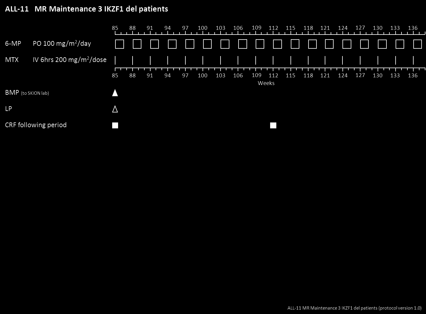
257 257/272
258 258/272
259 259/272
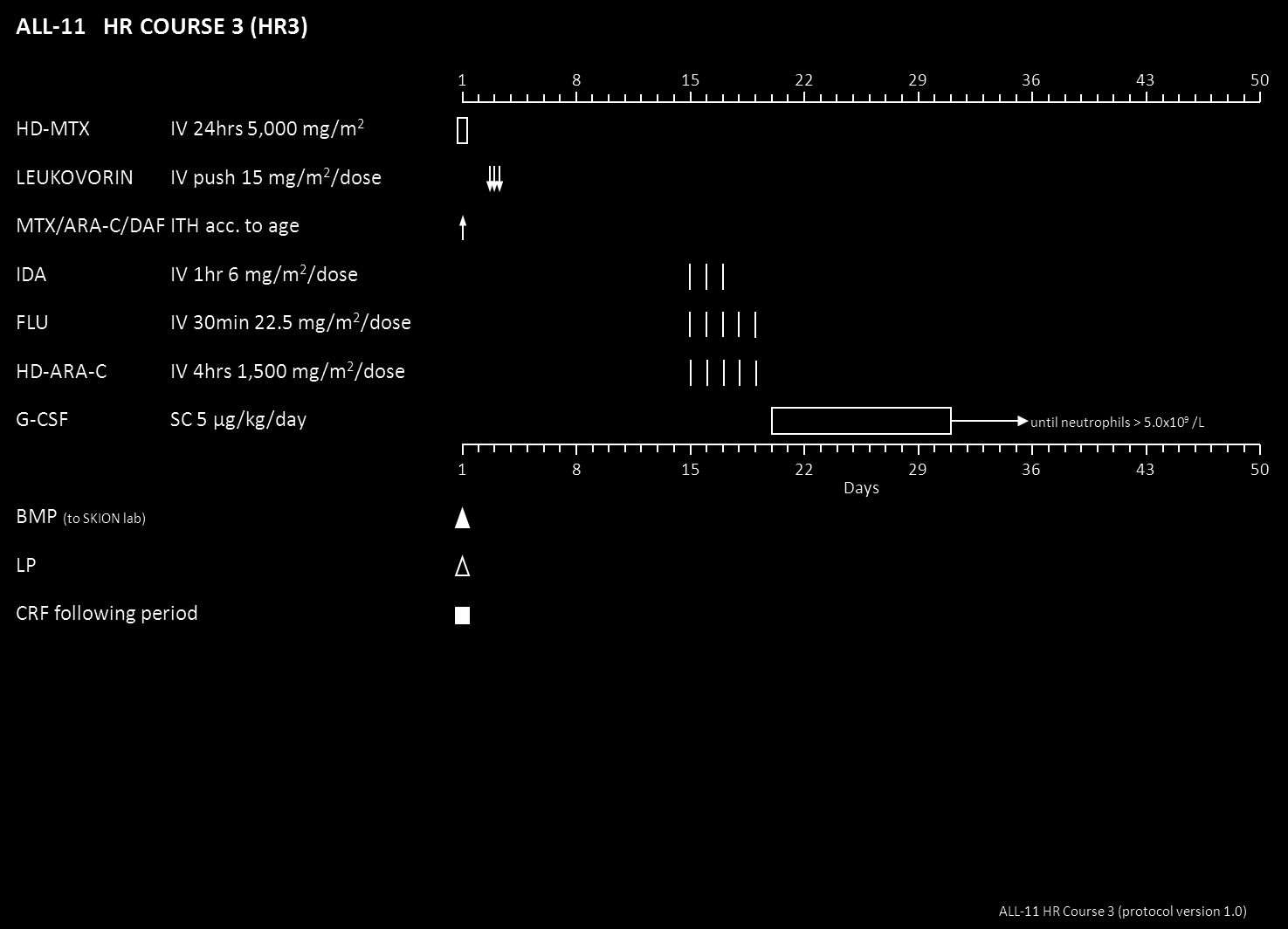
260 260/272
261 261/272
262 262/272

263 263/272
264 264/272
265 265/272
266 266/272
267 ALL-11 Version 2.0 (29 August 2012) Attachment I Logistics and processing guidelines PEGasparaginase Asparaginase trough levels and antibodies Blood samples (3 ml whole blood) are collected just before asparaginase infusion. The whole blood collected in vials without additive can be stored at room temperature. This whole blood sample should be processed for serum and sent together with the asparaginase measurement form to ErasmusMC Sophia, Rotterdam (attachment II). Twice weekly on Monday and Thursday, a courier will transport the samples to ErasmusMC, Rotterdam. Serum should be stored at -20 C till transport, but shipment is possible at room temperature. The whole blood is collected in vials without additive. Blood samples have to be centrifuged at 800 G for 10 min. The serum will then be harvested and divided into two Eppendorf tubes: 1. Asparaginase activity test (> 200 ul) 2. anti Asparaginase antibodies (> 200ul) The tubes should be send to: ErasmusMC, Sophia Tav R Stigter/ dr IM van der Sluis Speciaal laboratorium hematologie Sk 2282 Dr Molewaterplein GJ Rotterdam tel: (laboratory hemato-oncology) tel: (secretary hemato-oncology) fax: The bioanalytical part of the study will be conducted according to the applicable principles of Good Laboratory Practice (GLP). Serum levels of Asparaginase are determined according to standard operating procedures (SOPs) with a sensitive microplate reader-based method. 28 This method has already routinely been used for the therapeutic drug monitoring of Asparaginase in human serum and has been validated. Asparaginase levels will be determined in the laboratory of Pediatric hematology/oncology of the ErasmusMC Sophia (reference laboratory Prof Boos, Muenster). Asparagine (ErasmusMC only) Only when asparagine level has to be assessed a blood sample has to be put on ice immediately and rapidly transferred to the laboratory where it will be immediately processed for serum. For this purpose, blood samples have to be centrifuged at 800 G and 4 C for 10 min. The serum will then be harvested and divided into two aliquots with 200 ul serum each. The samples for amino acid determination are deproteinated (to avoid further enzymatic activity) by adding 1 part of 10% sulphosalicylic acid to 4 parts of serum, thoroughly mixed and centrifuged for 5 minutes at 10,000-13,000 rpm, and the supernatant harvested. This sample will be immediately frozen at -80 o C or colder and stored at this temperature until analysis. 267/272
268 Attachment II Logistics and processing guidelines Erwinase Asparaginase trough levels and antibodies Blood samples (3 ml whole blood) are collected just before asparaginase infusion. The whole blood collected in vials without additive has to be put on ice immediately and rapidly transferred to the laboratory where it will be immediately processed for serum. For this purpose, blood samples have to be centrifuged at 800 G and 4 C for 10 min. The serum will then be harvested and divided into four Eppendorf tubes with 200ul serum each for: a) Asparaginase level (200 ul) b) Amino acid determination (200 ul) c) Anti asparaginase antibodies (2 x 100ul) The samples for amino acid determination were then deproteinated (to avoid further enzymatic activity) by adding 1 part of 10% sulphosalicylic acid to 4 parts of serum, thoroughly mixed and centrifuged for 5 minutes at 10,000-13,000 rpm, and the supernatant harvested. All serum and bone marrow samples will be immediately frozen at -80 o C or colder and stored at this temperature until analysis. Serum for asparaginase and antibody levels has to be send together with the asparaginase measurement form (attachment IV) to Rotterdam. Twice weekly on Monday and Thursday, a courier will transport the samples to the laboratory of pediatric hematology/oncology of the ErasmusMC Sophia, Rotterdam. Serum should be stored at -20 C till transport, but shipment is possible at room temperature. The tubes should be send to: ErasmusMC, Sophia Tav R Stigter/ Dr IM van der Sluis Speciaal laboratorium hematologie Sk 2282 Dr Molewaterplein GJ Rotterdam tel: (laboratory hemato-oncology) tel: (secretary hemato-oncology) fax: Serum for asparagine levels should be stored at -80 C. The transport of these vials will be arranged in batches because transport on ice is necessary. The bioanalytical part of the study will be conducted according to the applicable principles of Good Laboratory Practice (GLP). Serum levels of asparaginase are determined according to standard operating procedures (SOPs) with a sensitive microplate reader-based method. 28 This method has already routinely been used for the therapeutic drug monitoring of asparaginase in human serum and has been validated. Asparaginase levels will be determined in the laboratory of Pediatric hematology/oncology in the ErasmusMC Sophia (reference laboratory Prof Boos, Muenster). 268/272
269 Attachment III Asparaginase measurement form Form needed for asparaginase level measurements (as used in the ALL-11 protocol to assess asparaginase levels for determining an adequate individual dosing scheme). 269/272
270 Attachment IV Wekelijkse online-vragenlijst voor deelnemers aan de IVIG-studie en hun ouders 270/272
271 271/272
272 272/272
Stem Cell Transplantation for Acute Lymphoblastic Leukemia
 Stem Cell Transplantation for Acute Lymphoblastic Leukemia Mona Shafey MD, FRCPC Bone Marrow Transplant Fellow Alberta Blood and Marrow Transplant Program 1 of 14 Stem Cell Transplantation for Acute Lymphoblastic
Introduction. About 10,500 new cases of acute myelogenous leukemia are diagnosed each
 Introduction 1.1 Introduction: About 10,500 new cases of acute myelogenous leukemia are diagnosed each year in the United States (Hope et al., 2003). Acute myelogenous leukemia has several names, including
If several different trials are mentioned in one publication, the data of each should be extracted in a separate data extraction form.
 General Remarks This template of a data extraction form is intended to help you to start developing your own data extraction form, it certainly has to be adapted to your specific question. Delete unnecessary
Treating Minimal Residual Disease in Acute Leukemias: How low should you go?
 Treating Minimal Residual Disease in Acute Leukemias: How low should you go? Ramsie Lujan, Pharm.D. PGY1 Pharmacy Practice Resident Methodist Hospital, San Antonio, Texas Pharmacotherapy Education and
FastTest. You ve read the book... ... now test yourself
 FastTest You ve read the book...... now test yourself To ensure you have learned the key points that will improve your patient care, read the authors questions below. Please refer back to relevant sections
PROGNOSIS IN ACUTE LYMPHOBLASTIC LEUKEMIA PROGNOSIS IN ACUTE MYELOID LEUKEMIA
 PROGNOSIS IN ACUTE LYMPHOBLASTIC LEUKEMIA UNFAVORABLE Advanced age High leukocyte count at diagnosis Presence of myeloid antigens Late achievement of CR Chromosomal abnormalities: t(9:22)(q34:q11) t(4;11)(q21;q23)
DECISION AND SUMMARY OF RATIONALE
 DECISION AND SUMMARY OF RATIONALE Indication under consideration Clinical evidence Clofarabine in the treatment of relapsed acute myeloid leukaemia (AML) The application was for clofarabine to remain in
Stakeholder Insight: Acute Leukemias - Reaching the Limits of Cytotoxic Chemotherapy
 Brochure More information from http://www.researchandmarkets.com/reports/1088137/ Stakeholder Insight: Acute Leukemias - Reaching the Limits of Cytotoxic Chemotherapy Description: The drug therapy of acute
Long Term Low Dose Maintenance Chemotherapy in the Treatment of Acute Myeloid Leukemia
 Long Term Low Dose Chemotherapy in the Treatment of Acute Myeloid Leukemia Murat TOMBULO LU*, Seçkin ÇA IRGAN* * Department of Hematology, Faculty of Medicine, Ege University, zmir, TURKEY ABSTRACT In
Adult Medical-Surgical Nursing H A E M A T O L O G Y M O D U L E : L E U K A E M I A 2
 Adult Medical-Surgical Nursing H A E M A T O L O G Y M O D U L E : L E U K A E M I A 2 Leukaemia: Description A group of malignant disorders affecting: White blood cells (lymphocytes or leucocytes) Bone
Corporate Medical Policy
 Corporate Medical Policy File Name: Origination: Last CAP Review: Next CAP Review: Last Review: hematopoietic_stem-cell_transplantation_for_epithelial_ovarian_cancer 2/2001 11/2015 11/2016 11/2015 Description
Outline of thesis and future perspectives.
 Outline of thesis and future perspectives. This thesis is divided into two different sections. The B- section involves reviews and studies on B- cell non- Hodgkin lymphoma [NHL] and radioimmunotherapy
I was just diagnosed, so my doctor and I are deciding on treatment. My doctor said there are several
 Track 3: Goals of therapy I was just diagnosed, so my doctor and I are deciding on treatment. My doctor said there are several factors she ll use to decide what s best for me. Let s talk about making treatment
Management of low grade glioma s: update on recent trials
 Management of low grade glioma s: update on recent trials M.J. van den Bent The Brain Tumor Center at Erasmus MC Cancer Center Rotterdam, the Netherlands Low grades Female, born 1976 1 st seizure 2005,
January 2013 LONDON CANCER NEW DRUGS GROUP RAPID REVIEW. Summary. Contents
 LONDON CANCER NEW DRUGS GROUP RAPID REVIEW Paclitaxel albumin (Abraxane ) as a substitute for docetaxel/paclitaxel for cancer Paclitaxel albumin (Abraxane ) as a substitute for docetaxel/ paclitaxel for
National Cancer Institute Research on Childhood Cancers. In the United States in 2005, approximately 9,510 children under age 15 will be
 CANCER FACTS N a t i o n a l C a n c e r I n s t i t u t e N a t i o n a l I n s t i t u t e s o f H e a l t h D e p a r t m e n t o f H e a l t h a n d H u m a n S e r v i c e s National Cancer Institute
Guideline for the Management of Acute Lymphoblastic Leukaemia (ALL) in Adults
 in Adults") Guideline for the Management of Acute Lymphoblastic Leukaemia (ALL) in Adults Version History Version Date Summary of Change/Process 1.0 The UKALL trial protocol was approved as version 1. 1.1 22.07.10
Guideline for the Management of Acute Lymphoblastic Leukaemia (ALL) in Adults Version History Version Date Summary of Change/Process 1.0 The UKALL trial protocol was approved as version 1. 1.1 22.07.10
Aggressive lymphomas. Michael Crump Princess Margaret Hospital
 Aggressive lymphomas Michael Crump Princess Margaret Hospital What are the aggressive lymphomas? Diffuse large B cell Mediastinal large B cell Anaplastic large cell Burkitt lymphoma (transformed lymphoma:
PROTOCOLS FOR TREATMENT OF MALIGNANT LYMPHOMA
 2012 1 31,, PROTOCOLS FOR TREATMENT OF MALIGNANT LYMPHOMA Version 1.0 2012 DIVISION OF HAEMATOLOGY / ONCOLOGY DEPARTMENT OF MEDICINE KAOHSING VETERAN GENERAL HOSPTIAL General Guide Diagnosis 1.Adequate
Corporate Medical Policy
 Corporate Medical Policy Hematopoietic Stem-Cell Transplantation for CLL and SLL File Name: Origination: Last CAP Review: Next CAP Review: Last Review: hematopoietic_stem-cell_transplantation_for_cll_and_sll
Acute Lymphoblastic Leukemia (Adult) Including Lymphoblastic lymphoma
 Including Lymphoblastic lymphoma") Acute Lymphoblastic Leukemia (Adult) Including Lymphoblastic lymphoma Updated April 2008 by Dr. Richard Wells* Updates: Minor changes only Introduction Acute lymphocytic leukemia (ALL) is a relatively
Acute Lymphoblastic Leukemia (Adult) Including Lymphoblastic lymphoma Updated April 2008 by Dr. Richard Wells* Updates: Minor changes only Introduction Acute lymphocytic leukemia (ALL) is a relatively
Acute myeloid leukemia (AML)
") Acute myeloid leukemia (AML) Adult acute myeloid leukemia (AML) is a type of cancer in which the bone marrow makes abnormal myeloblasts (a type of white blood cell), red blood cells, or platelets. Adult
Acute myeloid leukemia (AML) Adult acute myeloid leukemia (AML) is a type of cancer in which the bone marrow makes abnormal myeloblasts (a type of white blood cell), red blood cells, or platelets. Adult
Acute leukemias and myeloproliferative neoplasms
 Acute leukemias and myeloproliferative neoplasms GERGELY SZOMBATH SEMMELWEIS UNIVERSITY OF MEDICINE IIIRD. DEPARTMENT OF INTERNAL MEDICINE Basics of acute leukemia Neoplastic disease Cell of origin is
Why discuss CLL? Common: 40% of US leukaemia. approx 100 pa in SJH / MWHB 3 inpatients in SJH at any time
 Why discuss CLL? Common: 40% of US leukaemia approx 100 pa in SJH / MWHB 3 inpatients in SJH at any time Median age of dx is 65 (30s. Incurable, survival 2-202 20 years Require ongoing supportive care
What is a Stem Cell Transplantation?
 What is a Stem Cell Transplantation? Guest Expert: Stuart, MD Associate Professor, Medical Oncology www.wnpr.org www.yalecancercenter.org Welcome to Yale Cancer Center Answers with Drs. Ed and Ken. I am
INFORMATION ON STEM CELLS/BONE MARROW AND REINFUSION/TRANSPLANTATION SUR703.002
 INFORMATION ON STEM CELLS/BONE MARROW AND REINFUSION/TRANSPLANTATION SUR703.002 COVERAGE: SPECIAL COMMENT ON POLICY REVIEW: Due to the complexity of the Peripheral and Bone Marrow Stem Cell Transplantation
Trials in Elderly Melanoma Patients (with a focus on immunotherapy)
") Trials in Elderly Melanoma Patients (with a focus on immunotherapy) Where we were Immunotherapy Trials: past and present Relevance for real world practice Where we are SIOG October 2012 James Larkin FRCP
Trials in Elderly Melanoma Patients (with a focus on immunotherapy) Where we were Immunotherapy Trials: past and present Relevance for real world practice Where we are SIOG October 2012 James Larkin FRCP
Estimated New Cases of Leukemia, Lymphoma, Myeloma 2014
 ABOUT BLOOD CANCERS Leukemia, Hodgkin lymphoma (HL), non-hodgkin lymphoma (NHL), myeloma, myelodysplastic syndromes (MDS) and myeloproliferative neoplasms (MPNs) are types of cancer that can affect the
GUIDELINES FOR THE MANAGEMENT OF LUNG CANCER
 GUIDELINES FOR THE MANAGEMENT OF LUNG CANCER BY Ali Shamseddine, MD (Coordinator); [email protected] Fady Geara, MD Bassem Shabb, MD Ghassan Jamaleddine, MD CLINICAL PRACTICE GUIDELINES FOR THE TREATMENT
Project Lead: Stephen Forman, M.D. PI: Elizabeth Budde, M.D., Ph.D
 Phase I study using T cells expressing a CD123-specific chimeric antigen receptor and truncated EGFR for patients with relapsed or refractory acute myeloid leukemia Project Lead: Stephen Forman, M.D. PI:
An overview of CLL care and treatment. Dr Dean Smith Haematology Consultant City Hospital Nottingham
 An overview of CLL care and treatment Dr Dean Smith Haematology Consultant City Hospital Nottingham What is CLL? CLL (Chronic Lymphocytic Leukaemia) is a type of cancer in which the bone marrow makes too
Sonneveld, P; de Ridder, M; van der Lelie, H; et al. J Clin Oncology, 13 (10) : 2530-2539 Oct 1995
 : 2530-2539 Oct 1995") Comparison of Doxorubicin and Mitoxantrone in the Treatment of Elderly Patients with Advanced Diffuse Non-Hodgkin's Lymphoma Using CHOP Versus CNOP Chemotherapy. Sonneveld, P; de Ridder, M; van der Lelie,
Comparison of Doxorubicin and Mitoxantrone in the Treatment of Elderly Patients with Advanced Diffuse Non-Hodgkin's Lymphoma Using CHOP Versus CNOP Chemotherapy. Sonneveld, P; de Ridder, M; van der Lelie,
Hodgkin Lymphoma Disease Specific Biology and Treatment Options. John Kuruvilla
 Hodgkin Lymphoma Disease Specific Biology and Treatment Options John Kuruvilla My Disclaimer This is where I work Objectives Pathobiology what makes HL different Diagnosis Staging Treatment Philosophy
Acute Myeloid Leukemia
 Acute Myeloid Leukemia Introduction Leukemia is cancer of the white blood cells. The increased number of these cells leads to overcrowding of healthy blood cells. As a result, the healthy cells are not
Lauren Berger: Why is it so important for patients to get an accurate diagnosis of their blood cancer subtype?
 Hello, I m Lauren Berger and I m the Senior Director of Patient Services Programs at The Leukemia & Lymphoma Society. I m pleased to welcome Dr. Rebecca Elstrom. Dr. Elstrom is an Assistant Professor in
SMALL CELL LUNG CANCER
 Protocol for Planning and Treatment The process to be followed in the management of: SMALL CELL LUNG CANCER Patient information given at each stage following agreed information pathway 1. DIAGNOSIS New
CHAPTER 26 LATE BREAKING DEVELOPMENTS: IMPACT OF ANTI-CD20 MONOCLONAL ANTIBODIES ON LYMPHOMA THERAPY
 CHAPTER 26 LATE BREAKING DEVELOPMENTS: IMPACT OF ANTI-CD20 MONOCLONAL ANTIBODIES ON LYMPHOMA THERAPY 26.1 Introduction rituximab Subsequent to the completion of drafts for the guidelines earlier in 2004,
Elements for a Public Summary
 VI.2 Elements for a Public Summary VI.2.1 Overview of Disease Epidemiology Hunter syndrome is a rare genetic disease which mainly affects males of all ethnicities. The incidence rate ranges from 0.6 to
SWOG ONCOLOGY RESEARCH PROFESSIONAL (ORP) MANUAL VOLUME I RESPONSE ASSESSMENT LEUKEMIA CHAPTER 11A REVISED: OCTOBER 2015
 MANUAL VOLUME I RESPONSE ASSESSMENT LEUKEMIA CHAPTER 11A REVISED: OCTOBER 2015") LEUKEMIA Response in Acute Myeloid Leukemia (AML) Response criteria in Acute Myeloid Leukemia for SWOG protocols is based on the review article Diagnosis and management of acute myeloid leukemia in adults:
LEUKEMIA Response in Acute Myeloid Leukemia (AML) Response criteria in Acute Myeloid Leukemia for SWOG protocols is based on the review article Diagnosis and management of acute myeloid leukemia in adults:
Cure versus control: Which is the best strategy?
 Cure versus control: Which is the best strategy? Barcelona 8-9-2012 Mario Boccadoro DIVISIONE UNIVERSITARIA DI EMATOLOGIA AZIENDA OSPEDALIERA SAN GIOVANNI TORINO, ITALY MULTIPLE MYELOMA Cure versus control
STUDY PROTOCOL. Fabio Ciceri M.D. Istituto Scientifico H. San Raffaele Dept. of of Oncology, Haematology/Transplant Unit I-20132 Milan
 STUDY PROTOCOL Clinical phase II trial to evaluate the safety and efficacy of treosulfan combined with cytarabine and fludarabine prior to autologous haematopoietic stem cell transplantation in elderly
Bone Marrow/Stem Cell Transplant
 Blue Distinction Centers for Transplants Program Selection Criteria for 2010 Mid-Point Designations To qualify as a Blue Distinction Center for Transplants (), each facility must satisfy s quality based
Acute myeloid leukaemia (AML) in children
 in children") 1 61.02 Acute myeloid leukaemia (AML) in children AML can affect children of any age, and girls and boys are equally affected. Leukaemia Acute myeloid leukaemia (AML) FAB classification of AML Causes of
1 61.02 Acute myeloid leukaemia (AML) in children AML can affect children of any age, and girls and boys are equally affected. Leukaemia Acute myeloid leukaemia (AML) FAB classification of AML Causes of
Update in Hematology Oncology Targeted Therapies. Mark Holguin
 Update in Hematology Oncology Targeted Therapies Mark Holguin 25 years ago Why I chose oncology People How to help people with possibly the most difficult thing they may have to deal with Science Turning
MOLOGEN AG. Q1 Results 2015 Conference Call Dr. Matthias Schroff Chief Executive Officer. Berlin, 12 May 2015
 Q1 Results 2015 Conference Call Dr. Matthias Schroff Chief Executive Officer Berlin, 12 May 2015 V1-6 Disclaimer Certain statements in this presentation contain formulations or terms referring to the future
Pro Cure in Multiple Myeloma. Nicolaus Kröger Dept. of Stem Cell Transplantation University Hospital Hamburg Hamburg, Germany
 Pro Cure in Multiple Myeloma Nicolaus Kröger Dept. of Stem Cell Transplantation University Hospital Hamburg Hamburg, Germany Pro Cure in Multiple Myeloma Several hematological malignancies can be cured
Cyrine-Eliana Haidar, Pharm.D., BCPS, BCOP; and Jennifer L. Pauley, Pharm.D., BCPS
 Pediatric Leukemias Cyrine-Eliana Haidar, Pharm.D., BCPS, BCOP; and Jennifer L. Pauley, Pharm.D., BCPS Reviewed by Betsy Bickert Poon, Pharm.D.; Todd P. Semla, Pharm.D., M.S., FCCP, BCPS, AGSF; and Maya
Hematopoietic Stem Cell Transplantation. Imad A. Tabbara, M.D. Professor of Medicine
 Hematopoietic Stem Cell Transplantation Imad A. Tabbara, M.D. Professor of Medicine Hematopoietic Stem Cells Harvested from blood, bone marrow, umbilical cord blood Positive selection of CD34 (+) cells
treatments) worked by killing cancerous cells using chemo or radiotherapy. While these techniques can
 worked by killing cancerous cells using chemo or radiotherapy. While these techniques can") Shristi Pandey Genomics and Medicine Winter 2011 Prof. Doug Brutlag Chronic Myeloid Leukemia: A look into how genomics is changing the way we treat Cancer. Until the late 1990s, nearly all treatment methods
Shristi Pandey Genomics and Medicine Winter 2011 Prof. Doug Brutlag Chronic Myeloid Leukemia: A look into how genomics is changing the way we treat Cancer. Until the late 1990s, nearly all treatment methods
Feline Lymphoma Chemotherapy and Chemotherapy Protocols
 Feline Lymphoma Chemotherapy and Chemotherapy Protocols If you have reached this page, your cat probably has a definite diagnosis of feline lymphoma from your veterinarian. The information below is not
Immunoablative therapy with autologous hematopoietic stem cell transplantation in the treatment of poor risk multiple sclerosis
 Immunoablative therapy with autologous hematopoietic stem cell transplantation in the treatment of poor risk multiple sclerosis T Kozák, P Lhotáková Department of Clinical Haematology, 3r d School of Medicine,
亞 東 紀 念 醫 院 Follicular Lymphoma 臨 床 指 引
 前 言 : 惡 性 淋 巴 瘤 ( 或 簡 稱 淋 巴 癌 ) 乃 由 體 內 淋 巴 系 統 包 括 淋 巴 細 胞 淋 巴 管 淋 巴 腺 及 一 些 淋 巴 器 官 或 組 織 如 脾 臟 胸 腺 及 扁 桃 腺 等 所 長 出 的 惡 性 腫 瘤 依 腫 瘤 病 理 組 織 型 態 的 不 同 可 分 為 何 杰 金 氏 淋 巴 瘤 (Hodgkin s disease) 與 非 何 杰 金
Guidance for Industry
 Guidance for Industry Cancer Drug and Biological Products Clinical Data in Marketing Applications U.S. Department of Health and Human Services Food and Drug Administration Center for Drug Evaluation and
Proceedings of the World Small Animal Veterinary Association Sydney, Australia 2007
 Proceedings of the World Small Animal Sydney, Australia 2007 Hosted by: Next WSAVA Congress Rescue Chemotherapy Protocols for Dogs with Lymphoma Kenneth M. Rassnick, DVM, DACVIM (Oncology) Cornell University
Sommaire projets sélectionnés mesure 29: Soutien à la recherche translationnelle
 Sommaire projets sélectionnés mesure 29: Soutien à la recherche translationnelle TITLE PROJET NOM HOPITAL Assessment of tumor angiogenesis using PET/CT with 18 F-Galacto- RGD. (PNC_29_001) Division of
Are CAR T-Cells the Solution for Chemotherapy Refractory Diffuse Large B-Cell Lymphoma? Umar Farooq, MD University of Iowa Hospitals and Clinics
 Are CAR T-Cells the Solution for Chemotherapy Refractory Diffuse Large B-Cell Lymphoma? Umar Farooq, MD University of Iowa Hospitals and Clinics Disclosure(s) I do not intend to discuss an off-label use
LEUKEMIA LYMPHOMA MYELOMA Advances in Clinical Trials
 LEUKEMIA LYMPHOMA MYELOMA Advances in Clinical Trials OUR FOCUS ABOUT emerging treatments Presentation for: Judith E. Karp, MD Advancements for Acute Myelogenous Leukemia Supported by an unrestricted educational
A Career in Pediatric Hematology-Oncology? Think About It...
 A Career in Pediatric Hematology-Oncology? Think About It... What does a pediatric hematologist-oncologist do? What kind of training is necessary? Is there a future need for specialists in this area? T
Stem Cell Transplantation
 Harmony Behavioral Health, Inc. Harmony Behavioral Health of Florida, Inc. Harmony Health Plan of Illinois, Inc. HealthEase of Florida, Inc. Ohana Health Plan, a plan offered by WellCare Health Insurance
Disclosures. Consultant and Speaker for Biogen Idec, TEVA Neuroscience, EMD Serrono, Mallinckrodt, Novartis, Genzyme, Accorda Therapeutics
 Mitzi Joi Williams, MD Neurologist MS Center of Atlanta, Atlanta, GA Disclosures Consultant and Speaker for Biogen Idec, TEVA Neuroscience, EMD Serrono, Mallinckrodt, Novartis, Genzyme, Accorda Therapeutics
Malignant Lymphomas and Plasma Cell Myeloma
 Malignant Lymphomas and Plasma Cell Myeloma Dr. Bruce F. Burns Dept. of Pathology and Lab Medicine Overview definitions - lymphoma lymphoproliferative disorder plasma cell myeloma pathogenesis - translocations
ACUTE MYELOID LEUKEMIA (AML),
,") 1 ACUTE MYELOID LEUKEMIA (AML), ALSO KNOWN AS ACUTE MYELOGENOUS LEUKEMIA WHAT IS CANCER? The body is made up of hundreds of millions of living cells. Normal body cells grow, divide, and die in an orderly
1 ACUTE MYELOID LEUKEMIA (AML), ALSO KNOWN AS ACUTE MYELOGENOUS LEUKEMIA WHAT IS CANCER? The body is made up of hundreds of millions of living cells. Normal body cells grow, divide, and die in an orderly
The Clinical Trials Process an educated patient s guide
 The Clinical Trials Process an educated patient s guide Gwen L. Nichols, MD Site Head, Oncology Roche TCRC, Translational and Clinical Research Center New York DISCLAIMER I am an employee of Hoffmann-
AML- new studies. Moderator Prof. Edo Vellenga. 1st author / speaker Mojca Jongen-Lavrencic
 AML- new studies Moderator Prof. Edo Vellenga 1st author / speaker Mojca Jongen-Lavrencic Belangenverklaring In overeenstemming met de regels van de Inspectie van de Gezondheidszorg (IGZ) Naam: Organisatie:
Experience of a Pediatric Oncology Clinical Research Center in Brazil. Antonio Sergio Petrilli, MD, PhD
 Experience of a Pediatric Oncology Clinical Research Center in Brazil Antonio Sergio Petrilli, MD, PhD Children and adolescents Heterogeneous in many aspects 38% of Brazilian population (IBGE, 2000) 11,530
MEDICAL COVERAGE POLICY
 Important note Even though this policy may indicate that a particular service or supply is considered covered, this conclusion is not necessarily based upon the terms of your particular benefit plan. Each
Stem Cell Transplantation In Patients with Fanconi Anemia
 Stem Cell Transplantation In Patients with Fanconi Anemia FARF Annual Family Meeting 6/28/15 Casco, ME Parinda A. Mehta, M.D. Cincinnati Children s Hospital Medical Center Improvements in Unrelated Donor
Shaji Kumar, M.D. Multiple Myeloma: Multiple myeloma (MM) is the second most common hematological
 is the second most common hematological") An update on the management of multiple myeloma and amyloidosis Shaji Kumar, M.D. Multiple Myeloma: Multiple myeloma (MM) is the second most common hematological malignancy in this country affecting nearly
An update on the management of multiple myeloma and amyloidosis Shaji Kumar, M.D. Multiple Myeloma: Multiple myeloma (MM) is the second most common hematological malignancy in this country affecting nearly
Avastin in Metastatic Breast Cancer
 Non-interventional study Avastin in Metastatic Breast Cancer ML 21165 / 2007 Clinical Study Report Synopsis ROCHE ML21165 / WiSP Project RH09 / V. 1.0 / 24.06.2013 ROCHE ML21165-2 - Name of Sponsor Roche
GRANIX (tbo-filgrastim)
") RATIONALE FOR INCLUSION IN PA PROGRAM Background Neutropenia is a hematological disorder characterized by an abnormally low number of neutrophils. A person with severe neutropenia has an absolute neutrophil
RATIONALE FOR INCLUSION IN PA PROGRAM Background Neutropenia is a hematological disorder characterized by an abnormally low number of neutrophils. A person with severe neutropenia has an absolute neutrophil
UMHS-PUHSC JOINT INSTITUTE
 Imaging Biomarkers for Staging and Assessing Response to Therapy in Multiple Myeloma Qian Dong, MD. Radiology University of Michigan Wei Guo, MD. Orthopedic Oncology Peking University Second Hospital Team
Advances In Chemotherapy For Hormone Refractory Prostate Cancer. TAX 327 study results & SWOG 99-16 study results presented at ASCO 2004
 Ronald de Wit Rotterdam Cancer Institute The Netherlands Advances In Chemotherapy For Hormone Refractory Prostate Cancer TAX 327 study results & SWOG 99-16 study results presented at Slide 1 Prostate Cancer
Cytogenetics for the Rest of Us: A Primer
 Cytogenetics for the Rest of Us: A Primer James J. Stark, MD, FACP Medical Director Cancer Program Maryview Medical Center Diane Maia, M.D. Pathologist, Bon Secours Hampton Roads Case #1 78 y.o. lady seen
Acute Lymphoblastic Leukemia in Children: A Fifteen Year Experience at the Princess Nourah Oncology Center, Jeddah, Saudi Arabia.
 Acute Lymphoblastic Leukemia in Children: A Fifteen Year Experience at the Princess Nourah Oncology Center, Jeddah, Saudi Arabia. 1 Dr. C. Fryer FRCP(C), 2 Dr. S. Felimban FACHARTZ, 3 C. Paltiel MS 4 Dr.
CHAPTER 2. Neoplasms (C00-D49) March 2014. 2014 MVP Health Care, Inc.
 March 2014. 2014 MVP Health Care, Inc.") Neoplasms (C00-D49) March 2014 2014 MVP Health Care, Inc. CHAPTER SPECIFIC CATEGORY CODE BLOCKS C00-C14 Malignant neoplasms of lip, oral cavity and pharynx C15-C26 Malignant neoplasms of digestive organs
Neoplasms (C00-D49) March 2014 2014 MVP Health Care, Inc. CHAPTER SPECIFIC CATEGORY CODE BLOCKS C00-C14 Malignant neoplasms of lip, oral cavity and pharynx C15-C26 Malignant neoplasms of digestive organs
Health Disparities in Multiple Myeloma. Kenneth R. Bridges, M.D. Senior Medical Director Onyx Pharmaceuticals, Inc.
 Health Disparities in Multiple Myeloma Kenneth R. Bridges, M.D. Senior Medical Director Onyx Pharmaceuticals, Inc. Multiple Myeloma Overview Multiple myeloma (MM) is a type of blood cancer that develops
L Lang-Lazdunski, A Bille, S Marshall, R Lal, D Landau, J Spicer
 Pleurectomy/decortication, hyperthermic pleural lavage with povidone-iodine and systemic chemotherapy in malignant pleural mesothelioma. A 10-year experience. L Lang-Lazdunski, A Bille, S Marshall, R Lal,
What is Glioblastoma? How is GBM classified according to the WHO Grading System? What risk factors pertain to GBM?
 GBM (English) What is Glioblastoma? Glioblastoma or glioblastoma multiforme is one of the most common brain tumors accounting for approximately 12 to 15 percent of all brain tumors. The name of the tumor
Bendamustine for the fourth-line treatment of multiple myeloma
 LONDON CANCER NEW DRUGS GROUP RAPID REVIEW Bendamustine for the fourth-line treatment of multiple myeloma Contents Summary 1 Background 2 Epidemiology 3 Cost 6 References 7 Summary There is no standard
Daiichi Sankyo to Acquire Ambit Biosciences
 For Immediate Release Company name: DAIICHI SANKYO COMPANY, LIMITED Representative: Joji Nakayama, Representative Director, President and CEO (Code no.: 4568, First Section, Tokyo Stock Exchange) Please
Treatment of Low Risk MDS. Overview. Myelodysplastic Syndromes (MDS)
") Overview Amy Davidoff, Ph.D., M.S. Associate Professor Pharmaceutical Health Services Research Department, Peter Lamy Center on Drug Therapy and Aging University of Maryland School of Pharmacy Clinical
Overview Amy Davidoff, Ph.D., M.S. Associate Professor Pharmaceutical Health Services Research Department, Peter Lamy Center on Drug Therapy and Aging University of Maryland School of Pharmacy Clinical
NATIONAL CANCER DRUG FUND PRIORITISATION SCORES
 NATIONAL CANCER DRUG FUND PRIORITISATION SCORES Drug Indication Regimen (where appropriate) BORTEZOMIB In combination with dexamethasone (VD), or with dexamethasone and thalidomide (VTD), is indicated
Histopathologic results
 Self evaluation 1 Clinical Case 55-year-old woman Bilateral enlargement of cervical, axillary and inguinal lymph nodes, largest diameter > 6 cm Hepatosplenomegaly. Enlargement of retroperitoneal, mesenteric
Specific Standards of Accreditation for Residency Programs in Pediatric Hematology/Oncology
 Specific Standards of Accreditation for Residency Programs in Pediatric Hematology/Oncology INTRODUCTION 2009 A university wishing to have an accredited program in Pediatric Hematology/Oncology must also
Infosheet. Allogeneic stem cell transplantation in myeloma. What is the principle behind stem cell transplantation?
 Infosheet Allogeneic stem cell transplantation in myeloma High-dose therapy and autologous stem cell transplantation is currently the first-line treatment standard of care for younger/fitter myeloma patients.
Cytotoxic and Biotherapies Credentialing Programme Module 2
 Cytotoxic and Biotherapies Credentialing Programme Module 2 1. The Cell Cycle 2. Cancer Therapies 3. Adjunctive Therapies On completion of this module the RN will State the difference between a normal
CML. cure. A Patient s Guide. Molecular Biology Diagnosis Stem Cell Transplant Monitoring New Drugs Questions to Ask and More
 A Patient s Guide to CML Molecular Biology Diagnosis Stem Cell Transplant Monitoring New Drugs Questions to Ask and More cure C a n c e r U p d at e s, R e s e a r c h & E d u c at i o n Based on science,
Reference: NHS England B04/P/a
 Clinical Commissioning Policy: Haematopoietic Stem Cell Transplantation (HSCT) (All Ages): Revised Reference: NHS England B04/P/a 1 NHS England Clinical Commissioning Policy: Haematopoietic Stem Cell Transplantation
Emerging New Prognostic Scoring Systems in Myelodysplastic Syndromes 2012
 Emerging New Prognostic Scoring Systems in Myelodysplastic Syndromes 2012 Arjan A. van de Loosdrecht, MD, PhD Department of Hematology VU University Medical Center VU-Institute of Cancer and Immunology
Disclosures. I have no disclosures.
 Not Your Own Marrow Jenni Krajewski, MD Clinical Assistant Professor, Rutgers New Jersey Medical School Attending Physician, Pediatric Blood and Marrow Transplantation The Institute for Pediatric Cancer
Acute Lymphoblastic Leukemia in Adults and Children
 Acute Lymphoblastic Leukemia in Adults and Children Session Chair: Charles Linker, MD Speakers: Stephen E. Sallan, MD; Nicola Gökbuget, MD; and Ching-Hon Pui, MD Myths and Lessons from the Adult/Pediatric
Pan Birmingham Cancer Network: Haematology NSSG Chemotherapy Regimens AML intensive v1.0 August 2010
 AML - TRIALS It is best practise for all patients to be treated on the appropriate NCRI national trial for AML. It is outside of the remit for this regimen list to provide a comprehensive detail of the
guides BIOLOGY OF AGING STEM CELLS An introduction to aging science brought to you by the American Federation for Aging Research
 infoaging guides BIOLOGY OF AGING STEM CELLS An introduction to aging science brought to you by the American Federation for Aging Research WHAT ARE STEM CELLS? Stem cells are cells that, in cell cultures
Acute Myeloid Leukemia
 Acute Myeloid Leukemia Jeffrey E. Rubnitz, MD, PhD a, *, Brenda Gibson, MD b, Franklin O. Smith, MD c KEYWORDS Myeloid Leukemia Childhood Acute myeloid leukemia (AML) is a heterogeneous group of leukemias