Use of Ultrasound in the Provision of Abortion. Juan E. Vargas, MD Assistant Professor of Clinical Obstetrics and Gynecology and Radiology, UCSF
|
|
|
- Anastasia Summers
- 7 years ago
- Views:
Transcription
1 Use of Ultrasound in the Provision of Abortion Juan E. Vargas, MD Assistant Professor of Clinical Obstetrics and Gynecology and Radiology, UCSF

2 Overview Uses and indications of ultrasound in the provision of abortion Basic principles of pelvic sonography Ultrasonographic correlations in normal and abnormal early embryonic development, including ectopic pregnancy Uses and limitations of sonography in the provision of 1 st trimester abortions Uses and limitations of ultrasound in post MAB management

3 First trimester US allows: Confirmation of IUP Confirmation embryonic life Determination of gestational age Determination of singleton or multiple gestations Essential component in managing 1 st trimester vaginal bleeding and ectopic pregnancy

4 Applications of Ultrasound in the Provision of Abortion Determination of gestational age Determination of multiple pregnancies Confirmation of uterine position Assessment of ectopic pregnancies Assessment of uterine malformations or tumors that may interfere with the abortion procedure Direct ultrasound guidance 2 nd trimester procedures Difficult procedures (Uterine malformations, obstructive lesions, failed prior attempts) Managing post procedure complications Establishing placental location and abnormal placentation (accreta) in 2 nd trimester procedures
 Managing post procedure complications Establishing placental location and abnormal placentation (accreta) in 2 nd trimester")
5 Is ultrasound always necessary in the provision of abortion? No Ultrasound scanning is not necessary for the provision of early abortion (RCOG 2000). Where it is available, ultrasound can aid the detection of ectopic pregnancies beyond about 6 weeks of pregnancy. Some providers find the technology helpful before or during abortion procedures at later stages of pregnancy Safe Abortion: Technical and Policy Guidance for Health Systems. WHO 2003

6 Ultrasound and Medical Abortion Standard in US medical abortion trials Usage varies in international studies and settings Should be available for specific indications Useful, but not mandatory, with FDA-approved regimen More accurate dating Can monitor progress of Med AB Detection of early pregnancy abnormalities Useful in conjunction with hcg testing to diagnose ectopic pregnancy

7 PPGG Indications for Sonography for Medical Abortion Pre-abortion: 1. Gestational age > 8 weeks 2. Size/dates discrepancy 3. Uncertain LMP (or no menses - after delivery, abortion, stopping depo, etc) 4. Adnexal mass or pain 5. LMP at the end of a pack of oral contraceptives 6. Provider uncertainty with exam 7. History of previous ectopic pregnancy Post-abortion: 1. History not consistent with successful medical abortion (no bleeding, no cramping) 2. Woman still feels pregnant 3. HCG not declining 4. Provider uncertainty with history Courtesy Dr. Suzan Goodman, MD, MPH
 2. Woman still feels pregnant 3. HCG not declining 4.")
8 Abortion Surveillance --- United States, % were performed at <8 weeks' gestation 88% at <13 weeks. Steady increases have occurred in the percentage of abortions performed at <6 weeks' gestation (26%) 4.1% at weeks 1.4% at >21 weeks. MMWR November 24, 2006 / 55(SS11);1-32
 4.1% at 16--20 weeks 1.4% at >21 weeks.")
9 Basic principles of pelvic sonography

10 Guidelines for 1 st trimester ultrasound Basic Principles Begin by scanning transabdominally. If TAUS fails to provide all the necessary info, TVUS should be done. Never omit doing TAUS, as large pelvic/adnexal masses may be missed. The converse is not true, you may not need to do a TVUS if all the diagnostic information was obtained by TAUS.

11 Guidelines for 1 st trimester ultrasound Basic Principles Transabdominal ultrasound can be use solely or in conjunction with TV US beyond 7-8 weeks gestation A full bladder enhances visualization of the uterus and adnexa before 12 weeks gestation. Beyond weeks it is not necessary During transvaginal US a full bladder displaces the uterus and is not advantageous.

12 Guidelines for 1 st trimester ultrasound Basic Principles Begin by evaluation for presence or absence of gestational sac in the uterus For positive identification of an IUP, the gestational sac (GS) must be seen in relation to the cervix. This is best seen in a sagittal view. Absence of GS needs to be correlated with serum HCG level (> 2000 highly suggests ectopic pregnancy)

13 Guidelines for 1 st trimester ultrasound Basic Principles Use the mean GS diameter for calculation of GA when embryo/crl is not seen GA is best determined by measurement of CRL up to 12 weeks of GA

14 Determination of Gestational Age mean gestational sac diameter Several formulas exist for estimating gestational age based on sac measurement Ultrasound machines have built-in software to calculate gestational age based on these measurements MGS diameter is less than 10 mm, many machines read as out of range. MGS diameter (mm) + 30 = gestational age (days) For example, MGS 7 mm, then = 37 days or 5 weeks and 2 days

15 US correlation of embryonic development Gestational sac can be seen as early as 4 weeks and 1-3 days (2-3 mm sac) Yolk sac can be seen as early as mean GS size of 8mm, but no later than GS 20 mm The embryo can be seen as early as 5 to 6 weeks GA, which corresponds to a embryonic size of 1-2 mm. This correlates with a mean GS diameter of 5-12 mm. The absence of a identifiable embryo with a MGS of > mm is usually consistent with a failed IUP

16 Guidelines for 1 st trimester US Fetal number should be always reported Only sacs that contain embryonic structures should be reported as multiples (incomplete fusion of the amnion and chorion and sometimes intrachorionic hemorrhage may be mistaken for an additional GS. Adequately assess chorionicity (best seen before 12 weeks. Difficult after 20 weeks).

17 Ultrasonographic correlations in normal and abnormal early embryonic development Presence or absence of fetal heart activity should be reported With a CRL of 5 mm fetal heart motion should be clearly appreciated. If CRL is < than 5 mm, a f/u US is necessary for confirmation of fetal viability The embryo grows 1 mm per day (CRL) in the first trimester

18 Guidelines for 1 st trimester US Evaluation of the uterus, adnexa and culde-sac should be noted Any abnormalities in the uterus (for example fibroids) and adnexa should be reported Normal ovaries maybe difficult to visualize after weeks If fluid is seen in the cul-de-sac, evaluation of Morrison s pouch may be important in order to semi quantify the amount of free fluid.

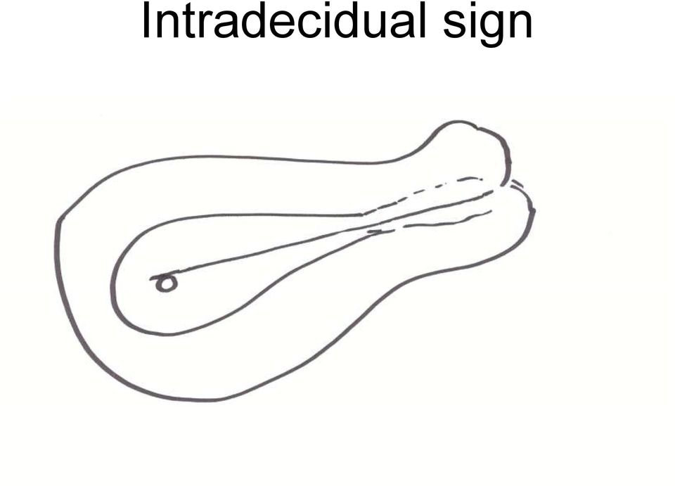
19 Early gestational sac or pseudo sac? The intradecidual sign Does not displace or deform the central cavity complex Echogenic rim should be at last 2 mm thick

20 Intradecidual sign
21 Double decidual sign Useful before yolk sac or embryo can be seen Always present when MGS diameter is >10 mm 2 echogenic lines: smooth chorion & decidua capsularis and decidua parietalis separated by an hypoechoic line that represents the virtual uterine cavity
22 Double Decidual Sign
23 Ultrasound / β -HCG Correlations Discrimination zone: β-hcg > 2000 If no IUP, ectopic pregnancy likely. DDx includes incomplete abortion or abnormal IUP
24 β-hcg doubling time Normal IUP : 66% rise in 48 hours 15% of normal IUP have abnormal rise Ectopics: 17% have a normal rise
25 Symptomatic patients with an early viable intrauterine pregnancy: HCG curves redefined The slowest or minimal rise for a normal viable intrauterine pregnancy was 24% at 1 day and 53% at 2 days Obstetrics & Gynecology 2004;104:50-55
26 Ultrasonographic correlations in normal and abnormal early embryonic development If embryo or yolk sac cannot be identified, the presence of an IUP cannot be confirmed 100% A small fluid collection may give a similar appearance (pseudo GS) which can be seen in ectopic pregnancies. Pseudo sacs don t have intradecidual sign (double decidual ring) and are located within the uterine cavity (early IUPs are slightly extrinsic), and have a tear drop shape
27 Gestational sac vs. Pseudo sac
28 Ectopic pregnancy Symptoms 90% lower abdominal pain (1/3 unilateral) 50-80% abnormal vaginal bleeding 60-70% amenorrhea Classic triad (pain, amenorrhea, VB) present in less than 50%of patients!
29 Ectopic Pregnancy Symptoms Most ectopics present between 6-7 weeks from LMP..before they are ASYMPTOMATIC
30 Clinically suspected ectopic IUP* 0% No IUP 55% Negative exam 7-33% Adnexal mass 75% Any FF 75% Adnexal mass & FF 85% Adnexal GS & embryo Or YS; adnexal ring 100% Large amount or echogenic FF 100% * Rare heterotopic pregnancy
31 Ectopic pregnancy- Unusual locations The great majority of ectopic pregnancies occur in the fallopian tubes. Ectopic pregnancies in other locations such as the cervix, the uterine cornua, or abdominal cavity can be associated with a very high maternal morbidity if not recognized and treated appropriately.
32 Classification of ectopic pregnancies
33 Interstitial (Cornual) Pregnancy 1-6% of all ectopic pregnancies Historically associated with late diagnosis, often with catastrophic complications such as uterine rupture and need for hysterectomy ULTRASOUND DIAGNOSIS: Regular endometrial stripe with no gestational sac products of conception located outside the endometrial echo, surrounded by a contiguous rim of myometrium, within the interstitial area Obstetrics & Gynecology 2004;103:47-50
34
35 Uses and limitations of ultrasound in post medical abortion (MAB) management
36 Uses and limitations of ultrasound in post MAB management The pre-dominant questions are. Is the patient still pregnant? How is the patient doing? Clinical picture Mary Fjerstad, NP, PPFA/CAPS
37 Several studies have failed to demonstrate a relationship between endometrial thickness and clinical outcome (i.e., need for aspiration, predictor of subsequent hemorrhage, etc.)
38 Post-Abortion Ultrasound: Absent Gestational Sac Thickened endometrial stripe Debris
39 Post-Abortion Ultrasound: Absent Gestational Sac Anterior lip of cervix Posterior lip of cervix Cervical canal closed Internal os of cervix Uterine cavity with minimal debris
40 Post-Abortion Ultrasound: Absent Gestational Sac Fundus of uterus Endometrial stripe
41 Ultrasound Evaluation of the Endometrium After Medical Termination of Pregnancy Prospective descriptive study of patients undergoing MAB Ultrasound examination between days 7-13post MAB were available for 525 of 684 patients Endometrial thickness and the presence of gestational sac, fluid interface, or complex echoes on postprocedure ultrasonogram were recorded. Repeat doses of medication, surgical intervention, and complications were noted. Success was defined as an abortion completed after a single course of medical therapy Obstetrics & Gynecology 2004;103:
42 Ultrasound Evaluation of the Endometrium After Medical Termination of Pregnancy Obstetrics & Gynecology 2004;103:
43 Ultrasound Evaluation of the Endometrium After Medical Termination of Pregnancy Obstetrics & Gynecology 2004;103:
44 Ultrasound Evaluation of the Endometrium After Medical Termination of Pregnancy Obstetrics & Gynecology 2004;103: Endometrial thickness after administration of a single dose of mifepristone and misoprostol for medical termination should not dictate clinical intervention. The decision to treat should be based on the presence of a persistent gestational sac or compelling clinical signs and symptoms. LEVEL OF EVIDENCE: II-3
45 19 year-old G1 day one day after misoprostol administration (three days after mifepristone administration) She reports light vaginal bleeding, no pain, fevers or chills Dx: The absence of the gestational sac and the presence of intrauterine debris are typical of a complete abortion. Ultrasound image courtesy of the National Abortion Federation
46 J Clin Ultrasound Jan;35(1):42-7 A 38-year-old woman had intermittent scant vaginal bleeding for 1 month after therapeutic abortion. The pathology report from the therapeutic abortion confirmed products of conception. The patient was referred for pelvic sonographic examination to exclude RPOC. She had positive pregnancy Serum beta-hcg level of 25 miu/ml.
47 Second Trimester Ultrasound in the Provision of Abortion
48 Panel I: Documentation Includes: Fetal number (singleton, multiple) fetal heart activity presentation cervix placenta location amniotic fluid volume.
49 Fetal heart rate (FHR) Real time observation is sufficient in most cases. The presence or absence of cardiac activity should always be reported. M-mode should be reserved for fetal demise, IUFD, or suspected FHR abnormalities. Measure the distance between 2 waves.
50 Lower uterine segment Sagital view of the cervix. Bladder should be empty or nearly empty This view serves to determine: 1) presentation, 2) r/o previa 3) cervical length TVUS may be helpful in some cases of placenta previa where transabdominal ultrasound (TAUS) has not been definitive in establishing the diagnosis.
51 Amniotic fluid (AF) Sagital image Amniotic fluid index (AFI): the sum of largest vertical pocket in each quadrant olygo < 5 cm, poly >24cm Manning: single largest vertical pocket. oligo< 2 cm, poly >8 cm Use this method in twins Subjective assessment of AF has also been shown to be a reliable method but requires more operator experience Avoid presence of cord in selected pockets (use color Doppler when in doubt.
52 Amniotic fluid (2) longitudinal image of largest vertical pocket. You just rotate the transducer in 90 to obtain this image. In this way you will avoid overestimation of films of AF that are tangent to the uterus. You cannot use AFI in assessing in twins. Use single largest pocket method. You must see the dividing membrane in your image. Subjective assessment (and hence more operator experience) is particularly important in multiples.
53 Panel II: fetal biometry Obtain 2 different measurements for each parameter. Use the average of the 2 measurements unless one of the pictures is clearly better than the other. Blow up your image as big as you can get it in the screen
54 Biometry Head The BPD can be measured from >12 weeks on BPD is the single best parameter for determining GA after 12 wks.
55
56
57
58
59 Biometry Head How to get the perfect BPD? Midline Falx Both thalami on each side of the falx Cavum septum pellucidum Caliper placement BPD: outer to inner HC: outer to outer (use the ellipse for HC if this option is available)
60 Biometry Abdomen Landmarks: Stomach should be in the left mid 1/3 of the abdomen Left portal vein that is equidistant from lateral sides Lungs, kidneys or heart should NOT be in the picture!
61
62 Biometry: Abdomen. AP and transverse diameters perpendicular to each other. Or use the ellipse mode. Should be nice and round, with fat pad clearly visible. You may want to increase the contrast (Gain) of your picture so you can see the fat pad more clearly. Don t place the transducer right over the spine. If the spine is up, go from the side.
63 Biometry: Femur The femur should be laying close to a horizontal plane (US beam perpendicular to bone). Measure the femur that is up.
64
65 Placental location Sagital image. You cannot exclude placenta previa unless the cervix was properly visualized
66
67 Placenta Accreta Has increased dramatically over the last three decades (10th fold increase in 3 decades) Observed in 9.3% of women with placenta previa or in 1 of 533 deliveries. UCSD Study: 453 women with placenta previa, previous cesarean delivery and low-lying anterior placenta, or previous myomectomy 39 had placenta accreta confirmed by pathological examination US accurately predicted placenta accreta in 30 of 39 correctly ruled out placenta accreta in 398 of 414 (sensitivity 0.77, specificity 0.96) Obstetrics & Gynecology 2006;108:
68 12 week IUP, posterior placenta previa?
69 Placenta Accreta US DIAGNOSIS: 1) loss of the hypoechoic retroplacental myometrial zone-uterine interface 2) adjacent placental sonolucent spaces 3) increased vascularity proximate to the bladder wall by color Doppler
70 The routine intraoperative use of ultrasonographic imaging to guide intrauterine forceps during uterine evacuation for second trimester elective abortion resulted in a significant reduction in uterine perforation, the rate declining from 1.4% to.2%. J Ultrasound Med Feb;8(2):71-5.
71 More cases
Ultrasound in the First Trimester of Pregnancy. Elizabeth Lipson, HMS III
 Ultrasound in the First Trimester of Pregnancy Elizabeth Lipson, HMS III First Trimester Sonography Localization of Gestational Sac Intrauterine vs. ectopic Identification of abnormalities Embryonic demise
1 st Trimester OB Ultrasound
 Indications/Goals: 1 st Trimester OB Ultrasound Geoffrey E. Hayden, MD Director of Emergency Ultrasonography Vanderbilt Emergency Medicine Primary objective is to identify an intrauterine pregnancy Secondary
Placenta, Cord, & Fluid
 , Cord, & Fluid Abruption Accreta/Increta/Percreta Chorioangioma Complete Partial Not generally Relevant to U/S Gestational Age (Weeks) Distance from 16-23.9 24 to Internal Os >20 mm No No 11-20 mm 0-10
Cornual ruptured pregnancy with placenta increta CORNUAL RUPTURED PREGNANCY WITH PLACENTA INCRETA A RARE CASE
 142 CORNUAL RUPTURED PREGNANCY WITH PLACENTA INCRETA A RARE CASE Agarwal NR 1, Rani A 1 *, Batra S 1 1. Department of Obststetrics and Gynaecology, Institute of Medical Sciences, Banares Hindu Univarsity.
Gynecology Abnormal Pelvic Anatomy and Physiology: Cervix. Cervix. Nabothian cysts. cervical polyps. leiomyomas. Cervical stenosis
 Gynecology Abnormal Pelvic Anatomy and Physiology: (Effective February 2007) pediatric, reproductive, and perimenopausal/postmenopausal (24-28 %) Cervix Nabothian cysts result from chronic cervicitis most
The following chapter is called "Follow-ups with a Positive or a Negative Pregnancy Test".
 Slide 1 Welcome to chapter 7. The following chapter is called "Follow-ups with a Positive or a Negative Pregnancy Test". The author is Professor Pasquale Patrizio. Slide 2 This chapter has the following
What is the diagnostic value of ultrasound for determining a viable intrauterine pregnancy?
 What is the diagnostic value of ultrasound for determining a viable intrauterine pregnancy? Full citation Sample size Tests Methods Results Limitations Steinkampf,M.P., Guzick,D.S., Hammond,K.R., Blackwell,R.E.,
Prognosis of Very Large First-Trimester Hematomas
 Case Series Prognosis of Very Large First-Trimester Hematomas Juliana Leite, MD, Pamela Ross, RDMS, RDCS, A. Cristina Rossi, MD, Philippe Jeanty, MD, PhD Objective. The aim of this study was to evaluate
Outcome of Patients with an Indeterminate Emergency Department First-trimester Pelvic Ultrasound to Rule Out Ectopic Pregnancy
 912 Tayal et al. d INDETERMINATE US AND ECTOPIC PREGNANCY Outcome of Patients with an Indeterminate Emergency Department First-trimester Pelvic Ultrasound to Rule Out Ectopic Pregnancy Abstract Vivek S.
Ultrasound Examinations Performed by Nurses in Obstetric, Gynecologic, and Reproductive Medicine Settings: Clinical Competencies and Education Guide
 Ultrasound Examinations Performed by Nurses in Obstetric, Gynecologic, and Reproductive Medicine Settings: Clinical Competencies and Education Guide 3rd Edition The Association of Women s Health, Obstetric
LINCOLN UNIVERSITY DI 281 B Practicum / Externship II in Sonography Summer 2015 Course Syllabus
 LINCOLN UNIVERSITY DI 281 B Practicum / Externship II in Sonography Summer 2015 Course Syllabus Course Number: DI 281 B Course Title: Practicum / Externship II in Sonography Course Credit: 3 units = 135
Fetal size and dating: charts recommended for clinical obstetric practice
 Fetal size and dating: charts recommended for clinical obstetric practice Pam Loughna 1, Lyn Chitty 2, Tony Evans 3 & Trish Chudleigh 4 1 Academic Division of Obstetrics and Gynaecology, Nottingham University
BELIEVE MIDWIFERY SERVICES, LLC
 , LLC TITLE: ESTABLISHING the GESTATIONAL AGE & ROUTINE ULTRASOUND EFFECTIVE DATE: November 11th, 2013 POLICY STATEMENT Establishing accurate pregnancy dating impacts the management of normal and abnormal
Objective. Indications for IUDs. IUDs 3 types. ParaGard IUD. Mirena IUD. Sonographic Evaluation of Intrauterine Devices (IUDs) Inert
 Inert") Sonographic Evaluation of Intrauterine Devices (IUDs) Anna S. Lev-Toaff, MD FACR Department of Radiology Hospital of the University of Pennsylvania Philadelphia, Pennsylvania Leading Edge in Diagnostic
Sonographic Evaluation of Intrauterine Devices (IUDs) Anna S. Lev-Toaff, MD FACR Department of Radiology Hospital of the University of Pennsylvania Philadelphia, Pennsylvania Leading Edge in Diagnostic
First-Trimester Cesarean Scar Pregnancy Evolving Into Placenta Previa/Accreta at Term
 Case Report First-Trimester Cesarean Scar Pregnancy Evolving Into Placenta Previa/Accreta at Term Jara Ben Nagi, MD, Dede Ofili-Yebovi, MD, Mike Marsh, MD, Davor Jurkovic, MD Placenta accreta is a rare
Interrupted Pregnancy Coding
 Interrupted Pregnancy Coding American College of Obstetricians and Gynecologists Terry Tropin, RHIA, CPC, CCS-P, ACS-OB, PCS Content Development Expert, DecisionHealth ACOG Committee on Coding and Nomenclature
CONFIDENT CODING FOR OB/GYN CONFIDENT CODING FOR OB/GYN
 Arlene J. Smith, CPC AAPC National Advisory Board 2007-2009 1 So when exactly does the global period start? Unraveling the confusion in antepartum care coding Correct coding for multiple gestations! Vaginal
A Guide to Hysteroscopy. Patient Education
 A Guide to Hysteroscopy Patient Education QUESTIONS AND ANSWERS ABOUT HYSTEROSCOPY Your doctor has recommended that you have a procedure called a hysteroscopy. Naturally, you may have questions about
Clinical Interruption of Pregnancy (Medical/Surgical Abortion)
") Clinical Interruption of Pregnancy (Medical/Surgical Abortion) Approximately one fifth of all pregnancies in the United States end in abortion (Ventura et al., 2009). According to the CDC (2011a), there
Clinical Interruption of Pregnancy (Medical/Surgical Abortion) Approximately one fifth of all pregnancies in the United States end in abortion (Ventura et al., 2009). According to the CDC (2011a), there
First, Do No Harm... to Early Pregnancies
 Editorial First, Do No Harm... to Early Pregnancies Peter M. Doubilet, MD, PhD Carol B. Benson, MD Department of Radiology Brigham and Women s Hospital Harvard Medical School Boston, Massachusetts USA
Uterus myomatosus. 10-May-15. Clinical presentation. Incidence. Causes? 3 out of 4 women. Growth rate vary. Most common solid pelvic tumor in women
 Uterus myomatosus A.J. Henriquez March 14, 2015 Uterus myomatosus Definition, incidence, clinical presentation and diagnosis. New FIGO classification for uterine leiomyomas Brief description on treatment
Free Echogenic Pelvic Fluid: Correlation with Hemoperitoneum
 Free Echogenic Pelvic Fluid: Correlation with Hemoperitoneum G. Kimberly Sickler, MD, Phebe C. Chen, MD, Theodore J. Dubinsky, MD, Nabil Maklad, MD, PhD Echogenic fluid is an important extrauterine finding
PREGNANCY OF UNKNOWN LOCATION (PUL) - CLINICAL GUIDELINE 1. Aim/Purpose of this Guideline
 - CLINICAL GUIDELINE 1. Aim/Purpose of this Guideline") PREGNANCY OF UNKNOWN LOCATION (PUL) - CLINICAL GUIDELINE 1. Aim/Purpose of this Guideline All clinical staff working in the Division of women, children & sexual health to provide evidence based guidance
PREGNANCY OF UNKNOWN LOCATION (PUL) - CLINICAL GUIDELINE 1. Aim/Purpose of this Guideline All clinical staff working in the Division of women, children & sexual health to provide evidence based guidance
Uterine fibroids (Leiomyoma)
") Uterine fibroids (Leiomyoma) What are uterine fibroids? Uterine fibroids are fairly common benign (not cancer) growths in the uterus. They occur in about 25 50% of all women. Many women who have fibroids
Uterine fibroids (Leiomyoma) What are uterine fibroids? Uterine fibroids are fairly common benign (not cancer) growths in the uterus. They occur in about 25 50% of all women. Many women who have fibroids
Laparoscopy and Hysteroscopy
 AMERICAN SOCIETY FOR REPRODUCTIVE MEDICINE Laparoscopy and Hysteroscopy A Guide for Patients PATIENT INFORMATION SERIES Published by the American Society for Reproductive Medicine under the direction of
Abnormal Uterine Bleeding
 Abnormal Uterine Bleeding WOMENCARE A Healthy Woman is a Powerful Woman (407) 898-1500 Abnormal uterine bleeding is one of the most common reasons women see their doctors. It can occur at any age and has
Ovarian Torsion: Sonographic Evaluation
 J Clin Ultrasound 17:327-332, June 1989 Ovarian Torsion: Sonographic Evaluation Mark A. Helvie, MD,* and Terry M. Silver, MDI Abstract: The sonographic and clinical findings of 13 patients with surgically
Prediction of Pregnancy Outcome Using HCG, CA125 and Progesterone in Cases of Habitual Abortions
 Prediction of Pregnancy Outcome Using HCG, CA125 and Progesterone in * (MBChB, FICMS, CABOG) **Sawsan Talib Salman (MBChB, FICMS, CABOG) ***Huda Khaleel Ibrahim (MBChB) Abstract Background: - Although
Migration of an intrauterine contraceptive device to the sigmoid colon: a case report
 The European Journal of Contraception and Reproductive Health Care 2003;8:229 232 Case Report Migration of an intrauterine contraceptive device to the sigmoid colon: a case report Ü. S. nceboz, H. T. Özçakir,
School of Diagnostic Medical Sonography
 Semester 1 Orientation - 101 This class is an introduction to sonography which includes a basic anatomy review, introduction to sonographic scanning techniques and physical principles. This curriculum
CAR Standard for Performing Diagnostic Obstetric Ultrasound Examinations
 CAR Standard for Performing Diagnostic Obstetric Ultrasound Examinations The standards of the Canadian Association of Radiologists (CAR) are not rules, but are guidelines that attempt to define principles
CHAPTER 10 Uterine Synechiae
 CHAPTER 10 Uterine Synechiae Uterine synechiae are intrauterine adhesions. They may involve small focal areas of the endometrium (Figures 10.1a e), or they can be so extensive that they obliterate the
Patient information on soft markers
 Patient information on soft markers Before you read this section remember the following important points. The vast majority of babies with soft markers are normal. Soft markers are frequently seen in healthy
Assessment of Fetal Growth
 Assessment of Fetal Growth Unit / Trust: 1. INTRODUCTION The aim of this guideline template is to outline the methods used to assess fetal growth and the referral pathways utilising customised antenatal
Ultrasound Billing CPT Codes Summary and Notes
 Ultrasound Billing CPT Codes Summary and Notes CPT codes for ultrasound examinations are considered to be complete studies unless specified as limited studies in their code definitions. A limited study
A report of 300 cases using vacuum aspiration for the termination of pregnancy
 A report of 300 cases using vacuum aspiration for the termination of pregnancy Wu, Yuantai and Wu, Xianzhen Chinese Journal of Obstetrics and Gynaecology (1958:447-9) More than 100 years after Recamier
Review Article Pitfalls in Emergency Department Focused Bedside Sonography of First Trimester Pregnancy
 Emergency Medicine International Volume 2013, Article ID 982318, 4 pages http://dx.doi.org/10.1155/2013/982318 Review Article Pitfalls in Emergency Department Focused Bedside Sonography of First Trimester
SUBSEROSAL FIBROIDS TREATMENT
 INTRODUCTION Uterine fibroids, also known as leiomyomas, are the most common pelvic mass found in women. Fibroids are benign tumors that arise from the uterine muscular tissue (myometrium). They occur
Up to 25% of all women in the early stages of. Vaginal bleeding in the early stages of pregnancy CME CE
 feature n Learning objectives: n complete the posttest: Page xx n additional CME/: Pages xx Turn to page 27 for additional information on this month s CME/ courses. Kimberly D. Walker; Kathy Dexter, MLS,
ALTERNATIVE TREATMENT PLAN AND CONSENT FOR MEDICAL ABORTION WITH MIFEPREX (MIFEPRISTONE) AND MISOPROSTOL
 AND MISOPROSTOL") ALTERNATIVE TREATMENT PLAN AND CONSENT FOR MEDICAL ABORTION WITH MIFEPREX (MIFEPRISTONE) AND MISOPROSTOL The FDA gave its approval status to Mifepristone in 1996 based on research up to that time. Extensive
ALTERNATIVE TREATMENT PLAN AND CONSENT FOR MEDICAL ABORTION WITH MIFEPREX (MIFEPRISTONE) AND MISOPROSTOL The FDA gave its approval status to Mifepristone in 1996 based on research up to that time. Extensive
Hysterectomy. What is a hysterectomy? Why is hysterectomy done? Are there alternatives to hysterectomy?
 ROBERT LEVITT, MD JESSICA BERGER-WEISS, MD ADRIENNE POTTS, MD HARTAJ POWELL, MD, MPH COURTNEY LEVENSON, MD LAUREN BURNS, MSN, RN, WHNP OBGYNCWC.COM What is a hysterectomy? Hysterectomy Hysterectomy is
INTERGROWTH-21 st International Fetal and Newborn Growth Standards for the 21 st Century
 INTERGROWTH-21 st CRL standardization 1 INTERGROWTH-21 st International Fetal and Newborn Growth Standards for the 21 st Century The International Fetal and Newborn Growth Consortium Correct measurement
Acute pelvic inflammatory disease: tests and treatment
 Acute pelvic inflammatory disease: tests and treatment Information for you Information for you Published August 2010 Published in August 2010 (next review date: 2014) Acute What is pelvic inflammatory
ProSono Copyright 2006. Ovarian Pathology
 Ovarian Pathology Physiologic cysts: Functional cysts Pathology: A simple cyst is a sac containing fluid or semi-solid material. Physiologic cysts are generic types of hormonally active cysts that result
WOMENCARE A Healthy Woman is a Powerful Woman (407) 898-1500. Endometriosis
 898-1500. Endometriosis") Endometriosis WOMENCARE A Healthy Woman is a Powerful Woman (407) 898-1500 The lining of the uterus is called the endometrium. Sometimes, endometrial tissue grows elsewhere in the body. When this happens
Endometriosis WOMENCARE A Healthy Woman is a Powerful Woman (407) 898-1500 The lining of the uterus is called the endometrium. Sometimes, endometrial tissue grows elsewhere in the body. When this happens
Early Pregnancy Assessment Unit EPAU
 Early Pregnancy Assessment Unit EPAU Introduction Miscarriage occurs in 20 30% of clinical pregnancies and accounts for 55,000 couples experiencing early pregnancy loss each year in Australia. With the
Sonography. 1. Introduction. 2. Documentation of Compliance. 3. Didactic Competency Requirements. 4. Clinical Competency Requirements
 PRIMARY CERTIFICATION Sonography 1. Introduction Candidates for certification and registration are required to meet the Professional Education Requirements specified in the ARRT Rules and Regulations.
Information for you A low-lying placenta (placenta praevia) after 20 weeks
 after 20 weeks") Information for you A low-lying placenta (placenta praevia) after 20 weeks Published in December 2011 Who is this information for? This information is intended to help you if you have, or have been told
Information for you A low-lying placenta (placenta praevia) after 20 weeks Published in December 2011 Who is this information for? This information is intended to help you if you have, or have been told
Pregnancy-related complications are, unfortunately, a common
 Complications In Pregnancy Part I: Early Pregnancy It is Sunday evening and the place is dead. You re thinking about napping when the charge nurse lets you know about a new patient in room 9, the dreaded
IMAP Statement on Safe Abortion
 International Planned Parenthood Federation IMAP Statement on Safe Abortion Key points: When performed early in pregnancy by trained health personnel in adequate facilities, abortion is a very safe procedure
Sonographic Evaluation of the Lower Uterine Segment in Patients With Previous Cesarean Delivery
 Article Sonographic Evaluation of the Lower Uterine Segment in Patients With Previous Cesarean Delivery Vincent Y. T. Cheung, MBBS, FRCOG, FRCSC, RDMS, Oana C. Constantinescu, MD, RDMS, Birinder S. Ahluwalia,
POSTMENOPAUSAL ASSESS AND WHAT TO DO
 POSTMENOPAUSAL OVARIAN CYSTS:HOW TO ASSESS AND WHAT TO DO Steven R. Goldstein, MD Professor of Obstetrics and Gynecology Director of Gynecologic Ultrasound Co-Director, Bone Densitometry New York University
Prenatal screening and diagnostic tests
 Prenatal screening and diagnostic tests Contents Introduction 3 First trimester routine tests in the mother 3 Testing for health conditions in the baby 4 Why would you have a prenatal test? 6 What are
Understanding Your Diagnosis of Endometrial Cancer A STEP-BY-STEP GUIDE
 Understanding Your Diagnosis of Endometrial Cancer A STEP-BY-STEP GUIDE Introduction This guide is designed to help you clarify and understand the decisions that need to be made about your care for the
Three-Dimensional Inversion Rendering
 Image Presentation Three-Dimensional Inversion Rendering New Sonographic Technique and Its Use in Gynecology Ilan E. Timor-Tritsch, MD, RDMS, na Monteagudo, MD, RDMS, Tanya Tsymbal,, RDMS, Irina Strok,
Introduction to Ultrasound for Obstetrics and Gynecology, 2 nd Session: Post-training report
 Introduction to Ultrasound for Obstetrics and Gynecology, 2 nd Session: Post-training report From May 19 22 nd, 2009, the 2 nd session of Introduction to Ultrasound for Obstetrics and Gynecology was held
EARLY PREGNANCY LOSS A Patient Guide to Treatment
 EARLY PREGNANCY LOSS A Patient Guide to Treatment You have a pregnancy that has stopped growing, or you have started to miscarry and the process has not completed. If so, there are four ways to manage
Why would you need a hysterectomy?
 Why would you need a hysterectomy? Removal of the uterus is performed to prevent, alleviate, or treat pain, pressure, bleeding, or cancer. Each reason is described in detail in the following pages. Benign
New approaches to management of early pregnancy loss (miscarriage) Larry Leeman MD MPH UNM MCH Resident School September 5, 2012
 Larry Leeman MD MPH UNM MCH Resident School September 5, 2012") New approaches to management of early pregnancy loss (miscarriage) Larry Leeman MD MPH UNM MCH Resident School September 5, 2012 Disclosure Statement No conflicts of interest Misoprostol is not FDA approved
New approaches to management of early pregnancy loss (miscarriage) Larry Leeman MD MPH UNM MCH Resident School September 5, 2012 Disclosure Statement No conflicts of interest Misoprostol is not FDA approved
The position of hysteroscopy in current fertility practice is under debate.
 The position of hysteroscopy in current fertility practice is under debate. The procedure is well tolerated. No consensus on effectiveness of HSC in improving prognosis of subfertile women. systematic
Changing Patterns of Ultrasound-Related Litigation
 Commemoration Changing Patterns of Ultrasound-Related Litigation A Historical Survey Roger C. Sanders, MD Los Alamos Women s Health Center Los Alamos Medical Center Los Alamos, New Mexico USA Abbreviations
K Raja/N Varol FPA 2013. FPA Sydney August 31 2013
 FPA Sydney August 31 2013 Ms wilson 32 year old woman Presents with worsening, heavy menstrual and intermenstrual bleeding and pain for 6 months. Ms Wilson What is the differential diagnosis What are the
Assessment and management of miscarriage
 Assessment and management of miscarriage Dawn Miller is a Senior Lecturer in Women s Health at the Dunedin School of Medicine, University of Otago. She is also a doctor at Family Planning, Dunedin, and
Treating heavy menstrual bleeding caused by fibroids or polyps
 Treating heavy menstrual bleeding caused by fibroids or polyps With today s medical advances the outlook for successful treatment of fibroids and polyps has never been better. You don t have to live with
Ultrasonographic Determination of Equine Fetal Gender (31 Mar 2000)
") In: Recent Advances in Equine Theriogenology, B.A. Ball (Ed.) Publisher: International Veterinary Information Service (www.ivis.org) Ultrasonographic Determination of Equine Fetal Gender (31 Mar 2000)
In: Recent Advances in Equine Theriogenology, B.A. Ball (Ed.) Publisher: International Veterinary Information Service (www.ivis.org) Ultrasonographic Determination of Equine Fetal Gender (31 Mar 2000)
da Vinci Myomectomy Changing the Experience of Surgery Are you a candidate for the latest treatment option for uterine fibroids?
 da Vinci Myomectomy Changing the Experience of Surgery Are you a candidate for the latest treatment option for uterine fibroids? Your doctor may be able to offer you a new, minimally invasive surgical
Kate O Hanlan, M. D. F. A. C. O. G., F. A. C. S.
 Kate O Hanlan, M. D. F. A. C. O. G., F. A. C. S. Gynecologic Oncology, Surgery and Endoscopy 4370 Alpine Road Portola Valley, CA 94028-7523 Phone: (650)-851-6669 FAX: (650) 851-9747 Regarding Ovarian Cancer,
About the Uterus. Hysterectomy may be done to treat conditions that affect the uterus. Some reasons a hysterectomy may be needed include:
 Hysterectomy removal of the uterus is a way of treating problems that affect the uterus. Many conditions can be cured with hysterectomy. Because it is major surgery, your doctor may suggest trying other
Fetal Development, Abortion And Adoption
 INFORMATION ON Fetal Development, Abortion And Adoption Written Materials in Compliance with West Virginia Law [Section 16-2I-1, et. seq.] as enacted by Senate Bill No. 170 of the year 2003 WEST VIRGINIA
Hysterosalpingography
 Scan for mobile link. Hysterosalpingography Hysterosalpingography uses a real-time form of x-ray called fluoroscopy to examine the uterus and fallopian tubes of a woman who is having difficulty becoming
This is Jaydess. Patient Information. What is Jaydess? How does Jaydess work?
 , Patient Information This is Jaydess We hope that this brochure will answer your questions and concerns about Jaydess. What is Jaydess? Jaydess is an intrauterine device consisting of a hormone capsule
UPDATE. OB/GYN SONOGRAPHY An Illustrated Review. Study Alert for RDMS Candidates
 UPDATE 1 OB/GYN SONOGRAPHY An Illustrated Review Study Alert for RDMS Candidates DAVIES PUBLISHING INC. Ob/Gyn Sonography Study Update UPDATED SEPTEMBER 16, 2011 Marie De Lange, BS, RT, RDMS, RVT, FSDMS,
Pain and bleeding in early pregnancy: assessment and initial management of ectopic pregnancy and miscarriage in the first trimester
 Pain and bleeding in early pregnancy: assessment and initial management of ectopic pregnancy and miscarriage in the first trimester National Collaborating Centre for Women s and Children s Health Commissioned
School of Diagnostic Medical Sonography Course Catalog
 School of Diagnostic Medical Sonography Course Catalog 2 School of Diagnostic Medical Sonography Course Schedule Our program provides a broad base of education and performance- based clinical experience
Advanced ICD-10-CM/PCS Coding for OB/Pregnancy
 Advanced ICD-10-CM/PCS Coding for OB/Pregnancy October 14, 2014 Karen Feltner, RHIA, CCS Plan for Today What are we discussing today? What is different in ICD-10-CM for pregnancy? What about ICD-10-PCS
Applications of Doppler Ultrasound in Fetal Growth Assessment. David Cole
 Applications of Doppler Ultrasound in Fetal Growth Assessment David Cole Aims The aim of this presentation is to consider the use of Doppler ultrasound to investigate and monitor those pregnancies at risk
How to Deal with Rumors and Misconceptions about IUDs
 How to Deal with Rumors and Misconceptions about IUDs Rumors are unconfirmed stories that are transferred from one person to another by word of mouth. In general, rumors arise when: an issue or information
Understanding Your Risk of Ovarian Cancer
 Understanding Your Risk of Ovarian Cancer A WOMAN S GUIDE This brochure is made possible through partnership support from Project Hope for Ovarian Cancer Research and Education. Project HOPE FOR OVARIAN
Laparoscopic Assisted Vaginal Hysterectomy
 Department of Obstetrics and Gynecology, Chang Gung Memorial Hospital at ChiaYi 嘉 義 長 庚 紀 念 醫 院 婦 產 科 Clinical Guideline Laparoscopic Assisted Vaginal Hysterectomy By Dr. CJ Tseng Laparoscopic assisted
CHLAMYDIA SCREENING IN WOMEN
 CHLAMYDIA SCREENING IN WOMEN APPLICATIONS OBJECTIVE Purpose of Measure: ELIGIBLE POPULATION Which members are included? STANDARD OF CARE What screening should be done? NCQA ACCEPTED CODES DOCUMENTATION
MHRI IUD Protocol. Migraine with aura Current DVT or PE History of or current breast cancer Active viral hepatitis Severe cirrhosis or liver tumors
 Table of Contents A. Indications B. Contraindications C. Prior to Insertion D. Insertion E. Follow-Up Visit F. Removal G. Re-Insertion H. Complications/Side Effects I. Appendices MHRI IUD Protocol A. Indications
Summa Health System. A Woman s Guide to Hysterectomy
 Summa Health System A Woman s Guide to Hysterectomy Hysterectomy A hysterectomy is a surgical procedure to remove a woman s uterus (womb). The uterus is the organ which shelters and nourishes a baby during
Gynecology Abnormal Physiology of the ovaries. Simple Cystic Masses
 Gynecology Abnormal Physiology of the ovaries (Effective February 2007) pediatric, reproductive, and perimenopausal/postmenopausal (24-28 %) Simple Cystic Masses ovary s function is to mature oocytes until
Ultrasound and Hysteroscopy in Infertility
 Ultrasound and Hysteroscopy in Infertility James M. Shwayder, M.D., J.D. Professor and Chair Department of Obstetrics and Gynecology University of Mississippi Medical Center Jackson, Mississippi Ultrasound
The Mysterious World of OB Ultrasound Coding
 The Mysterious World of OB Ultrasound Coding The Mysterious World of OB Ultrasound Coding Presented by: Lori-Lynne A. Webb CPC, CCS-P, CCP, CHDA, COBGC, AHIMA Accredited ICD-10 Trainer AHIMA ACE mentor
WHAT YOU SHOULD KNOW ABOUT ABORTION
 WHAT YOU SHOULD KNOW ABOUT ABORTION It is the public policy of the state of Idaho to prefer live childbirth over abortion: "The Supreme Court of the United States having held that the states have a "profound
Medical criteria for IUCD s Based on the WHO MEC (2004- Annexure 3) system a woman s eligibility for IUCD insertion falls in 4 categories. These categ
 system a woman s eligibility for IUCD insertion falls in 4 categories. These categ") CLIENT ASSESSMENT Ensure that the woman is not pregnant Determine the length and direction of uterus. Ensure that she does not have gonorrhea and chlamydia, and is not a high risk case of STI s Identify
CLIENT ASSESSMENT Ensure that the woman is not pregnant Determine the length and direction of uterus. Ensure that she does not have gonorrhea and chlamydia, and is not a high risk case of STI s Identify
Uterine Fibroid Symptoms, Diagnosis and Treatment
 Fibroids and IR Uterine Fibroid Symptoms, Diagnosis and Treatment Interventional radiologists use MRIs to determine if fibroids can be embolised, detect alternate causes for the symptoms and rule out misdiagnosis,
Coding for the OB/GYN Practice
 Coding for the OB/GYN Practice NAMAS 5 th Annual Auditing Conference Atlanta, GA December 10, 2013 Peggy Y. Green, CMA(AAMA), CPC, CPMA, CPC I Coding Principals Correct coding implies the selection is
X-ray (Radiography) - Abdomen
 - Abdomen") Scan for mobile link. X-ray (Radiography) - Abdomen Abdominal x-ray uses a very small dose of ionizing radiation to produce pictures of the inside of the abdominal cavity. It is used to evaluate the stomach,
Scan for mobile link. X-ray (Radiography) - Abdomen Abdominal x-ray uses a very small dose of ionizing radiation to produce pictures of the inside of the abdominal cavity. It is used to evaluate the stomach,
Ovarian Cyst. Homoeopathy Clinic. Introduction. Types of Ovarian Cysts. Contents. Case Reports. 21 August 2002
 Case Reports 21 August 2002 Ovarian Cyst Homoeopathy Clinic Check Yourself If you have any of the following symptoms call your doctor. Sense of fullness or pressure or a dull ache in the abdomen Pain during
Transcervical Resection of the Endometrium (TCRE)
") Oxford University Hospitals NHS Trust Women s Centre Transcervical Resection of the Endometrium (TCRE) Information for women This leaflet is for women who have been advised to have a transcervical resection
Oxford University Hospitals NHS Trust Women s Centre Transcervical Resection of the Endometrium (TCRE) Information for women This leaflet is for women who have been advised to have a transcervical resection
CHAPTER 2 ANATOMY AND PHYSIOLOGY OF UTERUS AND FOETAL HEART
 10 CHAPTER 2 ANATOMY AND PHYSIOLOGY OF UTERUS AND FOETAL HEART the foetal heart. This chapter describes the anatomy and physiology of the uterus and 2.1 ANATOMY OF THE UTERUS The uterus is a pear shaped
Beverly E Hashimoto, M.D. Virginia Mason Medical Center, Seattle, WA
 Pelvic Floor Relaxation Beverly E Hashimoto, M.D. Virginia Mason Medical Center, Seattle, WA Disclosures Beverly Hashimoto: GE Medical Systems: research support and consultant (all fees given to Virginia
An OB US protocol book is available in the reading room to help you learn what is needed for problem cases.
 HIGH RISK OBSTETRIC ULTRASOUND GUIDELINES Dolores H. Pretorius, M.D., Mary K. O Boyle M.D. and Lori Romine M.D. The obstetric ultrasound rotation is designed to emphasize an experience that relies on a
Placenta Accreta: Clinical Risk Factors, Accuracy of Antenatal Diagnosis and Effect on Pregnancy Outcome
 ORIGINAL ARTICLE Placenta Accreta: Clinical Risk Factors, Accuracy of Antenatal Diagnosis and Effect on Pregnancy Outcome S Sofiah, MMed*, Late Y C Fung, FRCOG** *Department of O & G, Medical Faculty,
WHAT YOU SHOULD KNOW ABOUT ABORTION
 WHAT YOU SHOULD KNOW ABOUT ABORTION It is the public policy of the state of Idaho to prefer live childbirth over abortion: "The Supreme Court of the United States having held that the states have a "profound
School of Diagnostic Medical Sonography Course Catalog
 School of Diagnostic Medical Sonography Course Catalog 2 School of Diagnostic Medical Sonography Course Schedule Our program provides a broad base of education and performance- based clinical experience
LIPPES LOOP TRADEMARK. your intrauterine contraceptive
 LIPPES LOOP TRADEMARK your intrauterine contraceptive LIPPES LOOP Patient Information This brochure provides information on the use of In trauterine Contraceptive Devices (lud s). There are other birth
Lippes Loop intrauterine device left in the uterus for 50 years. Case report
 1 2 3 4 5 6 7 8 9 10 11 12 13 14 15 16 Lippes Loop intrauterine device left in the uterus for 50 years Case report Background.The first Lippes Loop intrauterine device was distributed in 1962. It was a