Epithelial and Connective Tissues. Danil Hammoudi.MD
|
|
|
- Lindsey Dawson
- 7 years ago
- Views:
Transcription
1 Epithelial and Connective Tissues Danil Hammoudi.MD

2

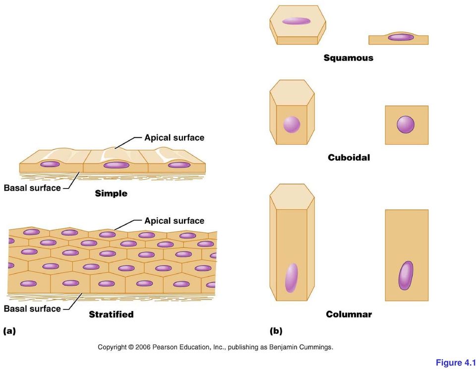
3 Figure 4.1
4 4 types of human tissues Epithelium Connective tissue Muscle tissue Skeletal Cardiac Smooth muscles Nerve tissue

5 Tissue derivation Ectoderm Mesoderm Endoderm

6 Epithelium derivation Ectoderm forms epidermis Mesoderm form mesothelium Endoderm forms the lining of the GI

7 CONNECTIVE TISSUE AND MUSCLES DERIVATION MESODERM

8 NERVES DERIVATION ECTODERM

9
10 EPITHELIUM epithelium is a tissue composed of a layer of cells. Epithelium lines both the outside (skin) and the inside cavities and lumen of bodies. The outermost layer of our skin is composed of dead stratified squamous epithelial cells, as are the mucous membranes lining the inside of mouths and body cavities. Other epithelial cells line the insides of the lungs, the gastrointestinal tract, the reproductive and urinary tracts, and make up the exocrine and endocrine glands.

11 Functions of epithelial cells include : secretion, absorption, protection, EXCEPT JOINTS CAVITIES, ANTERIOR SURFACE OF THE IRIS [NECKED CONNECTIVE TISSUE] transcellular transport, sensation detection, and selective permeability. Endothelium (the inner lining of blood vessels) is not related to epithelium except by name.
 is not related to epithelium")
12 Functions of Epithelial Tissue Protection Epithelial cells from the skin protect underlying tissue from mechanical injury, harmful chemicals, invading bacteria and from excessive loss of water. Sensation Sensory stimuli penetrate specialised epithelial cells. Specialised epithelial tissue containing sensory nerve endings is found in the skin, eyes, ears, nose and on the tongue. Secretion In glands, epithelial tissue is specialised to secrete specific chemical substances such as enzymes, hormones and lubricating fluids. Absorption Certain epithelial cells lining the small intestine absorb nutrients from the digestion of food. Excretion Epithelial tissues in the kidney excrete waste products from the body and reabsorb needed materials from the urine. Sweat is also excreted from the body by epithelial cells in the sweat glands. Diffusion Simple epithelium promotes the diffusion of gases, liquids and nutrients. Because they form such a thin lining, they are ideal for the diffusion of gases (eg. walls of capillaries and lungs). Cleaning Ciliated epithelium assists in removing dust particles and foreign bodies which have entered the air passages. Reduces Friction The smooth, tightly-interlocking, epithelial cells that line the entire circulatory system reduce friction between the blood and the walls of the blood vessels.

13 ENDOTHELIUM = CARDIOVASCULAR SYSTEM MESOTHELIUM = PERITONEAL CAVITY, THORACIC CAVITY, PERICARDIAL CAVITY

14 Epithelial cells: Sit on a basal lamina (formerly called a basement membrane). Have a free surface (if a cell has all of the other properties, but no free surface, it is called an epithelioid). Are avascular (contain no blood vessels).nourishment BY DIFFUSION Have almost no extracellular space. Renew basally. Are derived from all three germ layers. Are named for the most superficial live (nucleuscontaining) layer.

15 (1) Each epithelium has an apical surface, which is exposed to the outside of the body or to the lumen of an internal cavity. (2) The cells of an epithelium are held together with specialized contacts, including tight junctions and desmosomes. A cell junction is a structure within a tissue of a multicellular organism. Cell junctions are especially abundant in epithelial tissues. They consist of protein complexes and provide contact between neighbouring cells, between a cell and the extracellular matrix, or they built up the paracellular barrier of epithelia and control the paracellular transport. (3) A basement membrane connects the basal surface of the epithelium to an underlying layer of connective tissue. (4) Epithelial tissues are avascular.

16 There are three major types of cell junctions: Adherens junctions and Desmosomes Gap junctions Tight junctions

17 OTHERS Adhesion Plaques Actin filaments from the cell protrude and serve as the cell's anchor to the surrounding extracellular matrix. Chemical Synapses These occur among adjecent cells when one cell releases chemicals into the extracellular space that bind receptors on the adjacent cell. This variety of cell-to-cell communication is common among nerve cells Neurotransmission Neurotransmission is the passage of an electrical signal from a nerve cell (neuron) to another nerve cell, muscle or gland. In the process, chemicals called neurotransmitters, are released into the synapse (cleft between two neurons), where they bind receptors, changing the electrical potential in the membrane of the adjacent cell.

18

19

20 Cell Junctions Tight Junctions = zonula occludens, This is an occluding type of junction, where the plasma membranes of adjacent cells pinch tightly together. This creates a selective barrier between the spaces, allowing only certain materials to pass. Found often between cells of animal intestine, tight junctions assure that the correct chemical environment of intestinal lumen is maintained.

21 TIGHT JUNCTION
22
23 Desmosomes form links between cells, and provide a connection between intermediate filaments of the cell cytoskeletons of adjacent cells. This structure gives strength to tissues. DESMOSOME
24 Desmosomes
25 the need for signaling is a function of gap junctions that form pores connecting adjacent cells. Small molecules and electrical signals in one cell can pass through the gap junctions to adjacent cells. This process allows tissues to coordinate responses to stimuli. For example, gap junctions permit coordinated movements of muscles leading to childbirth. Gap junctions
26 Microvilli A microvillus (usually not occurring alone, so usually referred to as the plural microvilli) is a small (0.08 µm in diameter, 1 µm long) extension of the cell surface of absorptive and secretory epithelial cells, such as kidney and intestinal cells. These structures increase the surface area of cells by approximately 600 fold (human), thus facilitating absorption and secretion. There are several thousand microvilli present on the apical surface of a single cell in human small intestinal cells. Microvilli also occur in sensory cells of the inner ear (as stereocilia), in the cells of taste buds, and in olfactory receptor cells. They are observed on the plasma surface of eggs, aiding in the anchoring of sperm cells that have penetrated the extracellular coat of egg cells. Clustering of elongated microtubules around a sperm allows for it to be drawn closer and held firmly so fusion can occur. Microvilli are also of importance on the cell surface of white blood cells, as they aid in the migration of white blood cells.
27
28
29
30 Microvilli Microvilli are shorter and more uniform in length. These structures reminded histologists of a brush with bristles viewed from the side. They named this border in the small intestine the "brush border". Note, you can't see through the brush border. Cilia In this view of the epithelium lining the trachea, note the long filamentous cilia along the apical surface. You can usually see through a layer of cilia!
31 Specialized goblet cells
32 Classification Epithelial cells are classified by the following three factors: Shape Stratification (number of layers) Specialization
33 Shape Squamous: Cuboidal Columnar:. Transitional:
34 Squamous: Squamous cells are flat cells with an irregular flattened shape. The one-cell layer of simple squamous epithelium that forms the alveoli of the respiratory membrane, and the endothelium of capillaries, and is a minimal barrier to diffusion. Places where squamous cells can be found include the alveoli of the lungs, the filtration tubules of the kidneys, and the major cavities of the body. These cells are relatively inactive metabolically, and are associated with the diffusion of water, electrolytes, and other substances.
35 Cuboidal: As the name suggests, these cells have a shape similar to a cube, meaning its width is the same size as its height. The nuclei of these cells are usually located in the center
36 Columnar: These cells are taller than they are wide. Simple columnar epithelium is made up of a single layer of cells that are longer than they are wide. The nucleus is also closer to the base of the cell. The small intestine is a tubular organ lined with this type of tissue. Unicellular glands called goblet cells are scattered throughout the simple columnar epithelial cells and secrete mucus. The free surface of the columnar cell has tiny hairlike projections called microvilli. They increase the surface area for absorption
37 Transitional This is a specialized type of epithelium found lining organs that can stretch, such as the urothelium that lines the bladder and ureter of mammals. Since the cells can slide over each other, the appearance of this epithelium depends on whether the organ is distended or contracted: if distended, it appears as if there are only a few layers; when contracted, it appears as if there are several layers.
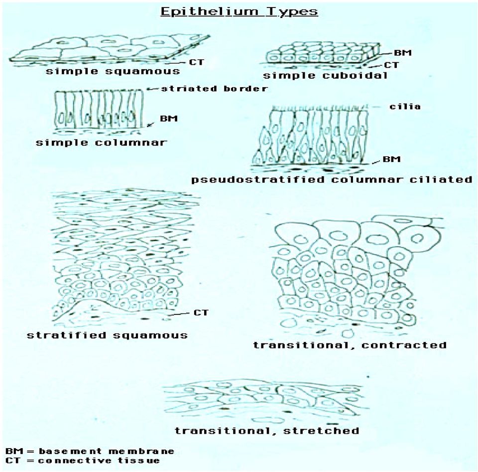
38 Stratification Simple: There is a single layer of cells. Stratified: More than one layer of cells. The superficial layer is used to classify the layer. Only one layer touches the basal lamina. Stratified cells can usually withstand large amounts of stress. Pseudostratified with cilia: This is used mainly in one type of classification (pseudostratified columnar epithelium). There is only a single layer of cells, but the position of the nuclei gives the impression that it is stratified. If a specimen looks stratified, but you can identify cilia, the specimen is pseudostratified ciliated epithelium since stratified epithelium cannot have cilia.
39 Specializations Keratinized cells contain keratin (a cytoskeletal protein). While keratinized epithelium occurs mainly in the skin, it is also found in the mouth and nose, providing a tough, impermeable barrier. Ciliated cells have apical plasma membrane extensions composed of microtubules capable of beating rhythmically to move mucus or other substances through a duct. Cilia are common in the respiratory system and the lining of the oviduct.
40 Simple squamous: Found in blood vessels & lymph channels (called endothelium) and body cavities (called mesothelium) Keratinized stratified squamous: Found in human skin (specifically, the dead superficial layer); also found in masticatory oral mucosa (attached gingiva, dorsum of tongue, hard palate, etc.) Non-Keratinised stratified squamous: Found in human oesophagus (Oral Mucosa) specfically non-masticatory "movable" mucosa, and vagina Simple cuboidal: Found in thyroid follicles Exclusively found in sweat gland ducts Ciliated Stratified cuboidal: simple columnar: Found in intestine and kidney (specifically, proximal convoluted tubule) Stratified columnar: Ducts of submandibular glands Transitional: Specialized to distend (stretch) as the urinary bladder fills
41 Simple epithelium,
42 Figure 4.2a
43 small blood vessel.
44 thyroid follicles Simple cuboidal epithelium
45 Figure 4.2b
46 kidney
47 Simple cuboidal eplithelia are cells in a single layer found on the surface of ovaries, the lining of nephrons parts of the eye thyroid. On these surfaces, the cells perform secretion and absorption. Stratified cuboidal epithelia are multi-layered. They protect such areas as ducts of sweat glands and the male urethra.
48 columnar The small intestine
49 Figure 4.2c
50 gallbladder
51
52 Columnar epithelial cells occur in one or more layers. The cells are elongated and column-shaped. The nuclei are elongated and are usually located near the base of the cells. Columnar epithelium forms the lining of the stomach and intestines. Some columnar cells are specialised for sensory reception such as in the nose, ears and the taste buds of the tongue. Goblet cells (unicellular glands) are found between the columnar epithelial cells of the duodenum. They secrete mucus or slime, a lubricating substance which keeps the surface smooth.
53 Ciliated Columnar Epithelium Pseudostratified Ciliated Columnar Epithelium TRACHEA
54
55
56 These are simple columnar epithelial cells, but in addition, they posses fine hair-like outgrowths, cilia on their free surfaces. These cilia are capable of rapid, rhythmic, wavelike beatings in a certain direction. This movement of the cilia in a certain direction causes the mucus, which is secreted by the goblet cells, to move (flow or stream) in that direction. Ciliated epithelium is usually found in the air passages like the e nose. It is also found in the uterus and Fallopian tubes of females. The movement of the cilia propel the ovum to the uterus.
57
58 Glandular Epithelium
59 Columnar epithelium with goblet cells is called glandular epithelium. Some parts of the glandular epithelium consist of such a large number of goblet cells that there are only a few normal epithelial cells left. Columnar and cuboidal epithelial cells often become specialised as gland cells which are capable of synthesising and secreting certain substances such as enzymes, hormones, milk, mucus, sweat, wax and saliva. Unicellular glands consist of single, isolated glandular cells such as the goblet cells. Sometimes a portion of the epithelial tissue becomes invaginated and a multicellular gland is formed. Multicellular glands are composed of clusters of cells. Most glands are multicellular including the the salivary glands
60
61
62
63 Stratified Epithelium. epidermis (outer part of the skin), typical stratified squamous epithelium and the dermis showing connective tissue.
64 stratified squamous epithelium from the esophagus
65
66 Where body linings have to withstand wear and tear, the epithelia are composed of several layers of cells and are then called compound or stratified epithelium. The top cells are flat and scaly and it may or may not be keratinised (i.e. containing a tough, resistant protein called keratin). The mammalian skin is an example of dry, keratinised, stratified epithelium. The lining of the mouth cavity is an example of an unkeratinisied, stratified epithelium.
67
68 TRANSITIONAL transitional epithelium from the urinary bladder. The arrows are on the actual transitional cell layer. Below the dark layer of transitional cell nuclei is the subucosa.
69 Transitional epithelium contains cells that are flattened and cells that are cuboidal; hence the name "transitional". You can find transitional epithelium in the bladder and in the first expansion of the ureters as they leave the kidneys (called a calyx)
70
71 TO REVIEW Simple Columnar Epithelium Colon Duodenum Jejunum Simple cuboidal epithelium thyroid kidney tubules Simple squamous epithelium spinal cord (capillaries) kidney glomerulus Pseudostratified columnar epithelium olfactory trachea
72 Stratified squamous epithelium esophagus skin Stratified columnar/cuboidal epithelium salivary gland ducts (excretory) sweat gland ducts Transitional epithelium major calyx bladder
73 EXERCISE WHAT TYPE OF TISSUE, WHERE,ANY GOBLET CELLS
74
75 What is the function of the ileum
76 The ileum, the last section of the small intestine, is different from the duodenum and the jejunum by having more goblet cells in the mucosa and lymph nodules called Peyer's patches.
77
78
79 Absorb vitamin B 12 and bile salts The wall itself is made up of folds, each of which has many tiny finger-like projections known as villi, on its surface. In turn, the epithelial cells which line these villi possess even larger numbers of microvilli. Therefore the ileum has an extremely large surface area both for the adsorption (attachment) of enzyme molecules and for the absorption of products of digestion. The cells that line the ileum contain the protease and carbohydrase enzymes responsible for the final stages of protein and carbohydrate digestion. These enzymes are present in the cytoplasm of the epithelial cells. The villi contain large numbers of capillaries which take the amino acids and glucose produced by digestion to the hepatic portal vein and the liver.
80
81 Connective tissues Dense connective tissue or Fibrous connective tissue forms ligaments and tendons. Its densely packed collagen fibers have great tensile strength. Loose connective tissue or Areolar connective tissue holds organs and epithelia in place, and has a variety of proteinaceous fibers, including collagen and elastin. It is also important in inflammation. Reticular connective tissue is a network of reticular fibers (fine collagen) that form a soft skeleton to support the lymphoid organs (lymph nodes, bone marrow, and spleen.) Adipose tissue contains adipocytes, used for cushioning, thermal insulation, lubrication (primarily in the pericardium) and energy storage. Fiber types as follows: Collagenous fibers, elastic fibers, retucular fibers
82 Connective tissues function primarily to support the body and to bind or connect together all types of tissue. This tissue also provide a mechanical framework (the skeleton) which plays an important role in locomotion. Unlike epithelial tissue, connective tissue is characterised by the large amounts of intercellular substance (also called ground substance or the matrix) that it contains. Connective tissue are relatively few cells which are widely seperated from each other. These living cells are responsible for secreting the large amounts of intercellular ground substance (matrix). The matrix is a non-living material which may be liquid (eg. blood), semi-solid (eg. connective tissue) or solid (eg. bone). Embedded in the matrix are a variety of connecting and supporting fibres,eg. collagen fibres and elastic fibres. Classification of the basic connective tissue depends on the predominant fibre type present in each
83 1). binding 2). support 3). binding 4). insulation 5). transportation. Accordingly, connective tissues are divided among four main classes: (1) connective tissue proper, (2) cartilage, (3) bone, (4) blood.
84 Characteristics of Connective Tissues A). Not adjacent to other cells B). Extra Cellular Matrix C). Common origin in embryonic development. D). Vary in degrees of vascularity
85 All connective tissues have a common origin during embryonic development, and all connective tissues contain extracellular matrix. The extracellular matrix (ECM) is a complex structural entity surrounding and supporting cells that are found within mammalian tissues. The ECM is often referred to as the connective tissue. The ECM is composed of 3 major classes of biomolecules: 1. Structural proteins: collagen and elastin. 2. Specialized proteins: e.g. fibrillin, fibronectin, and laminin. 3. Proteoglycans: these are composed of a protein core to which is attached long chains of repeating disaccharide units termed of glycosaminoglycans (GAGs) forming extremely complex high molecular weight components of the ECM.
86
87 Areolar connective tissue is found throughout the body underlying epithelial tissues, surrounding organs, and wrapped around blood vessels and nerves.you may think of it as the body s packing material. The predominant cell type in areolar connective tissue is the fibroblast, which secretes fibers into the extracellular matrix. Macrophages and mast cells are also present, and they act as parts of the immune system to protect the body. Three types of fibers can be found in the matrix of areolar connective tissue: Collagen fibers provide strength, elastic fibers can stretch and recoil, and reticular fibers may help connect the tissue to organs or other types of tissue.
88
89 loose connective tissue
90
91
92 loose connective tissue
93 dense irregular CT
94 dense regular CT from a tendon.
95 Bronchioles: the elastic CT is stained black and is arranged in loose lines around the open spaces as indicated by the arrows.
96 This silver stain of a lymph node stains the stromal reticular fibers black.
97 Adipose tissue in the renal pelvis. The adipocytes are the large lipidfilled cells
98
99
100 Irregular Dense Collagenous Connective Tissue - Human Dura mater
101 hyaline cartilage
102 The type of protein fiber embedded within the matrix of cartilage determines the cartilage type. In hyaline cartilage protein fibers are large and predominantly collagen
103 Hyaline cartilage is semi-transparent and appears bluish-white in colour. It is extremely strong, but very flexible and elastic. Hyaline cartilage consists of living cells, chondrocytes, which are situated far apart in fluid-filled spaces, the lacunae. There is an extensive amount of rubbery matrix between the cells and the matrix contains a number of collagenous fibres. Hyaline cartilage occurs in trachea, the larynx, the tip of the nose, in the connection between the ribs and the breastbone and also the ends of bone where they form joints. Temporary cartilage in mammalian embryos also consists of hyaline cartilage. Functions Reduces friction at joints. By virtue of the smooth surface of hyaline cartilage, it provides a sliding area which reduces friction, thus facilitating bone movement. Movement Hyaline cartilage joins bones firmly together in such a way that a certain amount of movement is still possible between them. Support The c-shaped cartilagenous rings in the windpipes (trachea and bronchi) assist in keeping those tubes open. Growth Hyaline cartilage is responsible for the longitudinal growth of bone in the neck regions of the long bones.
104 White fibrocartilage is an extremely tough tissue. The orientation of the bundles depends upon the stresses acting on the cartilage. The collagenous bundles take up a direction parallel to the cartilage. Fibrocartilage is found as discs between the vertebrae between the pubic bones in front of the pelvic girdle and around the edges of the articular cavities such as the glenoid cavity in the shoulder joint. Functions Shock absorbers. The cartilage between the adjacent vertebrae absorbs the shocks that will otherwise damage and jar the bones while we run or walk. Provides sturdiness without impeding movement. The white fibrocartilage forms a firm joint between bones but still allows for a reasonable degree of movement. Deepens sockets. In articular cavities (such as the ball-and-socket joints in the hip and shoulder regions) white fibrocartilage deepens the sockets to make dislocation less possible.
105
106
107 Elastic cartilage. Basically elastic cartilage is similar to hyaline cartilage, but in addition to the collagenous fibres, the matrix of the elastic also contains an abundant network of branched yellow elastic fibres. They run through the matrix in all directions. This type of cartilage is found in the lobe of the ear, the epiglottis and in parts of the larynx. Functions Maintain shape. In the ear, for example, elastic cartilage helps to maintain the shape and flexibility of the organ. Support Elastic cartilage also strengthens and supports these structures.
108 elastic.
109 Epiglottis have elastic cartilage
110
111 hyaline cartilage from a trachea. Notice the lacunae. Try saying "lacunae" 10 times in a row really fast
112 Fibrocartilage
113 The cartilage starts at the bottom and develops into trabecular bone towards the top
114 Blast suffix bud or germ Examples: osteoblast (osteo-blast) - a cell from which bone is derived Cyte suffix = New Latin -cyta, from Greek kutos, hollow vessel. used to form cell names and classifications adipocyte astrocyte blastocyte choanocyte coenocyte erythrocyte gametocyt
115 Disorders of connective tissue Various connective tissue conditions have been identified; these can be both inherited and environmental. Marfan syndrome - a genetic disease causing abnormal fibrillin. Scurvy - caused by a dietary deficiency in vitamin C, leading to abnormal collagen. Ehlers-Danlos syndrome - a genetic disease causing progressive deterioration of collagens, with different EDS types affecting different sites in the body, such as joints, heart valves, organ walls, arterial walls, etc. Osteogenesis imperfecta (brittle bone disease) - caused by insufficient production of good quality collagen to produce healthy, strong bones. Spontaneous pneumothorax - collapsed lung, believed to be related to subtle abnormalities in connective tissue. Sarcoma - a neoplastic process originating in connective tissue.
The Tissue Level of Organization
 The Tissue Level of Organization Tissues A groups of similar cells, usually having similar embryonic origin and specialized function Histology: the study of tissues Four general types Epithelial Muscle
Animal Tissues. I. Epithelial Tissue
 Animal Tissues There are four types of tissues found in animals: epithelial tissue, connective tissue, muscle tissue, and nervous tissue. In this lab you will learn the major characteristics of each tissue
Lab Exercise 4. Epithelial Tissues. Connective Tissue Proper. What you need to be able to do on the exam after completing this lab exercise:
 Lab Exercise 4 Epithelial Tissues Connective Tissue Proper Textbook Reference: See Chapter 4 What you need to be able to do on the exam after completing this lab exercise: Be able to identify each type
Section B: Epithelial Tissue 1. Where are epithelial tissues found within the body? 2. What are the functions of the epithelial tissues?
 Tissue worksheet Name Section A: Intro to Histology Cells are the smallest units of life. In complex organisms, cells group together with one another based on similar structure and function to form tissues.
Human Anatomy & Physiology I with Dr. Hubley. Practice Exam 1
 Human Anatomy & Physiology I with Dr. Hubley Practice Exam 1 1. Which definition is the best definition of the term gross anatomy? a. The study of cells. b. The study of tissues. c. The study of structures
Histology. Epithelial Tissue
 Histology Epithelial Tissue Epithelial Tissue Lines internal and external body surfaces Forms glands Epithelial Tissue Little extracellular matrix Attached on one side Avascular Basement membrane Apical
Vertebrate Body Organization
 Vertebrate Body Organization Digestive tube suspended in coelom from mouth to anus Body supported by internal skeleton of jointed bones Vertebrae and Cranium protects nervous system Diaphragm divides coelom
Tissues (Histology) Ch. 3 Human Anatomy lecture
 Ch. 3 Human Anatomy lecture") I. Histology the study of tissues A. 4 basic tissue types epithelial connective muscle nervous Tissues (Histology) Ch. 3 Human Anatomy lecture B. Usually found in combinations to form organs. C. As you
I. Histology the study of tissues A. 4 basic tissue types epithelial connective muscle nervous Tissues (Histology) Ch. 3 Human Anatomy lecture B. Usually found in combinations to form organs. C. As you
Tissue Types. 1. Epithelial Tissue (or epithelium) is the lining, covering, and glandular tissue of the body
 is the lining, covering, and glandular tissue of the body") Tissue Types A. Tissues 1. Tissues: groups of cells similar in structure and function 2. Four Types of Tissues: a. Epithelium: for covering b. Connective Tissue: for support c. Muscle: for movement d.
Tissue Types A. Tissues 1. Tissues: groups of cells similar in structure and function 2. Four Types of Tissues: a. Epithelium: for covering b. Connective Tissue: for support c. Muscle: for movement d.
CHAPTER 5: TISSUES. 2. Name the four primary adult tissue types, and give a brief description of each.
 OBJECTIVES: 1. Define the term tissue. 2. Name the four primary adult tissue types, and give a brief description of each. 3. Describe the functions and types of extracellular fluid (ECF). 4. Compare and
Introduction to Anatomy and Physiology: Tissues and Integumentary System. Biology 105 Lecture 7 Chapter 4
 Introduction to Anatomy and Physiology: Tissues and Integumentary System Biology 105 Lecture 7 Chapter 4 Outline I. Tissues A. Epithelial B. Connective C. Muscle D. Nervous tissues II. Cell-to-cell contact
Laboratory 3 Histology
 Laboratory 3 Histology Goals: For epithelial tissues: o discuss the major features; o classify based on simple/stratified and squamous/cubodial/columnar; o identify each type by microscopy; o identify
Biology 105 Human Biology PRACTICE MIDTERM EXAM 1. Essentials of Anatomy and Physiology, 5e (Martini/Nath) Chapter 4 The Tissue Level of Organization
 Chapter 4 The Tissue Level of Organization") Essentials of Anatomy and Physiology, 5e (Martini/Nath) Chapter 4 The Tissue Level of Organization Multiple-Choice Questions 1) The four main types of tissues include A) epithelium. B) muscle. C) nerve.
Essentials of Anatomy and Physiology, 5e (Martini/Nath) Chapter 4 The Tissue Level of Organization Multiple-Choice Questions 1) The four main types of tissues include A) epithelium. B) muscle. C) nerve.
ORGAN SYSTEMS OF THE BODY
 ORGAN SYSTEMS OF THE BODY DEFINITIONS AND CONCEPTS A. Organ a structure made up of two or more kinds of tissues organized in such a way that they can together perform a more complex function that can any
Chapter 48. Nutrients in Food. Carbohydrates, Proteins, and Lipids. Carbohydrates, Proteins, and Lipids, continued
 Carbohydrates, Proteins, and Lipids The three nutrients needed by the body in the greatest amounts are carbohydrates, proteins, and lipids. Nutrients in Food All of these nutrients are called organic compounds,
Ground substance is the component of connective tissue between the cells and the fibers
 Connective Tissues Directions: Insert and install your Interactions: Foundations CD. a. Click the "Contents" button. b. Open the Tissue Level of Organization file. c. Click on Anatomy Overviews. d. Work
Name Class Date Laboratory Investigation 24A Chapter 24A: Human Skin
 Name Class Date Laboratory Investigation 24A Chapter 24A: Human Skin Human Anatomy & Physiology: Integumentary System You may refer to pages 386-394 in your textbook for a general discussion of the integumentary
Location: air sacs of lungs; nephrons of kidney; lining of circulatory system, lymphatic vessels, & ventral body cavity
 Bio. 2304 - Human Anatomy HISTOLOGY (STUDY OF TISSUES) - Lab & Lecture Objectives Important: For each slide know 1.) specific tissue type 2.) any specialized structures or cells in the tissue (& know their
THE HUMAN BODY SYSTEMS
 Name Period Date THE HUMAN BODY SYSTEMS System Function Diagram Major Organs Digestive 1. take in food (ingestion) 2. digest food into smaller molecules and absorb nutrients 3. remove undigestable food
BIO 137: CHAPTER 1 OBJECTIVES
 BIO 137: CHAPTER 1 OBJECTIVES 1. Define the terms anatomy and physiology, and explain their relationship using an example of a human structure with its corresponding function. A. ANATOMY = the study of
10.2 The Human Digestive System pg. 411
 10.2 The Human Digestive System pg. 411 The human digestive system is made up of a group of organs working together. The digestive tract is made up of the mouth, esophagus, stomach, small intestine, and
2161-1 - Page 1. Name: 1) Choose the disease that is most closely related to the given phrase. Questions 10 and 11 refer to the following:
 Choose the disease that is most closely related to the given phrase. Questions 10 and 11 refer to the following:") Name: 2161-1 - Page 1 1) Choose the disease that is most closely related to the given phrase. a disease of the bone marrow characterized by uncontrolled production of white blood cells A) meningitis B)
Name: 2161-1 - Page 1 1) Choose the disease that is most closely related to the given phrase. a disease of the bone marrow characterized by uncontrolled production of white blood cells A) meningitis B)
By Casey Schmidt and Wendy Ford
 By Casey Schmidt and Wendy Ford Body systems Digestive System Circulatory System Respiratory System Excretory System Immune System Reproductive System Nervous System Muscular System Skeletal System Endocrine
Biology 2401 - Anatomy and Physiology I Exam 1 notes - Introduction, Cell and Tissue Structure
 Biology 2401 - Anatomy and Physiology I Exam 1 notes - Introduction, Cell and Tissue Structure Two major principles in study of animal bodies: (humans, like other living organisms are product of evolutionary
Biology 13A Lab #3: Cells and Tissues
 Biology 13A Lab #3: Cells and Tissues Lab #3 Table of Contents: Expected Learning Outcomes.... 28 Introduction...... 28 Activity 1: Eukaryotic Cell Structure... 29 Activity 2: Perspectives on Tissue Preparations.
5. Secretion: release of water, acids. Enzymes, buffers by digestive tract.
 Digestive System CH-16 Lecture topics Functions of the digestive system: p. 488. 1. Ingestion: Taking food in 2. Propulsion: movement of food thru alimentary canal p.490. voluntary: swalloing : skeletal
Eating, pooping, and peeing THE DIGESTIVE SYSTEM
 THE DIGESTIVE SYSTEM Ingested food is not technically in the body until it is absorbed so it needs to be: Mechanically and chemically reduced Transported by the blood to the cells Large portions are not
Introduction to Animal Systems
 Human Body Systems Introduction to Animal Systems Recurring Themes in Biology 1. Correlation between structure and function( seen at many levels) 2. Life is organized at many levels from Smallest ----
Lesson Aim To explain the human body at a microscopic level, including the structure and function of cells, tissues and membranes.
 LESSON 1. CELLS & TISSUES Lesson Aim To explain the human body at a microscopic level, including the structure and function of cells, tissues and membranes. THE CELL All living matter is composed of functional
The Digestive System. Chapter 16. Introduction. Histological Organization. Overview of Digestive System. Movement and Mixing of Digestive Materials
 The Digestive System Chapter 16 Introduction Structure of the digestive system A tube that extends from mouth to anus Accessory organs are attached Functions include Ingestion Movement Digestion Absorption
Animal Systems: The Musculoskeletal System
 Animal Systems: The Musculoskeletal System Tissues, Organs, and Systems of Living Things Cells, Cell Division, and Animal Systems and Plant Systems Cell Specialization Human Systems The Digestive The Circulatory
67 The Human Skeleton
 67 The Human Skeleton Skull SCIENCE EXPLORER Focus on Life Science Prentice-Hall, Inc. Clavicle (collarbone) Scapula (shoulder blade) Carpals Metacarpals Phalanges Femur Tibia Humerus Ulna Sternum (breastbone)
Small & Large Intestines
 Small & Large Intestines Small Intestine: principal site for digestion of food and absorption of the products of digestion Large Intestine: reabsorption of water and elimination of undigested food and
Integumentary System Digestive System. Outline. Integumentary System 11/4/2008. Week 11 BA & BP November 4, 2008 Nadia Arora, ND
 Integumentary System Digestive System Week 11 BA & BP November 4, 2008 Nadia Arora, ND Outline Integumentary system and body membranes Types of body membranes and their function General structure and main
Lect 01 - Epithelial Tissue
 Structure & Function Introduction Prof Kumlesh K. Dev Department of Physiology What is Structure & Function? Integration is key structure and function requires integration of physiology, anatomy and biochemistry
Engage: Brainstorming Body Systems. Record the structures and function of each body system in the table below.
 Engage: Brainstorming Body s Record the structures and function of each body system in the table below. Body Nervous Circulatory Excretory Immune Digestive Respiratory Skeletal Muscular Endocrine Integumentary
Digestion, Absorption. How & where?
 Digestion, Absorption How & where? What happens to food? Three processes Digestion Absorption Elimination Where do they occur? GI tract Overview of Digestion GI tract Gastrointestinal (GI) tract: series
THE GI TRACT IS A CONTINUOUS MULTILAYERED TUBE EXTENDING FROM THE MOUTH TO THE ANUS THAT IS SUPPORTED AND PARTIALLY COVERED BY THE PERITONEUM.
 THE GI TRACT IS A CONTINUOUS MULTILAYERED TUBE EXTENDING FROM THE MOUTH TO THE ANUS THAT IS SUPPORTED AND PARTIALLY COVERED BY THE PERITONEUM. OVERVIEW OF THE DIGESTIVE SYSTEM Two groups of organs compose
RAD 223. Radiography physiology. Lecture Notes. First lecture: Cell and Tissue
 RAD 223 Radiography physiology Lecture Notes First lecture: Cell and Tissue Physiology: the word physiology derived from a Greek word for study of nature. It is the study of how the body and its part work
Human Anatomy and Physiology The Respiratory System
 Human Anatomy and Physiology The Respiratory System Basic functions of the respiratory system: as a Gas exchange supply oxygen to aerobic tissues in the body and remove carbon dioxide waste product. in-
Human Body Systems Project By Eva McLanahan
 Human Body Systems Project By Eva McLanahan Students will work in groups to research one of the eleven body systems as found in Holt, Rinehart, and Winston Modern Biology (2002). Research will focus on
Outline Digestive System
 Outline Digestive System The Digestive System Digestive System Lecture Packet 19 Chapter 15 I. Function II. Layers of the GI tract III. Major parts: mouth, pharynx, esophagus, stomach, small intestine,
Digestive system Review
 Digestive system Review 1. Distinguish between chemical digestion and mechanical digestion. The physical breakdown of food begins in the mouth with two types of processes. The mouth is a complex structure
The Digestive System. Chapter 14. The Digestive System and Body Metabolism. Metabolism. Organs of the Digestive System. Digestion.
 Chapter 14 The Digestive System The Digestive System and Body Metabolism Digestion of ingested food of nutrients into the blood Metabolism Production of Constructive and degradative cellular activities
CHAPTER 9 BODY ORGANIZATION
 CHAPTER 9 BODY ORGANIZATION Objectives Identify the meaning of 10 or more terms relating to the organization of the body Describe the properties of life Describe the function for the structures of the
Fundamentals of Anatomy & Physiology Course Outline, Objectives and Accreditation Information
 201 Webster Building 3411 Silverside Road Wilmington, DE 19810 Phone: 1-888-658-6641 Fax: 1-302-477-9744 learn@corexcel.com www.corexcel.com Course Outline, Objectives and Accreditation Information Chapter
Chetek-Weyerhaeuser High School
 Chetek-Weyerhaeuser High School Anatomy and Physiology Units and Anatomy and Physiology A Unit 1 Introduction to Human Anatomy and Physiology (6 days) Essential Question: How do the systems of the human
Chapter 20: Tissues & Organ Systems
 Chapter 20: Tissues & Organ Systems The Importance of Homeostasis The maintenance of a relatively constant internal environment, i.e., homeostasis, is essential for life. Cellular conditions that need
Digestive System AKA. GI System. Overview. GI Process Process Includes. G-I Tract Alimentary Canal
 Digestive System AKA G-I Tract Alimentary Canal Overview GI System Consists of Mouth, pharynx, esophagus, stomach, small intestine, large intestine, anus About 30 in length Accessory Organs Teeth, tongue,
Paramedic Program Anatomy and Physiology Study Guide
 Paramedic Program Anatomy and Physiology Study Guide Define the terms anatomy and physiology. List and discuss in order of increasing complexity, the body from the cell to the whole organism. Define the
SAMPLE LECTURE EXAM 1 -- HUMAN ANATOMY
 SAMPLE LECTURE EXAM 1 -- HUMAN ANATOMY 1. The subcutaneous layer consists mostly of. a. smooth muscle c. areolar and adipose connective tissues d. melanin e. keratin 2. Which of the following statements
Investigating the Human Body On-site student activities: Years 7-8 Investigating the Human Body On-site student activities Years 7 8
 Investigating the Human Body On-site student activities Years 7 8 Student activity (and record) sheets have been developed with alternative themes for students to use as guides and focus material during
Cells, tissues and organs
 Chapter 8: Cells, tissues and organs Cells: building blocks of life Living things are made of cells. Many of the chemical reactions that keep organisms alive (metabolic functions) take place in cells.
Divisions of Digestive System. Organs of the Alimentary Canal. Anatomy of the Digestive System: Organs of the Alimentary Canal. CHAPTER 14 p.
 Divisions of Digestive System Anatomy of the Digestive System: Organs of the Alimentary Canal CHAPTER 14 p. 412-423 1. Alimentary Canal or Gastrointestinal Tract (GI)-digests and absorbs food coiled hollow
Digestive System Digestive Tract
 Digestive System Digestive Tract Dept. of Histology and Embryology 周 莉 教 授 Introduction of digestive system * a long tube extending from the mouth to the anus, and associated with glands. * its main function:
The Digestive System. Chapter 15
 The Digestive System Chapter 15 Introduction Digestion refers to the mechanical and chemical breakdown of food so the nutrients can be absorbed by cells Carried out by the digestive system Consists of
Chapter 49 - Nutrients and the Digestive System I. Nutrients (chemical substances necessary for organisms to grow and function properly)
") Chapter 49 - Nutrients and the Digestive System I. Nutrients (chemical substances necessary for organisms to grow and function properly) 6 basic nutrients - 4 food groups (milk, meat, fruit and vegetable,
Chapter 49 - Nutrients and the Digestive System I. Nutrients (chemical substances necessary for organisms to grow and function properly) 6 basic nutrients - 4 food groups (milk, meat, fruit and vegetable,
Smooth Muscle. Learning Objectives.
 Smooth Muscle. Learning Objectives. At the end of this course, you should be able to : 1. describe the structure of smooth muscle 2. describe where smooth muscle occurs within the body 3. discuss the structural
Skeletal, Muscular, and Integumentary Systems
 Chapter 36 Skeletal, Muscular, and Integumentary Systems Section 36 1 The Skeletal System (pages 921 925) This section describes the skeletal system and its functions. Introduction (page 921) 1. What forms
Essentials of Human Anatomy & Physiology. 7 th edition Marieb, Elaine, 2003. Chapters 10-11. Lab Manual, 2 nd edition and coloring book, 7 th edition
 Topic/Unit: Anatomy & Physiology Circulatory System Curricular Goals/ Learning Outcomes: Students will be able to identify the composition of blood and its function. Students will be able to differentiate
The Vertebrate (mostly human) Digestive System
 Digestive System") The Vertebrate (mostly human) Digestive System Mouth - mastication, lubrication, digestion Pharynx and Esophagus - swallowing Stomach - some digestion Small intestine - most digestion and absorption Large
The Vertebrate (mostly human) Digestive System Mouth - mastication, lubrication, digestion Pharynx and Esophagus - swallowing Stomach - some digestion Small intestine - most digestion and absorption Large
h. Large intestine 3
 (1) General features (a) Large intestine is last organ of digestive tract proper divided into 3 or 4 regions cecum appendix in humans colon rectum 1 b) No villi lumenal epithelium has microvilli This brush
Human Anatomy & Physiology I with Dr. Hubley. Practice Exam #2
 Human Anatomy & Physiology I with Dr. Hubley Practice Exam #2 For questions 1 through 3, select your answers from the following responses: a. stratified squamous epithelium b. reticular connective tissue
The Respiratory System
 Human Anatomy III: Respiratory, Urinary & Digestive Systems The Respiratory System Major functions include: Obtaining oxygen Removing carbon dioxide Maintenance of ph balance Respiration may be accomplished
Digestive System Functions
 Digestive System Functions A. Gastrointestinal Processes 1. Ingestion: placing food in mouth (voluntary) 2. Propulsion: moving food through GI tract a. Peristalsis: alternating waves of contraction and
BIOL 1108 Vertebrate Anatomy Lab
 BIOL 1108 Vertebrate Anatomy Lab This lab explores major organs associated with the circulatory, excretory, and nervous systems of mammals. Circulatory System Vertebrates are among the organisms that have
The Integumentary System Dr. Ali Ebneshahidi
 The Integumentary System Dr. Ali Ebneshahidi The Skin The integument system consists of the skin (cutaneous membrane) and its accessory organs. The skin is composed of three layers of tissue: the outer
North Bergen School District Benchmarks
 Grade: 10,11, and 12 Subject: Anatomy and Physiology First Marking Period Define anatomy and physiology, and describe various subspecialties of each discipline. Describe the five basic functions of living
The digestive system eliminated waste from the digestive tract. But we also need a way to eliminate waste from the rest of the body.
 Outline Urinary System Urinary System and Excretion Bio105 Lecture 20 Chapter 16 I. Function II. Organs of the urinary system A. Kidneys 1. Function 2. Structure III. Disorders of the urinary system 1
A. function: supplies body with oxygen and removes carbon dioxide. a. O2 diffuses from air into pulmonary capillary blood
 A. function: supplies body with oxygen and removes carbon dioxide 1. ventilation = movement of air into and out of lungs 2. diffusion: B. organization a. O2 diffuses from air into pulmonary capillary blood
The Lymphatic System. Dr. Naim Kittana, PhD
 The Lymphatic System Dr. Naim Kittana, PhD 1 Disclosure The material and the illustrations are adopted from the textbook Human Anatomy and Physiology / Ninth edition/ Eliane N. Marieb 2013 Dr. Naim Kittana,
HISTOLOGY: THE FORM AND FUNCTION OF TISSUES
 Biology E-65C Lab #1 HISTOLOGY: THE FORM AND FUNCTION OF TISSUES Objectives: 1. To gain an appreciation of tissue types in terms of their functional significance 2. To gain experience with the use of a
Digestive System. Gross Anatomy and Physiology
 Digestive System Gross Anatomy and Physiology I. Introduction A. Base Function: Working with the circulatory system the digestive system provides the body with fuel. B. Main players: 1. Digestive tract:
The Human Digestive System
 The Human Digestive System Name: Section: Date: Page 1 of 10 Page 2 of 10 Page 3 of 10 Page 4 of 10 Page 5 of 10 Page 6 of 10 Putting it All Together Digestive Enzymes Page 7 of 10 Page 8 of 10 Page 9
Digestive System Why is digestion important? How is food digested? Physical Digestion and Movement
 Digestive System The digestive system is made up of the digestive tract a series of hollow organs joined in a long, twisting tube from the mouth to the anus and other organs that help the body break down
Human Body Vocabulary Words Week 1
 Vocabulary Words Week 1 1. arteries Any of the blood vessels that carry blood away from the heart to all parts of the body 2. heart The muscular organ inside the chest that pumps blood through the body
Special organ structures and functions conduct these tasks through the successive parts of the overall system.
 Chapter 5 Digestion, Absorption, and Metabolism Chapter 5 Lesson 5.1 Key Concepts Through a balanced system of mechanical and chemical digestion, food is broken down into smaller substances and the nutrients
CHAPTER 6: INTEGUMENTARY SYSTEM. 1. Explain why the skin is called the cutaneous membrane.
 OBJECTIVES: 1. Explain why the skin is called the cutaneous membrane. 2. Name the layers of the skin, describe the structure (tissues) of each, and name a general function of each. 3. Discuss the four
Two main classes: Epithelial Connective (synovial) Epithelial. Cutaneous Mucous Serous
 Epithelial. Cutaneous Mucous Serous") Two main classes: Epithelial Connective (synovial) Epithelial Cutaneous Mucous Serous Epithelial Membranes = sheet of epithelia + connective tissue base 1. Cutaneous membrane: outer skin layer (stratified
Two main classes: Epithelial Connective (synovial) Epithelial Cutaneous Mucous Serous Epithelial Membranes = sheet of epithelia + connective tissue base 1. Cutaneous membrane: outer skin layer (stratified
Functions of the GI Tract. Chapter 18. Functions of the GI Tract (continued)
") Functions of the GI Tract Chapter 18 The Digestive System Motility: Movement of of food through the GI tract. Ingestion: Taking food into the mouth. Mastication: Chewing the food and mixing it with saliva.
Functions of the GI Tract Chapter 18 The Digestive System Motility: Movement of of food through the GI tract. Ingestion: Taking food into the mouth. Mastication: Chewing the food and mixing it with saliva.
Respiratory System. Chapter 21
 Respiratory System Chapter 21 Structural Anatomy Upper respiratory system Lower respiratory system throat windpipe voice box Function of Respiratory System Gas exchange Contains receptors for sense of
I. The basic function of the digestive system is
 Chapter 15, Digestive System - ANATOMY OF THE DIGESTIVE SYSTEM I. The basic function of the digestive system is. This process is called. II. List 2 other names for the digestive tract: A. B. III. The digestive
Chapter 15 Digestion and Nutrition
 Chapter 15 Digestion and Nutrition Digestive System: Digestion refers to the mechanical and chemical breakdown of foods so that nutrients can be absorbed by cells. Consists of the canal which is all of
The cells of the human body do not operate independently of
 4 Tissues I. Epithelial Tissue 65 Special Characteristics of Epithelia 66 Classification of Epithelia 66 Glands 72 Epithelial Surface Features 74 II. Connective Tissue 77 Special Characteristics of Connective
Anatomy and Physiology
 Learning Activities It is important that you do not lecture all of the time. If you employ a variety of teaching styles, your students will stay focused better and they will find it easier to process the
Questions on The Nervous System and Gas Exchange
 Name: Questions on The Nervous System and Gas Exchange Directions: The following questions are taken from previous IB Final Papers on Topics 6.4 (Gas Exchange) and 6.5 (Nerves, hormones and homeostasis).
Anatomy & Physiology Bio 2401 Lecture. Instructor: Daryl Beatty Day 1 Intro to Lecture 1
 Anatomy & Physiology Bio 2401 Lecture Instructor: Daryl Beatty Day 1 Intro to Lecture 1 Introduction: Daryl Beatty M.S. Microbiology 28 Years Dow, Research & TS&D. Family BC since 2007 More importantly:
Medical Physiology Z.H.Al-Zubaydi
 Lec.13 Medical Physiology Z.H.Al-Zubaydi Functions of the Digestive System The major functions of the digestive tract include the following six processes, summarized in Figure 1: 1. Ingestion Food must
Anatomy and Physiology
 Anatomy and Physiology UNIT I: Introduction to Anatomy and Physiology The student will demonstrate an understanding of the anatomic and physiological basis of life and the ability to explain the interdependence
7. Skeletal System: Bone Structure and Function
 7. Skeletal System: Bone Structure and Function For the next two chapters (7 and 9) we will study the skeletal system. Although the major feature of this system is the bones, the skeletal system also consists
The Gastrointestinal System It consists of: The digestive tract Mouth Pharynx Oesophagus Stomach Small intestine Large intestine
 The Gastrointestinal System It consists of: The digestive tract Mouth Pharynx Oesophagus Stomach Small intestine Large intestine The digestive organs Teeth Tongue Salivary glands Liver Gall bladder Pancreas
Welcome back. Today, we embark on Lesson 6 where we ll study the human digestive system.
 Basic Human Anatomy Lesson 6: The Human Digestive System Welcome back. Today, we embark on Lesson 6 where we ll study the human digestive system. After completing this lesson, you should be able to define
THE DIGESTIVE SYSTEM
 THE DIGESTIVE SYSTEM What is digestion? Digestion is the process of breaking down food so that it's small enough to be absorbed and used by the body for energy or in other bodily functions. Digestion involves
The Cell Interior and Function
 The Cell Interior and Function 5 5.0 CHAPTER PREVIEW Investigate and understand the organization and function of the cell interior. Define the differences between eukaryotic and prokaryotic cell structure.
chemicals > transported from outside to in > waste products created > they need to be removed
 1 Transport systems chemicals > transported from outside to in > waste products created > they need to be removed Simple organisms Diffusion the free movement of particles in a liquid or a gas down a concentration
Biology 224 Human Anatomy and Physiology II Week 8; Lecture 1; Monday Dr. Stuart S. Sumida. Excretory Physiology
 Biology 224 Human Anatomy and Physiology II Week 8; Lecture 1; Monday Dr. Stuart S. Sumida Excretory Physiology The following ELEVEN slides are review. They will not be covered in lecture, but will be
Chapter 2 - Anatomy & Physiology of the Respiratory System
 Chapter 2 - Anatomy & Physiology of the Respiratory System Written by - AH Kendrick & C Newall 2.1 Introduction 2.2 Gross Anatomy of the Lungs, 2.3 Anatomy of the Thorax, 2.4 Anatomy and Histology of the
ANATOMY AND PHYSIOLOGY OF THE PULMONARY SYSTEM Section 1 Part B Reading Assignment: Des Jardins - Chapter 1, pp. THE LOWER AIRWAY I.
 ANATOMY AND PHYSIOLOGY OF THE PULMONARY SYSTEM Section 1 Part B Reading Assignment: Des Jardins - Chapter 1, pp. THE LOWER AIRWAY I. Cartilaginous Airways A. Trachea 1. extends from the cricoid cartilage
CHAPTER 20: URINARY SYSTEM
 OBJECTIVES: 1. Name the major function of the urinary system, and name and locate (on a diagram) the organs that compose the system. 2. Explain what the term renal refers to. 3. Define the term retroperitoneal.
SKELETON AND JOINTS G.C.S.E. PHYSICAL EDUCATION. Unit 1. Factors Affecting Participation and Performance. G.C.S.E. P.E. Teacher:.
 G.C.S.E. PHYSICAL EDUCATION Unit 1 Factors Affecting Participation and Performance SKELETON AND JOINTS Name: G.C.S.E. P.E. Teacher:. By the end of this booklet you should be able to: Understand what the