Histology. Epithelial Tissue
|
|
|
- Hubert West
- 7 years ago
- Views:
Transcription
1 Histology Epithelial Tissue
2 Epithelial Tissue Lines internal and external body surfaces Forms glands

3 Epithelial Tissue Little extracellular matrix Attached on one side Avascular Basement membrane Apical surface

4 Epithelial Tissue Cell Junctions Desmosomes: Anchors Tight junctions Seals Gap junctions Communication

5 Classification Number of layers, cell shape
6 Classification Number of layers, cell shape Number of layers: Simple (one layer) Stratified (multiple layers)
 Stratified (multiple")
7 Classification Number of layers, cell shape Cell shape Squamous (flat) Cuboidal (cube-shaped) Columnar (elongated)
")
8 Classification
9 Classification Check your understanding
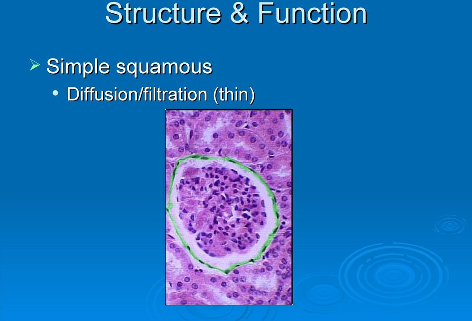
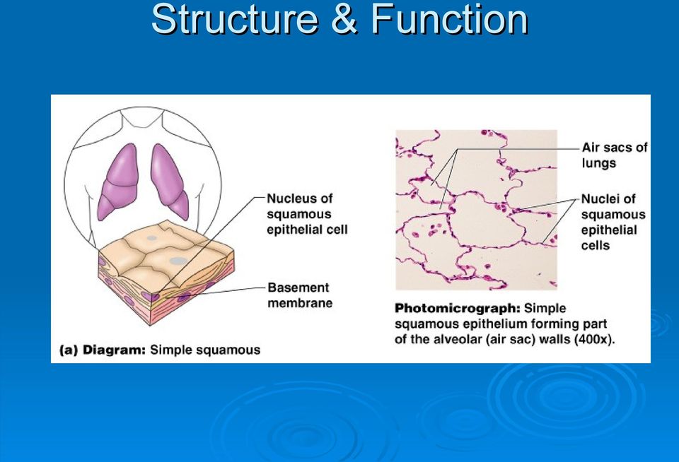
10 Structure & Function Simple squamous Diffusion/filtration (thin)
11 Structure & Function
12 Structure & Function Simple cuboidal & simple columnar Secretion, absorption, movement.

13 Structure & Function

14 Structure & Function Pseudostratified columnar Secretion (mucous), movement

15 Structure & Function

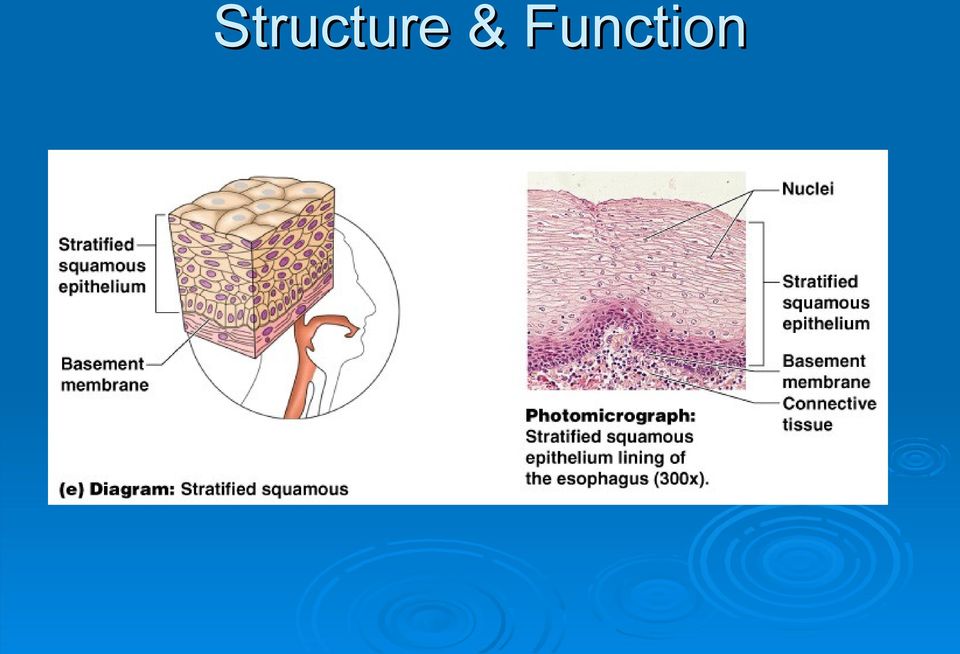
16 Structure & Function Stratified Squamous Protection Can be keratinized or moist.

17 Structure & Function
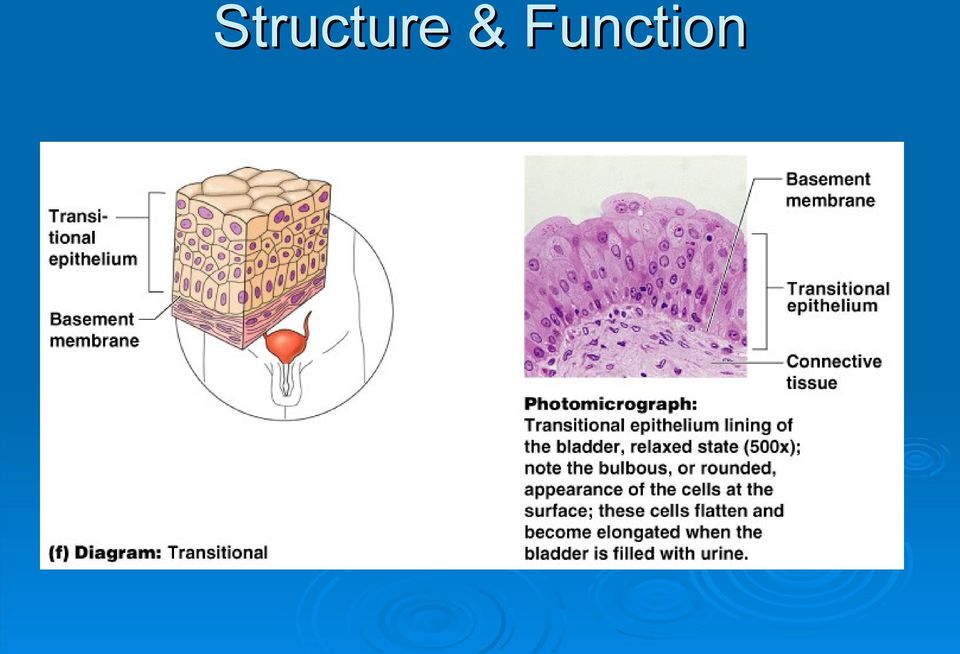
18 Structure & Function Transitional Stratified & stretchable

19 Structure & Function
20 Glands Secrete substances onto a surface, into a cavity or into the blood. Exocrine vs. endocrine glands

21 Histology Epithelial Tissue 1
22 Epithelial Tissue Lines internal and external body surfaces Forms glands 2 There are four types of tissues in the human body (and we re going to march through them all in this unit): epithelial tissue, connective tissue, muscular tissue, and nervous tissue. Epithelial tissue is composed of cells that line all body surfaces both external surfaces (skin and mucous membranes) and internal surfaces (lining all body cavities, blood vessels, organs, etc.) Epithelial tissue also form glands, which secrete substances into a cavity (such as the stomach), onto a surface (such as skin, mucous membrane, serous membrane, etc.) or into the blood.
23 Epithelial Tissue Little extracellular Apical surface matrix Attached on one side Avascular Basement membrane 3 All tissues are composed of two components: the living cells themselves and nonliving extracellular matrix. The extracellular matrix is composed of structural protein fibers, minerals, and fluid. One characteristic of epithelium is that it has very little matrix. In other words, epithelium consists mostly of cells packed very closely together with very little space between the cells. This makes sense since the job of epithelium is to line the body s surfaces. Structurally, since epithelium lines surfaces, all epithelial tissues are connected to other body tissues on exactly one side. The side of the epithelium that is not attached is called the apical surface (or, simply, free surface) while the other side is attached to a basement membrane, usually made of connective tissue.
24 Epithelial Tissue Cell Junctions Desmosomes: Anchors Tight junctions Seals Gap junctions Communication 4
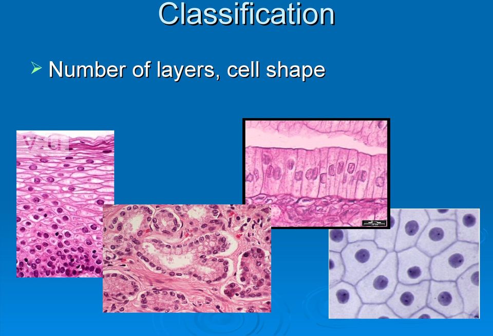
25 Classification Number of layers, cell shape 5 We classify most epithelium in two ways: by the number of layers of cells the tissue has and the shape of the cells. In the figures, notice that cells can be flat, squarish, or elongated. Also note that some epithelium has many layers (like the one on the left) while some has just one layer (all the rest). From left to right, these figures show vaginal epithelium, kidney tubules, the lining of the small intestine, and the lining of an alveolus in the lungs.
26 Classification Number of layers, cell shape Number of layers: Simple (one layer) Stratified (multiple layers) 6 If an epithelial tissue has just one layer of cells attached to a basement membrane, we call it simple epithelium. Simple epithelium is useful when we want to move substances across the tissue. Simple epithelium is found, for example, lining the capillary walls (we want to move gasses and nutrients), the lining of alveoli in the lungs (we want to move gasses), the lining of the small intestine (we want to move nutrients), the kidney tubules (we want to move ions and water), etc. If an epithelial tissue has more than one layer, we called it stratified epithelium (stratified means layered. ) We find stratified epithelium in places where we need to protect against abrasion and prevent the movement of substances (such as water and pathogens) across the tissue, most notably in the skin (cutaneous membrane) and mucous membranes.
27 Classification Number of layers, cell shape Cell shape Squamous (flat) Cuboidal (cube-shaped) Columnar (elongated) 7 If the cells in epithelial tissue appear flat, kind of stretched-out and spindleshaped, we call the tissue squamous epithelium. When stratified, sqamous epithelium is good for protecting against abrasion because the top layers can be scraped off without harming deeper layers. When simple, squamous epithelium is good for allowing substances to diffuse across. When epithelial cells are more-or-less square-shaped or round, we call the tissue cuboidal epithelium (don t confuse this with cubital, which means elbow. ). The larger size of cuboidal cells means they have more organelles (ER and Golgi apparatus primarily). They re found in places where we want to absorb or secrete substances. If epithelial cells are fat and elongated, we call the tissue columnar epithelium. Columnar epithelium has even bigger cells than cuboidal epithelium so you can probably imagine they are great for diffusion and secretion. A special type of columnar epithelium is a goblet cell. Goblet cells are single-celled glands that secrete mucous in various areas of the body (stomach, respiratory tract, vagina). The apical surface of columnar cells may have cilia attached (like in the respiratory tract to move mucous up and out) or have microvilli (like in the small intestine to increase cell surface area).
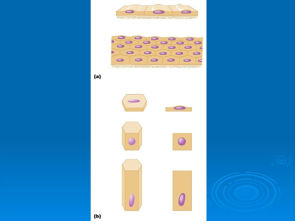
28 Classification 8 Quiz yourself about what is represented by each picture. In part A, the top figure shows simple epithelium and the bottom figure shows stratified epithelium. In part B, the top pair of figures shows squamous cells. The middle pair shows columnar cells. The bottom pair shows columnar cells.
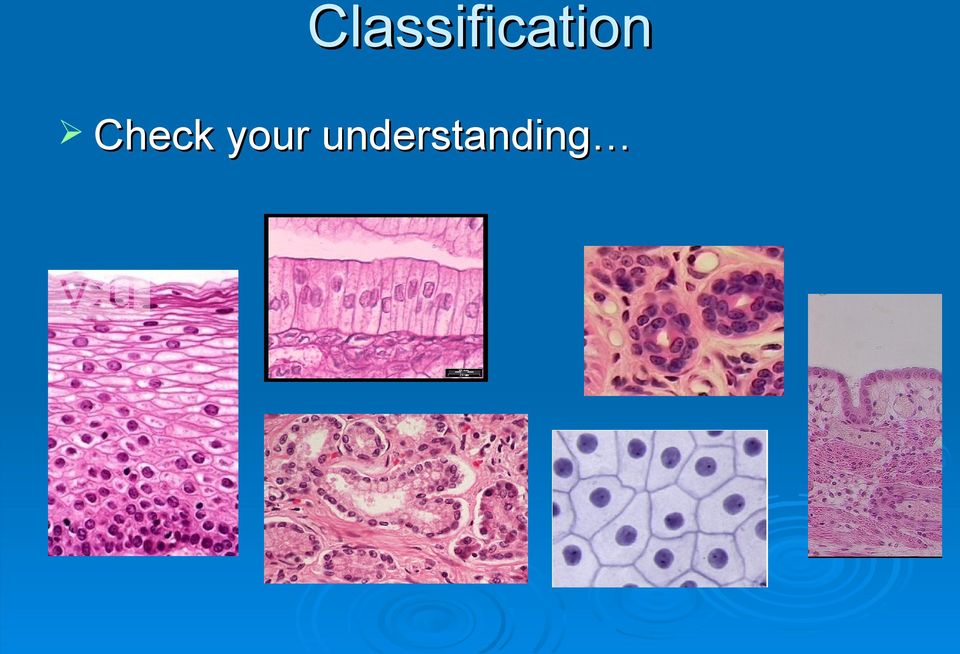
29 Classification Check your understanding 9 Name these tissues. Going from left-to-right and top-to bottom: stratified squamous, simple columnar, simple cuboidal, stratified cuboidal, simple squamous (this slide is a little different because it s a surface view rather than a cross-section), simple cuboidal.
30 Structure & Function Simple squamous Diffusion/filtration (thin) 10 There are five specific types of epithelium that you should be familiar with. The things I want you to know for each type are: 1) you should be able to identify the epithelium on sight, from micrographs like the ones in this lecture, 2) you should be able to deduce a location in the body (an organ) where the tissue might be found, and 3) you should be able to explain the function of the tissue (based on its structure). Simple squamous epithelium is found lining surfaces where diffusion or filtration are important. Since the tissue has just one layer, and since that layer is so thin, simple squamous epithelium is ideal for letting substances cross the epithelial layer without processing. For example, it lines the alveoli of the lungs, where nonpolar oxygen and carbon dioxide need to cross between the blood and the atmosphere; it lines the walls of capillaries, where gasses, nutrients, hormones, etc. need to cross between the blood and the interstitial space of tissues; etc. Since squamous cells are so thin and so small, they are not good for absorption or secretion they just don t have room for enough of the organelles (Golgi apparatus, ER, ribosomes, and lysosomes) that would be involved in absorption or secretion.
31 Structure & Function Click to add an outline 11 In the lungs, we don t want to prevent the exchange of gasses between the capillaries an the atmosphere, to simple squamous epithelium is ideal!
32 Structure & Function Simple cuboidal & simple columnar Secretion, absorption, movement. 12 Simple cuboidal and simple columnar epithelium are similar because their cells are larger than squamous cells (columnar is larger than cuboidal). Since the cells are larger, they can have more Golgi apparatus, ER and ribosomes (for secretion) as well as more lysosomes (for absorption). Consequently, epithelial tissues that are involved in absorption or secretion will often have simple cuboidal or simple columnar epithelium. Some simple cuboidal epithelium consists of goblet cells, which secrete mucous to trap junk that might otherwise get into the body. Goblet cells are usually ciliated (have cilia) to move the dirty mucous into the mouth, where it is eventually swallowed. Organs that are lined with simple cuboidal or simple columnar epithelium include the respiratory passages (usually simple columnar, to secrete mucous), the intestines (usually simple columnar, to absorb nutrients), the endocrine glands (usually simple cuboidal, to secrete hormones), etc.
33 Structure & Function Click to add an outline 13 Kidney tubules absorb ions and water; the stomach lining secretes acids and enzymes to aid in digestion. These are both ideal places for simple cuboidal or simple columnar epithelium.
34 Structure & Function Pseudostratified columnar Secretion (mucous), movement 14 Pseudostratified columnar epithelium is a strange tissue. When you look at it, it almost looks stratified because the nuclei of the cells are found at all sorts of different heights. In reality, though, it s simple epithelium because it s just one layer of epithelial cells. Pseudostratified columnar epithelium almost always secretes mucous and is almost always ciliated. The mucous traps particles, which are then swept away. This type of tissue is found in the trachea (ciliated; part of the respiratory tract) and the male urethra (unciliated; part of the male reproductive system).
35 Structure & Function Click to add an outline 15 Since the nuclei are at all different levels, pseudostratified epithelium looks stratified, but it s really just one layer (simple).
36 Structure & Function Stratified Squamous Protection Can be keratinized or moist. 16 Stratified squamous epithelium is found lining many surfaces that are exposed to the outside world (both cutaneous membrane [skin] and mucous membrane). As we ll learn later, the superficial layers of stratified squamous epithelium are really non-living cells that are filled with a tough, waterproof protein called keratin. There are multiple layers to protect against abrasion rubbing against stratified squamous tissue may pull off or damage a few layers, but the underlying tissues are still safe. The cutaneous membrane (outer skin) consists of dead cells filled with keratin, so we call it keratinized. It s tough, strong, and waterproof. Mucous membranes, though, do not have much keratin (and have fewer layers). We call them moist. Recall that mucous membranes line most cavity surfaces that are inside the body, but open the outside world (oral cavity, nasal cavity, orbital cavity, vaginal cavity, anal cavity) We find stratified squamous epithelium wherever movement and abrasion is an issue the skin, the esophagus, the oral cavity, etc.
37 Structure & Function Click to add an outline 17 The esophagus takes a lot of abuse. The cells are assaulted every time we swallow or cough. Stratified squamous epithelium ensures that even if the top layers of cells flake off, underlying tissue is not hurt.
38 Structure & Function Transitional Stratified & stretchable 18 Transitional epithelium is another weird tissue. The cells in transitional tissue have the ability to change shape when the organ it s lining expands. When the organ is empty, transitional epithelium looks columnar (pseudostratified, in fact). As the organ expands, though, transitional epithelium starts to look more stretched out and squamous. Transitional epithelium lines organs that need to stretch out, such as the urinary bladder and the female uterus.
39 Structure & Function Click to add an outline 19 Transitional epithelium lining the urinary bladder changes shape as the bladder fills. There aren t many micrographs of expanded transitional tissue. Can you imagine why?
40 Glands Secrete substances onto a surface, into a cavity or into the blood. Exocrine vs. endocrine glands 20 Glands are organs that secrete a substance (usually a protein, but sometimes water, ions or lipids) out of the cell. These substances can be secreted onto a surface, into a cavity or into the blood. Endocrine glands line the capillaries of certain organs and secrete their substances (hormones; proteins, amino acid derivatives or cholesterols) into the blood stream. Exocrine glands secrete their substances (proteins, ions and water) into cavities or onto surfaces. Most exocrine glands line ducts pockets in the epithelium lined with exocrine epithelial cells. Ducts increase the surface area of the gland and result in more secretion. For example, the pores in your face are mostly sebaceous glands pockets lined with exocrine cells that secrete an oily substance called sebum.
Section B: Epithelial Tissue 1. Where are epithelial tissues found within the body? 2. What are the functions of the epithelial tissues?
 Tissue worksheet Name Section A: Intro to Histology Cells are the smallest units of life. In complex organisms, cells group together with one another based on similar structure and function to form tissues.
Animal Tissues. I. Epithelial Tissue
 Animal Tissues There are four types of tissues found in animals: epithelial tissue, connective tissue, muscle tissue, and nervous tissue. In this lab you will learn the major characteristics of each tissue
Human Anatomy & Physiology I with Dr. Hubley. Practice Exam 1
 Human Anatomy & Physiology I with Dr. Hubley Practice Exam 1 1. Which definition is the best definition of the term gross anatomy? a. The study of cells. b. The study of tissues. c. The study of structures
Lab Exercise 4. Epithelial Tissues. Connective Tissue Proper. What you need to be able to do on the exam after completing this lab exercise:
 Lab Exercise 4 Epithelial Tissues Connective Tissue Proper Textbook Reference: See Chapter 4 What you need to be able to do on the exam after completing this lab exercise: Be able to identify each type
The Tissue Level of Organization
 The Tissue Level of Organization Tissues A groups of similar cells, usually having similar embryonic origin and specialized function Histology: the study of tissues Four general types Epithelial Muscle
Tissues (Histology) Ch. 3 Human Anatomy lecture
 Ch. 3 Human Anatomy lecture") I. Histology the study of tissues A. 4 basic tissue types epithelial connective muscle nervous Tissues (Histology) Ch. 3 Human Anatomy lecture B. Usually found in combinations to form organs. C. As you
I. Histology the study of tissues A. 4 basic tissue types epithelial connective muscle nervous Tissues (Histology) Ch. 3 Human Anatomy lecture B. Usually found in combinations to form organs. C. As you
Introduction to Anatomy and Physiology: Tissues and Integumentary System. Biology 105 Lecture 7 Chapter 4
 Introduction to Anatomy and Physiology: Tissues and Integumentary System Biology 105 Lecture 7 Chapter 4 Outline I. Tissues A. Epithelial B. Connective C. Muscle D. Nervous tissues II. Cell-to-cell contact
Tissue Types. 1. Epithelial Tissue (or epithelium) is the lining, covering, and glandular tissue of the body
 is the lining, covering, and glandular tissue of the body") Tissue Types A. Tissues 1. Tissues: groups of cells similar in structure and function 2. Four Types of Tissues: a. Epithelium: for covering b. Connective Tissue: for support c. Muscle: for movement d.
Tissue Types A. Tissues 1. Tissues: groups of cells similar in structure and function 2. Four Types of Tissues: a. Epithelium: for covering b. Connective Tissue: for support c. Muscle: for movement d.
Biology 13A Lab #3: Cells and Tissues
 Biology 13A Lab #3: Cells and Tissues Lab #3 Table of Contents: Expected Learning Outcomes.... 28 Introduction...... 28 Activity 1: Eukaryotic Cell Structure... 29 Activity 2: Perspectives on Tissue Preparations.
Biology 105 Human Biology PRACTICE MIDTERM EXAM 1. Essentials of Anatomy and Physiology, 5e (Martini/Nath) Chapter 4 The Tissue Level of Organization
 Chapter 4 The Tissue Level of Organization") Essentials of Anatomy and Physiology, 5e (Martini/Nath) Chapter 4 The Tissue Level of Organization Multiple-Choice Questions 1) The four main types of tissues include A) epithelium. B) muscle. C) nerve.
Essentials of Anatomy and Physiology, 5e (Martini/Nath) Chapter 4 The Tissue Level of Organization Multiple-Choice Questions 1) The four main types of tissues include A) epithelium. B) muscle. C) nerve.
BIO 137: CHAPTER 1 OBJECTIVES
 BIO 137: CHAPTER 1 OBJECTIVES 1. Define the terms anatomy and physiology, and explain their relationship using an example of a human structure with its corresponding function. A. ANATOMY = the study of
2161-1 - Page 1. Name: 1) Choose the disease that is most closely related to the given phrase. Questions 10 and 11 refer to the following:
 Choose the disease that is most closely related to the given phrase. Questions 10 and 11 refer to the following:") Name: 2161-1 - Page 1 1) Choose the disease that is most closely related to the given phrase. a disease of the bone marrow characterized by uncontrolled production of white blood cells A) meningitis B)
Name: 2161-1 - Page 1 1) Choose the disease that is most closely related to the given phrase. a disease of the bone marrow characterized by uncontrolled production of white blood cells A) meningitis B)
Vertebrate Body Organization
 Vertebrate Body Organization Digestive tube suspended in coelom from mouth to anus Body supported by internal skeleton of jointed bones Vertebrae and Cranium protects nervous system Diaphragm divides coelom
ORGAN SYSTEMS OF THE BODY
 ORGAN SYSTEMS OF THE BODY DEFINITIONS AND CONCEPTS A. Organ a structure made up of two or more kinds of tissues organized in such a way that they can together perform a more complex function that can any
Laboratory 3 Histology
 Laboratory 3 Histology Goals: For epithelial tissues: o discuss the major features; o classify based on simple/stratified and squamous/cubodial/columnar; o identify each type by microscopy; o identify
RAD 223. Radiography physiology. Lecture Notes. First lecture: Cell and Tissue
 RAD 223 Radiography physiology Lecture Notes First lecture: Cell and Tissue Physiology: the word physiology derived from a Greek word for study of nature. It is the study of how the body and its part work
CHAPTER 5: TISSUES. 2. Name the four primary adult tissue types, and give a brief description of each.
 OBJECTIVES: 1. Define the term tissue. 2. Name the four primary adult tissue types, and give a brief description of each. 3. Describe the functions and types of extracellular fluid (ECF). 4. Compare and
By Casey Schmidt and Wendy Ford
 By Casey Schmidt and Wendy Ford Body systems Digestive System Circulatory System Respiratory System Excretory System Immune System Reproductive System Nervous System Muscular System Skeletal System Endocrine
Lesson Aim To explain the human body at a microscopic level, including the structure and function of cells, tissues and membranes.
 LESSON 1. CELLS & TISSUES Lesson Aim To explain the human body at a microscopic level, including the structure and function of cells, tissues and membranes. THE CELL All living matter is composed of functional
Chapter 48. Nutrients in Food. Carbohydrates, Proteins, and Lipids. Carbohydrates, Proteins, and Lipids, continued
 Carbohydrates, Proteins, and Lipids The three nutrients needed by the body in the greatest amounts are carbohydrates, proteins, and lipids. Nutrients in Food All of these nutrients are called organic compounds,
Human Body Systems Project By Eva McLanahan
 Human Body Systems Project By Eva McLanahan Students will work in groups to research one of the eleven body systems as found in Holt, Rinehart, and Winston Modern Biology (2002). Research will focus on
North Bergen School District Benchmarks
 Grade: 10,11, and 12 Subject: Anatomy and Physiology First Marking Period Define anatomy and physiology, and describe various subspecialties of each discipline. Describe the five basic functions of living
Name Class Date Laboratory Investigation 24A Chapter 24A: Human Skin
 Name Class Date Laboratory Investigation 24A Chapter 24A: Human Skin Human Anatomy & Physiology: Integumentary System You may refer to pages 386-394 in your textbook for a general discussion of the integumentary
The Digestive System. Chapter 16. Introduction. Histological Organization. Overview of Digestive System. Movement and Mixing of Digestive Materials
 The Digestive System Chapter 16 Introduction Structure of the digestive system A tube that extends from mouth to anus Accessory organs are attached Functions include Ingestion Movement Digestion Absorption
Biology 2401 - Anatomy and Physiology I Exam 1 notes - Introduction, Cell and Tissue Structure
 Biology 2401 - Anatomy and Physiology I Exam 1 notes - Introduction, Cell and Tissue Structure Two major principles in study of animal bodies: (humans, like other living organisms are product of evolutionary
Topic 3.0 Healthy human function depends on a variety of interacting and reacting systems
 Topic 3.0 Healthy human function depends on a variety of interacting and reacting systems Organ Systems Organ systems must have the ability to to changes within and outside of your body to maintain life
Cells & Cell Organelles
 Cells & Cell Organelles The Building Blocks of Life H Biology Types of cells bacteria cells Prokaryote - no organelles Eukaryotes - organelles animal cells plant cells Cell size comparison Animal cell
Eating, pooping, and peeing THE DIGESTIVE SYSTEM
 THE DIGESTIVE SYSTEM Ingested food is not technically in the body until it is absorbed so it needs to be: Mechanically and chemically reduced Transported by the blood to the cells Large portions are not
Review of the Cell and Its Organelles
 Biology Learning Centre Review of the Cell and Its Organelles Tips for most effective learning of this material: Memorize the names and structures over several days. This will help you retain what you
Human Anatomy and Physiology The Respiratory System
 Human Anatomy and Physiology The Respiratory System Basic functions of the respiratory system: as a Gas exchange supply oxygen to aerobic tissues in the body and remove carbon dioxide waste product. in-
Digestive system Review
 Digestive system Review 1. Distinguish between chemical digestion and mechanical digestion. The physical breakdown of food begins in the mouth with two types of processes. The mouth is a complex structure
Small & Large Intestines
 Small & Large Intestines Small Intestine: principal site for digestion of food and absorption of the products of digestion Large Intestine: reabsorption of water and elimination of undigested food and
5. Secretion: release of water, acids. Enzymes, buffers by digestive tract.
 Digestive System CH-16 Lecture topics Functions of the digestive system: p. 488. 1. Ingestion: Taking food in 2. Propulsion: movement of food thru alimentary canal p.490. voluntary: swalloing : skeletal
The Cell Interior and Function
 The Cell Interior and Function 5 5.0 CHAPTER PREVIEW Investigate and understand the organization and function of the cell interior. Define the differences between eukaryotic and prokaryotic cell structure.
CHAPTER 9 BODY ORGANIZATION
 CHAPTER 9 BODY ORGANIZATION Objectives Identify the meaning of 10 or more terms relating to the organization of the body Describe the properties of life Describe the function for the structures of the
The Respiratory System
 Human Anatomy III: Respiratory, Urinary & Digestive Systems The Respiratory System Major functions include: Obtaining oxygen Removing carbon dioxide Maintenance of ph balance Respiration may be accomplished
THE GI TRACT IS A CONTINUOUS MULTILAYERED TUBE EXTENDING FROM THE MOUTH TO THE ANUS THAT IS SUPPORTED AND PARTIALLY COVERED BY THE PERITONEUM.
 THE GI TRACT IS A CONTINUOUS MULTILAYERED TUBE EXTENDING FROM THE MOUTH TO THE ANUS THAT IS SUPPORTED AND PARTIALLY COVERED BY THE PERITONEUM. OVERVIEW OF THE DIGESTIVE SYSTEM Two groups of organs compose
Animal Systems: The Musculoskeletal System
 Animal Systems: The Musculoskeletal System Tissues, Organs, and Systems of Living Things Cells, Cell Division, and Animal Systems and Plant Systems Cell Specialization Human Systems The Digestive The Circulatory
Homeostasis and Transport Module A Anchor 4
 Homeostasis and Transport Module A Anchor 4 Key Concepts: - Buffers play an important role in maintaining homeostasis in organisms. - To maintain homeostasis, unicellular organisms grow, respond to the
Digestive System AKA. GI System. Overview. GI Process Process Includes. G-I Tract Alimentary Canal
 Digestive System AKA G-I Tract Alimentary Canal Overview GI System Consists of Mouth, pharynx, esophagus, stomach, small intestine, large intestine, anus About 30 in length Accessory Organs Teeth, tongue,
Chetek-Weyerhaeuser High School
 Chetek-Weyerhaeuser High School Anatomy and Physiology Units and Anatomy and Physiology A Unit 1 Introduction to Human Anatomy and Physiology (6 days) Essential Question: How do the systems of the human
Anatomy and Physiology
 Learning Activities It is important that you do not lecture all of the time. If you employ a variety of teaching styles, your students will stay focused better and they will find it easier to process the
Essentials of Human Anatomy & Physiology. 7 th edition Marieb, Elaine, 2003. Chapters 10-11. Lab Manual, 2 nd edition and coloring book, 7 th edition
 Topic/Unit: Anatomy & Physiology Circulatory System Curricular Goals/ Learning Outcomes: Students will be able to identify the composition of blood and its function. Students will be able to differentiate
The Digestive System
 The Digestive System What do you know?? quiz-digestive-health Digestion Videos The Digestive System Inside-Dr-Ozs-Digestive-System-Video Now it is your turn to recreate the digestive system. How is food
The cells of the human body do not operate independently of
 4 Tissues I. Epithelial Tissue 65 Special Characteristics of Epithelia 66 Classification of Epithelia 66 Glands 72 Epithelial Surface Features 74 II. Connective Tissue 77 Special Characteristics of Connective
6023-1 - Page 1. Name: 4) The diagram below represents a beaker containing a solution of various molecules involved in digestion.
 The diagram below represents a beaker containing a solution of various molecules involved in digestion.") Name: 6023-1 - Page 1 1) Which one of the following situations indicates a serious organ system malfunction? A) Mitochondria stop functioning in a unicellular organism exposed to pollutants. B) White blood
Name: 6023-1 - Page 1 1) Which one of the following situations indicates a serious organ system malfunction? A) Mitochondria stop functioning in a unicellular organism exposed to pollutants. B) White blood
Introduction to the Cell: Plant and Animal Cells
 Introduction to the Cell: Plant and Animal Cells Tissues, Organs, and Systems of Living Things Cells, Cell Division, and Animal Systems and Plant Systems Cell Specialization Human Systems All organisms
Chapter 3. Cellular Structure and Function Worksheets. 39 www.ck12.org
 Chapter 3 Cellular Structure and Function Worksheets (Opening image copyright by Sebastian Kaulitzki, 2010. Used under license from Shutterstock.com.) Lesson 3.1: Introduction to Cells Lesson 3.2: Cell
Fundamentals of Anatomy & Physiology Course Outline, Objectives and Accreditation Information
 201 Webster Building 3411 Silverside Road Wilmington, DE 19810 Phone: 1-888-658-6641 Fax: 1-302-477-9744 learn@corexcel.com www.corexcel.com Course Outline, Objectives and Accreditation Information Chapter
Digestion, Absorption. How & where?
 Digestion, Absorption How & where? What happens to food? Three processes Digestion Absorption Elimination Where do they occur? GI tract Overview of Digestion GI tract Gastrointestinal (GI) tract: series
Engage: Brainstorming Body Systems. Record the structures and function of each body system in the table below.
 Engage: Brainstorming Body s Record the structures and function of each body system in the table below. Body Nervous Circulatory Excretory Immune Digestive Respiratory Skeletal Muscular Endocrine Integumentary
Outline Digestive System
 Outline Digestive System The Digestive System Digestive System Lecture Packet 19 Chapter 15 I. Function II. Layers of the GI tract III. Major parts: mouth, pharynx, esophagus, stomach, small intestine,
Chapter 4: A Tour of the Cell. 1. Cell Basics. Limits to Cell Size. 1. Cell Basics. 2. Prokaryotic Cells. 3. Eukaryotic Cells
 Chapter 4: A Tour of the Cell 1. Cell Basics 2. Prokaryotic Cells 3. Eukaryotic Cells 1. Cell Basics Limits to Cell Size There are 2 main reasons why cells are so small: If cells get too large: 1) there
The Integumentary System Dr. Ali Ebneshahidi
 The Integumentary System Dr. Ali Ebneshahidi The Skin The integument system consists of the skin (cutaneous membrane) and its accessory organs. The skin is composed of three layers of tissue: the outer
The Vertebrate (mostly human) Digestive System
 Digestive System") The Vertebrate (mostly human) Digestive System Mouth - mastication, lubrication, digestion Pharynx and Esophagus - swallowing Stomach - some digestion Small intestine - most digestion and absorption Large
The Vertebrate (mostly human) Digestive System Mouth - mastication, lubrication, digestion Pharynx and Esophagus - swallowing Stomach - some digestion Small intestine - most digestion and absorption Large
Date: Student Name: Teacher Name: Jared George. Score: 1) A cell with 1% solute concentration is placed in a beaker with a 5% solute concentration.
 A cell with 1% solute concentration is placed in a beaker with a 5% solute concentration.") Biology Keystone (PA Core) Quiz Homeostasis and Transport - (BIO.A.4.1.1 ) Plasma Membrane, (BIO.A.4.1.2 ) Transport Mechanisms, (BIO.A.4.1.3 ) Transport Facilitation Student Name: Teacher Name: Jared
Biology Keystone (PA Core) Quiz Homeostasis and Transport - (BIO.A.4.1.1 ) Plasma Membrane, (BIO.A.4.1.2 ) Transport Mechanisms, (BIO.A.4.1.3 ) Transport Facilitation Student Name: Teacher Name: Jared
Lab 18 The Digestive System
 Lab 18 The Digestive System Laboratory Objectives Identify on a diagram, model or cadaver the parts of the digestive system and accessory organs. Describe the general histology of the digestive system.
Digestive System Why is digestion important? How is food digested? Physical Digestion and Movement
 Digestive System The digestive system is made up of the digestive tract a series of hollow organs joined in a long, twisting tube from the mouth to the anus and other organs that help the body break down
Cells, tissues and organs
 Chapter 8: Cells, tissues and organs Cells: building blocks of life Living things are made of cells. Many of the chemical reactions that keep organisms alive (metabolic functions) take place in cells.
I. The basic function of the digestive system is
 Chapter 15, Digestive System - ANATOMY OF THE DIGESTIVE SYSTEM I. The basic function of the digestive system is. This process is called. II. List 2 other names for the digestive tract: A. B. III. The digestive
Exchange and transport
 Exchange and transport Examples of things which need to be interchanged between an organism and its environment include: Respiratory gases Nutrients Excretory products Heat This exchange can take place
10.2 The Human Digestive System pg. 411
 10.2 The Human Digestive System pg. 411 The human digestive system is made up of a group of organs working together. The digestive tract is made up of the mouth, esophagus, stomach, small intestine, and
Lect 01 - Epithelial Tissue
 Structure & Function Introduction Prof Kumlesh K. Dev Department of Physiology What is Structure & Function? Integration is key structure and function requires integration of physiology, anatomy and biochemistry
CHAPTER 20: URINARY SYSTEM
 OBJECTIVES: 1. Name the major function of the urinary system, and name and locate (on a diagram) the organs that compose the system. 2. Explain what the term renal refers to. 3. Define the term retroperitoneal.
Hole s Human Anatomy and Physiology Eleventh Edition. Mrs. Hummer Hanover Area Jr./Sr. High School. Chapter 1 Introduction to Anatomy and Physiology
 Hole s Human Anatomy and Physiology Eleventh Edition Mrs. Hummer Hanover Area Jr./Sr. High School Chapter 1 Introduction to Anatomy and Physiology 1 Chapter 1 Introduction to Human Anatomy and Physiology
Human Anatomy & Physiology II with Dr. Hubley
 Human Anatomy & Physiology II with Dr. Hubley Practice Exam III Name: Instructions This exam consists of 50 questions. You may write on the exam itself, but be sure to answer all your questions on a Scantron
Maxillary Sinus. (Antrum of Higmore)
") Maxillary Sinus (Antrum of Higmore) The maxillary sinus is a pneumatic space. It is the largest bilateral air sinus located in the body of the maxilla and opens in the middle nasal meatus of the nasal
Maxillary Sinus (Antrum of Higmore) The maxillary sinus is a pneumatic space. It is the largest bilateral air sinus located in the body of the maxilla and opens in the middle nasal meatus of the nasal
Digestive System Functions
 Digestive System Functions A. Gastrointestinal Processes 1. Ingestion: placing food in mouth (voluntary) 2. Propulsion: moving food through GI tract a. Peristalsis: alternating waves of contraction and
Skeletal, Muscular, and Integumentary Systems
 Chapter 36 Skeletal, Muscular, and Integumentary Systems Section 36 1 The Skeletal System (pages 921 925) This section describes the skeletal system and its functions. Introduction (page 921) 1. What forms
Paramedic Program Anatomy and Physiology Study Guide
 Paramedic Program Anatomy and Physiology Study Guide Define the terms anatomy and physiology. List and discuss in order of increasing complexity, the body from the cell to the whole organism. Define the
Location: air sacs of lungs; nephrons of kidney; lining of circulatory system, lymphatic vessels, & ventral body cavity
 Bio. 2304 - Human Anatomy HISTOLOGY (STUDY OF TISSUES) - Lab & Lecture Objectives Important: For each slide know 1.) specific tissue type 2.) any specialized structures or cells in the tissue (& know their
B2 Revision. Subject Module Date Biology B2 13 TH May (am)
") B2 Revision Subject Module Date Biology B2 13 TH May (am) Useful websites www.aqa.org.uk This website contains the specifications that we follow and also has a large number of past papers and mark schemes
B2 Revision Subject Module Date Biology B2 13 TH May (am) Useful websites www.aqa.org.uk This website contains the specifications that we follow and also has a large number of past papers and mark schemes
Fifth Grade Cells: Structures and Processes Assessment
 Fifth Grade Cells: Structures and Processes Assessment 1a. All living things are made up of. a. cells b. tissues c. organisms d. systems 1b. All living things are made up of. 1c. Explain what cells are
Topic 4: Digestion and Nutrition
 Topic 4: Digestion and Nutrition THE CONTENTS OF FOOD Food contains nutrients: Nutrients include: 1. 2. 3. 4. 5. Nutrients must be small enough to enter our cells. If they are too large they must be digested
Pre-requisites: Successful completion of 4th grade science and the 4th grade science assessment.
 Throughout each unit, assessments are incorporated into lessons. These assessments are activities that occur within the context of each lesson providing the guidelines for assessing students' progress.
4. Biology of the Cell
 4. Biology of the Cell Our primary focus in this chapter will be the plasma membrane and movement of materials across the plasma membrane. You should already be familiar with the basic structures and roles
67 The Human Skeleton
 67 The Human Skeleton Skull SCIENCE EXPLORER Focus on Life Science Prentice-Hall, Inc. Clavicle (collarbone) Scapula (shoulder blade) Carpals Metacarpals Phalanges Femur Tibia Humerus Ulna Sternum (breastbone)
A. function: supplies body with oxygen and removes carbon dioxide. a. O2 diffuses from air into pulmonary capillary blood
 A. function: supplies body with oxygen and removes carbon dioxide 1. ventilation = movement of air into and out of lungs 2. diffusion: B. organization a. O2 diffuses from air into pulmonary capillary blood
Water Homeostasis. Graphics are used with permission of: Pearson Education Inc., publishing as Benjamin Cummings (http://www.aw-bc.
 Water Homeostasis Graphics are used with permission of: Pearson Education Inc., publishing as Benjamin Cummings (http://www.aw-bc.com) 1. Water Homeostasis The body maintains a balance of water intake
Comparing Plant And Animal Cells
 Comparing Plant And Animal Cells http://khanacademy.org/video?v=hmwvj9x4gny Plant Cells shape - most plant cells are squarish or rectangular in shape. amyloplast (starch storage organelle)- an organelle
Tissue: The Living Fabric
 CHAPTER4 I Tissue: The Living Fabric Chapter Outline and Student Objectives Preview of Selected Key Terms Epithelial Tissue (pp. 101-112) 1. List several characteristics that typify epithelial tissue.
Investigating the Human Body On-site student activities: Years 7-8 Investigating the Human Body On-site student activities Years 7 8
 Investigating the Human Body On-site student activities Years 7 8 Student activity (and record) sheets have been developed with alternative themes for students to use as guides and focus material during
Integumentary System Digestive System. Outline. Integumentary System 11/4/2008. Week 11 BA & BP November 4, 2008 Nadia Arora, ND
 Integumentary System Digestive System Week 11 BA & BP November 4, 2008 Nadia Arora, ND Outline Integumentary system and body membranes Types of body membranes and their function General structure and main
Anatomy & Physiology Bio 2401 Lecture. Instructor: Daryl Beatty Day 1 Intro to Lecture 1
 Anatomy & Physiology Bio 2401 Lecture Instructor: Daryl Beatty Day 1 Intro to Lecture 1 Introduction: Daryl Beatty M.S. Microbiology 28 Years Dow, Research & TS&D. Family BC since 2007 More importantly:
Smooth Muscle. Learning Objectives.
 Smooth Muscle. Learning Objectives. At the end of this course, you should be able to : 1. describe the structure of smooth muscle 2. describe where smooth muscle occurs within the body 3. discuss the structural
BSC 2010 - Exam I Lectures and Text Pages. The Plasma Membrane Structure and Function. Phospholipids. I. Intro to Biology (2-29) II.
 II.") BSC 2010 - Exam I Lectures and Text Pages I. Intro to Biology (2-29) II. Chemistry of Life Chemistry review (30-46) Water (47-57) Carbon (58-67) Macromolecules (68-91) III. Cells and Membranes Cell structure
BSC 2010 - Exam I Lectures and Text Pages I. Intro to Biology (2-29) II. Chemistry of Life Chemistry review (30-46) Water (47-57) Carbon (58-67) Macromolecules (68-91) III. Cells and Membranes Cell structure
ANATOMY AND PHYSIOLOGY OF THE PULMONARY SYSTEM Section 1 Part B Reading Assignment: Des Jardins - Chapter 1, pp. THE LOWER AIRWAY I.
 ANATOMY AND PHYSIOLOGY OF THE PULMONARY SYSTEM Section 1 Part B Reading Assignment: Des Jardins - Chapter 1, pp. THE LOWER AIRWAY I. Cartilaginous Airways A. Trachea 1. extends from the cricoid cartilage
THE HUMAN BODY SYSTEMS
 Name Period Date THE HUMAN BODY SYSTEMS System Function Diagram Major Organs Digestive 1. take in food (ingestion) 2. digest food into smaller molecules and absorb nutrients 3. remove undigestable food
Special organ structures and functions conduct these tasks through the successive parts of the overall system.
 Chapter 5 Digestion, Absorption, and Metabolism Chapter 5 Lesson 5.1 Key Concepts Through a balanced system of mechanical and chemical digestion, food is broken down into smaller substances and the nutrients
Chapter 15 Digestive System.
 Chapter 15 Digestive System. I. The Gastrointestinal Tract. a. The digestive system mechanically and chemically breaks down food into molecules that can be absorbed into the bloodstream or lymph. Residues
PCB 4023 Cell Biology. Lab 8: Organology II Digestive tract and accessory organs
 PCB 4023 Cell Biology Lab 8: Organology II Digestive tract and accessory organs Name: Name: SSN: SSN: N.B. Since this document is in pdf format, the URLs (web addresses) cannot be linked. To use them,
The chemical reactions inside cells are controlled by enzymes. Cells may be specialised to carry out a particular function.
 12.1 What are animals and plants built from? All living things are made up of cells. The structures of different types of cells are related to their functions. to relate the structure of different types
Two main classes: Epithelial Connective (synovial) Epithelial. Cutaneous Mucous Serous
 Epithelial. Cutaneous Mucous Serous") Two main classes: Epithelial Connective (synovial) Epithelial Cutaneous Mucous Serous Epithelial Membranes = sheet of epithelia + connective tissue base 1. Cutaneous membrane: outer skin layer (stratified
Two main classes: Epithelial Connective (synovial) Epithelial Cutaneous Mucous Serous Epithelial Membranes = sheet of epithelia + connective tissue base 1. Cutaneous membrane: outer skin layer (stratified
Chapter 49 - Nutrients and the Digestive System I. Nutrients (chemical substances necessary for organisms to grow and function properly)
") Chapter 49 - Nutrients and the Digestive System I. Nutrients (chemical substances necessary for organisms to grow and function properly) 6 basic nutrients - 4 food groups (milk, meat, fruit and vegetable,
Chapter 49 - Nutrients and the Digestive System I. Nutrients (chemical substances necessary for organisms to grow and function properly) 6 basic nutrients - 4 food groups (milk, meat, fruit and vegetable,
Medical Physiology Z.H.Al-Zubaydi
 Lec.13 Medical Physiology Z.H.Al-Zubaydi Functions of the Digestive System The major functions of the digestive tract include the following six processes, summarized in Figure 1: 1. Ingestion Food must
Cell and Membrane Practice. A. chromosome B. gene C. mitochondrion D. vacuole
 Name: ate: 1. Which structure is outside the nucleus of a cell and contains N?. chromosome. gene. mitochondrion. vacuole 2. potato core was placed in a beaker of water as shown in the figure below. Which
Magic School Bus Digestive System Brainpop Digestive System
 The Digestive System Magic School Bus Digestive System Brainpop Digestive System 1 Functions of the Digestive System: 1. Break up food into smaller pieces 2. Absorbing nutrients into the blood 3. Excreting
Questions on The Nervous System and Gas Exchange
 Name: Questions on The Nervous System and Gas Exchange Directions: The following questions are taken from previous IB Final Papers on Topics 6.4 (Gas Exchange) and 6.5 (Nerves, hormones and homeostasis).
Cardiovascular System. Blood Components
 Cardiovascular System Blood Components 1 Components of Blood Formed elements: erythrocytes, leukocytes, platelets Plasma: water, proteins, other solutes The components of blood can be divided into two
Mechanical digestion: physical breaking of food chewing by teeth churning by stomach segmentation by intestines (= mixing food) p.611/ Fig. 22.
 p.611/ Fig. 22.") The Digestive System 1. Describe the general functions of the digestive system Ingestion: Taking food in Propulsion: movement of food thru alimentary canal voluntary: swalloing involuntary: peristalsis
The Digestive System 1. Describe the general functions of the digestive system Ingestion: Taking food in Propulsion: movement of food thru alimentary canal voluntary: swalloing involuntary: peristalsis