GROSS ANATOMY. Lecture Syllabus ANAT Gross Anatomy Department of Neurobiology and Anatomy University of Utah School of Medicine
|
|
|
- Mary Fitzgerald
- 8 years ago
- Views:
Transcription
1 GROSS ANATOMY Lecture Syllabus 2008 ANAT Gross Anatomy Department of Neurobiology and Anatomy University of Utah School of Medicine David A. Morton K. Bo Foreman Kurt H. Albertine Andrew S. Weyrich Kimberly Moyle 1

2 GROSS ANATOMY (ANAT 6010) ORIENTATION, FALL 2008 Welcome to Human Gross Anatomy! Course Director David A. Morton, Ph.D. Office: 223 Health Professions Education Building; Phone: ; Faculty Kurt H. Albertine, Ph.D., (Assistant Dean for Faculty Administration) K. Bo Foreman, PT, Ph.D, (Gross and Neuro Anatomy Course Director in Dept. of Physical Therapy) (bo. David A. Morton, Ph.D. (Gross Anatomy Course Director, School of Medicine) edu) Andrew S. Weyrich, Ph.D. (Professor of Human Molecular Biology and Genetics) utah.edu) Kerry D. Peterson, L.F.P. (Body Donor Program Director) Cadaver Laboratory staff Jordan Barker, Blake Dowdle, Christine Eckel, MS (Ph.D.), Nick Gibbons, Richard Homer, Heather Homer, Nick Livdahl, Kim Moyle, Neal Tolley, MS, Rick Webster Course Objectives The study of anatomy is akin to the study of language. Literally thousands of new words will be taught throughout the course. Success in anatomy comes from knowing the terminology, the three-dimensional visualization of the structure(s) and using that knowledge in solving problems. The discipline of anatomy is usually studied in a dual approach: Regional approach - description of structures regionally and their relationships to each other (back, thorax, abdomen, pelvis, perineum, neck, head, upper limb, lower limb) Systemic approach - description of the major systems of the body musculoskeletal, nervous cardiovascular, lymphatic, digestive, respiratory, endocrine, urinary and reproductive The gross anatomy course (ANAT 6010) is organized around a regional approach to anatomy, but time is taken throughout the course to review information from a systemic standpoint because true understanding requires that both approaches be used. The regional approach is divided into the following 4 Units: Unit #1 Back and Thorax Unit #2 Abdomen, Pelvis and Perineum Unit #3 Neck and Head Unit #4 Upper and Lower Limb 2
 (andrew.weyrich@hmbg. utah.edu) Kerry D. Peterson, L.F.P. (Body Donor Program Director) Cadaver Laboratory staff Jordan Barker, Blake Dowdle, Christine Eckel, MS (Ph.")
3 Lecture Syllabus The goal of the lecture syllabus was to: Create lecture outlines that are practical and easy to follow during self study and during lecture Provide students with clear expectations of the anatomic content to be mastered Each lecture session of the syllabus begins with a list of Objectives that identifies anatomic structures and concepts to be mastered. The goal of the objectives list is to help students focus study time on required content instead of guessing what the instructor expects them to memorize and learn. Similarly to learning a new language there is a large amount of memorization required. However, the end goal is not in memorizing long lists of terms and verbs but of communicating. Anatomy is much the same. There are long lists of structures and concepts to memorize and learn but that is not the end goal. The end goal is applying the anatomic content to clinical/analytical problems. The syllabus is meant to help serve as a guide in this study process. The syllabus and associated objectives are meant to serve as a focus to study time. In addition, homework exercises are provided to aid students in applying anatomic concepts in clinical situations to assess self-mastery of anatomic knowledge and to prepare for the end-unit examinations. The syllabus does not supplant the required textbooks. The authoritative source for lectures (written exams) is Gray s Anatomy for Students, not the syllabus. The authoritative source for the laboratory practical exam is Gray s Dissection Guide for Human Anatomy, not the syllabus. If you discover format mistakes or inconsistencies between the content of the syllabus and text books, please let us know, constructively, so that we may revise the syllabus for next year. Grading and Testing Cumulative scores for exams, and cadaver autopsy report for a total of 455 possible points. 4-Midterm lecture exams consist of multiple choice and constructed response questions (50 points each). Hard copies of the written exams will not be returned. 4-Cadaver practical exams (50 points each). 4-Dissection area cleanliness and Dissection completeness (5 points each) 1-Written cadaver autopsy report (35 points... 5 group report and 30 individual report) Medical students: Honors/Pass/Fail; Dental students: Letter grade (A-F) Cheating is unacceptable. Please do not wear baseball caps or other brimmed hats during exams. Exam dates: Unit #1 - Monday September 15th, Unit #2 - Friday October 10th, Unit #3 - Wednesday November 12th and finally Friday November 5. Changing dates of exams is not an option, without prior written approval from the Dean s Offi ce and Course Director. Remediation Approved medical school summer anatomy courses (i.e., University of Louisville in Kentucky or PT gross anatomy course at UofU) Materials Scrubs and/or white laboratory coat, latex gloves, scalpel blades, scissors, forceps/hemostats (2 sets of other dissection tools are included for each table). 3

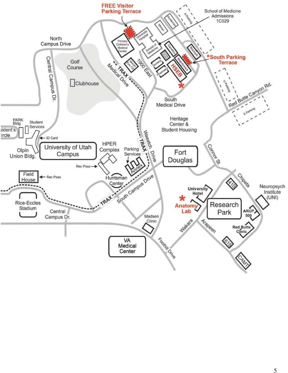
4 Required texts Drake et al., Gray s Anatomy for Students (2005) Morton et al., Gray s Dissection Guide for Human Anatomy (2007) Atlas: Your choice (recommend Netter s, or Thieme) Dissection Groups 6 medical students are assigned to each cadaver. They are further subdivided into group A and group B. Each group will alternate each laboratory period between cadaver dissection and small group activities in the HSEB (i.e., osteology tutorials, radiology review, practice problem solving etc...). The dissecting group will teach the dissected material to the non-dissecting group. For example, when Group A is dissecting the triangles of the neck in the cadaver lab, Group B will be reviewing the osteology of the head and neck. Group A will be responsible for teaching Group B the anatomy of the triangles of the neck to Group A. Learning Aids The official anatomy website is DIGANAT. The site contains photographic dissections for each session, osteology tutorials, radiographic tutorials, homework questions and other computer assisted instructional aids. username: gross password: anatomy Bone boxes: A bone box is assigned to each cadaver table (to be shared between groups A and B) Skulls: A skull is assigned to each dissection group (Group A and Group B both are assigned skulls) Health Professions Education Building (HPEB) Address: 520 Wakara Way, Research Park between the Marriott Hotel and Orthopedic Building 15 walk from HSEB. The HPEB is shared with the College of Health s Physical and Occupational Therapy programs (good citizenship is therefore obligatory). The building has 3 levels with a mezzanine between levels and 2 and 3. (Level 1 is the bottom level). Each student is given access to the building 7 days a week from 6AM to 11PM. For the students protection against liability, we advise that more than one student be in the laboratory at a time. The cadaver laboratories are located on Level 2 (middle level). The labs have keypad locks with the code for each door being... 2 and 4 at the same time followed by 3. The student commons area is on Level 3 (top floor). Please feel free to take advantage of the common areas. Changing-Rest rooms are located on all three floors. Shuttle bus: To see a schedule for shuttles to and from the medical school and cadaver lab please refer to Parking: Limited parking is available at the HPEB facility. Additional parking is located at 615 on the map on the following page. Individual property managers control all other lots in Research Park. Unauthorized use is subject to private, not University, enforcement. Please do not park in the Patient Parking at the bottom of the hill in HPEB. Those spots are reserved for patients with multiple sclerosis and Parkinson s. If student s take their parking spots they are forced to park else where and will be unable get to the rehab room on Level 1 and will miss their session. Thank-you for your consideration in this matter. 4
. The dissecting group will teach the dissected material to the non-dissecting group.")
5 5
6 Cadaver Laboratory Respect the cadavers Their donation is for your benefit. No tissues are to leave the laboratory. All inorganic materials accompanying the body (e.g., false teeth, fillings, pacemakers, artificial joints) are to remain with the cadaver. No eating, drinking smoking, cameras or visitors are allowed in the laboratory. For sanitary reasons Scrubs and/or white laboratory coat, close toed shoes and gloves are required in the laboratory Sandals are discouraged (no protection from dropped scalpels) Do not touch anything while you are wearing soiled gloves, except your cadaver, texts and instruments Keep the dissection table surface and the floor in your immediate area clean of cadaver wastes At the end of each dissection, sweep and mop the area around your table, spray and wipe down your table edges, wash all dissection tools and place them in the plastic container underneath your cadaver table. Place the lab stools under the table before you leave the laboratory The anatomy laboratory must always look clean out of respect for the donors and their families. When laboratory equipment (saw, mallet, chisel) is used: Clean and return the equipment to the proper place immediately after use... Care must be used with all instruments to prevent injury When first aid is needed please find one of the laboratory staff Cadaver Maintenance The cadaver must remain wrapped in moist shrouds when not being dissected to prevent dehydration Dissection is enhanced when the area being dissected is sprayed periodically with wetting agent; this also retards dehydration Refill the bottles from container marked wetting agent in the supply room Keep areas of the cadaver not being dissected covered with moist shrouds and plastic; additional shrouds are available upon request Do not allow body fluids to pool (build up) in the plastic covers or on the dissection table surfaces Disposal - 4 Containers White pail - Body tissues and body tissues only (i.e., fat, skin, muscle etc...). Located under dissection table Red Sharps container - Scalpel blades and any item that can puncture the skin. Located in each room. Grey pail - All fluids from the cadaver are drained into the grey bucket at the foot of each cadaver table. Black Trash Can - Gloves, paper towels and anything else that is not a body tissue, a sharp item or a fluid. Adherence to these policies sustains the integrity of the body donor program, improves the laboratory environment and reduces the risk of illness or injury to you and your fellow students. For more information on the body donor program go to 6
 Do not touch")
7 Cadaver Autopsy Report Project Description Introduction In the human gross anatomy course, the primary teaching and learning tool is the human cadaver. Often, the cadaver is introduced as the medical student s first patient. Because the cadaver was at one time a living, breathing human being, the cadaver was in fact a real patient at some point in time. For some reason, often unknown to the student, the patient passed away and his or her body was sent to the University of Utah s Body Donor Program per the patient s wishes. As a student, the only information you receive about the cause of death of the cadaver you are dissecting comes from the death certificate. Keep in mind, however, that the cause of death listed on the death certificate is often listed as the immediate cause of death. However, the immediate cause of death may have been preceded by some other long-term disease process(es). As you dissect the cadaver, you may discover pathology or variations from normal anatomy that may or may not have contributed to the death of the individual. You will use these discoveries to compose an autopsy report for your cadaver. A benefit of this exercise is integration of coursework within the medical curriculum. In addition, this exercise will assist you in developing an understanding about normal and pathological structure and function of the human body. The cadaver autopsy report is modeled after a typical autopsy report from a hospital pathologist or coroner s office. A pathologist who performs an autopsy will usually be privy to the medical record of the individual and this can be of great assistance in guiding the pathologist in his or her exploration of the body. You, however, do not have that information, nor do you likely have experience in human pathology. Thus, your cadaver autopsy report will be limited in scope. To assist you in this endeavor, pathology faculty and residents will come to the gross anatomy laboratory during particular laboratory sessions (such as when the thoracic and abdominal cavities are opened) and assist you in finding pathologies in the cadaver. They will also take tissue samples from the cadaver and prepare histology slides of those samples for you to analyze in the histology course. As you perform your dissections, you will take notes regarding abnormal or pathological conditions in an Anatomic Pathology Notes packet that will be handed out to each dissection group. At the end of the course, each student will compose a written Cadaver Autopsy Report that summarizes his or her findings (and interpretations) from the cadaver. In addition, you will provide a suspected cause of death (based on your observations) for your cadaver in your written report. A. Overview and Components of the Project Grading and Evaluation This project will comprise 8% of your overall grade in Anatomy There are two components to the exercise that you will be graded on: Cadaver Autopsy Notes 1 completed packet per group (5 points). Cadaver Autopsy Report 1 written report per individual (30 points). 7

8 1. Cadaver Autopsy Notes Each dissection group will receive an Anatomic Pathology Notes packet at the beginning of the course. As you perform your dissections on the cadaver, you will enter pertinent observations of normal and/or pathologic anatomy that you observe in your cadaver. We recommend having one notebook at each table throughout the term. At the end (or during) each dissection period, read through the appropriate section of the notes packet and answer the questions related to you cadaver s anatomy/pathology. You must be diligent and record your notes after every dissection period. The activity cannot be done accurately in a single day at, or near, the end of the term because much of the pathology you notice early on will be removed and/or destroyed by subsequent dissection of your cadaver. 2. Cadaver Autopsy Report Each student will turn in an original written Cadaver Autopsy Report based on observations made on the cadaver and the anatomic pathology notes taken by the dissection group. An example of an autopsy report has been posted to the web site to help you compose your report. In addition, a template for your report (in Microsoft Word format)is posted to the web site for you to download. Due Date: FRIDAY, DECEMBER 5, 2008 (no later than midnight). The report will be docked 5 points for being late. If the report is not received by the following Wednesday no credit will be awarded. Submission: your personal autopsy report to Dr. Morton (david.morton@hsc.utah.edu) Required Formatting: The report must be typed, contain no more than two pages of single-spaced text, onesided with 1-inch margins and 12 point font. B. Instructions for the Cadaver Autopsy Report Each cadaver autopsy report will have two sections. You will organize Section I according to the directions below so that all reports include the same general information about the cadavers. The same general information text may be written by the group and copied by every student who worked on a particular cadaver. However, each student must write an original report summarizing the anatomic pathologies (Section II). You have two choices concerning how you organize Section II of your report. Directions and suggestions are listed on the next page. Section I General Information (Shared text by each student per cadaver) A. Technical Information 1. Table number 2. Cadaver number 3. Cause of death: State your hypothesis regarding the cause of death. B. Body habitus C. Observations of surface anatomy 8
 each dissection period, read through the appropriate section of the notes packet and answer the questions related to you cadaver s anatomy/pathology.")
9 Section II Anatomic Pathology Summary (Original text by each student per cadaver) We recognize that you are not experienced with anatomic pathology and its technical terms, some of which are defined in the Anatomic Pathology Notes booklet that you used in the laboratory. Therefore, we would like you to describe, in plain language, the appearance of the region or organ system you are writing about, and to describe how that appearance was different from normal anatomy. If the region or organ/organ system you are writing about has no abnormality, please describe the normal anatomy for that region or organ/organ system. You may organize your report based on one of the following options: A. Summary of regional anatomic pathology with integrated histopathology. Each group member will choose one anatomic pathology finding for the cadaver and describe the finding. The description should include where the pathology is located, the type of pathology, and the size of the pathology. Compare the regional pathology to normal anatomy for the same region. Repeat those descriptions for the tissue slides, as guided below. This may be a good choice if you found multiple abnormalities involving several organ systems in your cadaver (e.g. metastatic tumors located in multiple organ systems). If you choose this approach, have each member of your dissecting group write about a different finding (e.g. student A describes lung anatomy and tumors in lung; student B describes liver anatomy and tumors in liver; both reports reference the common origin of the tumors). If you choose this approach, all of the reports will collectively provide a summary of your group s findings. B. Summary of organ system pathology with integrated histopathology. Each group member will choose one organ system and describe the abnormal and normal anatomy of the organ system. Compare the organ system pathology to normal anatomy for the same system. Repeat those descriptions for the tissue slides, as guided below. This may be a more interesting, informative, or practical approach if your cadaver died as a result of a disease that predominantly affected one organ system (e.g. cardiovascular disease). For some cadavers, only one organ system may be affected. Nonetheless, each group member should write his or her own report (do not copy each other s reports). If more than one organ system is affected, each group member may write about a different organ system. Guidance for the histopathology section of the Cadaver Autopsy Report. You may copy and paste the histology report that you submitted to Dr. Ash. However, we expect you to expand that report by relating the histopathology to the anatomic pathology for your cadaver. We understand that you are not experienced with histopathology. Therefore, we would like you to describe, in plain language, how the tissue samples you obtained from your cadaver differ histologically from normal (if at all). For example, in a slide prepared from a tissue sample taken from a healthy heart ventricle, the cardiac muscle cells should have one to two centrally located nuclei. In contrast, a slide prepared from a tissue sample taken from a pathologic region of the heart, where the gross observations showed a cardiac infarct, the histopathology might show cardiac muscle cells devoid of nuclei. Your report does not have to state the cause of the abnormality. Rather, state your observations. Given this example, you might state, Cardiac muscle cells appear to be the normal size and shape, but they are devoid of nuclei. Once again, remember that you will be revisiting this summary in the pathology course next term so a thorough summary of your current observations will be beneficial to you in the future. 9

10 UNIT #1 - BACK AND THORAX 10

11 UNIT #1 BACK AND THORAX Reading Gray s Anatomy for Students (GAFS), Chapters 2-3 Gray s Dissection Guide for Human Anatomy (GDGHA), Labs 1-9 Lectures G01 Back (Dr. Weyrich) G02 Overview of CNS and PNS (Dr. Morton) G03 Overview of PNS and ANS (Dr. Morton) G04 Anterior Thoracic Wall (Dr. Weyrich) G05 Lungs (Dr. Weyrich) G06 Heart (Dr. Weyrich) G07 - Mediastina (Dr. Weyrich) 11
 G04 Anterior Thoracic Wall (Dr. Weyrich) G05 Lungs (Dr. Weyrich) G06 Heart (Dr.")
12 G01: Back (Dr. Weyrich) At the end of this lecture, students should be able to master the following: 1) Skin of the back a) Identify basic anatomical landmarks of the back (external occipital protuberance, C7 vertebra, angles of the scapula, medial margin of the scapula, spine of the scapula, iliac crest, and boundaries of the trapezius, latissimus dorsi, and erector spinae muscles) b) Identify the vertebral level of the following bony land marks i) Root of the Spine of the scapula- T3 ii) Inferior angle of the scapula- T7 iii) Iliac crest- L5 c) Understand the segmental cutaneous innervation (dorsal rami) and vascular supply (posterior intercostal branches) to the back 12
 b) Identify the vertebral level of the following bony land marks i) Root of the Spine of the scapula- T3 ii) Inferior angle of the")
13 2) Back Muscles a) Identify and describe specifi c attachments, actions, innervation, vascularization, and relations of superficial back muscles (See table) i) Trapezius (1) Innervation: (a) Motor: Spinal accessory nerve (CN XI)- arises from upper segments of the spinal cord, ascends through the foramen magnum to enter the cranial cavity, then exits through the jugular foramen; descends through the posterior triangle of the neck to the sternocleidomastoid and trapezius (i) Accessory nerve syndromes can occur as it crosses the posterior triangle of the neck resulting in a medial winged scapula, droopy shoulder, or the inability to raise shoulder or arm above the head (b) Sensory: propriopection through C3 and C4 spinal nerves ii) Latissimus Dorsi iii) Levator and Table of Superficial Back Muscles Muscle Trapezius Latissimus dorsi Levator scapulae Rhomboid major Rhomboid minor Proximal Attachment Occipital bone, nuchal ligament, C7 T12 vertebrae T7 sacrum, thoracolumbar fascia, iliac crest and inferior 3 ribs Transverse processes of C1 C4 vertebrae Spinous processes of T2 T5 vertebrae Spinous processes of C7- T1 Distal Attachment Clavicle, acromion and spine of scapula Intertubercular groove of humerus Superior angle of the scapula Medial margin of scapula Action Innervation Vascularization Elevates, retracts, depresses, and rotates scapula Extends, adducts, and medially rotates humerus Elevates and rotates the scapula; inclines the neck to the same side of contraction Retract and rotate scapula Spinal root of accessory n. (CN XI) cervical nn (C3 C4) Thoracodorsal n. (C6 C8) Cervical nn. (C3 C4) and dorsal scapular n. (C5) Dorsal scapular n. (C4 C5) Superficial branch of the transverse cervical artery Thoracodorsal artery Transverse cervical artery Transverse cervical artery 13
 Sensory: propriopection through C3 and C4 spinal nerves ii) Latissimus Dorsi iii) Levator and Table of")
14 3) Deep Back Muscles a) Identify and describe general attachments, actions, segmental innervation, and relations of the deep back muscles (splenius capitis and cervicis, erector spinae, and transversospinalis muscles) (See table) 4) Suboccipital region a) Identify and describe the boundaries and contents of the suboccipital triangle i) Boundaries: rectus capitis posterior major, obliquus capitis superior, obliquus capitis inferior muscles ii) Contents: suboccipital nerve (posterior ramus of C1) and vertebral artery b) Identify and describe the general attachments, functions, innervation, and vascularization of the suboccipital muscles (rectus capitis posterior major, rectus capitis posterior minor, obliquus capitis superior, obliquus capitis inferior) (See table) 14
")
15 Table of Deep Muscles Muscle Proximal Attachment Distal Attachment Action Innervation Erector spinae group - series of muscles that extends from the sacrum to the skull Iliocostalis Iliac crest, sacrum, ribs Thoracolumbar fascia, ribs, cervical vertebrae Longissimus Thoracodorsal fascia, transverse and cervical vertebrae Vertebrae and mastoid process of temporal bone Bilaterally extend vertebral column Unilaterally, lateral flexion of vertebral column Segmentally innervated by dorsal rami of spinal nn. at each vertebral level where they attach Spinalis Spinous processes of vertebrae Transversospinalis group Semispinalis Transverse processes of thoracic vertebrae Multifidus Sacrum and transverse processes of lumbar, thoracic and cervical vertebrae Rotatores Transverse processes of C2 vertebra to the sacrum Splenius capitis Nuchal ligament, spinous processes of C7 T4 vertebrae Spinous processes of vertebrae Spinous processes of thoracic and cervical vertebrae and occipital bone Spinous processes of lumbar, thoracic, and lower cervical vertebrae Lamina immediately above vertebra of origin Mastoid process of temporal bone and occipital bone Splenius cervicis Spinous processes of T3 T6 Transverse processes of C2 C3 Extend vertebral column and rotates vertebral column contralaterally Extend vertebral column and rotates vertebral column contralaterally Acting alone, laterally bends and rotates head Acting together, extend head and neck Segmentally innervated by dorsal rami of spinal nn. at each vertebral level where they attach Table of Subboccipital Muscles Suboccipitals Attachments Actions Innervation Vascularization Rectus capitis, posterior major, rectus capitis posterior minor, obliquus capitis superior, obliquus capitis inferior Occiput, C1, and C2 Extension and rotation of the head (but in reality stabilization and minor adjustments of the position of the head) Posterior ramus of C1 Vertebral and suboccipital arteries 15

16 G02: Introduction to the CNS and PNS (Dr. Morton) At the end of this lecture, students should be able to master the following: 1) Overview of the Nervous System (NS) a) Discuss the anatomical organization and basic functions of the NS b) Define and list the components of the central nervous system (CNS) and peripheral nervous system (PNS); explain the structural and functional divisions of each 2) Spinal Meninges a) Describe the three layers of spinal meninges Dura mater The dura mater is the most superficial layer of meninges. It forms a sheath around the spinal cord that extends from the internal surface of the skull to the S2 vertebral level. The dura mater evaginates into each intervertebral foramen to surround the spinal nerve and becomes continuous with the connective tissue covering each spinal nerve (epineurium). - The epidural space is located between the dura mater and the vertebral canal. A liquid anesthetic agent can be injected into the epidural space to anesthetize the spinal nerve roots surrounding it. Arachnoid mater The arachnoid mater is the intermediate meningeal layer, which is attached to the underlying pia by numerous arachnoid trabeculae. The cavity between the arachnoid and the pial layers defines this subarachnoid space. This space contains the cerebrospinal fluid (CSF), which suspends the spinal cord, brain, and nerve roots. Large blood vessels pass within this space. The dural sac is that portion of the subarachnoid space between the conus medullaris (approximately L1 vertebra) and the point at which the coccygeal ligament begins (approximately S2 vertebra). By definition, the dural sac contains only spinal roots suspended in CSF. At the caudal end of the spinal cord, at about the L2 vertebral level, the pia mater surrounding the cord continues as a strand of connective tissue that attaches to the coccyx. -The spinal cord terminates in an adult at the L1 L2 vertebral level, whereas it ends at L3 in a newborn. The subarachnoid space extends to about S2. Therefore, using the L4 vertebral spine as reference (located by using the iliac crest as a reference), a needle can be passed with relative safety into the subarachnoid space to sample CSF, as in a lumbar puncture. Pia mater The pia mater is the deepest meningeal layer. - The denticulate ligaments are lateral extensions of the pia mater that support the entire spinal cord by attaching to the dura mater and maintaining a centralized location of the spinal cord in the subarachnoid space. These ligaments are located in the coronal plane, between the ventral and dorsal roots, and project through the arachnoid mater to attach to the dura mater, thereby creating a series of sawtooth projections of the arachnoid mater. - The filum terminale is an extension of the pia beyond the tip of the spinal cord (conus medullaris) that attaches to the coccyx in the vertebral canal. 16

17 Central Nervous System (CNS) Peripheral Nervous System (PNS) Sensory Motor Somatic Autonomic Nervous System (ANS) Symphathetic Parasymphathetic 17
 Symphathetic")
18 3) The Spinal Cord a) Define the boundaries of the spinal cord from top to bottom and its protection b) Compare and contrast white and gray matter c) Identify the regions of gray mater on a cross section of spinal cord d) Distinguish between various regions of the spinal cord using gray and white mater markings Topography and Overview The spinal cord receives sensory input from the body tissues via spinal nerves, processes these messages with the brain and sends out appropriate motor responses through spinal nerves. The spinal cord is located within the vertebral (spinal) canal, and extends from the medulla oblongata at the C1 vertebral level and terminates as the conus medullaris at the L1 and L2 vertebral level. In a newborn child, the spinal cord terminates at the L3 vertebral level; in a fetus, it continues all of the way to the sacrum. In cross-section, the spinal cord consists of both white and gray matter. The white matter consists of neuronal axons, with the myelin appearing white. The gray matter consists of aggregates of neuronal cell bodies, which do not contain myelin, and thus it appears gray. White matter of the spinal cord White matter is composed of columns surrounding the gray matter. Axons are arranged in the white matter so that those of similar functions are grouped together to form a tract. These tracts are not sharply demarcated from each other and, therefore, there may be some overlap between them. Bundles of axons in the white matter carry impulses up to the brain from sensory tracts and, conversely, bundles of axons carrying impulses in the white matter down from the brain to neurons in the gray matter of the spinal cord from motor tracts. The amount of white mater increases at each successive higher spinal segment. Cervical spinal cord levels therefore, possess more white matter as all neurons descending from the brain inferiorly or from body tissues to the brain will pass through the cervical spinal cord. As a result, the sacral spinal cord has the least white mater as most ascending or descending fibers have terminated their trip before that region. Gray matter of the spinal cord The gray matter forms the letter H in cross-section of the spinal cord. The thoracic and upper lumbar levels have relatively small amounts of gray mater because they only innervate the thoracic and abdominal regions. The gray mater consists of the following regions: Ventral horn. Contains cell bodies of lower motor neurons (LMN) innervating skeletal muscle. The ventral horns are largest in those parts of the spinal cord that serve regions of the body with many muscles. For example, the spinal cord segments C5 T1 that serve the upper limbs (brachial plexus) has a cervical spinal cord swelling, and L4 S3 spinal cord segments serving the lower limbs (lumbosacral plexus) forms a lumbar spinal cord swelling. Lateral horn. Contains cell bodies of preganglionic sympathetic motor neurons of the autonomic nervous system. The lateral horn is only present in the spinal cords between the T1 and L2 region. Dorsal horn. Handles sensory impulses entering via the dorsal root. 18
 canal, and extends from the medulla oblongata at the C1 vertebral level and terminates as the conus medullaris at the L1 and L2 vertebral")
19 19

20 4) PNS - Spinal Nerves a) Compare and contrast the structure and function of spinal nerves (roots, ganglia, nerves and rami) b) Define and identify the 31 pairs of spinal nerves and describe how they form the major plexuses d) Distinguish between sensory, motor and mixed nerves e) Contrast the difference between vertebral, spinal cord and spinal nerve levels f) Trace a nervous impulse through all the parts of a spinal nerve Spinal Roots At each spinal cord segment, paired dorsal and ventral roots exit the lateral sides of the cord to form left and right spinal nerves. As a result of unequal growth between the vertebral canal and spinal cord (the vertebral canal is longer than the spinal cord in adults), the nerve roots follow an oblique course from superior to inferior, medial to lateral. Only in the cervical region are the segments of the spinal cord at the same level with the corresponding cervical vertebrae. Inferior to the cervical region, each spinal nerve from the thoracic, lumbar, and sacral spinal cord segments exits inferior to its similarly numbered vertebra. Dorsal roots convey sensory (afferent) information from body tissues to the spinal cord (i.e., skin to spinal cord). The dorsal root ganglion, a swelling in the dorsal root, houses the cell bodies of all sensory neurons entering the spinal cord for that specific body segment. Ventral roots convey motor (efferent) information away from the spinal cord to the body tissues (i.e., spinal cord to the biceps brachii muscle). The spinal cord terminates at the L1 vertebral level in adults. Therefore, the lumbar and sacral nerve roots descending in the vertebral canal below the L1 vertebral level form a mass of nerve roots that resembles a horses-tail, hence the name cauda equina. Because the cauda equina floats in the CSF, needle introduced into the subarachnoid space will displace the roots with little possibility of punture damage. Spinal Nerves The spinal roots unite in or near the intervertebral foramen to form a spinal nerve. There are 31 pairs of spinal nerves, formed by the dorsal and ventral roots. They are organized as follows: Eight cervical spinal nerves. The first seven cervical spinal nerves, C1 C7, exit the vertebral canal superior to each respective cervical vertebra. The last cervical nerve, C8, exits inferior to the seventh cervical vertebra. The remaining spinal nerves segmentally exit the spinal cord inferior to their respective vertebra, as follows: Twelve thoracic spinal nerves exit inferior to the twelve thoracic vertebrae Five lumbar spinal nerves exit inferior to the five lumbar vertebrae Five sacral spinal nerves exit inferiorly through the dorsal sacral foramina of the sacrum One coccyx spinal nerve exits by the coccyx bone Rami The spinal nerve exits the vertebral canal through the intervertebral foramen. Each spinal nerve bifurcates into a dorsal primary ramus and an ventral primary ramus. Dorsal rami segmentally supply the skin of the back (in a dermatomal pattern) as well as provide motor innervation to the deep vertebral muscles of the median portion of the back (e.g., the erector spinae and transversospinalis muscles). The dorsal rami do not contribute to the innervation of the limbs or face. Ventral primary rami supply the dermatomes and myotomes of the anterolateral portions of the torso as well as the upper and lower limbs. Note: Dorsal and ventral roots are not the same as dorsal and ventral rami. Dorsal roots convey sensory impulses, whereas ventral roots convey motor impulses. Once these roots unite to form the spinal nerve, all subsequent branches, including the rami, convey both sensory and motor impulses (mixed nerves). 20
, the nerve roots follow an oblique course from superior to")
21 21
22 5) Sensory and Motor Neurons a) Contrast the difference between general and visceral sensory neurons b) Contrast the difference between somatic and visceral motor neurons Sensory Neurons Conduct sensory (afferent) information from the tissues of the body to the CNS; there are two modalities of spinal sensory neurons: General (somatic afferent) sensory neurons - convey sensations such as pain, temperature, touch and pressure from body tissues to the spinal cord. Also included are proprioceptive sensory neurons that measure changes in joint position and tension of tendons. General sensory neurons are found in all areas of the skin and general body parts. Visceral (visceral afferent) sensory neurons - convey impulses from viscera such as glands, heart, blood vessels, gut tube and other body organs to the spinal cord. These neurons are responsible for the perception of hunger, nausea, sexual excitement, bladder distension, pulmonary airway irritation, blood pressure changes etc... Visceral sensory neurons accompany visceral motor neurons. Motor Neurons Conduct motor (efferent) information from the CNS to tissues of the body; there are two modalities of spinal motor neurons: Somatic motor (efferent) neurons - innervate skeletal muscles (voluntary). Visceral motor (efferent) neurons - innervate smooth and cardiac muscle and glandular tissue (involuntary). They belong to the ANS and can be classified as either sympathetic or parasympathetic visceral motor neurons. 22
23 23
24 G03: Introduction to the PNS and ANS (Dr. Morton) At the end of this lecture, students should be able to master the following: 1) Overview of the Autonomic Nervous System (ANS) a) Describe the general functions of the ANS b) Describe the function of the sympathetic ( fight-or-flight ) and the parasympathetic nervous system ( restand-digest ) in general and with the heart, blood vessels, GI tract, lungs, adrenal medulla and genitalia. The Autonomic Nervous System (ANS) is the involuntary division of the PNS. The ANS controls homeostasis, cardiovascular, digestive and respiratory functions, as well as salivation, perspiration, pupil diameter, urination, and reproductive functions through innervation of smooth and cardiac muscle and glands. The ANS consists of the sympathetic division and parasympathetic division which typically function in opposition to each other. Sympathetic Division - typically functions in actions requiring quick responses (fight or flight) Parasympathetic Division - Actions do not require immediate reaction (rest and digest) Some typical actions and features of both systems are listed on the next page. 2) Anatomy of the ANS a) Understand that there is one ANS sensory but two ANS motor neurons (pre- and post-ganglionic) b) Compare and contrast somatic, sympathetic and parasympathetic divisions of PNS relative to sensory pathways, CNS origin, motor pathways, effectors, roots, rami, ganglia and neurotransmitters released The ANS possesses both motor and sensory neurons. Sensory neurons monitor changes in the viscera (organs). Motor neurons innervate smooth and cardiac muscle and glands. ANS motor response contains a two-neuron circuit of distribution consisting of pre-ganglionic cell bodies in the CNS and post-ganglionic cell bodies in a PNS ganglion. 24
25 Organ, Tract or System Sympathetic Pupil Dilates Skin Arrector pili m. contraction Vasoconstriction Sweat and sebaceous gland secretion Lacrimal and Salivary Glands Decreases secretion Heart Increase rate and strength of contraction Dilates coronary vessels Lung Bronchodilation Digestive tract Inhibits peristalsis Constricts blood vessels Reproductive System Ejaculation Adrenal gland (medulla) Release adrenaline Overview Fight or Flight Exercise, Excitement, Emergency Features Neurotransmitters Acetylcholine (ACh) Parasympathetic Constricts No effect Increases secretion Decreases rate and strength of contraction Constricts coronary vessels Bronchoconstriction Stimulates peristalsis Erection No effect Rest and Digest Digestion, Defecation, Diuresis Preganglionic: ACh Postganglionic: ACh, Norepinephrine and Epinephrine Origin Lateral horn of gray matter of T1-L2 Brainstem (CN III, VII, IX, X) and spinal cord segments spinal cord segments S2-S4 Location of Ganglia Close to or inside the end organs Ganglia are close to CNS (paravertebral and prevertebral ganglia) Rami Communicantes Gray and white rami communicantes None Relative length of pre and post ganglionic fibers Short preganglionic; long postganglionic Long preganglionic; short postganglionic 25
26 2) Sympathetics a) Understand pre- and post-ganglionic sympathetic neurons b) Describe the concept of referred pain Sympathetic neurons are responsible for the fight or flight response. Just as in the parasympathetic division, the sympathetic pathways in the PNS require a two neuron circuit of pre- and postganglionic sympathetic motor neurons. Preganglionic sympathetic neuron: The cell bodies originate in the lateral horns of gray matter at spinal cord levels T1-L2. Once preganglionic sympathetic neurons exit the spinal cord via the ventral root, the axons pass through a white ramus communicans to enter an adjoining paravertebral ganglion forming part of the sympathetic trunk (the sympathetic trunks flank each side of the vertebral column and look like strands of glistening white beads) Although the sympathetic trunks extend from the neck to the pelvis, remember sympathetic fibers arise only from spinal cord segments T1-L2. The ganglia vary in size, position and number. Once a preganglionic sympathetic neuron reaches a paravertebral ganglion via the white ramus communicans, one of three things happen... the preganglionic sympathetic neuron can: 1. Synapse with a postganglionic sympathetic neuron within the same paravertebral ganglion and exits via the gray ramus communicans and travels along the ventral ramus to innervate blood vessels, arrector pili muscles and/or sweat glands associated with the dermatome. 2. Ascends or descends the sympathetic chain to synapse in either a supra- or infradjacent paravertebral ganglion. The postganglionic sympathetic neuron will exit at the same level of synapse via a gray ramus communicans and travel along the ventral ramus or exit the paravertebral ganglion in some type of visceral ramus (cardiac splanchnic nerve) to course to an end organ. 3. Course through the ganglion without a synapse and the preganglionic sympathetic neuron will enter a splanchnic nerve to synapse in a prevertebral ganglion (i.e., celiac ganglion) in the abdominal cavity. Once the preganglionic sympathetic fiber synapses in the prevertebral ganglion the postganglionic fiber will course along nerves to innervate end organs such as the liver. 26
27 27
28 3) Parasympathetics a) Understand pre- and post-ganglionic parasympathetic neurons The parasympathetic division is concerned with vegetative functions. Encourages secretory activity on the body s mucous and serous membranes, promotes digestion by increased peristalsis and glandular secretion, and induces contraction of the urinary bladder. Brain stem: preganglionic parasympathetic neuron cell bodies originate in the brain stem associated with cranial nerves III, VII, IX and X. They leave the brain stem and synapse in one of the cranial or intramural ganglia. The post ganglionic parasympathetic neurons tend to be short, terminating in smooth or cardiac muscle or glands. Sacral region: preganglionic parasympathetic neuron cell bodies originate in spinal cord segments S2, S3 and S4. Their axons leave the cord via the ventral rami but form their own nerves called pelvic splanchnic nerves. These nerves project to the pelvis, mix with sympathetic postganglionics in the pelvic plexuses and depart for their target end organs. They synapse with the postganglionic parasympathetic neurons near or in the wall of the end organ. These fibers stimulate contraction of rectal and bladder musculature, and induce vasodilation of vessels to the penis and clitoris (erection). 28
29 29
30 G04: Anterior Thoracic Wall (Dr. Weyrich) At the end of this lecture, students should be able to master the following: 1) Skin of the Anterior Thoracic Wall a) Dermatomes i) Describe the segmental innervation (ventral rami) of the anterior skin of the thoracic wall including the following key landmark levels (1) T4- nipple (2) T6- xiphoid process b) Thoracic topography i) Locate the lines of the thoracic wall (midsternal, midclavicular, anterior axillary, midaxillary, posterior axillary, midvertebral, scapular lines) ii) Locate major thoracic surface landmarks (jugular notch, sternal angle, infrasternal angle) 30
31 2) Breast a) Identify and describe the structure and function of female breast components (areola, nipple, suspensory ligaments, lactiferous ducts, and mammary glands) and their relationship to other thoracic structures b) The nipples usually lie in the fourth intercostal space in the T4 dermatome b) Trace the vascular pathways from the aorta to the breast and then back to the superior vena cava i) Internal thoracic (mammary) artery/vein ii) Lateral thoracic artery/vein iii) Posterior intercostal arteries/vein c) Contrast differences in lymphatic drainage from right and left breasts (right lymphatic duct and thoracic lymphatic duct) i) Medial breast- parasternal nodes ii) Lateral breast- pectoral nodes of the axillary lymph nodes 31
32 3) Thoracic muscles a) Learn the attachments, actions, relations, vascularization, and innervation for the thoracic muscles as listed in the table Table of Thoracic Muscles Muscle Proximal Distal Action Innervation attachment attachment Pectoralis major Clavicle, sternum and ribs Lateral intertubercular groove of humerus Flexion, adduction, medial rotation of humerus Medial (C8-T1) and lateral (C5- C7) pectoral nerves Pectoralis minor Ribs 3-5 Coracoid process Protraction and Medial pectoral of the scapula stabilization of nerve (C8-T1) scapula Serratus anterior Lateral border of Medial margin of Protraction and Long thoracic ribs 1-8 the scapula stabilization of nerve (C5-C7) Subclavius Ribs one Clavicle scapula Stabilize clavicle Nerve to the subclavius (C5- C6) Intercostals External Internal Innermost Transversus thoracis Subcostalis Diaphragm Lower border of ribs Posterior, lower sternum and xiphoid Inner surface of lower ribs Xiphoid, costal margin, ribs11and 12, lumbar vertebrae Upper border of inferior rib Inner surface of costal cartilage 2-6 Upper borders of ribs 2-3 Elevate ribs Depress ribs Elevates ribs Increases the volume of the thoracic cavity for respiration Segmental innervation by intercostal nerves (T1-T11) and the subcostal nerve (T12) Phrenic nerve (C3-5) Pectoral branch of the thoracoacromial trunk Lateral thoracic artery Superior thoracic artery, Clavicular branch of the thoracoacromial trunk Intercostal branches 32
33 4) Thoracic vasculature a) Arteries of the thorax i) Describe the origin, course and distribution of the following thoracic arteries (1) Internal thoracic artery- branches from the subclavian artery, travels along the internal surface of the ribcage just lateral to the sternum, gives rise to the anterior intercostal and musculophrenic arteries (a) Anterior intercostal arteries- arise from the internal thoracic and musculophrenic arteries, travel posteriorly between the ribs, anastamose with the posterior intercostal arteries (2)Posterior intercostal arteries- the first two arise from the superior intercostals artery, the remaining arise from the thoracic aorta, travel anteriorly between the ribs, anastamose with the anterior intercostal arteries (3) Subcostal artery- branches from the thoracic aorta, travel anteriorly inferior to the 12th rib ii) Describe the arterial collateral circuitry (aorta, internal thoracic, anterior intercostal, posterior intercostal, and subcostal arteries) 33
34 b) Veins of the thorax i) Describe the course and drainage pathway of the following thoracic veins (1) Internal thoracic vein- travels along internal surface of the rib cage just lateral to the sternum, drains into the brachiocephalic vein (a) Anterior intercostal veins- travel anteriorly between the ribs to drain into the internal thoracic vein (2) Posterior intercostal veins- travel posteriorly between the ribs; the first three join to form the superior intercostals vein and drain into the brachiocephalic vein; the remaining drain into the azygos system ii) Describe the venous collateral circuitry (brachiocephalic, internal thoracic, anterior intercostal, posterior intercostals, superior intercostals, and azygos veins) 34
35 G05: Lungs (Dr. Weyrich) At the end of this lecture, students should be able to master the following: 1) Diaphragm a) Describe the attachments vascularization, and innervation of the diaphragm (See table from G04) b) Explain the function of the diaphragm- Respiration The diaphragm is the prime mover of inspiration. It flattens on contraction, thus increasing the vertical dimensions of the thoracic cavity. i) Increase intra-abdominal pressure - In addition to its rhythmic contractions during respiration, one can also take a deep breath to fix the diaphragm to push down on the abdominal viscera and increase the pressure in the abdominal cavity. This may be done to help expel vomit, feces, and urine from the body by increasing intra-abdominal pressure, and preventing acid reflux by exerting pressure on the esophagus as it passes through the esophageal hiatus. c) Describe the vertebral levels and anatomical contents of the apertues of the diphragm i) Caval opening (T8)- Inferior vena cava and right phrenic nerve ii) Esophagal hiatus (T10)- esophagus and right and left vagus nerves iii) Aortic hiatus(t12)- aorta, azygos vein, thoracic lymphatic duct 35
36 2) Pleura a) Describe the location of the visceral and parietal (costal, mediastinal, diaphragmatic, cervical) layers of the pleura b) Describe the location and significance of the pleural cavity, reflections (pleural and diphragmatic), and recesses (costodiaphragmatic and costomediastinal) c) Contrast the differences in the sensory innervation of the parietal and visceral pleura i) Parietal pleural- innervated by spinal nerves (intercostal nerves and the phrenic nerve) ii) Visceral pleura- autonomic nerves from the anterior and posterior pulmonary plexuses (parasympathetic fibers from the vagus nerve and sympathetic fibers) 36
Chapter 15. Autonomic Nervous System (ANS) and Visceral Reflexes. general properties Anatomy. Autonomic effects on target organs
 and Visceral Reflexes. general properties Anatomy. Autonomic effects on target organs") Chapter 15 Autonomic Nervous System (ANS) and Visceral Reflexes general properties Anatomy Autonomic effects on target organs Central control of autonomic function 15-1 Copyright (c) The McGraw-Hill Companies,
Chapter 15 Autonomic Nervous System (ANS) and Visceral Reflexes general properties Anatomy Autonomic effects on target organs Central control of autonomic function 15-1 Copyright (c) The McGraw-Hill Companies,
Autonomic Nervous System Dr. Ali Ebneshahidi
 Autonomic Nervous System Dr. Ali Ebneshahidi Nervous System Divisions of the nervous system The human nervous system consists of the central nervous System (CNS) and the Peripheral Nervous System (PNS).
Chapter 15. Sympathetic Nervous System
 Chapter 15 Sympathetic Nervous System Somatic versus Autonomic Pathways Somatic efferent innervation ACh Myelinated fiber Somatic effectors (skeletal muscles) Autonomic efferent innervation ACh ACh or
Please read chapter 15, The Autonomic Nervous System, complete this study guide, and study this material BEFORE coming to the first class.
 Please read chapter 15,, complete this study guide, and study this material BEFORE coming to the first class. I. Introduction to the autonomic nervous system: Briefly describe the autonomic nervous system.
Chapter 15 Anatomy and Physiology Lecture
 1 THE AUTONOMIC NERVOUS SYSTEM Chapter 15 Anatomy and Physiology Lecture 2 THE AUTONOMIC NERVOUS SYSTEM Autonomic Nervous System (ANS) regulates the activity of smooth muscles, cardiac muscles, and certain
The Nervous System: The Spinal Cord and Spinal Nerves
 14 The Nervous System: The Spinal Cord and Spinal Nerves PowerPoint Lecture Presentations prepared by Steven Bassett Southeast Community College Lincoln, Nebraska Introduction The Central Nervous System
D.U.C. Assist. Lec. Faculty of Dentistry General Physiology Ihsan Dhari. The Autonomic Nervous System
 The Autonomic Nervous System The portion of the nervous system that controls most visceral functions of the body is called the autonomic nervous system. This system helps to control arterial pressure,
Human Anatomy & Physiology Spinal Cord, Spinal Nerves and Somatic Reflexes 13-1
 Human Anatomy & Physiology Spinal Cord, Spinal Nerves and Somatic Reflexes 13-1 Spinal Cord, Spinal Nerves and Somatic Reflexes Spinal cord Spinal nerves Somatic reflexes 13-2 Overview of Spinal Cord Information
Chapter 13: The Spinal Cord, Spinal Nerves, and Spinal Reflexes
 Chapter 13: The Spinal Cord, Spinal Nerves, and Spinal Reflexes I. General Organization of the Nervous System, p. 422 Objectives 1. Describe the basic structural and organizational characteristics of the
Anatomicalintroduction
 3rd INTERVENTIONAL HANDS-ON PAIN RELIEF & NEUROMODULATION CADAVER WORKSHOP Anatomicalintroduction Gdansk-Poland Programmeofthelecture Shortintroductionto theanatomyof: 1.Vertebralcolumn 2.Intervertebraldiscs
MD 2016. Back Muscles & Movements Applied Anatomy. A/Prof Chris Briggs Anatomy & Neuroscience
 MD 2016 Back Muscles & Movements Applied Anatomy A/Prof Chris Briggs Anatomy & Neuroscience WARNING This material has been provided to you pursuant to section 49 of the Copyright Act 1968 (the Act) for
AUTONOMIC NERVOUS SYSTEM
 AUTONOMIC NERVOUS SYSTEM Somatic efferent and ANS Somatic Efferent Control is over skeletal muscles. External environment This division of the PNS responds to some change in the external environment. single
Nervous System: Spinal Cord and Spinal Nerves (Chapter 13) Lecture Materials for Amy Warenda Czura, Ph.D. Suffolk County Community College
 Lecture Materials for Amy Warenda Czura, Ph.D. Suffolk County Community College") Nervous System: Spinal Cord and Spinal Nerves (Chapter 13) Lecture Materials for Amy Warenda Czura, Ph.D. Suffolk County Community College Primary Sources for figures and content: Eastern Campus Marieb,
Nervous System: Spinal Cord and Spinal Nerves (Chapter 13) Lecture Materials for Amy Warenda Czura, Ph.D. Suffolk County Community College Primary Sources for figures and content: Eastern Campus Marieb,
3) Cerebral Cortex & Functions of the 4 LOBES. 5) Cranial Nerves (Nerves In the Cranium, i.e., Head)
 Cerebral Cortex & Functions of the 4 LOBES. 5) Cranial Nerves (Nerves In the Cranium, i.e., Head)") Lecture 5 (Oct 8 th ): ANATOMY and FUNCTION OF THE NERVOUS SYSTEM Lecture Outline 1) Basic Divisions (CNS vs. PNS, Somatic vs. Autonomic) and Directional Terms 2) The Brain (Hindbrain/ Midbrain/ Forebrain)
Lecture 5 (Oct 8 th ): ANATOMY and FUNCTION OF THE NERVOUS SYSTEM Lecture Outline 1) Basic Divisions (CNS vs. PNS, Somatic vs. Autonomic) and Directional Terms 2) The Brain (Hindbrain/ Midbrain/ Forebrain)
CSE511 Brain & Memory Modeling. Lect04: Brain & Spine Neuroanatomy
 CSE511 Brain & Memory Modeling CSE511 Brain & Memory Modeling Lect02: BOSS Discrete Event Simulator Lect04: Brain & Spine Neuroanatomy Appendix of Purves et al., 4e Larry Wittie Computer Science, StonyBrook
CHAPTER 11: NERVOUS SYSTEM II: DIVISIONS OF THE NERVOUS SYSTEM OBJECTIVES: 1. Outline the major divisions of the nervous system.
 CHAPTER 11: NERVOUS II: DIVISIONS OF THE NERVOUS OBJECTIVES: 1. Outline the major divisions of the nervous system. NERVOUS CENTRAL NERVOUS (BRAIN & SPINAL CORD) (INTERNEURONS) PERIPHERAL NERVOUS (CRANIAL
Nervous System: PNS and CNS
 Nervous System: PNS and CNS Biology 105 Lecture 10 Chapter 8 Outline I. Central Nervous System vs Peripheral Nervous System II. Peripheral Nervous System A. Somatic Nervous System B. Autonomic Nervous
Chapter 15. The Autonomic Nervous. The Autonomic Nervous System. Autonomic Motor Pathways. ANS vs. SNS
 The Autonomic Nervous System Chapter 15 The subconscious involuntary nervous system Regulates activity of smooth muscle, cardiac muscle & certain glands The Autonomic Nervous System 1 2 ANS vs. SNS Somatic
Peripheral Nervous System
 Nervous system consists of: Peripheral Nervous System CNS = brain and spinal cord ~90% (90 Bil) of all neurons in body are in CNS PNS = Cranial nerves and spinal nerves, nerve plexuses & ganglia ~10% (10
Transverse Sections of the Spinal Cord
 Transverse Sections of the Spinal Cord The spinal cord is perhaps the most simply arranged part of the CNS. Its basic structure, indicated in a schematic drawing of the eighth cervical segment (Figure
Human Anatomy & Physiology
 PowerPoint Lecture Slides prepared by Barbara Heard, Atlantic Cape Community College Ninth Edition Human Anatomy & Physiology C H A P T E R 7 The Skeleton: Part B Annie Leibovitz/Contact Press Images Vertebral
Clarification of Terms
 Shoulder Girdle Clarification of Terms Shoulder girdle = scapula and clavicle Shoulder joint (glenohumeral joint) = scapula and humerus What is the purpose (or function) of the shoulder and entire upper
Anatomy of the Spine. Figure 1. (left) The spine has three natural curves that form an S-shape; strong muscles keep our spine in alignment.
 The spine has three natural curves that form an S-shape; strong muscles keep our spine in alignment.") 1 2 Anatomy of the Spine Overview The spine is made of 33 individual bony vertebrae stacked one on top of the other. This spinal column provides the main support for your body, allowing you to stand upright,
1 2 Anatomy of the Spine Overview The spine is made of 33 individual bony vertebrae stacked one on top of the other. This spinal column provides the main support for your body, allowing you to stand upright,
Autonomic Nervous System of the Neck. Adam Koleśnik, MD Department of Descriptive and Clinical Anatomy Center of Biostructure Research, MUW
 Autonomic Nervous System of the Neck Adam Koleśnik, MD Department of Descriptive and Clinical Anatomy Center of Biostructure Research, MUW Autonomic nervous system sympathetic parasympathetic enteric Autonomic
Muscles of the Spinal Column. Chapter 12
 Muscles of the Spinal Column Chapter 12 Cervical Muscles Splenius Splenius (capitis and cervicis) Origin: Cervicis spinous process of T3-T6 Capitis - lower half of ligmentum nuchea & spinous process of
Anatomy and Terminology of the Spine. Bones of the Spine (Vertebrae)
") Anatomy and Terminology of the Spine The spine, also called the spinal column, vertebral column or backbone, consists of bones, intervertebral discs, ligaments, and joints. In addition, the spine serves
Anatomy and Terminology of the Spine The spine, also called the spinal column, vertebral column or backbone, consists of bones, intervertebral discs, ligaments, and joints. In addition, the spine serves
Spine Anatomy and Spine General The purpose of the spine is to help us stand and sit straight, move, and provide protection to the spinal cord.
 Spine Anatomy and Spine General The purpose of the spine is to help us stand and sit straight, move, and provide protection to the spinal cord. Normal List Kyphosis The human spine has 7 Cervical vertebra
Chapter 12 The Trunk and Spinal Column
 Chapter 12 The Trunk and Spinal Column Manual of Structural Kinesiology R.T. Floyd, EdD, ATC, CSCS Vertebral Abdominal 24 31 most column complex some intricate & complex articulating vertebrae do pairs
by joseph e. muscolino, DO photography by yanik chauvin
 by joseph e. muscolino, DO photography by yanik chauvin body mechanics palpation of the anterior neck ESOUCES For more information go to www.medlineplus.gov and search under anterior neck. The anterior
CHAPTER 16: THE AUTONOMIC NERVOUS SYSTEM
 Unit 4: The Autonomic Nervous System 254 CHAPTER 16: THE AUTONOMIC NERVOUS SYSTEM Read: Chapter 16, pages 517-534. Read only the topics covered in lecture. We will not cover this chapter in detail. Complete
Vertebral anatomy study guide. Human Structure Summer 2015. Prepared by Daniel Schmitt, Angel Zeininger, and Karyne Rabey.
 Vertebral anatomy study guide. Human Structure Summer 2015 Prepared by Daniel Schmitt, Angel Zeininger, and Karyne Rabey. 1. Plan of Action: In this guide you will learn to identify these structures: Cervical
CHAPTER 9 BODY ORGANIZATION
 CHAPTER 9 BODY ORGANIZATION Objectives Identify the meaning of 10 or more terms relating to the organization of the body Describe the properties of life Describe the function for the structures of the
The Anatomy of Spinal Cord Injury (SCI)
") The Anatomy of Spinal Cord Injury (SCI) What is the Spinal Cord? The spinal cord is that part of your central nervous system that transmits messages between your brain and your body. The spinal cord has
The Anatomy of Spinal Cord Injury (SCI) What is the Spinal Cord? The spinal cord is that part of your central nervous system that transmits messages between your brain and your body. The spinal cord has
Lectures of Human Anatomy
 Lectures of Human Anatomy Vertebral Column-I By DR. ABDEL-MONEM AWAD HEGAZY M.B. with honor 1983, Dipl."Gynecology and Obstetrics "1989, Master "Anatomy and Embryology" 1994, M.D. "Anatomy and Embryology"
Chapter 7: The Nervous System
 Chapter 7: The Nervous System I. Organization of the Nervous System Objectives: List the general functions of the nervous system Explain the structural and functional classifications of the nervous system
ANATOMY & PHYSIOLOGY ONLINE COURSE - SESSION 2 ORGANIZATION OF THE BODY
 ANATOMY & PHYSIOLOGY ONLINE COURSE - SESSION 2 ORGANIZATION OF THE BODY Human beings are arguably the most complex organisms on this planet. Imagine billions of microscopic parts, each with its own identity,
Peripheral nervous system (PNS) Consists of: 12 pairs of cranial nerves 31 pairs of spinal nerves The autonomic nervous system
 Consists of: 12 pairs of cranial nerves 31 pairs of spinal nerves The autonomic nervous system") Peripheral nervous system (PNS) Consists of: 12 pairs of cranial nerves 31 pairs of spinal nerves The autonomic nervous system Nerves and neurons Cranial nerves There are 12 pairs They are numbered according
Peripheral nervous system (PNS) Consists of: 12 pairs of cranial nerves 31 pairs of spinal nerves The autonomic nervous system Nerves and neurons Cranial nerves There are 12 pairs They are numbered according
Introduction. I. Objectives. II. Introduction. A. To become familiar with the terms of direction and location.
 E X E R C I S E Introduction I. Objectives A. To become familiar with the terms of direction and location. B. To become familiar with different types of planes and sections. C. To learn the names and locations
Laboratory 1 Anatomical Planes and Regions
 Laboratory 1 Anatomical Planes and Regions Goals: Define the anatomical position, including the application of the terms right and left. List and correctly use the major directional terms used in anatomy.
Chapter 15. Neurotransmitters of the ANS
 Chapter 15 Neurotransmitters of the ANS Neurotransmitters and Receptors How can the same ANS neurons create different effects on different target tissue? Variety of neurotransmitters Secondly, different
An Introduction to Anatomy and Physiology
 An Introduction to Anatomy and Physiology Objectives Define anatomy and physiology Identify the levels of organization in organisms from simplest to most complex Identify the organ systems of the human
2401 : Anatomy/Physiology
 Dr. Chris Doumen Week 7 2401 : Anatomy/Physiology The Brain Central Nervous System TextBook Readings Pages 431 through 435 and 463-467 Make use of the figures in your textbook ; a picture is worth a thousand
General A&P Nervous Tissues, Nerves, Spinal Cord and Reflexes Lab Exercises
 1 General A&P Nervous Tissues, Nerves, Spinal Cord and Reflexes Lab Exercises Have someone in your group read the following out loud, while the others read along: In this "Lab Guide", we will be looking
UNIT 1 BODY PLAN AND ORGANIZATION LECTURE
 UNIT 1 BODY PLAN AND ORGANIZATION LECTURE 1.03 CONTRAST THE SCIENCES OF ANATOMY AND PHYSIOLOGY A. Anatomy Anatomy is the scientific study of structures and the relationship of.. structures to each other.
Chapter 15: The Autonomic Nervous System
 Chapter 15: The Autonomic Nervous System Chapter Objectives COMPARISON OF SOMATIC AND AUTONOMIC NERVOUS SYSTEMS 1. List the structural and functional characteristics of the autonomic nervous system. 2.
Basic techniques of pulmonary physical therapy (I) 100/04/24
 100/04/24") Basic techniques of pulmonary physical therapy (I) 100/04/24 Evaluation of breathing function Chart review History Chest X ray Blood test Observation/palpation Chest mobility Shape of chest wall Accessory
Basic techniques of pulmonary physical therapy (I) 100/04/24 Evaluation of breathing function Chart review History Chest X ray Blood test Observation/palpation Chest mobility Shape of chest wall Accessory
The Autonomic Nervous System and Visceral Sensory Neurons 15
 The Autonomic Nervous System and Visceral Sensory Neurons 15 Overview of the Autonomic Nervous System 468 Comparison of the Autonomic and Somatic Motor Systems 468 Divisions of the Autonomic Nervous System
Biology 141 Anatomy and Physiology I
 Fall 2016 Biology 141 Anatomy and Physiology I COURSE OUTLINE Faculty Name: Enter Faculty Name Here Program Head: Enter Program Head Here Dean s Review: Dean s Signature: Date Reviewed: / / Revised: Fall
CENTRAL NERVOUS SYSTEM. Sensory Pathway (PNS) OVERVIEW OF SPINAL CORD ANATOMY OF THE SPINAL CORD FUNCTIONS OF THE SPINAL CORD
 OVERVIEW OF SPINAL CORD ANATOMY OF THE SPINAL CORD FUNCTIONS OF THE SPINAL CORD") CENTRAL NERVOUS SYSTEM Central nervous system (CNS) brain and spinal cord enclosed in bony coverings Functions of the spinal cord spinal cord reflexes integration ti (summation of inhibitory and excitatory)
CENTRAL NERVOUS SYSTEM Central nervous system (CNS) brain and spinal cord enclosed in bony coverings Functions of the spinal cord spinal cord reflexes integration ti (summation of inhibitory and excitatory)
Reavis High School Anatomy and Physiology Curriculum Snapshot
 Reavis High School Anatomy and Physiology Curriculum Snapshot Unit 1: Introduction to the Human Body 10 days As part of this unit, students will define anatomy, physiology, and pathology. They will identify
Level 2 Certificate in Fitness Instructing Unit 1: Anatomy and Physiology
 Level 2 Certificate in Fitness Instructing Unit 1: Anatomy and Physiology These questions have been compiled based on the information available for the above qualification and unit. This mock should be
Thoracic Spine Anatomy
 A Patient s Guide to Thoracic Spine Anatomy 228 West Main, Suite C Missoula, MT 59802 Phone: info@spineuniversity.com DISCLAIMER: The information in this booklet is compiled from a variety of sources.
Diagram 2(i): Structure of the Neuron
: Structure of the Neuron") Diagram 2(i): Structure of the Neuron Generally speaking, we can divide the nervous system into different parts, according to location and function. So far we have mentioned the central nervous system
Diagram 2(i): Structure of the Neuron Generally speaking, we can divide the nervous system into different parts, according to location and function. So far we have mentioned the central nervous system
THE SPINAL CORD AND THE INFLUENCE OF ITS DAMAGE ON THE HUMAN BODY
 THE SPINAL CORD AND THE INFLUENCE OF ITS DAMAGE ON THE HUMAN BODY THE SPINAL CORD. A part of the Central Nervous System The nervous system is a vast network of cells, which carry information in the form
Anatomy and Physiology for Exercise and Health Level 3
 Anatomy and Physiology for Exercise and Health Level 3 A/600/9051 Mock Paper There are 25 questions within this paper To achieve a pass you will need to score 18 out of 25 marks All questions are multiple
Laerdal' Human Anatomy Manual The Skeleton
 Human Anatomy Manual The Skeleton Laerdal Texas P.O. Box 38.226 EM. 116 Gatesville,Texas U.S.A.76528 U.S.A.1-800-433-5539 IntemationaI1-254-865-7221 24 Hour Fax 254-865-8011 ~ Laerdal' TABLE OF CONTENTS
Dr. Weyrich G04: Anterior Thoracic Wall, Breast and Lymphatic System
 Dr. Weyrich G04: Anterior Thoracic Wall, Breast and Lymphatic System Reading: 1. Gray s Anatomy for Students, Chapter 3 2. Dissection Guide for Human Anatomy, Lab 4 Objectives: 1. Osteocartilaginous thoracic
Skeletal System. Axial Skeleton: Vertebral Column and Ribs
 Skeletal System Axial Skeleton: Vertebral Column and Ribs Functions Regions Cervical Thoracic Lumbar Sacral Primary & secondary curvatures There are three major functions of the vertebral column. First,
MOCK Level 3 Anatomy and Physiology for Exercise and Health
 MULTIPLE CHOICE QUESTION PAPER Paper number APEH 3.0 Please insert this reference number in the appropriate boxes on your candidate answer sheet Title Time allocation 50 minutes MOCK Level 3 Anatomy and
Lab Exercise 9. Nervous Tissue. Brain. Cranial Nerves. Spinal Cord. Spinal Nerves
 Lab Exercise 9 Nervous Tissue Brain Cranial Nerves Spinal Cord Spinal Nerves Textbook Reference: See Chapter 11 for histology of nerve tissue and spinal cord See Chapter 12 for brain and spinal cord anatomy
Lumbar Spine Anatomy. eorthopod.com 228 West Main St., Suite D Missoula, MT 59802-4345 Phone: 406-721-3072 Fax: 406-721-2619 info@eorthopod.
 A Patient s Guide to Lumbar Spine Anatomy 228 West Main St., Suite D Missoula, MT 59802-4345 Phone: 406-721-3072 Fax: 406-721-2619 info@eorthopod.com DISCLAIMER: The information in this booklet is compiled
BIOL 1108 Vertebrate Anatomy Lab
 BIOL 1108 Vertebrate Anatomy Lab This lab explores major organs associated with the circulatory, excretory, and nervous systems of mammals. Circulatory System Vertebrates are among the organisms that have
ANATOMY AND PHYSIOLOGY
 ANATOMY AND PHYSIOLOGY Placement: First Year Theory - Anatomy: 60 Physiology: 60 Course Description -The course is designed to assist students to acquire the knowledge of the normal Structure of human
Welcome to Anatomy & Physiology
 Welcome to Anatomy & Physiology Chapter 1 -Human Organization What do you need to do to pass this class? MEMORIZE! The Scope of Human Anatomy Human anatomy is the study of the structure of the human body.
Reflex Physiology. Dr. Ali Ebneshahidi. 2009 Ebneshahidi
 Reflex Physiology Dr. Ali Ebneshahidi Reflex Physiology Reflexes are automatic, subconscious response to changes within or outside the body. a. Reflexes maintain homeostasis (autonomic reflexes) heart
Medical Terminology, Anatompy & Physiology
 1. Which of the following BEST describes the anatomical position? a. Supine with arms crossed over the chest and knees slightly bent b. Standing, facing forward, with arms raised above the head c. Standing,
Parts of the Brain. Chapter 1
 Chapter 1 Parts of the Brain Living creatures are made up of cells. Groups of cells, similar in appearance and with the same function, form tissue. The brain is a soft mass of supportive tissues and nerve
Mammalian Physiology. Autonomic Nervous System UNLV. PHYSIOLOGY, Chapter 11 Berne, Levy, Koeppen, Stanton UNIVERSITY OF NEVADA LAS VEGAS
 Mammalian Physiology Autonomic Nervous System UNLV 1 UNIVERSITY OF NEVADA LAS VEGAS PHYSIOLOGY, Chapter 11 Berne, Levy, Koeppen, Stanton Objectives Describe the organization of the autonomic nervous system
Human Anatomy & Physiology General
 Human Anatomy & Physiology General Biology is the study of life but, what exactly is life? how are living things different from nonliving things eg. a human from a rock eg. a a human from a robot eg. a
North Bergen School District Benchmarks
 Grade: 10,11, and 12 Subject: Anatomy and Physiology First Marking Period Define anatomy and physiology, and describe various subspecialties of each discipline. Describe the five basic functions of living
Nervous System Organization. PNS and CNS. Nerves. Peripheral Nervous System. Peripheral Nervous System. Motor Component.
 Nervous System Organization PNS and CNS Chapters 8 and 9 Peripheral Nervous System (PNS) connects CNS to sensory receptors, muscles and glands Central Nervous System (CNS) control/integrating center brain
Divisions of the Skeletal System
 OpenStax-CNX module: m46344 1 Divisions of the Skeletal System OpenStax College This work is produced by OpenStax-CNX and licensed under the Creative Commons Attribution License 3.0 By the end of this
Central control of the autonomic nervous system
 Central control of the autonomic nervous system John Morris Hypothalamus as an integrative centre regulating the ANS Involved in homeostasis stress responses anticipatory responses Sympathetic and para
Introduction to A&P (Chapter 1) Lecture Materials for Amy Warenda Czura, Ph.D. Suffolk County Community College. Eastern Campus
 Lecture Materials for Amy Warenda Czura, Ph.D. Suffolk County Community College. Eastern Campus") Introduction to A&P (Chapter 1) Lecture Materials for Amy Warenda Czura, Ph.D. Suffolk County Community College Primary Sources for figures and content: Eastern Campus Marieb, E. N. Human Anatomy & Physiology
Introduction to A&P (Chapter 1) Lecture Materials for Amy Warenda Czura, Ph.D. Suffolk County Community College Primary Sources for figures and content: Eastern Campus Marieb, E. N. Human Anatomy & Physiology
Spinal Cord Injury Education. An Overview for Patients, Families, and Caregivers
 Spinal Cord Injury Education An Overview for Patients, Families, and Caregivers Spinal Cord Anatomy A major component of the Central Nervous System (CNS) It is 15 to 16 inches long, and weighs 1 to 2 ounces
Student Academic Learning Services Page 1 of 8 Nervous System Quiz
 Student Academic Learning Services Page 1 of 8 Nervous System Quiz 1. The term central nervous system refers to the: A) autonomic and peripheral nervous systems B) brain, spinal cord, and cranial nerves
Chapter 3 The Anatomy of the Nervous System
 Chapter 3 The Anatomy of the Nervous System Systems, Structures, and Cells That Make Up Your Nervous System 1 General Layout of the Nervous System Central Nervous System (CNS) Brain (in the skull) Spinal
MUSCULAR SYSTEM REVIEW. 1. Identify the general functions of the muscular system
 MUSCULAR SYSTEM REVIEW 1. Identify the general functions of the muscular system 2. Define the four characteristics of muscular tissue a. irritability (excitability) - b. extensibility- c. contractibility
Nerve Tissue. Muscle Tissue. Connective Tissue
 Human Body Tissues Levels of Organization 1. Cells 2. = groups of similar cells that perform a 3. Organ = 4. = group of organs Four Major Tissues 1. 2. 3. 4. Epithelial Tissue Nerve Tissue Muscle Tissue
SYLLABUS. SPINAL ANATOMY 1- ANAT- 118/618(lab)
") SYLLABUS Name of Course: Length of Course: Course Description: Prerequisites: SPINAL ANATOMY 1- ANAT- 118/618(lab) 3.5 units, 63 hours (6 hrs/wk) This course is an introduction to the study of the human
SYLLABUS Name of Course: Length of Course: Course Description: Prerequisites: SPINAL ANATOMY 1- ANAT- 118/618(lab) 3.5 units, 63 hours (6 hrs/wk) This course is an introduction to the study of the human
Biol 111 Comparative & Human Anatomy Lab 9: Circulatory System of the Cat Spring 2014
 Biol 111 Comparative & Human Anatomy Lab 9: Circulatory System of the Cat Spring 2014 Philip J. Bergmann Lab Objectives 1. To learn how blood flows through a dual circuit circulation with lungs. 2. To
CONTINUING EDUCATION COURSES. for Massage Therapists. Online!
 CONTINUING EDUCATION COURSES for Massage Therapists Online! ccmh Halifax Canadian College of Massage & Hydrotherapy Online Continuing Education Program CCMH Halifax offers a variety of Continuing Education
Lu L m u b m a b r a rta T p a Figure 12.30
 SPINAL CORD ANATOMY AND DANIL HAMMOUDI.MD FUNCTION SPINAL CORD ANATOMY Lumbar Tap Figure 12.30 Spinal Cord Figure 12.29a There are 31 spinal cord segments: 8 cervical segments 12 thoracic segments 5 lumbar
Overactive bladder is a common condition thought to. women, and is a serious condition that can lead to. significant lifestyle changes.
 Overactive bladder is a common condition thought to FADE UP TO WIDE SHOT OF FEMALE MODEL WITH TRANSPARENT SKIN. URINARY BLADDER VISIBLE IN PELVIC REGION affect over 16 percent of adults. It affects men
Chapter 2 - Anatomy & Physiology of the Respiratory System
 Chapter 2 - Anatomy & Physiology of the Respiratory System Written by - AH Kendrick & C Newall 2.1 Introduction 2.2 Gross Anatomy of the Lungs, 2.3 Anatomy of the Thorax, 2.4 Anatomy and Histology of the
Biology 224 Human Anatomy and Physiology II Week 8; Lecture 1; Monday Dr. Stuart S. Sumida. Excretory Physiology
 Biology 224 Human Anatomy and Physiology II Week 8; Lecture 1; Monday Dr. Stuart S. Sumida Excretory Physiology The following ELEVEN slides are review. They will not be covered in lecture, but will be
Chapter 9 Nervous System
 Chapter 9 Nervous System Nervous System function: The nervous system is composed of neurons and neuroglia. at the ends of peripheral nerves gather information and convert it into nerve impulses. When sensory
Introduction to Animal Systems
 Human Body Systems Introduction to Animal Systems Recurring Themes in Biology 1. Correlation between structure and function( seen at many levels) 2. Life is organized at many levels from Smallest ----
Republic Polytechnic. Continuing Education & Training. Course Structure: Anatomy & Physiology
 Republic Polytechnic Continuing Education & Training Course Structure: Anatomy & Physiology Module Anatomy and Physiology Description This module introduces the basic human anatomical organization, tissue
The Pilates Studio of Los Angeles / PilatesCertificationOnline.com
 Anatomy Review Part I Anatomical Terminology and Review Questions (through pg. 80) Define the following: 1. Sagittal Plane 2. Frontal or Coronal Plane 3. Horizontal Plane 4. Superior 5. Inferior 6. Anterior
Anatomy and Physiology
 Anatomy and Physiology UNIT I: Introduction to Anatomy and Physiology The student will demonstrate an understanding of the anatomic and physiological basis of life and the ability to explain the interdependence
THE BRAIN AND CRANIAL NERVES
 THE BRAIN AND CRANIAL NERVES The Brain - made up of a trillion neurons - weighs about 3 lbs - has four principle parts 1. Brain stem - medulla oblongata, pons, midbrain (mesencephalon) 2. Diencephalon
The Autonomic Nervous System
 Introduction (pp. 526 528) Comparison of the Somatic and Autonomic Nervous Systems (pp. 526 527) ANS Divisions (pp. 527 528) ANS Anatomy (pp. 528 535) Parasympathetic (Craniosacral) Division (pp. 529 530)
Paramedic Program Anatomy and Physiology Study Guide
 Paramedic Program Anatomy and Physiology Study Guide Define the terms anatomy and physiology. List and discuss in order of increasing complexity, the body from the cell to the whole organism. Define the
Lassen Community College Course Outline
 Lassen Community College Course Outline BIOL-25 Human Anatomy and Physiology I 4.0 Units I. Catalog Description First semester of a two semester sequence covering structure and function, integration and
Learning Objectives for Anatomy & Physiology
 Learning Objectives for Anatomy & Physiology Anatomy & Physiology by Visible Body contains 12 units. Below is a listing of each unit, the chapters within it, and the unit s associated learning objectives.
Lab 5 Overview of the Skeleton: Classification and Structure of Bones and Cartilages Exercise 9 The Axial Skeleton Exercise 10
 Lab 5 Overview of the Skeleton: Classification and Structure of Bones and Cartilages Exercise 9 The Axial Skeleton Exercise 10 Overview of the Skeleton Locate the important cartilages in the human skeleton
Common Regional Nerve Blocks Quick Guide developed by UWHC Acute Pain Service Jan 2011
 Common Regional Nerve Blocks Quick Guide developed by UWHC Acute Pain Service Jan 2011 A single shot nerve block is the injection of local anesthetic to block a specific nerve distribution. It can be placed
Bio 348 Human Anatomy & Physiology Lapsansky 2014
 LEARNING OBJECTIVES NOTE: Please review the objectives for both the lecture and lab in preparation for lecture exams. Although lab questions will not be on the lecture exams, many of the objectives overlap,