Lessons from a consultation practice
|
|
|
- Frank Campbell
- 8 years ago
- Views:
Transcription
1 Pitfalls in the Application of Immunohistochemistry in Diagnostic Pathology Lessons from a consultation practice Kevin O. Leslie, MD Professor and Consultant Mayo Clinic Arizona Scottsdale, Arizona

2 Presenter Disclosures Kevin O. Leslie, MD Personal financial relationships with commercial interests relevant to this presentation during the past 12 months: Personal financial relationships with non-commercial interests (e.g., government or other nonprofit funding) relevant to this presentation, within past 12 months: Relevant institutional financial interests Personal financial relationships with tobacco industry entities within the past 3 years: No Disclosures
 relevant to this presentation, within past 12 months: Relevant")
3 Mayo Clinic

4

5

6 The Territory Ahead Introduction The critical role of sample size and quality Tumors can be triaged by pattern Not all antibodies are created equal Top 10 Pitfalls you can avoid

7 Why is this presentation useful? Today, immunohistochemistry (IHC) is fully integrated into contemporary diagnostic pathology Technical challenges of the past have been largely eliminated by commercialization and automation The expertise of application is highly variable in practice

8 Why is this presentation useful? Problems in selection of antibodies and interpretation of staining results play a major role in the consultation practice A limited set of recurrent pitfalls emerge Examining these in detail may help us hone our individual mastery of this broad and complex ancillary technique in our daily practice

9 A word about cost containment The reality of contemporary IHC is that multiple antibody determinations are the rule. The trick is to avoid wasteful avenues of investigation. Today, it is becoming progressively difficult to identify proliferative histopathological lesions that rely on routine morphology alone!

10 AAH Metaplasia PB AdenoCa+BAC Metaplasia PB Metaplasia PB AdenoCa BAC AAH AdenoCa+BAC AdenoCa+BAC AdenoCa BAC

11 A word about cost containment The reality of contemporary IHC is that multiple antibody determinations are the rule. The trick is to avoid wasteful avenues of investigation. Today, it is becoming progressively difficult to identify proliferative histopathological lesions that rely on routine morphology alone! So, for all of those cases where we must rely on IHC, we should try to use a consistent strategy

12 IHC is most helpful when it Clarifies the phenotype of a neoplasm or pathogen Clarifies the origin of a metastatic tumor Clarifies tumor behavior, and possibly therapy Clarifies patient prognosis Clarifies cellular relationships

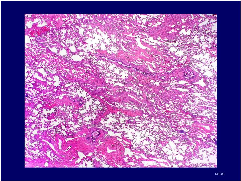
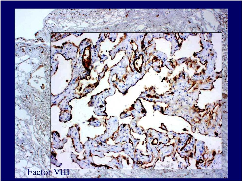
13 Case example Case courtesy of Dr. Fuad Al Dayal, Saudi Arabia A 10 year old boy presented with a hemorrhagic left pleural effusion. The child is hearing and speech impaired. The past medical history is significant for recurrent lung infections, recurrent hemorrhagic pleural effusions and a cerebrovascular accident. KOL03

14 KOL03

15 A surgical wedge lung biopsy was performed KOL03

16 KOL03
17 KOL03

18 KOL03

19 EMA KOL03

20 Factor VIII KOL03
21 TTF-1 KOL03
22 ASMA KOL03
23 MIB1 KOL03
24 Diagnosis Diffuse Pulmonary Lymphangiomatosis KOL03
25 The Territory Ahead Introduction The critical role of sample size and quality
26 The critical role of sample size There is a natural tendency for clinicians to expect more and more information from smaller and smaller biopsy samples. Case example: A 66 year old woman is found to have a 3 cm lung mass. A transbronchial biopsy is performed
27 You decide the included cell group is malignant, and nonsmall cell. and if it is please send for Her2 You attempt IHC to confirm lung origin. Results: TTF-1 neg, CK 7 pos neu. Oh, and if it is a lung cancer, we CK20, synapto, chromogranin, and P63 insufficient tumor in the recuts need to know if it is squamous. If not please send for EGFR After signing the case out as nonsmall cell carcinoma, the clinician calls to ask if it could be from her breast cancer analysis.
28 The Territory Ahead Introduction The critical role of sample size and quality Neoplasms can be triaged by pattern
29 A practical approach Neoplasms are the largest source of targets for diagnostic IHC 4 general morphological categories emerge Neoplasms of lymphoid cells (hematolymphoid) Neoplasms with organoid features Neoplasms with spindled features Neoplasms with undifferentiated epithelioid cells
30 Lymphoid Purpose: Confirm hematolymphoid, confirm neoplastic; subclassify The panel (s) (first consult a local hematopathologist!) CD45 CD20 CD3 CD43 Kappa and lambda (if cytoplasmic) BCL2 (if nodular) Unstained slides (many) CD5 CD10 Cyclin D1 CD79a CD138 CD30 CD15 Myeloperoxidase CD68 Lysozyme S100 protein CD21/CD35 Histiocytic and dendritic cell
31 A 47 year old man presents with weightloss, malaise and abdominal pain. Imaging shows multiple enlarged lymph nodes. A needle core biopsy is performed Another DDX: H&E Immunopanel: Lymphoma Carcinoma Diagnosis CD30 Melanoma CD3 CD45 CD20 CD3 CD43 Kappa and lambda Anaplastic large cell lymphoma, ALK-1 Other? negative (see discussion) CD43, kappa, lambda ALK-1CD20 LCA CD30 ALK-1 LCA
32 The panel CK7/20 Synaptophysin TTF-1 CDX2 PSA Organoid Purpose: Determine primary origin of metastasis, guide therapy Pattern assists general localization Neuroendocrine carcinoma, paraganglioma Lung and thyroid, other small cell Intestinal adenos, other mucinous, endometrioid Prostate, breast, salivary, sweat gland ca, ER/PR --other adeno, melanoma Breast, ovary, endometrium. ER in some lung, stoma and thyroid adenos Calret-CK5/6-WT1 Combined specificity for mesothelioma
33 A 62 year old patient with back pain is found to have a lytic lesion involving T11. A core biopsy is performed DDX: Diagnosis Metastatic adenocarcinoma Metastatic adenocarcinoma consistent with lung (TTF1) or breast (ER) origin of unknown primary origin panck and CK7 CK20 panck CK20 CDX2 TTF1 or and ER! CK 7 TTF1 or ER!
34 The panel Spindled cells Purpose: Determine primary origin/ phenotype, guide therapy PanCK S100 protein Defines epithelial phenotype, spurious in sarcomas, melanoma, dendritic cells. plasma cells Melanocytic, neural, myoepithelial, histiocytic/dendritic, Langerhans cells, liposarcoma, chondrosarcoma Melan-A Desmin CD99 CD31/34 Melanocytic, pre-melanosomes, adrenocortical, sex-cord ovarian Myogenous tumors, PNETs, epithelioid sarcoma Don t forget ER/PR for metastatic Lymphoblastic lymphoma, synovial sarc, EWS/PNET spindled cell tumors in women! Vascular tumors (CD34 better for KS); LFT/SFT (CD34) CD117 GIST
35 A 49 year old woman presents with chest pain and cough.. A 10 cm tumor is identified and removed from the RLL. DDX: Sarcomatoid Ca Primary sarcoma Diagnosis: S100, Desmin, CD117, ER Metastatic sarcoma Localized fibrous tumor Sarcomatoid mesothelioma CD34 + BCL2 Nerve sheath tumor Localized fibrous tumor CD34 + BCL2 S100, Desmin, CD117, ER
36 The panel Undifferentiated epithelioid Purpose: Determine primary origin of metastasis, guide therapy Pan CK S100 protein Synaptophysin CD45 ER/PR (female) Defines epithelial phenotype Melanocytic, neural, myoepithelial, histiocytic/dendritic, Langerhans cells Neuroendocrine cells Hematolymphoid cells Breast, ovary, endometrium. ER in some lung, stomach, and thyroid adenos
37 A 61 year old man presents with a large left axillary mass. His past medical history is remarkable for a previously resected skin appendage tumor from the left hand (said to have been a malignant poroma outside hospital). DDX The Panel panck Inflammatory Diagnosis: panck pseudotumor S100 protein K Lymphoma Metastatic LCA carcinoma Plasma cell Synaptophysin myeloma Melanoma SYN, LCA. Kappa S100 + lambda L panck
38 The Territory Ahead Introduction The critical role of sample size and quality Neoplasms can be triaged by pattern Not all antibodies are created equal
39 Who to trust. Sensitivity and specificity issues Example: synaptophysin and chromogranin Certain antigens in tissue are more resistant to fixation, processing, and tissue degredation Example: panck versus S100 protein The utility and specificity of some antibodies requires context Example: CD30 in ALCL versus carcinoma, or melanoma!
40 A 71 year old man, smoker, is found to have a large central lung mass. A transbronchial biopsy is performed TTF-1 and MIB-1 TTF-1 and MIB1 Small Cell Carcinoma
41 The Territory Ahead Introduction The critical role of sample size and quality Neoplasms can be triaged by pattern Not all antibodies are created equal Top 10 Pitfalls you can avoid
42 TOP TEN PITFALLS IN IHC 10. Incorrect panel of antibodies
43 A 57 year old woman presents with an enlarged groin lymph node. She has a history of node + breast cancer. A needle core biopsy is performed DDX: MelanA Diagnosis: Carcinoma Lymphoma Metastatic melanoma, amelanotic Melanoma Paraganglioma Sarcoma panck, HMB45, EMA, CK7, CK20 LCA S100 Protein LCA panck, HMB45, EMA, CK7, CK20 S100 Protein
44 A 26 year old African woman presents to the emergency room with cough and chest pain, 1 month after delivering a healthy baby. Bronchoscopy yields this biopsy PanCK Synapto, CD31 CD34, TTF1, Melan-A DDX Carcinoid Melanoma Sarcoma Sarcomatoid Ca S100 Vascular PanCK Smooth muscle IMFT CD31
45 TOP TEN PITFALLS IN IHC 9. Incomplete panel of antibodies 10. Incorrect panel of antibodies
46 Some diagnoses require a combination of IHC results for validity A 72 year old man, long time smoker, presents with right sided chest pain The and surgeon breathlessness. finds diffuse thickening of the pleura without a definite mass in underlying Imaging lung. reveals He a feels right the pleural changes are quite effusion. typical The for mesothelioma underlying lung in is his not experience well visualized. He gives a history of asbestos exposure. A VATS procedure obtains tissue
47 Some diagnoses require a combination of IHC results for validity A limited battery of IHC stains is performed, including calretinin CALRET A diagnosis of malignant mesothelioma, epithelial type. The family swears that the patient had no asbestos exposure CK 5/6 and requests that the biopsy be sent out for review BerEP4 B72.3 CALRET WT-1 BerEP4 CALRET B72.3
48 TOP TEN PITFALLS IN IHC 8. Excessive panel of antibodies 9. Incomplete panel of antibodies 10. Incorrect panel of antibodies
49 Thyroidectomy from a young patient with Hashimoto thyroiditis
50 TOP TEN PITFALLS IN IHC 7. Incorrect histopathological DDX 8. Excessive panel of antibodies 9. Incomplete panel of antibodies 10. Incorrect panel of antibodies
51 A 37 year old man presents with chest discomfort and is found to have several nodular lung lesions. A VATS biopsy is performed DDX: The case was sent out for consultation. The lesion was recognized as epithelioid hemangioendothelioma and confirmatory IHC was performed Carcinoma Mesothelioma Melanoma PanCK Chordoma TTF1 TTF1 CD34/CD31 PanCK Hamartoma? CD34/CD31
52 TOP TEN PITFALLS IN IHC 6. Undue pressure on speed of diagnosis 7. Incorrect histopathological DDX 8. Excessive panel of antibodies 9. Incomplete panel of antibodies 10. Incorrect panel of antibodies
53 A 22 year old college student notices a persistent swelling above her right knee. Imaging reveals involvement of the distal femur and additional lytic bone lesions are present. She is brought into the hospital for a percutaneous needle core biopsy She is the only daughter of the Chief of Surgery.
54
55 DDX: Ewing sarcoma/pnet Large cell lymphoma Melanoma of soft parts Epithelioid sarcoma Other? Recommended panel panck LCA S100 protein Synaptophysin 8 unstained Their panel Vimentin panck TdT CD99
56 DDX: Ewing sarcoma/pnet Large cell lymphoma Melanoma of soft parts Epithelioid sarcoma Other? Recommended panel panck LCA S100 protein Synaptophysin 8 unstained
57 DDX: Ewing sarcoma/pnet Large cell lymphoma Melanoma of soft parts Epithelioid sarcoma Other? Secondary panel CD20 CD3 MIB1 Final Diagnosis: Malignant lymphoma, diffuse large B-cell type, CD20 positive.
58 TOP TEN PITFALLS IN IHC 5. Overconfidence in the value of IHC 6. Undue pressure on speed of diagnosis 7. Incorrect histopathological DDX 8. Excessive panel of antibodies 9. Incomplete panel of antibodies 10. Incorrect panel of antibodies
59 A 52 year old man has a serum PSA drawn during a routine physical exam. This results in sextant needle core biopsies
60 TOP TEN PITFALLS IN IHC 3. Relying on tissue from another lab 4. Relying on the IHC of another lab 5. Overconfidence in the value of IHC 6. Undue pressure on speed of diagnosis 7. Incorrect histopathological DDX 8. Excessive panel of antibodies 9. Incomplete panel of antibodies 10. Incorrect panel of antibodies
61 Lymphoid Case in point A 65 year old man presents with a soft tissue mass adjacent to his clavicle and eroding bone. A biopsy is performed. VIM After a first round of IHC the diagnosis remained uncertain and the case was sent for consultation. LCA panck, S100 VIM LCA panck, S100
62 Lymphoid Case in point We restained the tissue block in our laboratory with the following results panck, S100, melana Repeat LCA Our diagnosis: Diffuse large B-cell lymphoma, CD20 positive. CD20 panck, S100, melana Repeat LCA CD20
63 From tissue acquisition to coverslip on your IHC slide, there are so many potential areas for mishap that it is remarkable how frequently IHC is successful! Crush injury Antigen retrival problems Delay in fixation IHC technical failures Improper fixative -reagent sequence Processing damage -incomplete slide flooding Overheating in paraffin -poor humidity control Infiltration contaminants -pipetting inaccuracy Rehydration damage -outdated reagents Poor section adherence -poor antibody quality Incomplete deparaffinization of sections
64 A 48 year old woman present with leg pain and is found to have a cystic lesion in the proximal tibia. Curettings of the lesion are performed at another hospital and a frozen section is requested by the surgeon. SYN
65
66 TOP TEN PITFALLS IN IHC 2. Not knowing the expected staining pattern 3. Relying on tissue from another lab 4. Relying on the IHC of another lab 5. Overconfidence in the value of IHC 6. Undue pressure on speed of diagnosis 7. Incorrect histopathological DDX 8. Excessive panel of antibodies 9. Incomplete panel of antibodies 10. Incorrect panel of antibodies
67 Before ordering, check the expected positive staining reaction! All of these are NUCLEAR stains! ER MyoD1 P63 Ki67 PR WT-1 P53 FLI-1 TTF-1 TDT PCNA Myogenin CDX-2 Cyclin D1
68 TOP TEN PITFALLS IN IHC 1. Not recognizing the histopathology 2. Not knowing the expected staining pattern 3. Relying on tissue from another lab 4. Relying on the IHC of another lab 5. Overconfidence in the value of IHC 6. Undue pressure on speed of diagnosis 7. Incorrect histopathological DDX 8. Excessive panel of antibodies 9. Incomplete panel of antibodies 10. Incorrect panel of antibodies
69 A 66 yr old man presents to the emergency room with left sided chest pain and is found to have a large left pleural effusion. His past medical history is remarkable for left sided pneumonia 3 months earlier for which he was hospitalized and treated empirically with broad spectrum antibiotics. Calret, CK5/6, TTF-1. CK,7, CK20, CEA all negative PanCK Our diagnosis: PanCK WT-1 Malignant mesothelioma, desmoplastic type Calret, CK5/6, TTF-1. CK7. CK20, CEA all neg WT-1
70 The Trail Behind Introduction The critical role of sample size and quality Tumors can be triaged by pattern Not all antibodies are created equal Top 10 Pitfalls you can avoid
71 Questions?
Immunohistochemical differentiation of metastatic tumours
 Immunohistochemical differentiation of metastatic tumours Dr Abi Wheal ST1. TERA 3/2/14 Key points from a review article written by Daisuke Nonaka Intro Metastatic disease is the initial presentation in
Ovarian tumors Ancillary methods
 Ovarian tumors Ancillary methods Ovarian tumor course Oslo, 24-25/11/14 Prof. Ben Davidson, MD PhD Department of Pathology, Norwegian Radium Hospital, Oslo University Hospital, Oslo, Norway Division of
Immunohistochemistry of soft tissue tumors
 Immunohistochemistry of soft tissue tumors Immunohistochemistry Major advances : antigen retrieval techniques (HIER) sensitive detection systems numerous antibodies of good quality Standardization : automated
Update on Mesothelioma
 November 8, 2012 Update on Mesothelioma Intro incidence and nomenclature Update on Classification Diagnostic specimens Morphologic features Epithelioid Histology Biphasic Histology Immunohistochemical
Seattle. Case Presentations. Case 1. 76 year old female with a history of breast cancer 12 years ago. Now presents with a pleural effusion.
 Seattle Montreal IAP September 2006 Case Presentations Allen M. Gown, M.D. Medical Director and Chief Pathologist PhenoPath Laboratories Clinical Professor of Pathology University of British Columbia Case
Neoplasms of the LUNG and PLEURA
 Neoplasms of the LUNG and PLEURA 2015-2016 FCDS Educational Webcast Series Steven Peace, BS, CTR September 19, 2015 2015 Focus o Anatomy o SSS 2000 o MPH Rules o AJCC TNM 1 Case 1 Case Vignette HISTORY:
Pathology of lung cancer
 Pathology of lung cancer EASO COURSE ON LUNG CANCER AND MESOTHELIOMA DAMASCUS (SYRIA), MAY 3-4, 2007 Gérard ABADJIAN MD Pathologist Associate Professor, Saint Joseph University Pathology Dept. Hôtel-Dieu
Index. F Factor VIII-related antigen, see VWF FactorXIIIa, for dermatofibroma, 272-275 5-HT, see Serotonin
 A Acantholytic squamous cell carcinoma vs epithelioid angiosarcoma, 56-57 Acinic cell carcinoma of pancreas, 76-77 vs ductal adenocarcinoma, 74-75 vs islet cell tumor, 78-81 Adenomatoid tumor vs hemangioma,
Practical Effusion Cytology
 Practical Effusion Cytology A Community Pathologist s Approach to Immunocytochemistry in Body Fluid Cytology Emily E. Volk, MD William Beaumont Hospital Troy, MI College of American Pathologists 2004.
Outline. Workup for metastatic breast cancer. Metastatic breast cancer
 Metastatic breast cancer Immunostain Update: Diagnosis of metastatic breast carcinoma, emphasizing distinction from GYN primary 1/3 of breast cancer patients will show metastasis 1 st presentation or 20-30
Diagnosis of Mesothelioma Pitfalls and Practical Information
 Diagnosis of Mesothelioma Pitfalls and Practical Information Mary Beth Beasley, M.D. Mt Sinai Medical Ctr Dept of Pathology One Gustave L Levy Place New York, NY 10029 (212) 241-5307 mbbeasleymd@yahoo.com
MALIGNANT MESOTHELIOMA UPDATE ON PATHOLOGY AND IMMUNOHISTOCHEMISTRY
 MALIGNANT MESOTHELIOMA CLASSIFICATION MALIGNANT MESOTHELIOMA UPDATE ON PATHOLOGY AND IMMUNOHISTOCHEMISTRY Sisko Anttila, MD, PhD Jorvi Hospital Laboratory of Pathology Helsinki University Hospital Espoo,
PATHOLOGY OF THE PLEURA: Mesothelioma and mimickers Necessity of Immunohistochemistry. M. Praet
 PATHOLOGY OF THE PLEURA: Mesothelioma and mimickers Necessity of Immunohistochemistry M. Praet Pathology of the Pleura Normal serosa: visceral and parietal layers Inflammation Neoplasia: Primary: mesothelioma
MALIGNANT MESOTHELIOMA UPDATE ON PATHOLOGY AND IMMUNOHISTOCHEMISTRY
 MALIGNANT MESOTHELIOMA UPDATE ON PATHOLOGY AND IMMUNOHISTOCHEMISTRY Sisko Anttila, MD, PhD Jorvi Hospital Laboratory of Pathology Helsinki University Hospital Espoo, Finland 2nd Nordic Conference on Applied
The develpemental origin of mesothelium
 Mesothelioma Tallinn 14.12.06 Henrik Wolff Finnish Institute of Occupational Health The develpemental origin of mesothelium Mesodermal cavities (pleura, peritoneum and pericardium ) are lined with mesenchymal
A 70-year old Man with Pleural Effusion
 Mesothelioma Diagnosis: Pitfalls and Latest Updates S Klebe and DW Henderson Recommendations Indisputable malignant cells on cytomorphological criteria which demonstrate a mesothelial phenotype, which
Immunohistochemistry on cytology specimens from pleural and peritoneal fluid
 Immunohistochemistry on cytology specimens from pleural and peritoneal fluid Dr Naveena Singh Consultant Pathologist Bart health NHS Trust London United Kingdom Disclosures and Acknowledgements I have
A23: Oncologic Disease- Tumor Markers
 A23: Oncologic Disease- Tumor Markers Diagnosis Tumor Markers and Genetic Markers Use for Specific Malignancy The following information is from multiple guideline sources as recommendations for use of
Disclosures. Learning Objectives. Effusion = Confusion. Diagnosis Of Serous Cavity Effusions - Beware The Mesothelial Cell!
 Disclosures Diagnosis Of Serous Cavity Effusions - Beware The Mesothelial Cell! No Relevant Financial Relationships with Commercial Interests Syed Z. Ali, M.D. Syed Z. Ali, M.D. Associate Professor of
How To Test For Cancer
 Diagnosis Of Serous Cavity Effusions - Beware The Mesothelial Cell! Effusion = Confusion Syed Z. Ali, M.D. Professor of Pathology and Radiology The Johns Hopkins Hospital Baltimore, Maryland Diagnostic
Effusions: Mesothelioma and Metastatic Cancers
 Effusions: Mesothelioma and Metastatic Cancers Malignant Mesothelioma Incidence: 2,500 cases/year ~60-80% pts with pleural MM relationship with asbestos exposure Other risk factors: radiation, other carcinogens,
Diagnostic Challenge. Department of Pathology,
 Cytology of Pleural Fluid as a Diagnostic Challenge Paavo Pääkkö,, MD, PhD Chief Physician and Head of the Department Department of Pathology, Oulu University Hospital,, Finland Oulu University Hospital
MODERN IMMUNOHISTOCHEMISTRY
 MODERN IMMUNOHISTOCHEMISTRY Cambridge Illustrated Surgical Pathology Peiguo G. Chu City of Hope National Medical Center, Duarte, California Lawrence M. Weiss City of Hope National Medical Center, Duarte,
HKCPath Anatomical Pathology Peer Review and Scores : PDF version for download
 AP2003R1 http://hkcpath.org. Correspondence: pkhui@ha.org.hk 1of 10 07/08/2003 HKCPath Anatomical Pathology Peer Review and Scores : PDF version for download AP141 Bone Marrow: Metastatic Carcinoma from
Lung Cancer. Ossama Tawfik, MD, PhD Professor, Vice Chairman Director of Anatomic &Surgical Pathology University of Kansas School of Medicine
 Lung Cancer Ossama Tawfik, MD, PhD Professor, Vice Chairman Director of Anatomic &Surgical Pathology University of Kansas School of Medicine Alexandria, Egypt July 1-1 3, 2008 OBJECTIVES Describe and
CHAPTER 2. Neoplasms (C00-D49) March 2014. 2014 MVP Health Care, Inc.
 March 2014. 2014 MVP Health Care, Inc.") Neoplasms (C00-D49) March 2014 2014 MVP Health Care, Inc. CHAPTER SPECIFIC CATEGORY CODE BLOCKS C00-C14 Malignant neoplasms of lip, oral cavity and pharynx C15-C26 Malignant neoplasms of digestive organs
Neoplasms (C00-D49) March 2014 2014 MVP Health Care, Inc. CHAPTER SPECIFIC CATEGORY CODE BLOCKS C00-C14 Malignant neoplasms of lip, oral cavity and pharynx C15-C26 Malignant neoplasms of digestive organs
Académie internationale de Pathologie - Division arabe XX ème congrès 24-26 novembre 2008 Alger. Immunohistochemistry in malignant mesotheliomas
 Académie internationale de Pathologie - Division arabe XX ème congrès 24-26 novembre 2008 Alger Immunohistochemistry in malignant mesotheliomas Françoise Thivolet-Béjui Groupement Hospitalier Est Lyon-Bron
Cytopathology Case Presentation #8
 Cytopathology Case Presentation #8 Emily E. Volk, MD William Beaumont Hospital, Troy, MI Jonathan H. Hughes, MD Laboratory Medicine Consultants, Las Vegas, Nevada Clinical History 44 year old woman presents
The term undifferentiated tumor has been used in reference
 Undifferentiated Tumor True Identity by Immunohistochemistry Armita Bahrami, MD; Luan D. Truong, MD; Jae Y. Ro, MD, PhD Context. Undifferentiated tumor refers to a heterogeneous group of neoplasms with
Changes in Breast Cancer Reports After Second Opinion. Dr. Vicente Marco Department of Pathology Hospital Quiron Barcelona. Spain
 Changes in Breast Cancer Reports After Second Opinion Dr. Vicente Marco Department of Pathology Hospital Quiron Barcelona. Spain Second Opinion in Breast Pathology Usually requested when a patient is referred
Male. Female. Death rates from lung cancer in USA
 Male Female Death rates from lung cancer in USA Smoking represents an interesting combination of an entrenched industry and a clearly drug-induced cancer Tobacco Use in the US, 1900-2000 5000 100 Per Capita
TUMORS OF THE TESTICULAR ADNEXA and SPERMATIC CORD
 TUMORS OF THE TESTICULAR ADNEXA and SPERMATIC CORD Victor E. Reuter, MD Memorial Sloan-Kettering Cancer Center reuterv@mskcc.org 66 th Annual Pathology Seminar California Society of Pathologists Short
Today s Topics. Tumors of the Peritoneum in Women
 Today s Topics Tumors of the Peritoneum in Women Charles Zaloudek, M.D. Department of Pathology 505 Parnassus Ave., M563 University of California, San Francisco San Francisco, CA USA charles.zaloudek@ucsf.edu
- Slide Seminar - Endocrine pathology in non-endocrine organs. Case 11. Stefano La Rosa, Gioacchino D Ambrosio, Fausto Sessa
 - Slide Seminar - Endocrine pathology in non-endocrine organs Case 11 Stefano La Rosa, Gioacchino D Ambrosio, Fausto Sessa Dept. of Pathology, Multimedica, Milan, Italy Dept. of Surgical and Morphological
YOUR LUNG CANCER PATHOLOGY REPORT
 UNDERSTANDING YOUR LUNG CANCER PATHOLOGY REPORT 1-800-298-2436 LungCancerAlliance.org A GUIDE FOR THE PATIENT 1 CONTENTS What is a Pathology Report?...3 The Basics...4 Sections of a Pathology Report...7
Cytology of Effusion Fluids. Cytology of Effusion Fluids. Types of Effusion Fluids. Anatomy. Causes of Effusions. Sampling of Effusion Fluids
 Cytology of Effusion Fluids John W. Wong, MD, FRCPC Sunnybrook Health Sciences Centre Assistant Professor, Laboratory Medicine and Pathobiology Faculty of Medicine, University of Toronto November 10, 2012
Report series: General cancer information
 Fighting cancer with information Report series: General cancer information Eastern Cancer Registration and Information Centre ECRIC report series: General cancer information Cancer is a general term for
Primary -Benign - Malignant Secondary
 TUMOURS OF THE LUNG Primary -Benign - Malignant Secondary The incidence of lung cancer has been increasing almost logarithmically and is now reaching epidemic levels. The overall cure rate is very low
Introduction: Tumor Swelling / new growth / mass. Two types of growth disorders: Non-Neoplastic. Secondary / adaptation due to other cause.
 Disorders of Growth Introduction: Tumor Swelling / new growth / mass Two types of growth disorders: Non-Neoplastic Secondary / adaptation due to other cause. Neoplastic. Primary growth abnormality. Non-Neoplastic
How To Diagnose And Treat A Tumour In An Effusion
 Effusions of the Serous Cavities Annika Dejmek Professor/Consultant in Cytopathology Clinical Pathology; Department of Laboratory Medicine, Malmö, Lund University 5th EFCS Tutorial Trondheim 2012 Pleura
Notice of Faculty Disclosure
 The Diagnosis of Malignant Mesothelioma Andrew Churg, MD Department of Pathology University of British Columbia Vancouver, BC, Canada achurg@mail.ubc.ca Notice of Faculty Disclosure In accordance with
Efficient Tumor Immunohistochemistry A Differential Diagnosis-Driven Approach
 Efficient Tumor Immunohistochemistry A Differential Diagnosis-Driven Approach Publishing Team Erik Tanck (production manager/designer) Joshua Weikersheimer (publisher) Copyright 2006 by the American Society
DESMOPLASTIC SMALL ROUND CELL TUMOR: A RARE PATHOLOGY PUZZLE
 DESMOPLASTIC SMALL ROUND CELL TUMOR: A RARE PATHOLOGY PUZZLE Ryan Granger University of Rhode Island Cytotechnology program May 2, 2015 ASCT Annual Meeting Nashville, Tennessee DESMOPLASTIC SMALL ROUND
Tumour Markers. What are Tumour Markers? How Are Tumour Markers Used?
 Dr. Anthony C.H. YING What are? Tumour markers are substances that can be found in the body when cancer is present. They are usually found in the blood or urine. They can be products of cancer cells or
 DIFFICULTIES IN THE PATHOLOGIC DIAGNOSIS OF LUNG CANCER Notice of Financial Disclosures (TV Colby MD) (Esp. in small biopsies) Personal Experience Thomas V. Colby, M.D. Mayo Clinic Arizona NONE This talk
The Diagnosis of Cancer in the Pathology Laboratory
 The Diagnosis of Cancer in the Pathology Laboratory Dr Edward Sheffield Christmas Select 74 Meeting, Queen s Hotel Cheltenham, 3 rd December 2014 Agenda Overview of the pathology of cancer How specimens
3-F. Pathology of Mesothelioma
 3-F. Pathology of Mesothelioma Kouki Inai Professor of Department of Pathology, Graduate School of Biomedical Science, Hiroshima University Introduction Mesothelioma is a peculiar type of malignancy, which
How To Test For Cancer With A Blood Test
 Histolab Products AB Eva Alströmer Jonas Falgén Helsingborg 7-8/10 2010 Table 1 Formalin Fixation Times and Estrogen Receptor Staining With 25 Minutes Antigen Retrieval Pretreatment Formalin Q-Score Difference
The Value of Thyroid Transcription Factor-1 in Cytologic Preparations as a Marker for Metastatic Adenocarcinoma of Lung Origin
 Anatomic Pathology / TTF-1 IN CYTOLOGY OF BODY FLUIDS The Value of Thyroid Transcription Factor-1 in Cytologic Preparations as a Marker for Metastatic Adenocarcinoma of Lung Origin Jonathan L. Hecht, MD,
Contents. 1. Introduction and Approach to Fine Needle Aspiration Cytology... 1. 2. Head, Neck, Orbit and Salivary Glands... 12
 Contents 1. Introduction and Approach to Fine Needle Aspiration Cytology... 1 Complications 1 Fine Needle Aspiration Technique 1 Evaluation of FNAC Smear 4 Cell Morphology 4 Nucleus 4 Cytoplasm 6 Background
Schedule of Accreditation issued by United Kingdom Accreditation Service 2 Pine Trees, Chertsey Lane, Staines-upon-Thames, TW18 3HR, UK
 Schedule of ccreditation United Kingdom ccreditation Service 2 Pine Trees, Chertsey Lane, Staines-upon-Thames, TW18 3HR, UK University College London, operating UK NEQS for ccredited to UK NEQS ICC & ISH
BAP1 germline mutations A new Cutaneous Nevus Melanoma Syndrome. Thomas Wiesner
 BAP1 germline mutations A new Cutaneous Nevus Melanoma Syndrome Thomas Wiesner Disclosure Listed as co-inventor US patent application US 61/463,389 BAP1 mutational analysis in determining susceptibility
Abstracts and References
 Abstracts and References Soft Tissue Pathology Professor Cyril Fisher, Royal Marsden Hospital Learning points CD34 and CK positivity coexist in epithelioid sarcoma and epithelioid endothelial tumours.
BIOBANK LPCE-NICE CHEST
 BIOBANK LE-NIE HEST athologist :. BUTORI 12/09/2013 LE / HU Unit atient : N LH13-3603 N LB 13-0691 ID : RO A onsent : YES Age : 54 Diagnosis and staging : chronic pleuresia 5x1000µL BIOBANK LE-NIE HEST
20 Diagnostic Cytopathology, Vol 36, No 1 ' 2007 WILEY-LISS, INC.
 Utility of WT-1, p63, MOC31, Mesothelin, and Cytokeratin (K903 and CK5/6) Immunostains in Differentiating Adenocarcinoma, Squamous Cell Carcinoma, and Malignant Mesothelioma in Effusions Robert T. Pu,
Understanding Pleural Mesothelioma
 Understanding Pleural Mesothelioma UHN Information for patients and families Read this booklet to learn about: What is pleural mesothelioma? What causes it? What are the symptoms? What tests are done to
Cytology : first alert of mesothelioma? Professor B. Weynand, UCL Yvoir, Belgium
 Cytology : first alert of mesothelioma? Professor B. Weynand, UCL Yvoir, Belgium Introduction 3 cavities with the same embryologic origin the mesoderme Pleura Exudates Pleura Peritoneum Pericardium 22%
This factsheet aims to outline the characteristics of some rare lung cancers, and highlight where each type of lung cancer may be different.
 There are several different kinds of lung cancer, often referred to as lung cancer subtypes. Some of these occur more often than others. In this factsheet we will specifically look at the subtypes of cancers
Objectives. Mylene T. Truong, MD. Malignant Pleural Mesothelioma Background
 Imaging of Pleural Tumors Mylene T. Truong, MD Imaging of Pleural Tumours Mylene T. Truong, M. D. University of Texas M.D. Anderson Cancer Center, Houston, TX Objectives To review tumors involving the
CASE OF THE MONTH AUGUST-2015 DR. GURUDUTT GUPTA HEAD HISTOPATHOLOGY
 CASE OF THE MONTH AUGUST-2015 DR. GURUDUTT GUPTA HEAD HISTOPATHOLOGY CASE HISTORY 52Y MALE RIGHT RADICAL NEPHERECTOMY Case of right renal mass with IVC thrombus. History of surgery and RT for right occipital
Surgeons Role in Symptom Management. A/Prof Cliff K. C. Choong Consultant Thoracic Surgeon Latrobe Regional Hospital GIPPSLAND
 Surgeons Role in Symptom Management A/Prof Cliff K. C. Choong Consultant Thoracic Surgeon Latrobe Regional Hospital GIPPSLAND Conditions PLEURAL Pleural effusion Pneumothorax ENDOBRONCHIAL Haemoptysis
ASBESTOS EXPOSURE AND SARCOMATOID MALIGNANT PLEURAL MESOTHELIOMA Gorantla Sambasivarao 1, Namballa Usharani 2, Tupakula Suresh Babu 3
 ASBESTOS EXPOSURE AND SARCOMATOID MALIGNANT PLEURAL MESOTHELIOMA Gorantla Sambasivarao 1, Namballa Usharani 2, Tupakula Suresh Babu 3 HOW TO CITE THIS ARTICLE: Gorantla Sambasivarao, Namballa Usharani,
Lung Carcinomas New 2015 WHO Classification. Spasenija Savic Pathology
 Lung Carcinomas New 2015 WHO Classification Spasenija Savic Pathology ***EXPECTED SPRING 2015*** This authoritative, concise reference book provides an international standard for oncologists and pathologists
Applications of immunohistology to non-heme tumor differential diagnosis R V Rouse 7/22/2014 http://surgpathcriteria.stanford.edu
 Applications of immunohistology to non-heme tumor differential diagnosis R V Rouse 7/22/2014 http://surgpathcriteria.stanford.edu Table of Contents Page Undifferentiated panel 1 CK7/20 table 2 Breast carcinoma
NEOPLASMS C00 D49. Presented by Jan Halloran CCS
 NEOPLASMS C00 D49 Presented by Jan Halloran CCS 1 INTRODUCTION A neoplasm is a new or abnormal growth. In the ICD-10-CM classification system, neoplastic disease is classified in categories C00 through
Lung Cancer: Diagnosis, Staging and Treatment
 PATIENT EDUCATION patienteducation.osumc.edu Lung Cancer: Diagnosis, Staging and Treatment Cancer begins in our cells. Cells are the building blocks of our tissues. Tissues make up the organs of the body.
Problem Cases in Surgical Pathology XXV Congreso de la Sociedad Española de Patologia (SEAP) - Zaragoza, Mayo 18-21, 2011.
 - Zaragoza, Mayo 18-21, 2011.") Problem Cases in Surgical Pathology XXV Congreso de la Sociedad Española de Patologia (SEAP) - Zaragoza, Mayo 18-21, 2011. Saul Suster, M.D. Medical College of Wisconsin Milwaukee, WI, USA Case - 2 Clinical
Problem Cases in Surgical Pathology XXV Congreso de la Sociedad Española de Patologia (SEAP) - Zaragoza, Mayo 18-21, 2011. Saul Suster, M.D. Medical College of Wisconsin Milwaukee, WI, USA Case - 2 Clinical
Oncology. Objectives. Cancer Nomenclature. Cancer is a disease of the cell Cancer develops when certain cells begin to grow out of control
 Oncology Objectives Describe the etiology and pathophysiological mechanisms of cancer Discuss medical and family history findings relevant to cancer Identify general signs and symptoms associated with
WORKPLACE SAFETY AND INSURANCE APPEALS TRIBUNAL DECISION NO. 1557/14
 WORKPLACE SAFETY AND INSURANCE APPEALS TRIBUNAL DECISION NO. 1557/14 BEFORE: M. Crystal: Vice-Chair HEARING: August 20, 2014 at Toronto Written DATE OF DECISION: December 4, 2014 NEUTRAL CITATION: 2014
Bovine heart LSA, Case #94.37374
 1. Bovine heart LSA, Case #94.37374 Page - 1 2. Bovine heart LSA Page - 2 3. Bovine heart malignant lymphoma Page - 3 4. Bovine heart LSA, Case #85.0616 Page - 4 5. Canine heart and pericardium mesothelioma,
FRCPath Part 2 Histopathology Short Cases, autumn 2014
 FRCPath Part 2 Histopathology Short Cases, autumn 2014 Commentary Case 1 Female age 52: palpable lesion, left breast Breast, fat necrosis. Average: 2.6/5 This case was chosen as a good example of fat necrosis
4/15/2013. bi/o carcin/ chem/o immun/o onc/o radi/o sarc/o. anabrachydysectoendoneo- -ectomy -genesis -oma -plasia -sarcoma
 Chapter Sixteen Oncology bi/o carcin/ chem/o immun/o onc/o radi/o sarc/o Combining Forms Prefixes and Suffixes Carcinogenesis anabrachydysectoendoneo- -ectomy -genesis -oma -plasia -sarcoma Causes of cancer
Immunohistochemistry in diagnostic pathology of tumors. Approach, benefits, limits and pitfalls
 Aus dem Institut für Pathologie der Medizinischen Fakultät Charité Universitätsmedizin Berlin DISSERTATION Immunohistochemistry in diagnostic pathology of tumors. Approach, benefits, limits and pitfalls
Mesothelioma. 1995-2013, The Patient Education Institute, Inc. www.x-plain.com ocft0101 Last reviewed: 03/21/2013 1
 Mesothelioma Introduction Mesothelioma is a type of cancer. It starts in the tissue that lines your lungs, stomach, heart, and other organs. This tissue is called mesothelium. Most people who get this
Case based applications part III
 Case based applications part III Los Angeles Society Of Pathologists January 25, 2014 Sanja Dacic, MD, PhD University of Pittsburgh Medical Center 1 CASE 1 A 44-year-old woman with multiple lung nodules.
Case presentation. Awatif Al-Nafussi
 Case presentation Awatif Al-Nafussi Case History 49 year old DVT & small PE June 08, Pelvic mass Ca125 33 Laparotomy-TAHBSO, drainage of ascites Ovarian carcinoma Clinical diagnosis Multiple specimens
Case Report A Cause of Bilateral Chylothorax: A Case of Mesothelioma without Pleural Involvement during Initial Diagnosis
 Case Reports in Pulmonology Volume 2015, Article ID 962504, 4 pages http://dx.doi.org/10.1155/2015/962504 Case Report A Cause of Bilateral Chylothorax: A Case of Mesothelioma without Pleural Involvement
DELRAY MEDICAL CENTER. Cancer Program Annual Report
 DELRAY MEDICAL CENTER Cancer Program Annual Report Cancer Statistical Data From 2010 TABLE OF CONTENTS Chairman s Report....3 Tumor Registry Statistical Report Summary...4-11 Lung Study.12-17 Definitions
Renal Cell Carcinoma: Advances in Diagnosis B. Iványi, MD
 Renal Cell Carcinoma: Advances in Diagnosis B. Iványi, MD Department of Pathology University of Szeged, Hungary ISUP Vancouver Classification of Renal Neoplasia Am J Surg Pathol 37:14691489, 2013 13 histologic
INTERNATIONAL ASSOCIATION FOR THE STUDY OF LUNG CANCER Prospective Mesothelioma Staging Project
 INTERNATIONAL ASSOCIATION FOR THE STUDY OF LUNG CANCER Prospective Mesothelioma Staging Project Data Forms and Fields in CRAB Electronic Data Capture System - Reduced Set - Pivotal data elements for developing
A Cytokeratin- and Calretinin-negative Staining Sarcomatoid Malignant Mesothelioma
 A Cytokeratin- and Calretinin-negative Staining Sarcomatoid Malignant Mesothelioma MICHAEL G. HURTUK and MICHELE CARBONE Cardinal Bernadin Cancer Center, Cancer Immunology Program, Department of Pathology,
The evolving pathology of solitary fibrous tumours. Luciane Dreher Irion MREH / CMFT / NSOPS
 The evolving pathology of solitary fibrous tumours Luciane Dreher Irion MREH / CMFT / NSOPS Historical review Haemangiopericytoma (HPC) first described primarily as a soft tissue vascular tumour of pericytic
Immunohistochemistry of clear cell tumours. What are clear cell tumours? Jan Klos 1
 Immunohistochemistry of clear cell tumours J. Klos MD Department of Pathology Stavanger University Hospital Norway What are clear cell tumours? Multiple factors in etiology of clear cell changes: - technical
How CanCer becomes critical in the claims
 How CanCer becomes critical in the claims arena Cancer is a disease in which cells in your body grow in an uncontrolled way and form a lump called a tumour. In a healthy individual cells grow and reproduce
Case Report Malignant Mesothelioma Mimicking Invasive Mammary Carcinoma in a Male Breast
 Case Reports in Oncological Medicine Volume 2015, Article ID 298523, 4 pages http://dx.doi.org/10.1155/2015/298523 Case Report Malignant Mesothelioma Mimicking Invasive Mammary Carcinoma in a Male Breast
FNA Cytology of Mediastinal Lesions. Presenters: Xiaoqi Lin, M.D., Ph.D. Ritu Nayar, M.D.
 Disclosure information The speakers have no relationship that represents a possible conflict of interest with respect to the content of this presentation. FNA Cytology of Mediastinal Lesions Presenters:
.org. Metastatic Bone Disease. Description
 Metastatic Bone Disease Page ( 1 ) Cancer that begins in an organ, such as the lungs, breast, or prostate, and then spreads to bone is called metastatic bone disease (MBD). More than 1.2 million new cancer
Frequently Asked Questions About Ovarian Cancer
 Media Contact: Gerri Gomez Howard Cell: 303-748-3933 gerri@gomezhowardgroup.com Frequently Asked Questions About Ovarian Cancer What is ovarian cancer? Ovarian cancer is a cancer that forms in tissues
LYMPHOMA. BACHIR ALOBEID, M.D. HEMATOPATHOLOGY DIVISION PATHOLOGY DEPARTMENT Columbia University/ College of Physicians & Surgeons
 LYMPHOMA BACHIR ALOBEID, M.D. HEMATOPATHOLOGY DIVISION PATHOLOGY DEPARTMENT Columbia University/ College of Physicians & Surgeons Normal development of lymphocytes Lymphocyte proliferation and differentiation:
Small Cell Lung Cancer
 Small Cell Lung Cancer Types of Lung Cancer Non-small cell carcinoma (NSCC) (87%) Adenocarcinoma (38%) Squamous cell (20%) Large cell (5%) Small cell carcinoma (13%) Small cell lung cancer is virtually
III II II 111111111111111111111111111111111111111111 1 1111111111111 III. ge: /ears sex: Male Access
 UUID : 10F97F63 - EE6F-448D - 98E1-8AAA21F55753 TCGA - TS-A7PB-01A-PR Redacted III II II 111111111111111111111111111111111111111111 1 1111111111111 III Date Receive III 11111111111111111 11111111111 II
page antibody Adipophilin (polyclonal) ALDH1A1 (44) c-myc (EP121) * Cadherin-17 (SP183) Cathepsin K (3F9)
 ALDH1A1 (44) c-myc (EP121) * Cadherin-17 (SP183) Cathepsin K (3F9)") New IHC Antibodies 2 Table of Contents page antibody 1 2 3 4 5 6 7 8 Adipophilin (polyclonal) ALDH1A1 (44) c-myc (EP121) * Cadherin-17 (SP183) Cathepsin K (3F9) Caveolin-1 (2297) CD13 (SP187) CD16 (SP175)
New IHC Antibodies 2 Table of Contents page antibody 1 2 3 4 5 6 7 8 Adipophilin (polyclonal) ALDH1A1 (44) c-myc (EP121) * Cadherin-17 (SP183) Cathepsin K (3F9) Caveolin-1 (2297) CD13 (SP187) CD16 (SP175)
NEU IM PROGRAMM. LOXO Antikörper für die Immunhistochemie (alphabetisch nach Bezeichnung sortiert) Art.Code Artikelbezeichnung Typ Einsatz Einheit
 Art.Code Artikelbezeichnung Typ Einsatz Einheit") A20002 Actin, Alpha-Smooth Muscle; Clone 1A4 monoklonal IHC 2 ml Ready-to-use A00002.0025 Actin, Alpha-Smooth Muscle; Clone 1A4 monoklonal IHC 25 ml Ready-to-use A00002 Actin, Alpha-Smooth Muscle; Clone
A20002 Actin, Alpha-Smooth Muscle; Clone 1A4 monoklonal IHC 2 ml Ready-to-use A00002.0025 Actin, Alpha-Smooth Muscle; Clone 1A4 monoklonal IHC 25 ml Ready-to-use A00002 Actin, Alpha-Smooth Muscle; Clone
Frozen Section Diagnosis
 Frozen Section Diagnosis Dr Catherine M Corbishley Honorary Consultant Histopathologist St George s Healthcare NHS Trust and lead examiner final FRCPath Practical 2008-2011 Frozen Section Diagnosis The
General Information About Non-Small Cell Lung Cancer
 General Information About Non-Small Cell Lung Cancer Non-small cell lung cancer is a disease in which malignant (cancer) cells form in the tissues of the lung. The lungs are a pair of cone-shaped breathing
بسم هللا الرحمن الرحيم
 بسم هللا الرحمن الرحيم Updates in Mesothelioma By Samieh Amer, MD Professor of Cardiothoracic Surgery Faculty of Medicine, Cairo University History Wagner and his colleagues (1960) 33 cases of mesothelioma
Plueral Malignancy: Radiologic-pathologic
 Plueral Malignancy: Radiologic-pathologic Correlation Ritu R. Gill, MD Pleural Malignancies: Radiologic-Pathologic Correlation Ritu R Gill MD Brigham and Women s Hospital Boston, Massachusetts Pleural
Malignant Lymphomas and Plasma Cell Myeloma
 Malignant Lymphomas and Plasma Cell Myeloma Dr. Bruce F. Burns Dept. of Pathology and Lab Medicine Overview definitions - lymphoma lymphoproliferative disorder plasma cell myeloma pathogenesis - translocations
Case of the. Month October, 2012
 Case of the Month October, 2012 Case The patient is a 47-year-old male with a 3-week history of abdominal pain. A CT scan of the abdomen revealed a suggestion of wall thickening at the tip of the appendix