Diagnosis and Treatment of Adult Flatfoot
|
|
|
- Jeffry McDaniel
- 8 years ago
- Views:
Transcription
1 CLINICAL PRACTICE GUIDELINE Diagnosis and Treatment of Adult Flatfoot Clinical Practice Guideline Adult Flatfoot Panel: Michael S. Lee, DPM, 1 John V. Vanore, DPM, 2 James L. Thomas, DPM, 3 Alan R. Catanzariti, DPM, 4 Geza Kogler, PhD, 5 Steven R. Kravitz, DPM, 6 Stephen J. Miller, DPM, 7 and Susan Couture Gassen 8 This clinical practice guideline (CPG) is based on the consensus of current clinical practice and review of the clinical literature. The guideline was developed by the Clinical Practice Guideline Adult Flatfoot Panel of the American College of Foot and Ankle Surgeons. The guideline and references annotate each node of the corresponding pathways. Introduction to Adult Flatfoot (Pathway 1) Foot and ankle specialists agree that flatfoot is a frequently encountered pathology in the adult population. For the purpose of this document, adult flatfoot is defined as a foot condition that persists or develops after skeletal maturity and is characterized by partial or complete loss (collapse) of the medial longitudinal arch. Adult flatfoot may present as an incidental finding or as a symptomatic condition with clinical consequences ranging from mild limitations to severe disability and pain causing major life impediments. Adult flatfoot encompasses a wide variety of pathologic etiologies that may include a benign process reflecting continuation of a congenital problem, trauma, or a condition associated with systemic pathology. The adult flatfoot is often a complex disorder with a diversity of symptoms and various degrees of deformity. Pathology and symptoms are caused by structural loading Address correspondence to: John V. Vanore, DPM, Gadsden Foot Clinic, 306 South 4th St, Gadsden, AL jvanore@ bellsouth.net 1 Chair, Adult Flatfoot Panel, Ankeny, IA; 2 Chair, Clinical Practice Guideline Core Committee, Gadsden, AL; 3 Board Liaison, Birmingham, AL; 4 Pittsburgh, PA; 5 Springfield, IL; 6 Richboro, PA; 7 Anacortes, WA; 8 Chicago, IL. Copyright 2005 by the American College of Foot and Ankle Surgeons /05/ $30.00/0 doi: /j.jfas changes along the medial foot and plantar arch, as well as by collapse through the midfoot and impingement along the lateral column and rearfoot (1 5). Muscles in the leg and foot tend to fatigue and cramp because of overuse (6 8). Peritalar subluxation defines the pathologic malalignment of the talus about the subtalar and midtarsal joints (9, 10). Literature on the incidence and symptomatology of adult flatfoot is limited (11 13). Ferciot (14) estimated a 5% incidence of flatfoot in all children and adults. Harris and Beath (15) studied 3,619 Royal Canadian Army recruits and found that 15% had a simple hypermobile flatfoot, 6% had simple hypermobile flatfoot with a tight heel cord, and 2% had a tarsal coalition. Significant History (Pathway 1, Node 1) The natural history of the adult flatfoot has not been clearly defined, as evidenced by the absence of reliable studies analyzing the long-term sequelae of this condition (16). The deformity may be associated with pain, instability, and severe functional limitations or it may be of little clinical significance (17, 18). The evaluation of adult flatfoot requires a pertinent patient history that includes onset of the deformity, timing of symptoms, and severity of past and current symptoms (with particular regard to arch and rearfoot pain). A family history of flatfoot deformity may be elicited. Associated conditions such as rheumatoid arthritis, seronegative arthropathies, hypertension, or diabetes may be significant in the patient with adult flatfoot (19, 20). Occupation, activity level, and obesity may also be contributory factors. Footwear, history of trauma, and previous treatment are significant. A pertinent review of systems should be performed. Extrapedal findings, such as knee, hip, or back pain, have also been associated with concurrent flatfoot (21). 78 THE JOURNAL OF FOOT & ANKLE SURGERY

2 PATHWAY 1 Significant Findings (Pathway 1, Node 2) Flatfoot versus a normal foot (Fig 1) is readily apparent with clinical evaluation. The appearance of the foot, both on and off weightbearing, will help define its deformed or compensated condition. Physical examination reveals 1 or more of the following characteristics: depression of the medial longitudinal arch, everted or valgus heel in relaxed stance, and abduction of the forefoot relative to the rearfoot (Figs 2 and 3). Areas of tenderness may be localized with careful palpation of the foot, ankle, and leg; particular attention should be paid to the posterior tibial tendon, lateral rearfoot, and plantar fascia. Range of motion evaluation differentiates the flexible from rigid flatfoot and identifies the degree of abnormal motion that may be present. Flexibility can also be assessed by using the Hubscher maneuver (Jack test) to determine if the deformity is reducible (22, 23). Manual muscle testing and the single heel-rise test assess muscle strength and tendon function (24). Additionally, the double heel-rise test determines reducibility of rearfoot valgus (Figs 1 and 3). Gait observation may show an increased angle of gait, delayed or absent supination of the foot, or decreased propulsion. Footwear patterns also can provide valuable information. Extrapedal manifestations may include genu valgum, shin splits, short tendo-achilles with calf tenderness, VOLUME 44, NUMBER 2, MARCH/APRIL
.")
3 FIGURE 1 Clinical examination of the foot includes inspection of the weightbearing foot, with attention to (A) the frontal plane position of the heel and (B) maintenance of the medial longitudinal arch. The patient should be able to perform (C) single and (D) double heel rises, which show flexibility and normal supinatory potential of the foot.
 single and (D) double heel rises, which show")
4 FIGURE 2 Examination of the flatfoot compares the (A) nonweightbearing and (B) weightbearing arch of the foot. As the arch depresses, (C) the forefoot abducts and (D) the lesser toes become visible upon posterior observation of the foot. The relaxed calcaneal stance position is viewed standing behind the patient. A flatfoot deformity will demonstrate heel eversion that is accentuated with apparent bowing of the tendo-achilles (Helbing sign). The too many toes sign, indicative of excessive forefoot abduction in the flatfoot, may also be noted. VOLUME 44, NUMBER 2, MARCH/APRIL
.")
5 FIGURE 3 The severe flatfoot deformity shows very excessive heel valgus, (A) forefoot abduction, and (B) medial collapse of the foot. (C) The patient may be able to perform a double heel rise, but the heel shows lack of supinatory varus.
 The patient may be able to perform a double heel rise,")
6 peroneal muscle spasm, medial knee tenderness, leg length discrepancy, and torsional problems. Frontal, sagittal, and transverse plane changes can be assessed in regard to deformity as well as compensation (25, 26). Radiographic Findings (Pathway 1, Node 3) Radiographic evaluation of the adult foot in the angle and base of gait allows assessment of the degree of deformity. Routine radiographs may include standing anterior-posterior (AP) (dorsoplantar), lateral, and oblique views, as well as Harris-Beath views if a tarsal coalition is suspected. Consideration should also be given to ankle radiographs if ankle valgus is a concern. A number of radiographic criteria are used in the assessment of the foot structure (Fig 4): Calcaneal pitch is decreased in a flatfoot deformity and may approach 0 or become less than 0 in the presence of a rocker-bottom deformity. The degree of talar head coverage by the navicular and an increased calcaneocuboid abduction angle are useful parameters to assess the degree of pronation and abduction of the forefoot on the rearfoot. The cyma line is the radiographic visualization of the talonavicular and calcaneocuboid joints on the lateral radiograph. The joints should be visualized as a continuum, whereas in a pronated foot type, the talonavicular joint space is positioned further anterior than the calcaneocuboid joint space. A talocalcaneal angle is formed by the long axis of the rearfoot and the midtalar line. This angle is increased in pronated feet on both the AP and lateral views. The talar first metatarsal angle, measured on both AP and lateral views, increases with the degree of pronation. Findings consistent with degenerative arthritis of 1 or more joints are significant and may be secondary to a longstanding flatfoot deformity or may represent a primary cause of the flatfoot. The hindfoot alignment view provides coronal plane evaluation of the hindfoot in relation to the distal tibia (27). Radiographs may show a tarsal coalition. Optional Ancillary Studies (Pathway 1, Node 4) Although not routinely indicated for evaluation of adult flatfoot, the following tests can provide additional information that may better define the condition and aid in treatment selection: Magnetic resonance imaging (MRI) (28). Computed tomography (29 32). Bone scan (29, 33). Gait analysis via video or computer studies (34). Nerve conduction velocities/electromyography studies. Ink print mats (35) and Harris mats (36). Computerized force plate analysis. Local anesthetic diagnostic injections. Radiographs with the foot in the neutral position (37). Diagnosis (Pathway 1, Node 5) Information obtained from the initial evaluation and diagnostic tests is correlated into a diagnosis. The differential diagnosis of the adult flatfoot foot includes the following: adult flexible flatfoot (non posterior tibial tendon dysfunction [PTTD]) (Pathway 2); PTTD (Pathway 3); tarsal coalition (Pathway 4); arthritic, posttraumatic, or iatrogenic deformity (Pathway 5); Charcot foot (Pathway 6); and neuromuscular flatfoot (Pathway 7). Adult Flexible Flatfoot (Non-PTTD) (Pathway 2) Adult flexible (non-pttd) flatfoot is generally a progression of a pediatric condition characterized by partial or complete loss of the medial arch. There are many terms used to describe the flexible flatfoot. The designation of flexible refers to the general qualitative stiffness properties of the foot evident during dynamic loading and/or physical examination (ie, flexible vs rigid). Flexible flatfoot in the adult may present as unilateral or, more commonly, as bilateral (38). It is frequently associated with a short or contracted Achilles muscle-tendon complex (39). In its late stages of progression, degenerative arthritis may occur, leading to loss of flexibility or ankylosis (37, 40). Additionally, peroneal spasm may result with rearfoot arthritis. Abnormal pronation of the rearfoot during weightbearing has been associated with collapse of the longitudinal arch in the adult flexible flatfoot. The talus adducts and plantarflexes on the calcaneus, which simultaneously everts and plantarflexes. Subtalar joint pronation unlocks the midtarsal joint, making it unstable and leading to various degrees of transverse plane abduction (41, 42). The tarsometatarsal and other joints may also be affected (2). Causative factors of pronation include the following: compensated forefoot varus, compensated flexible forefoot valgus, equinus, congenital talipes calcaneovalgus, torsional abnormalities of adduction or abduction, muscle imbalance, ligamentous laxity, neurotrophic feet, and anything that contributes to a medial shift in weightbearing (eg, genu valgum, obesity, wide base of gait) (39). Significant History (Pathway 2, Node 1) Patients with adult flexible flatfoot may present with postural symptoms as well as weakness and fatigue in the VOLUME 44, NUMBER 2, MARCH/APRIL
 (dorsoplantar), lateral, and oblique views, as well as Harris-Beath views if a tarsal coalition is suspected.")
7 FIGURE 4 Radiographic examination of the weightbearing foot allows for a more quantitative analysis of the flatfoot deformity. (A) Normal foot: line diagram with corresponding AP and lateral radiographs showing normal osseous relationships. Talocalcaneal angles may be drawn on both the AP and the lateral standing radiographs (dotted line, midtalar line; dashed line, longitudinal axis of hindfoot; solid line, first metatarsal bisection). The talocalcaneal angle (TC) increases with the degree of pronation. The talar first metatarsal angle should be nearly parallel on both the AP and lateral weightbearing radiographs. The AP radiograph shows good talar head coverage, whereas the lateral radiograph shows a normal calcaneal pitch or inclination angle with good height to the medial longitudinal arch. (B) Adult flatfoot: Deformity is shown on both the AP and the lateral radiographs, which show markedly increased talocalcaneal angles, decreased calcaneal pitch with depression of the medial longitudinal arch, and poor talar head coverage. Deviation of the talar first metatarsal angle is quite marked on the lateral film, showing a plantarflexed talus or talar ptosis. (Diagrams drawn by, and provided courtesy of, Maria Bidny, DPM, Hillside, MI.) 84 THE JOURNAL OF FOOT & ANKLE SURGERY
 increases with the degree of pronation. The talar first metatarsal angle should be nearly parallel on both the AP and lateral weightbearing radiographs.")
8 PATHWAY 2

9 foot or leg. Flexible flatfoot in the adult may manifest as a bilateral (more common) or unilateral condition with an onset of symptoms later in life and not attributable to PTTD (38). Arch, heel, and lateral foot pain may be primary complaints, with symptoms exacerbated by weightbearing activities (eg, running, walking, hiking). For further discussion, see Pathway 1. Significant Findings (Pathway 2, Node 2) By definition, the adult flexible flatfoot is supple, although it is not always completely reducible in its later stages. The heel may assume a valgus position during weightbearing. Areas tender to palpation might include the sinus tarsi, talonavicular joint, plantar arch and heel, posterior tibial tendon, anterior tibial tendon, and anterior or posterior tibia. Significant rearfoot eversion may result in subfibular impingement pain. Equinus is often present, and evaluation of the gastrosoleal complex is warranted. Clinical maneuvers as described in Pathway 1 may be performed to assess flexibility. For further discussion, see Pathway 1. Radiographic Findings (Pathway 2, Node 3) Radiographic findings consistent with flatfoot have been described in Pathway 1. These findings include various angular changes of the foot, peritalar subluxation, and degenerative arthritis in more advanced stages (Fig 4). Initial Treatment (Pathway 2, Node 7) For asymptomatic flatfoot, treatment entails patient education, discussion of the prognosis, and observation of the condition (Node 5). In the symptomatic flatfoot (Node 6), nonsurgical therapy is directed at resisting the deformity and limiting uncontrolled pronatory compensation (43). Correction of the deformities should not be anticipated with this line of treatment. Initial treatment options for adult flexible flatfoot include one or more of the following: activity modifications, weight loss, orthotic management, immobilization, and footwear modifications. Antiinflammatory medications and physical therapy may also be beneficial. Orthotic management (Node 8) encompasses a broad spectrum of devices that includes foot orthoses or ankle-foot orthoses (AFO), either prefabricated or custom molded (Fig 5) (44 55). FIGURE 5 Orthotic management of flatfoot is accomplished with foot orthoses, which often include custom-made devices to accommodate the unique topography and degree of deformity present in the individual. The orthotic device may possess features of (A) a medial arch flange or deep heel seat, or (B) a full AFO (ankle-foot orthosis) that will support the entire ankle-tarsal complex. Clinical Response (Pathway 2, Node 9) Nonsurgical therapy should be continued in patients who experience a favorable clinical response (Node 10). Surgical management should be considered if nonsurgical options fail to provide adequate relief from pain, if there is progression of deformity or instability, or if there is failure to return to acceptable function (39). 86 THE JOURNAL OF FOOT & ANKLE SURGERY

10 TABLE 1 Relative corrective ability of flatfoot procedures Surgical Procedure Type of Correction Able to Be Achieved Equinus Medial Column Sag Forefoot Abduction Rearfoot Eversion Peritalar Subluxation Ankle Valgus Osteotomies Evans o * * PCDO o Double Calcaneal o Cotton o o? Arthrodesis Medial Column o * Talonavicular o * CC Distraction o * Double (MTJ) o * Subtalar Joint o * Triple o * Pantalar * Ankle /o * TTCA * Arthroeresis Arthroeresis o * Soft Tissue Procedures TAL * Gastroc Recession o Scale, 0 to. Abbreviations: CC, calcaneocuboid; MTJ, midtarsal joint; PCDO, posterior calcaneal displacement osteotomy; TAL, tendo-achillis lengthening; TTCA, tibiotalocalcaneal arthrodesis *Indirect. Surgical Intervention (Pathway 2, Node 11) Various surgical techniques have been described for the treatment of adult flexible flatfoot. These may include osteotomy, arthroereisis, arthrodesis, and adjunctive soft tissue procedures (Table 1). Procedure selection should be based on clinical and radiographic findings, degree of arthritis, patient age, and activity level. Soft Tissue Procedures. For the correction of symptomatic flexible flatfeet with minimal deformity, soft tissue procedures can be considered. These procedures may include the Kidner posterior tibial tendon advancement, the flexor digitorum longus tendon transfer, the Young tenosuspension, reconstruction of the spring ligament, and the medial arch reconstruction combination (39, 56 65). Osteotomy. Various osteotomies for correction of the adult flatfoot have been recommended and studied. The main advantage of periarticular osteotomies is preservation of joint function while improving structural alignment. The Evans calcaneal osteotomy was developed to lengthen the lateral column and realign the midtarsal joint by reducing forefoot abduction (Fig 6) (66 69). This procedure has also been shown to plantarflex the first metatarsal and reduce talocalcaneal subluxation (66, 70 72). Cadaver studies have demonstrated increased pressures through the calcaneocuboid joint following lateral distraction; however, these increases in joint pressure have not been found to be significant in a flatfoot model (73 76). A posterior calcaneal displacement osteotomy may be indicated for reduction of significant rearfoot valgus (Fig 7). The rationale for performing this osteotomy includes restoring the gastrosoleal complex as a heel invertor, increasing supinatory ground reactive forces, and decreasing medial arch load (70, 77 84). Recent studies have shown good clinical results with this osteotomy (79, 80, 85 88). However, investigators have noted that the osteotomy alters motion and joint contact characteristics within the ankle joint, which may predispose this joint to premature arthrosis (89 93). In cases of more significant deformity, double calcaneal osteotomies (Evans and posterior calcaneal displacement osteotomy) have been performed concurrently to achieve adequate correction (Fig 8) (94 97). Other calcaneal osteotomies have been described but are less frequently used (98 101). The Cotton opening wedge osteotomy through the medial cuneiform has been promoted as a valuable adjunctive procedure to plantarflex the medial column ( ). Correction of deformity is achieved with a structural allograft or autograft. Arthroereisis. Several implant devices have been designed to limit pronatory motion by blocking movement between the talus and calcaneus ( ). In the adult, arthroereisis is seldom implemented as an isolated procedure. Because of the long-term compensation and adaptation of the foot and adjunctive structures for flatfoot function, other ancillary procedures are usually used for VOLUME 44, NUMBER 2, MARCH/APRIL

11 FIGURE 6 (A) This preoperative weightbearing AP radiograph of flatfoot deformity shows increased calcaneocuboid (CC) and talo-first metatarsal (TMT) angles. An Evans calcaneal osteotomy with bone graft lengthening was performed, and (B) the postoperative weightbearing AP radiograph shows reduction of forefoot abduction and reduction of respective calcaneocuboid and talo-first metatarsal angles. Lateral (C) preoperative and (D) postoperative radiographs show similar improvement in biomechanical relationships. 88 THE JOURNAL OF FOOT & ANKLE SURGERY

12 FIGURE 7 The posterior calcaneal osteotomy involves medial displacement of the posterior fragment to place the leg axis through the foot. Preoperative weightbearing (A) AP and (B) lateral radiographs show a flatfoot deformity with low calcaneal inclination or pitch and depression of the medial longitudinal arch. Forefoot abduction is limited. Postoperative weightbearing (C) AP and (D) lateral radiographs show a posterior calcaneal osteotomy with medial displacement. Improvement of talocalcaneal relationships can be appreciated as well as improved calcaneal inclination and arch height.
 AP and (D) lateral radiographs show a posterior calcaneal osteotomy with medial displacement.")
13 FIGURE 8 Multiple calcaneal osteotomies may be performed, as in this case, which included both an Evans-type procedure and a posterior calcaneal displacement osteotomy. (A) The preoperative weightbearing lateral radiograph of the flatfoot deformity shows a low calcaneal inclination angle, prominent talar declination, and depression of the longitudinal arch. (B) The postoperative weightbearing lateral radiograph shows the usefulness of performing an Evans bone graft lengthening of the lateral column combined with a posterior calcaneal osteotomy with medial displacement. Improvement of all biomechanical parameters is seen. appropriate stabilization (37). Long-term results of arthroereisis in the adult flexible flatfoot patient have not been established. Some surgeons advise against the subtalar arthroereisis procedure because of the risks associated with implantation of a foreign material, the potential need for further surgery to remove the implant, and the limited capacity of the implant to stabilize the medial column sag directly (119). Arthrodesis. Arthrodesis of the medial column, including the naviculocuneiform joint(s) and/or metatarsocuneiform joint, may be used when medial column collapse is noted (120, 121). More recently, medial column arthrodesis techniques have been implemented as adjunctive procedures (80, 122, 123). Isolated talonavicular joint arthrodesis provides very powerful correction of the subluxated talar head ( ). The reduction in subtalar joint range of motion after talonavicular joint arthrodesis has limited the use of this procedure (58, 127, 128). Calcaneocuboid joint distraction arthrodesis has been described for the correction of the flatfoot deformity with significant forefoot abduction (63, 129). Indications are similar to those of the Evans osteotomy, but the arthrodesis technique is thought to avoid the increased calcaneocuboid joint pressures/arthrosis that have been theorized in adult patients after Evans osteotomy (69, 74, 76, 130). Autogenous bone grafting is recommended for distraction arthrodesis because of higher nonunion rates (69). Double arthrodesis (talonavicular and calcaneocuboid joints) may also 90 THE JOURNAL OF FOOT & ANKLE SURGERY
.")
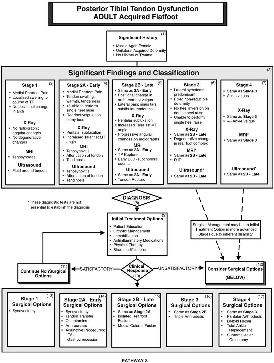
14 be used for correction of the flatfoot deformity and may provide less subtalar joint arthrosis than the isolated talonavicular joint arthrodesis (128, 131, 132). Isolated subtalar joint and triple arthrodesis provide viable options for flatfoot correction (Fig 11) ( ). A fixed forefoot varus necessitates the use of triple arthrodesis. Additionally, triple arthrodesis is more often implemented in the rigid flatfoot with rearfoot arthrosis in the subtalar and midtarsal joints ( ). Subtalar joint arthrodesis is more frequently indicated in the flatfoot with a reducible deformity without midtarsal joint arthrosis or fixed forefoot varus ( ). Union rates with subtalar joint arthrodesis have been reported to be high (144). Pantalar arthrodesis is used in longstanding deformities with rearfoot and ankle degenerative changes. Often, the ankle arthrosis is a result of a valgus tilt of the talus within the mortise. In situations in which the midtarsal joint has been spared of degenerative changes, tibiotalocalcaneal arthrodesis may be warranted. Retrograde intramedullary nail fixation may be used in these cases. Adjunctive Soft Tissue Procedures. The shortened Achilles muscle-tendon complex may be lengthened in conjunction with reconstructive flatfoot surgery (22, 83, 103, ). Common techniques include gastrocnemius recession, gastrocnemius-soleus recession, and Achilles-tendon lengthening. The relationship between flexible flatfoot and contracted Achilles muscle-tendon complex is well established, and the loss of muscle strength after lengthening has been shown to be insignificant (151). Posterior Tibial Tendon Dysfunction (Pathway 3) PTTD is the most common cause of the adult acquired flatfoot. Dysfunction of the posterior tibial tendon is typically a unilateral condition caused by pathologic changes within the tendon. The deformity is usually progressive and results in a flexible to rigid flatfoot, depending on the stage of the condition. Significant History (Pathway 3, Node 1) PTTD is common in women aged 45 to 65 years (152, 153). Patients usually present without a specific history of trauma. Symptoms are typically preceded by overuse activity. Patients with PTTD may also present with a preexisting flatfoot deformity, family history of flatfoot deformity, or other systemic conditions (154). Significant Findings and Classification (Pathway 3, Node 2) Patients with PTTD may present with symptoms at any stage of this progressive condition. Numerous PTTD classification systems have been developed (24, 155, 156). The classification originally described by Johnson and Strom (24) and later modified (157) was used in developing this guideline. Additionally, the guideline panel further divided stage 2 PTTD into early (stage 2A) and late (stage 2B). Significant findings are discussed in this document, according to the stages of the deformity. Stage 1 (Pathway 3, Node 3) In stage 1 PTTD, the foot may have an unchanged appearance with no deformity (24). Pain and edema along the medial aspect of the rearfoot corresponding to the posterior tibial tendon may be present. This is usually indicative of tenosynovitis or early tendinosis. Clinical assessment may demonstrate increased warmth, edema, and tenderness along the course of the tendon. Weakness is usually absent in stage 1 PTTD. Patients are typically able to perform a single heel raise without difficulty, although this maneuver may reproduce symptoms. Radiographs are usually unremarkable, whereas MRI and ultrasound studies may show tenosynovitis (156). Stage 2A (Pathway 3, Node 4) Stage 2 PTTD encompasses a wider spectrum of pathology. Stage 2A is usually characterized by medial rearfoot pain, edema, and tenderness along the course of the posterior tibial tendon, and mild valgus of the heel, with or without lowering of the medial longitudinal arch (24). There may be some abduction of the forefoot on the rearfoot (too many toes sign) (24). Although patients with stage 2A PTTD may be able to perform a single heel raise, they are more likely to have difficulty and pain completing this maneuver. Subtalar joint motion is supple with increased eversion. Radiographs typically reveal a flatfoot deformity with increased talo-first metatarsal angle, peritalar subluxation, and increased calcaneocuboid abduction angle. MRI and ultrasound studies may reveal tenosynovitis, tendinosis, or attenuation of the posterior tibial tendon (Fig. 9) (156) (Fig 9). Stage 2B (Pathway 3, Node 5) The findings in stage 2B PTTD are similar to those in stage 2A, with the addition of lateral pain (sinus tarsi, subfibular, cuboid), more severe valgus deformity, collapse of the medial longitudinal arch, and obvious abduction of the forefoot on the rearfoot. Forefoot supinatus may be present in stage 2B. There may also be decreased subtalar joint motion. Contracture of the posterior muscle group is typical. Radiographs show an increase in the talo-first meta- VOLUME 44, NUMBER 2, MARCH/APRIL
.")
15 PATHWAY 3
16 VOLUME 44, NUMBER 2, MARCH/APRIL

17 tarsal angle on AP and lateral views as well as peritalar subluxation. Degenerative changes are usually absent. MRI and ultrasound findings are similar to those found in stage 2A, although they may also reveal complete tendon rupture. Stage 3 (Pathway 3, Node 6) In stage 3 PTTD, deformities become more severe and fixed. Significant loss of subtalar joint motion and contracture of the posterior muscle group usually occur at this stage. Lateral symptoms predominate. The patient is unable to perform a single heel-raise test and the rearfoot remains everted with a double heel-raise test (Fig 3). A forefoot varus is often noted. Radiographs of stage 3 PTTD show continued increase in angular deformities and may show degenerative changes within the rearfoot. MRI and ultrasound studies reveal similar findings to those in stage 2. Stage 4 (Pathway 3, Node 7) Stage 4 PTTD has ankle involvement (Fig 10). The medial soft tissue constraints, including the deltoid ligament, become attenuated. An AP view of the ankle may show valgus tilt of the talus within the ankle mortise. Degenerative changes of the ankle may develop with advancing PTTD. Initial Treatment (Pathway 3, Node 9) Initial treatment options for most cases of PTTD include various types of nonsurgical therapy. Stage 1 PTTD may be treated with a short-leg cast or walking boot for 4 to 6 weeks. The patient may participate in physical therapy for rehabilitation of the posterior tibial tendon muscle after cast immobilization. Antiinflammatory medications may assist in reducing inflammation. Orthoses or ankle stirrup braces may also be used in stage 1 PTTD. Stage 2A PTTD is usually treated with orthoses. Stage 2A PTTD may involve some deformity or impending deformity, thus necessitating support of the longitudinal arch and heel. An orthotic device with a deep heel cup is usually sufficient. Patients in stage 2B PTTD may require a University of California Biomechanics Laboratory device or AFO (Fig 5). Modifications in footwear, such as an extended medial counter or medial heel wedge, may be helpful. In the acutely painful foot, immobilization may be warranted. Stage 3 PTTD may require some type of custom AFO or supramalleolar brace. The AFO should be articulated to permit ankle joint range of motion. Unfortunately, once the deformity is fixed, the patient may have difficulty tolerating various types of AFOs. This is especially true in cases of significant rearfoot valgus deformity. Stage 4 PTTD is usually treated with a nonarticulated AFO. Surgical management (Node 12) may be an initial treatment option when inherent disability is present. Clinical Response (Pathway 3, Node 10) When nonsurgical care is rendered, the clinical response is assessed. If the patient is doing well, initial treatment may be continued (Node 11). If there is little or no improvement or if initial improvement deteriorates, surgical management may be considered (Node 12). In cases in which the initial evaluation and treatment were performed by a primary care physician, referral to a podiatric foot and ankle surgeon is warranted. Surgical Intervention (Pathway 3, Node 12) Surgical management during stage 1 PTTD (Node 13) primarily consists of synovectomy. This should be considered if noninvasive therapy has been ineffective. Tendon decompression with synovectomy has been reported to provide relatively good results (153, 158). In stage 2A (Node 14), synovectomy, tendon debridement, and/or primary repair may be indicated. When tendon dysfunction is present, a tendon transfer should be considered as an adjunct procedure. Osteotomy has also been recommended when deformity is present. Calcaneal osteotomies may include posterior calcaneal displacement osteotomy or Evans calcaneal osteotomy (68, 78, 80, 82, 85, 87, 97, 159, 160). Posterior muscle group lengthening should be considered when equinus is present. Stage 2B PTTD (Node 15) may require synovectomy or tendon repair in combination with a tendon transfer. Advancing deformity is common in stage 2B, necessitating some type of realignment through an osteotomy or arthrodesis. The calcaneal osteotomies described for stage 2A, as 4 FIGURE 9 (A) Adult-acquired flatfoot deformity is often the result of posterior tibial tendon dysfunction with pathologies involving the integrity of the tendon with attenuation or rupture. (B) MRI (shown here) or other imaging modalities may be used to evaluate the integrity of the posterior tibial tendon. This patient s preoperative weightbearing (C) AP and (D) lateral radiographs of flatfoot deformity reveals degenerative changes at the first and second metatarsocuneiform joints. Surgical treatment included tarsometatarsal arthrodesis and tendon repair, as shown in postoperative weightbearing (E) AP and (F) lateral radiographs. 94 THE JOURNAL OF FOOT & ANKLE SURGERY

18 VOLUME 44, NUMBER 2, MARCH/APRIL

19 well as double calcaneal osteotomy, may be considered for the stage 2B deformity (94, 97). Medial column osteotomies (eg, the Cotton open-wedge osteotomy) may also be considered as adjunct procedures. Isolated rearfoot arthrodesis procedures have been recommended in stage 2B. These procedures may include subtalar joint, calcaneocuboid interpositional bone block, or medial column arthrodesis (62, 129, 131, ) (Fig 9). Selection of a single joint arthrodesis is based on the type and degree of deformity (Table 1), as well as on the goals and expectations of the patient and surgeon. Isolated rearfoot arthrodesis of any joint will subsequently affect kinematics and may contribute to adjacent joint arthrosis (165, 166). Arthroereisis procedures have also been recommended as an option in stage 2 PTTD (114, 167). Additionally, tendon transfers have been used extensively to augment these osseous procedures (80, ). Stage 2 invariably results in equinus deformity that should be addressed with posterior muscle group lengthening. Stage 3 PTTD (Node 16) usually requires an arthrodesis procedure. An isolated joint arthrodesis may be considered. However, if arthrosis is extensive and involves the entire rearfoot, triple arthrodesis is the procedure of choice (140, 143, 153, ). In longstanding, rigid deformities, a fixed forefoot varus often necessitates triple arthrodesis. Additionally, such patients require lengthening of the posterior muscle group to address the equinus deformity. Stage 4 (Node 17) involves the ankle joint. A triple arthrodesis in combination with deltoid ligament repair and a medializing calcaneal osteotomy may be considered in cases in which valgus deformity of the ankle exists with no degenerative joint disease (Fig 10). A pantalar arthrodesis may be indicated when degenerative changes involve not only the rearfoot but also the ankle joint. In addition, total ankle replacement or supramalleolar osteotomy may be used in conjunction with triple arthrodesis. Tarsal Coalition (Pathway 4) Tarsal coalition may present in the adult patient, and the evaluation and diagnosis of this developmental aberration is much the same as that for the child. This disorder is often an incidental finding to routine examination and radiographs in adult patients. Patients with tarsal coalition may be asymptomatic, display an otherwise normal examination, and not require treatment. When tarsal coalitions in adults become painful, treatment may be required, including surgical intervention. Symptoms are often initiated by trauma. Degenerative changes secondary to tarsal coalition are common in the adult. Conservative measures (Node 5) may be used, depending on the degree of symptoms and deformity as well as activity demands. In addition to resection of the coalition (Fig 11), surgical treatment is more likely to include arthrodesis in the adult patient (Node 9) (Fig 12). For further discussion, see Pathway 4 in the clinical practice guideline on pediatric flatfoot (176). Iatrogenic, Post-traumatic, or Arthritic Deformity (Pathway 5) Significant History (Pathway 5, Node 1) The patient with an iatrogenic, posttraumatic, or arthritic flatfoot presents a unique treatment challenge. Typically the healthy anatomy has been altered and there is a history of surgery, trauma, and/or arthritis. Severity of symptoms may vary depending on the cause and the patient s activity level. Pain may be isolated to one particular joint or region or may be global in nature. Duration and onset of pain should be determined. The iatrogenic flatfoot may result from over- or undercorrection of many different deformities, such as talipes equino varus, pes cavus, metatarsus adductus, pes planovalgus, or Achilles tendon lengthening (177). A complete review of systems and family history are important in cases of suspected systemic disease. A posttraumatic flatfoot may result from ankle fracture/ dislocations, calcaneal fractures, talar neck fractures, Lis Franc fracture/dislocations, and other dislocations of the rearfoot and midfoot. Soft tissue injuries such as laceration of the posterior tibial tendon may also lead to a flatfoot deformity (Fig 13) (178). Arthritic conditions such as rheumatoid arthritis may also result in a flatfoot condition (179). In such cases, patients will often relate an insidious onset, with deformity and pain progressively worsening. 4 FIGURE 10 (A) Posterior tibial tendon dysfunction may progress resulting in severe deformities as demonstrated by the heel eversion of the relaxed calcaneal stance position of this individual. Preoperative weightbearing (B) lateral and (C) AP radiographs show a large talocalcaneal angle with collapse of the medial longitudinal arch and low calcaneal pitch. The foot was reconstructed by triple arthrodesis to stabilize the entire hindfoot complex, as shown on postoperative weightbearing (D) lateral and (E) AP radiographs. The tendo-achilles must also be evaluated and lengthened as necessary. 96 THE JOURNAL OF FOOT & ANKLE SURGERY
, as well as on the goals and expectations of the patient and surgeon.")
20 PATHWAY 4 VOLUME 44, NUMBER 2, MARCH/APRIL

21 FIGURE 11 Tarsal coalitions may present as rearfoot pain in a pronated foot or a foot that may appear normal. Examination determining limitation of subtalar or midtarsal range of motion is the crucial to diagnosis. Preoperative weightbearing (A) lateral and (B) oblique radiographs reveal osseous extension of the anterior calcaneus in this adult with a mild pronatory architecture. The patient underwent resection of the bar, as shown on postoperative weightbearing (C) lateral and (D) oblique radiographs. FIGURE 12 Older individuals with tarsal coalitions are generally treated with arthrodesis. (A) This symptomatic pronated foot presented as a rigid deformity, and a work-up showed a talocalcaneal coalition. The preoperative weightbearing lateral radiograph shows a pronatory architecture, with inability to visualize the middle facet of the subtalar joint. Triple arthrodesis was performed to restore normal osseous relationships through fusion of the entire rearfoot complex, shown on (B) the postoperative weightbearing lateral radiograph. (C) In another case, a preoperative lateral radiograph of a middle-aged man reveals secondary osteoarthritis caused by a calcaneonavicular bar. (D) This patient was also treated with triple arthrodesis. 98 THE JOURNAL OF FOOT & ANKLE SURGERY
22 PATHWAY 5
23 Radiographic Findings (Pathway 5, Node 3) Radiographic evaluation of the iatrogenic or posttraumatic flatfoot is performed as previously described (Pathway 1, Node 3). Malunited or malreduced calcaneal fractures may show a loss of calcaneal height and decreased Böhler s angle (Fig 14). Evaluation of the distal tibia for malalignment is important, since post-traumatic ankle valgus may produce a flatfoot deformity. Lis Franc fracture/ dislocations may show malalignment of the forefoot to the rearfoot. Evidence of previously placed implants or hardware may be present. Plain radiographs are used to assess arthritic changes in the affected joints. However, in many cases, advanced imaging (eg, computed tomography, MRI) may prove valuable in determining the extent of arthritic involvement and evaluating any changes in anatomic relationships. Erosive lesions or hypertrophic changes may exist if an arthritic process is present. MRI may also be helpful in assessing the posttraumatic foot for osseous viability (ie, talar avascular necrosis) and for soft tissue pathology. Diagnosis (Pathway 5, Node 4) Once diagnosis of the causative factor(s) and the degree of deformity or arthrosis has been made, initial treatment may proceed. Initial Treatment (Pathway 5, Node 5) FIGURE 13 Flatfoot deformities may result from traumatic causes. (A) This clinical photograph shows a deep laceration just inferior to the medial malleolus. The tibialis posterior tendon was lacerated and the patient underwent surgical repair, as shown in this (B) intraoperative image. Significant Findings (Pathway 5, Node 2) Clinical maneuvers such as the single and double heel-raise tests (previously described) may be used to test tendon function and joint range of motion of the foot and ankle. Pain on range of motion, crepitus, locking, or guarding may be noted. In many cases of iatrogenic/post-traumatic/arthritic flatfeet, the quality and quantity of joint motion are limited. Diagnostic local anesthetic blocks are useful in determining symptomatic joints. Examination for previous incisions, lacerations, and deformity is performed. Palpation may isolate point tenderness or areas of inflammation, swelling, and joint effusions. The patient s gait is often antalgic. If the patient is already wearing orthoses or bracing devices, gait may be evaluated with and without these devices. Further weightbearing examination is performed as previously described in Pathway 1. Activity modifications may be recommended, as indicated, in the initial treatment of the iatrogenic, posttraumatic, or arthritic flatfoot. If the patient is overweight, weight loss is encouraged. Over-the-counter ankle supports, custom foot orthoses, or extended leg bracing may be used. Antiinflammatory medications, corticosteroid injections, physical therapy, and footwear modifications are used in selected cases. Clinical Response (Pathway 5, Node 6) If clinical response to nonsurgical measures is satisfactory, these modalities are continued as indicated (Node 7). If symptoms recur or if initial treatment is unsatisfactory, surgical treatment may be warranted. Surgical Intervention (Pathway 5, Node 8) Osteotomies, arthrodesis, and soft tissue procedures may be used alone or in combination when surgically treating the iatrogenic, posttraumatic, or arthritic flatfoot. Posterior and/or anterior calcaneal osteotomies may be useful when performing extraarticular correction of the flatfoot deformity. Cuneiform osteotomy is beneficial when addressing first ray pathology. Distal tibial osteotomy may be an option in cases of ankle valgus. 100 THE JOURNAL OF FOOT & ANKLE SURGERY
24 When arthrosis or significant deformity exists, arthrodesis of the ankle, rearfoot, or midfoot may be necessary. Arthrodesis can be performed in situ with lesser degrees of deformity. However, realignment arthrodesis with or without bone grafting is commonly required. Posterior bone block distraction arthrodesis may be needed in cases of the neglected or malreduced calcaneal fracture (Fig 14). When treating deformity secondary to calcaneal fractures, extraarticular osteotomy combined with arthrodesis procedures may be used to enhance realignment of the foot and ankle. Achilles-tendon lengthening or gastrocnemius recession may be needed to fully correct ankle equinus. Tendon transfer or ligament plications are performed as indicated. Adjunctive Measures (Pathway 5, Node 9) To maintain correction and prevent recurrent deformity, postsurgical orthotic or bracing management is used as needed. Charcot Foot (Pathway 6) Charcot foot, also called neuropathic osteoarthropathy, is a pathologic condition associated with peripheral neuropathy. Diabetes mellitus is the most common cause of this disorder ( ). Syphilis, alcoholism, leprosy, and idiopathic neuropathy are less common causes. Charcot foot is a deformity characterized by pathologic fractures, joint dislocation, and overt loss of normal pedal architecture (Figs 15 and 16). The prevalence of Charcot foot involvement ranges from 0.15% of all patients with diabetes to 29% of patients with both diabetes and neuropathy (186). Significant History (Pathway 6, Node 1) FIGURE 14 This patient sustained a joint depression calcaneal fracture, resulting in a painful flatfoot deformity, shown in (A) this lateral radiograph. The foot was reconstructed with a bone graft subtalar fusion. (B) An intraoperative image shows the technique of distracting the subtalar joint, which was followed with rigid internal fixation with 2 large cannulated screws, shown on (C) the postoperative lateral radiograph. Charcot joint involvement is most often seen in obese patients with longstanding diabetes who have peripheral neuropathy. Trauma may play a role in development, although, more commonly, the repetitive stress of daily weightbearing is enough to precipitate an episode (192, 193). Typically, the patient with an acute episode of Charcot foot presents with a generally painless, swollen extremity. Regional inflammation may lead to misdiagnosis. Charcot foot often results in a progressive flatfoot deformity that may be grossly unstable or rigid in nature. Ulceration and subsequent osteomyelitis may develop because of the resultant deformity ( ). Significant Findings (Pathway 6, Node 2) The acute Charcot foot has a dramatically different presentation than the chronic Charcot foot. The acute Charcot VOLUME 44, NUMBER 2, MARCH/APRIL
25 PATHWAY 6
26 FIGURE 15 Charcot foot episodes result in flatfoot deformities wherein the breech occurs at the midtarsal or tarsometatarsal articulations. This patient has involvement predominantly at the Lis Franc joints, as shown on preoperative (A) AP and (B) lateral radiographs. The patient underwent Charcot reconstruction with intertarsal and tarsometatarsal arthrodesis. Postoperative (C) AP and (D) lateral radiographs show realignment of the osseous segments. The tendo-achilles must also be lengthened as necessary.
27 FIGURE 16 Charcot collapse of the foot may occur at varied locations. This case shows deformity caused by a neuropathic ankle fracture dislocation with tarsometatarsal involvement, as revealed in preoperative (A) AP and (B) lateral radiographs. The patient was treated with tibiotalocalcaneal arthrodesis by using an intramedullary nail. Postoperative (C) AP and (D) lateral radiographs show impressive restoration of the pedal architecture and use of an internal bone stimulator. foot exhibits significant localized or regional changes, including erythema, warmth, and swelling. The condition is normally painless, despite the presence of significant deformity. Because of increased blood flow to the area, pulses in the foot and extremity are palpable and often bounding. Radiographs may show that the architecture appears normal, with the exception of soft tissue swelling or vascular calcifications (184, ). The stress of continued 104 THE JOURNAL OF FOOT & ANKLE SURGERY
28 weightbearing leads to joint subluxation and fractures, resulting in dislocation and deformity. The foot is clearly unstable in this phase. In the chronic Charcot foot, deformity and/or a plantar ulcer may be the most striking feature(s) (203). The limb may be enlarged and indurated because of osseous changes, but erythema will be absent unless there is infection or ulceration. An insensate extremity with gross deformity or instability without vascular deficit is the rule in chronic Charcot foot (182). Ancillary Diagnostic Procedures (Pathway 6, Nodes 3 and 4) Because of the differential diagnosis, identification of a Charcot process may require additional diagnostic studies as follows. Imaging studies. Radiographs may depict joint disorganization, osteolysis, and deformity (197). These findings, combined with the presence of ulceration associated with chronic Charcot foot, often produce a diagnostic dilemma regarding whether the pathology reflects infection or Charcot (201). Advanced imaging studies play a clarifying role. Bone scans, computed tomography, and MRI studies have been advocated to help differentiate the diagnosis (196, 201, 204, 205). Laboratory testing. Serologic testing may also help in the diagnostic process. In patients with no history of diabetes and with acute signs of inflammation, useful information may be obtained through routine blood work and tests that measure complete blood cell count, sedimentation rate, uric acid, blood glucose, and glycohemoglobin. Bone biopsy and bone culture. In the presence of apparent osteolysis or destructive bone changes, particularly when adjacent ulceration exists, bone biopsy and bone culture may be the best method for confirming or negating the presence of osteomyelitis (190). Diagnosis (Pathway 6, Node 5) The diagnosis of neuropathic osteoarthropathy generally provides distinction between acute and chronic Charcot foot. Acute Charcot foot. Recognition of acute Charcot foot is often based on clinical acumen and a high index of suspicion. Profound unilateral swelling with inflammation in a diabetic patient with neuropathy is the rule. Depending on the duration, bone changes (eg, joint effusion, pathologic fracture, osteolysis, dislocation) may or may not be present. The process may occur spontaneously, following trauma, or after surgery ( ). Chronic Charcot foot. The patient with chronic Charcot foot typically presents with overt deformity and resultant contiguous ulceration. The chronic Charcot foot is usually free of erythema and swelling, except in the ulcerated or infected foot. Treatment of Acute Charcot (Pathway 6, Node 6) Treatment of the acute Charcot limb must begin with absolute restriction of weightbearing that would otherwise lead to certain deformity (210). Avoidance of weightbearing of the extremity may be accomplished with the use of crutches or a wheelchair. Elevation and immobilization with a compressive wrap is helpful to reduce acute swelling and abate the hyperemic process. The patient may be immobilized in a splint, cast, or removable orthosis. Immobilization should be continued until quiescence (Node 6), with absence of inflammation and edema. More recently, treatment of the acute Charcot process with pharmacotherapy and/or bone stimulation has been advocated to aid in abatement (211). In some cases in which deformity is present, surgical management of the acute Charcot foot may be indicated. Treatment of Chronic Charcot (Pathway 6, Nodes 7 9) Treatment of the chronic Charcot foot varies, depending on whether instability or ulceration is present. For information on the treatment of infection and ulceration, refer to Diabetic Foot Disorders: A Clinical Practice Guideline (190). In the absence of infection or ulceration, the clinician must assess whether the foot is stable or unstable to reach a decision regarding allowance of weightbearing. Stable chronic Charcot foot. Patients with stable chronic Charcot foot (Node 8) may be provided with relatively simple supportive measures and therapeutic footwear, and allowed to bear weight with periodic evaluation. In some instances, extradepth or custom-molded shoes combined with in-shoe orthoses or multiple density insoles may provide enough support to allow daily activities and prevent ulceration ( ). These measures, combined with patient education, are important to prevent recurrence (217). In the presence of a nonhealing ulceration, isolated exostectomy may be considered (218). Unstable chronic Charcot foot. The unstable chronic Charcot foot (Node 8) provides a more difficult treatment challenge. Various forms of bracing patellar weightbearing, molded AFO, adjunctive footwear may be used. If these are unsuccessful, the foot may require surgical intervention with arthrodesis or amputation (189, ) (Node 9) (Figs 15 and 16). In summary, flatfoot deformity as a result of neuropathic osteoarthropathy is a diagnostic dilemma that deserves the full attention of the attending clinician. Recognition and timely management can prevent lifelong deformity or amputation (217, 223). VOLUME 44, NUMBER 2, MARCH/APRIL
Posttraumatic medial ankle instability
 Posttraumatic medial ankle instability Alexej Barg, Markus Knupp, Beat Hintermann Orthopaedic Department University Hospital of Basel, Switzerland Clinic of Orthopaedic Surgery, Kantonsspital Baselland
FORGET ME NOT: The Triple Arthrodesis
 C H A P T E R 1 5 FORGET ME NOT: The Triple Arthrodesis Andrea D. Cass, DPM INTRODUCTION The triple arthrodesis is a procedure that is performed much less commonly for the same conditions as it was 20
CPME Memorandum Proper Logging of Surgical Procedures November 15, 2012
 For the procedure codes listed below, the program director must review each entry to determine proper usage. 1.13 other osseous digital procedure not listed above 2.3.10 other first ray procedure not listed
The Flatfoot. Flatfoot: Terminology, Treatment, & Importance of Cobey View page 1 of 10. Are You Smarter Than a 5 th Grader? Podiatrist?
 & Importance of Cobey View page 1 of 10 Society of Skeletal Radiology 2010 The Flatfoot Adult PTT Acquired Surgeries Flatfoot Terminology, Treatment, & Importance of Cobey View I have nothing to disclose
.org. Posterior Tibial Tendon Dysfunction. Anatomy. Cause. Symptoms
 Posterior Tibial Tendon Dysfunction Page ( 1 ) Posterior tibial tendon dysfunction is one of the most common problems of the foot and ankle. It occurs when the posterior tibial tendon becomes inflamed
Council on Podiatric Medical Education
 CPME MEMORANDUM November 15, 2012 TO: Program Directors and Residents FROM: Council on Podiatric Medical Education SUBJECT: By conference call in October 2012, members of the Council's Residency Review
Semmelweis University Department of Traumatology Dr. Gál Tamás
 Semmelweis University Department of Traumatology Dr. Gál Tamás Anatomy Ankle injuries DIRECT INDIRECT Vertical Compression (Tibia plafond Pilon) AO 43-A,B,C Suppination (adduction + inversion) AO 44-A
RX: Custom Thermoplastic AFO Compliance Documentation
 RX: Custom Thermoplastic AFO Compliance Documentation Doctor Name: Phone: Patient Name: HICN: DOB: Indicate Quality ARIZONA THERMOPLASTIC ARTICULATED AFO, DORSI-ASSIST CROW L4631 A bivalved custom molded
Sports Injuries of the Foot and Ankle. Dr. Travis Kieckbusch August 7, 2014
 Sports Injuries of the Foot and Ankle Dr. Travis Kieckbusch August 7, 2014 Foot and Ankle Injuries in Athletes Lateral ankle sprains Syndesmosis sprains high ankle sprain Achilles tendon injuries Lisfranc
Osteoarthritis progresses slowly and the pain and stiffness it causes worsens over time.
 Arthritis of the Foot and Ankle Arthritis is the leading cause of disability in the United States. It can occur at any age, and literally means "pain within a joint." As a result, arthritis is a term used
Synopsis of Causation. Pes Planus
 Ministry of Defence Synopsis of Causation Pes Planus Author: Mr Matthew J Wilson, Ninewells Hospital and Medical School, Dundee Validator: Mr William Body, The Hillingdon Hospital, Uxbridge September 2008
The Five Most Common Pathomechanical Foot Types (Rearfoot varus, forefoot varus, equinus, plantarflexed first ray, forefoot valgus)
") The Five Most Common Pathomechanical Foot Types (Rearfoot varus, forefoot varus, equinus, plantarflexed first ray, forefoot valgus) Pathomechanical foot types usually refer to structural deformities that
The Five Most Common Pathomechanical Foot Types (Rearfoot varus, forefoot varus, equinus, plantarflexed first ray, forefoot valgus) Pathomechanical foot types usually refer to structural deformities that
PHYSICAL EXAMINATION OF THE FOOT AND ANKLE
 PHYSICAL EXAMINATION OF THE FOOT AND ANKLE Presenter Dr. Richard Coughlin AOFAS Lecture Series OBJECTIVES 1. ASSESS 2. DIAGNOSE 3. TREAT HISTORY TAKING Take a HISTORY What is the patient s chief complaint?
Chapter 5. Objectives. Normal Ankle Range of Motion. Lateral Ankle Sprains. Lateral Ankle Sprains. Assessment of Lateral Ankle Sprains
 Objectives Chapter 5 Assessment of Ankle & Lower Leg Injuries Review the following components of injury assessment related to the ankle and lower leg Stress tests Special tests Normal Ankle Range of Motion
Ankle Sports injuries. Ben Yates
 Ankle Sports injuries Ben Yates Common Extra-articular Conditions Lateral collateral ligament sprains (grades 1,2,3) Functional instability Mechanical instability Achilles tendonopathy (Achillodynia) superficial
.org. Lisfranc (Midfoot) Injury. Anatomy. Description
 Injury. Anatomy. Description") Lisfranc (Midfoot) Injury Page ( 1 ) Lisfranc (midfoot) injuries result if bones in the midfoot are broken or ligaments that support the midfoot are torn. The severity of the injury can vary from simple
Lisfranc (Midfoot) Injury Page ( 1 ) Lisfranc (midfoot) injuries result if bones in the midfoot are broken or ligaments that support the midfoot are torn. The severity of the injury can vary from simple
Biomechanical Explanations for Selective Sport Injuries of the Lower Extremity
 Biomechanical Explanations for Selective Sport Injuries of the Lower Extremity DR. LEE S. COHEN Podiatric Consultant: Philadelphia Eagles Philadelphia 76ers Philadelphia Wings Understanding Normalcy What
Imaging of Lisfranc Injury
 November 2011 Imaging of Lisfranc Injury Greg Cvetanovich, Harvard Medical School Year IV Agenda Case Presentation Introduction Anatomy Lisfranc Injury Classification Imaging Treatment 2 Case Presentation
Rheumatoid Arthritis of the Foot and Ankle
 Copyright 2011 American Academy of Orthopaedic Surgeons Rheumatoid Arthritis of the Foot and Ankle Rheumatoid arthritis is a chronic disease that attacks multiple joints throughout the body. It most often
PROTOCOLS FOR INJURIES TO THE FOOT AND ANKLE
 PROTOCOLS FOR INJURIES TO THE FOOT AND ANKLE I. DIGITAL FRACTURES A. Background Digital fractures commonly occur in the workplace and are usually the result of a crush injury from a falling object, or
Clinical Analysis of Foot Problems
 Clinical Analysis of Foot Problems by Karen S. Seale, M.D. Introduction Orthotists are vital members of the foot care team. Their expertise and special interests in materials and biomechanics add a unique
Acute Ankle Injuries, Part 1: Office Evaluation and Management
 t June 08, 2009 Each acute ankle injury commonly seen in the office has associated with it a mechanism by which it can be injured, trademark symptoms that the patient experiences during the injury, and
Podo Pediatrics Identifying Biomechanical Pathologies
 Podo Pediatrics Identifying Biomechanical Pathologies David Lee, D.P.M., D. A.B.P.S. Purpose Identification of mechanical foot and ankle conditions Base treatments Knowing when to refer to a podiatrist
George E. Quill, Jr., M.D. Louisville Orthopaedic Clinic Louisville, KY
 George E. Quill, Jr., M.D. Louisville Orthopaedic Clinic Louisville, KY The Ankle Sprain That Won t Get Better With springtime in Louisville upon us, the primary care physician and the orthopaedist alike
Calcaneus (Heel Bone) Fractures
 Fractures") Copyright 2010 American Academy of Orthopaedic Surgeons Calcaneus (Heel Bone) Fractures Fractures of the heel bone, or calcaneus, can be disabling injuries. They most often occur during high-energy collisions
Copyright 2010 American Academy of Orthopaedic Surgeons Calcaneus (Heel Bone) Fractures Fractures of the heel bone, or calcaneus, can be disabling injuries. They most often occur during high-energy collisions
WALKING BOOTS WALKING BOOTS. AFO s: Provider vs Prescriber? Provider. Prescriber
 Douglas H. Richie, Jr., D.P.M. 550 Pacific Coast Highway Suite 209 Seal Beach, California 90740 562.493.2451 phone 562.596.3157 fax DRichieJr@aol.com WALKING BOOTS Definitions: L 4360 (defined by HCPS):
WHEN TO BE CONCERNED: Leg Bowing, Intoeing and Flat Feet.
 WHEN TO BE CONCERNED: Leg Bowing, Intoeing and Flat Feet. Understanding normal childhood development and recognizing signs of pathology WHAT IS NORMAL? Very important: the average and normal are not the
LATERAL PAIN SYNDROMES OF THE FOOT AND ANKLE
 C H A P T E R 3 LATERAL PAIN SYNDROMES OF THE FOOT AND ANKLE William D. Fishco, DPM The majority of patient encounters with the podiatrist are secondary to pain in the foot and/or ankle. If we draw an
Predislocation syndrome
 Predislocation syndrome Sky Ridge Medical Center, Aspen Building Pre-dislocation syndrome, capsulitis, and metatarsalgia are all similar problems usually at the ball of the foot near the second and third
.org. Ankle Fractures (Broken Ankle) Anatomy
 Anatomy") Ankle Fractures (Broken Ankle) Page ( 1 ) A broken ankle is also known as an ankle fracture. This means that one or more of the bones that make up the ankle joint are broken. A fractured ankle can range
Ankle Fractures (Broken Ankle) Page ( 1 ) A broken ankle is also known as an ankle fracture. This means that one or more of the bones that make up the ankle joint are broken. A fractured ankle can range
APPENDIX 1: INTERDISCIPLINARY APPROACH TO PREVENTION AND MANAGEMENT OF DIABETIC FOOT COMPLICATIONS
 APPENDIX 1: INTERDISCIPLINARY APPROACH TO PREVENTION AND MANAGEMENT OF DIABETIC FOOT COMPLICATIONS Template: Regional Foot Programs should develop a list of available health professionals in the following
Rehabilitation Guidelines for Lateral Ankle Reconstruction
 UW HEALTH SPORTS REHABILITATION Rehabilitation Guidelines for Lateral Ankle Reconstruction The ankle is a very complex joint. There are actually three joints that make up the ankle complex: the tibiotalar
12. Physical Therapy (PT)
") 1 2. P H Y S I C A L T H E R A P Y ( P T ) 12. Physical Therapy (PT) Clinical presentation Interventions Precautions Activity guidelines Swimming Generally, physical therapy (PT) promotes health with a
1 2. P H Y S I C A L T H E R A P Y ( P T ) 12. Physical Therapy (PT) Clinical presentation Interventions Precautions Activity guidelines Swimming Generally, physical therapy (PT) promotes health with a
Integra. Subtalar MBA and bioblock Implant SURGICAL TECHNIQUE
 Integra Subtalar MBA and bioblock Implant SURGICAL TECHNIQUE Table of contents Introduction Description... 2 Indications... 2 Contraindications... 2 Surgical Technique Step 1: Incision and Dissection...3
Pathomechanics, Gait Deviations, and Treatment of the Rheumatoid Foot
 Pathomechanics, Gait Deviations, and Treatment of the Rheumatoid Foot A Clinical Report PHYLLIS DIMONTE and HOLLIS LIGHT This article describes the five major foot deformities or problems often seen in
Calcaneal Fracture and Rehabilitation
 1 Surgical Indications and Considerations Calcaneal Fracture and Rehabilitation Anatomic Considerations: The calcaneus articulates with the talus superiorly at the subtalar joint. The three articulating
November 2012 Case Study. Authors: Kyle Nagle, MD, MPH; Karl Fry, PT, DPT, OCS; David Bernhardt, MD
 CC: Right foot pain November 2012 Case Study Authors: Kyle Nagle, MD, MPH; Karl Fry, PT, DPT, OCS; David Bernhardt, MD HPI: A 17 year old female cross country runner presents with right foot pain. At a
Outline. The Agony of the Foot: Disclosure. Plantar Fasciitis. Top 5 Foot and Ankle Problems in Primary Care. Daniel Thuillier, M.D.
 The Agony of the Foot: Top 5 Foot and Ankle Problems in Primary Care Daniel Thuillier, M.D. Assistant Professor of Clinical Orthopaedics University of California San Francisco Plantar Fasciitis Achilles
PATHOLOGIC GAIT -- MUSCULOSKELETAL. Focal Weakness. Ankle Dorsiflexion Weakness COMMON GAIT ABNORMALITIES
 Pathological Gait I: Musculoskeletal - 1 PATHOLOGIC GAIT -- MUSCULOSKELETAL Normal walking is the standard against which pathology is measured Efficiency is often reduced in pathology COMMON GAIT ABNORMALITIES
J. M. Skamai, ATC R. L. Trenney, M.Ed., ATC. Department of Kinesiology, Health, and Sport Science Athletic Training Education Program
 J. M. Skamai, ATC R. L. Trenney, M.Ed., ATC Department of Kinesiology, Health, and Sport Science Athletic Training Education Program Evidence Category A Recommendation based on consistent and upper quality
ORTHOARIZONA Shelden L. Martin, M.D.
 ORTHOARIZONA Shelden L. Martin, M.D. Common Foot Procedures - Physical Therapy Guidelines 1. Hallux Rigidus: Cheilectomy with and without concomitant osteotomies. Hallux Rigidus refers to the limitation
The Journal of Foot & Ankle Surgery
 The Journal of Foot & Ankle Surgery 49 (2010) 517 522 Contents lists available at ScienceDirect The Journal of Foot & Ankle Surgery journal homepage: www.jfas.org A Two-Stage Percutaneous Approach to Charcot
Case Log Guidelines for Foot and Ankle Orthopaedic Surgery Review Committee for Orthopaedic Surgery
 Case Log Guidelines for Foot and Ankle Orthopaedic Surgery Review Committee for Orthopaedic Surgery The ACGME Case Log System for Foot and Ankle Orthopaedic Surgery allows fellows to document their operative
Deformities. Assessment of Foot. The majority of foot deformities occur in otherwise healthy infants. However most generalized.
 Assessment of Foot Deformities in the Infant By Maureen Baxter, MDCM, FRCS The majority of foot deformities occur in otherwise healthy infants. However most generalized neurologic disorders (spina bifida,
Objectives Learn the anatomy of the foot. Identify key terms associated with plantar fasciitis. Determine the causes of plantar fasciitis and understa
 Plantar Fasciitis Objectives Learn the anatomy of the foot. Identify key terms associated with plantar fasciitis. Determine the causes of plantar fasciitis and understand why it occurs. Recognize the injury
Integra. Subtalar MBA Implant
 Integra Subtalar MBA Implant Patient EDUCATION Overview: What is Flatfoot? Flatfoot is a physical deformity where there is an absence of the arch that runs from the heel of the foot to the toes. A common
SALVATION. Fusion Bolts and Beams SURGICAL TECHNIQUE
 SALVATION Fusion Bolts and Beams SURGICAL TECHNIQUE Contents Chapter 1 4 Introduction Chapter 2 4 Intended Use Chapter 3 4 Device Description 4 Fusion Beams 5 Fusion Bolts Chapter 4 5 Preoperative Planning
QUESTION I HAVE BEEN ASKED TO REHAB GRADE II AND III MCL INJURIES DIFFERENTLY BY DIFFERENT SURGEONS IN THE FIRST 6WEEKS FOLLOWING INJURY.
 QUESTION I HAVE BEEN ASKED TO REHAB GRADE II AND III MCL INJURIES DIFFERENTLY BY DIFFERENT SURGEONS IN THE FIRST 6WEEKS FOLLOWING INJURY. SOME ARE HINGE BRACED 0-90 DEGREES AND ASKED TO REHAB INCLUDING
Arches. Foot Injuries. Medial Longitudinal Arch. Lateral Longitudinal Arch. Transverse Arch. Arch Strains
 Arches Foot Injuries Three arches in the foot: 1) Lateral longitudinal arch 2) Medial longitudinal arch 3) Transverse arch These arches are maintained and supported by the wedging of the interlocking tarsal
Plantar fascia. Plantar Fasciitis (pain in the heel of the foot)
") ! Plantar fascia Plantar Fasciitis (pain in the heel of the foot) Plantar Fasciitis is the most common foot problem seen in runners and is often associated with an increase in running mileage. Typically
! Plantar fascia Plantar Fasciitis (pain in the heel of the foot) Plantar Fasciitis is the most common foot problem seen in runners and is often associated with an increase in running mileage. Typically
INJURIES OF THE HAND AND WRIST By Derya Dincer, M.D.
 05/05/2007 INJURIES OF THE HAND AND WRIST By Derya Dincer, M.D. Hand injuries, especially the fractures of metacarpals and phalanges, are the most common fractures in the skeletal system. Hand injuries
Syndesmosis Injuries
 Syndesmosis Injuries Dr. Alex Rabinovich Outline Anatomy Injury types and classification Treatment options Nonoperative vs. Operative Indications for operative Operative technique Postoperative management
The Ankle Sprain That Won t Get Better. By: George E. Quill, Jr., M.D. With springtime in Louisville upon us, the primary care physician and the
 The Ankle Sprain That Won t Get Better By: George E. Quill, Jr., M.D. With springtime in Louisville upon us, the primary care physician and the orthopaedist alike can expect to see more than his or her
Lower Leg and Ankle Injuries. Ankle Injuries. Ankle Anatomy. Jon DeBord, PT, MS, ATC, SCS. Rehab Summit 2008. Most common injury in sports.
 Lower Leg and Ankle Injuries Jon DeBord, PT, MS, ATC, SCS Rehab Summit 2008 Ankle Injuries Most common injury in sports 38-45% of all sportsrelated injuries 86% are sprains Mechanism Forceful inversion
Y O U R S U R G E O N S. choice of. implants F O R Y O U R S U R G E R Y
 Y O U R S U R G E O N S choice of implants F O R Y O U R S U R G E R Y Y O U R S U R G E O N S choice of implants F O R Y O U R S U R G E R Y Your Surgeon Has Chosen the C 2 a-taper Acetabular System The
A Simplified Approach to Common Shoulder Problems
 A Simplified Approach to Common Shoulder Problems Objectives: Understand the basic categories of common shoulder problems. Understand the common patient symptoms. Understand the basic exam findings. Understand
Foot Pain. Aching in arch of foot - worse after prolonged weight bearing
 1 Foot Pain "Pronatory Disorder" ICD-9-CM: 734 Flat foot (pes planus-acquired) Diagnostic Criteria History: Physical Exam: Aching in arch of foot - worse after prolonged weight bearing Excessive pronation
63 Tarsometatarsal arthritis
 63 Tarsometatarsal arthritis Contents Introduction Anatomy Biomechanics Clinical Presentation Pathogenesis Physical Examination Imaging Conservative Treatment Operative Treatment Controversy References
Posterior Tibialis Tendon Dysfunction (PTTD)
") Patient information leaflet Royal Surrey County Hospital NHS Foundation Trust Posterior Tibialis Tendon Dysfunction (PTTD) Physiotherapy This leaflet has been designed to provide information about the
Patient information leaflet Royal Surrey County Hospital NHS Foundation Trust Posterior Tibialis Tendon Dysfunction (PTTD) Physiotherapy This leaflet has been designed to provide information about the
.org. Plantar Fasciitis and Bone Spurs. Anatomy. Cause
 Plantar Fasciitis and Bone Spurs Page ( 1 ) Plantar fasciitis (fashee-eye-tiss) is the most common cause of pain on the bottom of the heel. Approximately 2 million patients are treated for this condition
.org. Arthritis of the Hand. Description
 Arthritis of the Hand Page ( 1 ) The hand and wrist have multiple small joints that work together to produce motion, including the fine motion needed to thread a needle or tie a shoelace. When the joints
MODIFIED STRAYER GASTROCNEMIUS RECESSION: A Technique Guide for the Supine Positioned Patient
 C H A P T E R 4 5 MODIFIED STRAYER GASTROCNEMIUS RECESSION: A Technique Guide for the Supine Positioned Patient M. Jay Groves, IV, DPM Gastrosoleal equinus is a common deforming force on the foot and ankle.
Anatomic Percutaneous Ankle Reconstruction of Lateral Ligaments (A Percutaneous Anti ROLL)
") Anatomic Percutaneous Ankle Reconstruction of Lateral Ligaments (A Percutaneous Anti ROLL) Mark Glazebrook James Stone Masato Takao Stephane Guillo Introduction Ankle stabilization is required when a patient
Anatomic Percutaneous Ankle Reconstruction of Lateral Ligaments (A Percutaneous Anti ROLL) Mark Glazebrook James Stone Masato Takao Stephane Guillo Introduction Ankle stabilization is required when a patient
Wrist and Hand. Patient Information Guide to Bone Fracture, Bone Reconstruction and Bone Fusion: Fractures of the Wrist and Hand: Carpal bones
 Patient Information Guide to Bone Fracture, Bone Reconstruction and Bone Fusion: Wrist and Hand Fractures of the Wrist and Hand: Fractures of the wrist The wrist joint is made up of the two bones in your
Hyperpronation and Foot Pain
 Hyperpronation and Foot Pain Steps Toward Pain-Free Feet Steven D. Stovitz, MD; J. Chris Coetzee, MD THE PHYSICIAN AND SPORTSMEDICINE - VOL 32 - NO. 8 - AUGUST 2004 For CME accreditation information, instructions
Malleolar fractures Anna Ekman, Lena Brauer
 Malleolar fractures Anna Ekman, Lena Brauer How to use this handout? The left column is the information as given during the lecture. The column at the right gives you space to make personal notes. Learning
& MEDICINE. Prescription Custom Foot Orthoses Practice Guidelines. December, 2006. www.acfaom.org info@acfaom.org
 TheAmerican College of FOOT&ANKLE ORTHOPEDICS & MEDICINE Prescription Custom Foot Orthoses Practice Guidelines December, 2006 www.acfaom.org info@acfaom.org 5272 River Road, Suite 630 Bethesda, MD 20816
A Patient s Guide to Diffuse Idiopathic Skeletal Hyperostosis (DISH)
") A Patient s Guide to Diffuse Idiopathic Skeletal Hyperostosis (DISH) Introduction Diffuse Idiopathic Skeletal Hyperostosis (DISH) is a phenomenon that more commonly affects older males. It is associated
A Patient s Guide to Diffuse Idiopathic Skeletal Hyperostosis (DISH) Introduction Diffuse Idiopathic Skeletal Hyperostosis (DISH) is a phenomenon that more commonly affects older males. It is associated
SUMMARY DECISION NO. 1008/98. Delay (onset of symptoms); Pensions (assessment) (ankle).
; Pensions (assessment) (ankle).") SUMMARY DECISION NO. 1008/98 Delay (onset of symptoms); Pensions (assessment) (ankle). The worker twisted his right ankle in 1989. He was granted a 3% pension for chronic right ankle strain. The worker
SUMMARY DECISION NO. 1008/98 Delay (onset of symptoms); Pensions (assessment) (ankle). The worker twisted his right ankle in 1989. He was granted a 3% pension for chronic right ankle strain. The worker
Elbow Injuries and Disorders
 Elbow Injuries and Disorders Introduction Your elbow joint is made up of bone, cartilage, ligaments and fluid. Muscles and tendons help the elbow joint move. There are many injuries and disorders that
Functional Anatomy and Lower Extremity Biomechanics
 Functional Anatomy and Lower Extremity Biomechanics Eric Folmar, MPT, OCS Functional Lower Extremity Biomechanics The science of foot, ankle, knee and hip biomechanics and their relationships and interactions
Foot and Ankle Complaints
 Foot and Ankle Complaints INTRODUCTION Anatomy and Function Foot Ankle Common complaints Common diagnoses FOOT AND ANKLE ANATOMY 26 bones and 2 sesamoids Forefoot Metatarsals phalanges Midfoot 5 tarsals
Ankle Arthritis Treatment in Worker Compensation The New Gold Standard ~ Total Ankle Replacement
 Ankle Arthritis Treatment in Worker Compensation The New Gold Standard ~ Total Ankle Replacement Aprajita Nakra, DPM, FACFAS Board Certified, Reconstructive Foot & Ankle Surgery Past President, Arizona
Ankle Injury/Sprains in Youth Soccer Players Elite Soccer Community Organization (ESCO) November 14, 2013
 November 14, 2013") Ankle Injury/Sprains in Youth Soccer Players Elite Soccer Community Organization (ESCO) November 14, 2013 Jeffrey R. Baker, DPM, FACFAS Weil Foot and Ankle Institute Des Plaines, IL Ankle Injury/Sprains
Ankle Injury/Sprains in Youth Soccer Players Elite Soccer Community Organization (ESCO) November 14, 2013 Jeffrey R. Baker, DPM, FACFAS Weil Foot and Ankle Institute Des Plaines, IL Ankle Injury/Sprains
.org. Shoulder Pain and Common Shoulder Problems. Anatomy. Cause
 Shoulder Pain and Common Shoulder Problems Page ( 1 ) What most people call the shoulder is really several joints that combine with tendons and muscles to allow a wide range of motion in the arm from scratching
Arthritis of the Foot and Ankle
 Arthritis of the Foot and Ankle Arthritis is inflammation of one or more of your joints. It can cause pain and stiffness in any joint in the body, and is common in the small joints of the foot and ankle.
University of South Florida
 University of South Florida Foot & Ankle Orthopaedics PGY 2 Competency Based Goals & Objectives Competency 1- Patient Care: Provide patient care that is compassionate, appropriate and effective for the
Treatment of Spastic Foot Deformities
 Penn Comprehensive Neuroscience Center Treatment of Spastic Foot Deformities Penn Neuro-Orthopaedics Service 1 Table of Contents Overview Overview 1 Treatment 2 Procedures 4 Achilles Tendon Lengthening
Clinical guidance for MRI referral
 MRI for cervical radiculopathy Referral by a medical practitioner (excluding a specialist or consultant physician) for a scan of spine for a patient 16 years or older for suspected: cervical radiculopathy
Hindfoot surgery for rheumatoid arthritis
 Hindfoot surgery for rheumatoid arthritis B. Valtin and Th. Leemrijse Introduction Rheumatoid arthritis is a common condition with an incidence ranging from 0.5 to 1.5%. Involvement of the foot and of
Posterior Tibial Tendon Tear/Insufficiency/Rupture PTTD Posterior Tibial Tendon Dysfunction Flatfeet Deformity, Pes Planus, Fallen Arches
 Jonathan Deland, MD Chief Foot and Ankle Service 535 East 70 th Street New York, NY 10021 212-606-1665 Posterior Tibial Tendon Tear/Insufficiency/Rupture PTTD Posterior Tibial Tendon Dysfunction Flatfeet
RUNNING INJURIES: PREVENTION AND REHABILITATION
 RUNNING INJURIES: PREVENTION AND REHABILITATION Topics of Tonight s s Lecture Common Injuries and Treatments Causes of Common Injuries Measures to Avoid Injury Most Common Running Injuries Plantar Fascitis
Name: (Please print/type name on all pages) QUALIFICATIONS FOR PODIATRY CORE PRIVILEGES
 QUALIFICATIONS FOR PODIATRY CORE PRIVILEGES") QUALIFICATIONS FOR PODIATRY CORE PRIVILEGES Training requirements are based on American Podiatric Medical Association/Counsel on Podiatric Medical Educationapproved school and residency. All requests for
QUALIFICATIONS FOR PODIATRY CORE PRIVILEGES Training requirements are based on American Podiatric Medical Association/Counsel on Podiatric Medical Educationapproved school and residency. All requests for
DARCO MRS. Locked Plating System for Reconstructive Rearfoot Surgery
 DARCO MRS Locked Plating System for Reconstructive Rearfoot Surgery DARCO MRS Locked plating system for reconstructive rearfoot surgery System Basics The DARCO MRS plating system for the rearfoot has been
Common Foot & Ankle Sports Injuries
 Common Foot & Ankle Sports Injuries Symptoms Related to Abnormal Foot Biomechanics & their Differential Diagnosis Daniel Pang BSc (Hon) P&O, Cped Certified Pedorthist (USA) Only 10% of foot having structure
Field Evaluation and Management of Non-Battle Related Knee and Ankle Injuries by the ATP in the
 Field Evaluation and Management of Non-Battle Related Knee and Ankle Injuries by the ATP in the JF Rick Hammesfahr, MD Editor s Note: Part Three consists of ankle injury evaluation and taping. Part Two
HEEL PAIN. Differential diagnosis. 1. Insertional Plantar fascitis. 2. Enthesis. 3. Heel pad atrophy. 4. Tarsal tunnel syndrome
 HEEL PAIN Differential diagnosis 1. Insertional Plantar fascitis 2. Enthesis 3. Heel pad atrophy 4. Tarsal tunnel syndrome 5. Calcaneal stress fracture 6. Haglund s syndrome 7. Posterior ankle impingement
AOBP with thanks to: Dawn Dillinger, DO Kyle Bodley, DO
 AOBP with thanks to: Dawn Dillinger, DO Kyle Bodley, DO Common maneuvers in some sports that can increase risk for injury Jumping Pivoting while running Sudden stopping while running Maneuvering a ball
ASOP Exams PO Box 7440 Seminole, FL 33775. The Manual of Fracture Casting & Bracing Exam 80% Passing ID # Name Title. Address. City State Zip.
 The Manual of Fracture Casting & Bracing Exam 80% Passing ID # Name Title Address City State Zip Tel# Email Certification Organization Cert# Mail a copy of your completed exam to: ASOP Exams PO Box 7440
Page 2 of 6 plantar fascia. This is called the windlass mechanism. Later, we'll discuss how this mechanism is used to treat plantar fasciitis with str
 Page 1 of 6 Plantar Fasciitis (Heel Pain) Plantar fasciitis is a painful condition affecting the bottom of the foot. It is a common cause of heel pain and is sometimes called a heel spur. Plantar fasciitis
The WalkOn Range. Dynamic Lower Leg Orthoses. NeW. Information for physicians, orthotists and physiotherapists
 The WalkOn Range Dynamic Lower Leg Orthoses NeW Information for physicians, orthotists and physiotherapists One Range Many Different Applications The WalkOn product range allows you to address the specific
Case Report Reconstructive Osteotomy for Ankle Malunion Improves Patient Satisfaction and Function
 Case Reports in Orthopedics Volume 2015, Article ID 549109, 5 pages http://dx.doi.org/10.1155/2015/549109 Case Report Reconstructive Osteotomy for Ankle Malunion Improves Patient Satisfaction and Function
C H A P T E R 3 5. Thomas J. Merrill, DPM Riquel Gonzalez, DPM INTRODUCTION CASE REPORT
 C H A P T E R 3 5 CORTICOSTEROID INDUCED AVASCULAR NECROSIS OF THE RIGHT MEDIAL CUNEIFORM TREATED WITH TRINITY EVOLUTION BONE GRAFT AND ARTHRODESIS: Case Report and Review of the Literature Thomas J. Merrill,
Steve Manning 2/10/2011
 Utilising Footwear Modification as a Treatment Modality Steve Manning Intraining Running Injury Clinic, Milton BHlSc Podiatry (Hons), Level IV Coach Athletics SMA Qld President, QUT Sports Medicine Clinic
Radial Head Fracture Repair and Rehabilitation
 1 Radial Head Fracture Repair and Rehabilitation Surgical Indications and Considerations Anatomical Considerations: The elbow is a complex joint due to its intricate functional anatomy. The ulna, radius
SPECIAL KAISER SECTION. Klaus J. Kernbach, DPM, 1 Neal M. Blitz, DPM, FACFAS, 2 and Shannon M. Rush, DPM, FACFAS 3
 SPECIAL KAISER SECTION Bilateral Single-stage Middle Facet Talocalcaneal Coalition Resection Combined with Flatfoot Reconstruction: A Report of 3 Cases and Review of the Literature. Investigations Involving
Flat foot and lower back pain
 Flat foot and lower back pain Dr James Tang, MBA, BDS, LDS RCS General Dental Practitioner, NASM Corrective Exercise Specialist with special interest in postural dysfunction & lower back problems, Level
Ligamentous and Tendon Injuries About the Ankle
 Ligamentous and Tendon Injuries About the Ankle Dec. 8, 2004 Brecht & Watnik 2004, OTA Presentations Krause 2003, Miller s Review Course Mizel et al 2004, ICL 53 OKU Foot & Ankle 3 Outline Anatomy Sprains
Spinal Arthrodesis Group Exercises
 Spinal Arthrodesis Group Exercises 1. Two surgeons work together to perform an arthrodesis. Dr. Bonet, a general surgeon, makes the anterior incision to gain access to the spine for the arthrodesis procedure.
ASSOCIATED LESIONS COMPLICATIONS OSTEOARTICULAR COMPLICATIONS
 Corrective Osteotomy of Distal Radius Malunion---New Horizons I certify that, to the best of my knowledge, no aspect of my current personal or profession situation might reasonably be expected to affect