What is an ECG? The electrocardiogram (ECG) is a representation of the electrical events of the cardiac cycle.
|
|
|
- Estella Henry
- 7 years ago
- Views:
Transcription

1 ECG A to Z
2 What is an ECG? The electrocardiogram (ECG) is a representation of the electrical events of the cardiac cycle. Each event has a distinctive waveform, the study of which can lead to greater insight into a patient s cardiac pathophysiology.
3 What types of pathology can we identify and study from ECGs? Arrhythmias Myocardial ischemia and infarction Pericarditis Chamber hypertrophy Electrolyte disturbances (i.e. hyperkalemia, hypokalemia) Drug toxicity (i.e. digoxin and drugs which prolong the QT interval)
4 ECG PAPER Light lines small squares- 1 X 1 mm Bold lines large squares 5 X 5 mm Horizontal axis=time 1. Distance across small square=0.04 sec. 2. Distance across large square=0.2 sec. Vertical axis=voltage 1. Distance across small square=0.1 mv 2. Distance across large square=0.5 mv

5


6 Anatomy of Heart and ECG signal Conducting System of Heart Normal ECG signal

7
 2.")
8 ECG Leads Leads are electrodes which measure the difference in electrical potential between either: 1. Two different points on the body (bipolar leads) 2. One point on the body and a virtual reference point with zero electrical potential, located in the center of the heart (unipolar leads)
9 ECG Leads The standard ECG has 12 leads: 3 Standard Limb Leads 3 Augmented Limb Leads 6 Precordial Leads The axis of a particular lead represents the viewpoint from which it looks at the heart.
10 Standard Limb Leads

11

12 Precordial Leads

13 Precordial Leads
14 Summary of Leads Bipolar Unipolar Limb Leads I, II, III (standard limb leads) avr, avl, avf (augmented limb leads) Precordial Leads - V 1 -V 6
15 Arrangement of Leads on the ECG

16 Anatomic Groups

17

18
19 Heart Rate measurements

20 For regular H.R. 15 cm
21 What is the heart rate? (300 / 6) = 50 bpm
22 What is the heart rate? (300 / ~ 4) = ~ 75 bpm
 = 200 bpm")
23 What is the heart rate? (300 / 1.5) = 200 bpm

24 What is the heart rate? 20 x 10 = 200 bpm
25 Normal H.R bpm Bradycardia less than 60 bpm Tachycardia less than 100 bpm
26 Rhythm

27

28

29

30

31
32
33 Regular rhythm If H.R. is normal ( normal, atrial flutter) If bradycardia # no P or inverted ( nodal rhythm ) # normal regular relation with QRS ( complete HB, sinus irregular relation with QRS ( partial HB) If tachycardia # Abnormal QRS ( vent. Tachycardia ) # normal Normal P (SVT atrial or inverted P ( SVT nodal )
34 Irregular rhythm If irregular irregularity ( AF) If occasional irregularity # normal QRS ( Supravent. extrasystole ) # abnormal ORS ( Vent. Extrasystole )
35 Normal Sinus Rhythm the rules! P before every QRS PR interval <0.2 seconds (5 baby squares) QRS after every P wave QRS <0.12 seconds (3 baby squares) Regular and identical Rate bpm <60 bpm sinus bradycardia >100 bpm sinus tachycardia
36 Sinus rhythm


37


38


39


40

41

42 Bigeminy VPC
43 Trigeminy VPC
44 PSVT
45 Left Bundle Branch Block Criteria QRS duration 120ms Broad R wave in I and V 6 Prominent QS wave in V 1 Absence of q waves (including physiologic q waves) in I and V 6
46 Left Bundle Branch Block
47 Right Bundle Branch Block Criteria QRS duration 110ms rsr pattern or notched R wave in V 1 Wide S wave in I and V 6
48 Right Bundle Branch Block

49
50
51

52
53 P wave
54 P waves It is important to remember that the P wave represents the sequential activation of the right and left atria, and it is common to see notched or biphasic P waves of right and left atrial activation. Does not exceed 2.5 mm (height) in lead II Less than 0.12 seconds (width) in lead II Abnormal P: RAE ( P Pulmonale ) LAE ( P mitrale ) Atrial flutter Nodal rhythm ( absent with regular rhythm ) AF( absent with irregular rhythm ) Dextrocardia
55 RAE LAE

56 Atrial flutter
57 Atrial fibrillation

58 Left Atrial Enlargement Criteria P wave duration in II 120ms or Negative component of biphasic P wave in V 1 1 small box in area

59 Right Atrial Enlargement Criteria P wave height in II 2.4mm or Positive component of biphasic P wave in V 1 1 small box in area

60
61

62 Dextrocardia
63 PR Interval Look at it!
64
65 PR interval measured from beginning of P to beginning of QRS s ( 3-5 small squares). Best seen in lead II.
66 Short PR: < 0.12s 1-. Preexcitation syndromes: PR interval *WPW (Wolff-Parkinson-White) Syndrome: An accessory pathway connects the right atrium to the right ventricle or the left atrium to the left ventricle, and this permits early activation of the ventricles (delta wave) and a short PR interval.

67

68 WPW syndrome
69 PR interval. 2- AV Junctional Rhythms with retrograde atrial activation (inverted P waves in II, III, avf): Retrograde P waves may occur before the QRS complex (usually with a short PR interval), in the QRS complex (i.e., hidden from view), or after the QRS complex (i.e., in the ST segment). 3- Ectopic atrial rhythms originating near the AV node (the PR interval is short because atrial activation originates close to the AV node; the P wave morphology is different from the sinus P) 4- Normal variant 5- tachycardia
70 Prolonged PR: >0.20s PR interval 1-First degree AV block (PR interval usually constant) 2-Second. degree AV block (PR interval may be normal or prolonged; some P waves do not conduct) Type I (Wenckebach): Increasing PR until nonconducted P wave occurs Type II (Mobitz): Fixed PR intervals plus nonconducted P waves 3-AV dissociation: Some PR's may appear prolonged, but the P waves and QRS complexes are dissociated. 4- Rheumatic fever 5- Digitalis

71 First degree AV block

72 Second degree AV block (mobitz I)-Wenckebach
73 QRS complex

74

75
76 Standard Limb Leads
77 Augmented Limb Leads
78 The QRS Axis The QRS axis represents the net overall direction of the heart s electrical activity. Abnormalities of axis can hint at: Ventricular enlargement Conduction blocks (i.e. hemiblocks)
 +90 to +180 is referred to as a")
79 By near-consensus, the normal QRS axis is defined as ranging from -30 to +90. The QRS Axis -30 to -90 is referred to as a left axis deviation (LAD) +90 to +180 is referred to as a right axis deviation (RAD)
80 Determining the Axis The Quadrant Approach The Equiphasic Approach
81 Determining the Axis Predominantly Positive Predominantly Negative Equiphasic
82 The Quadrant Approach 1. Examine the QRS complex in leads I and avf to determine if they are predominantly positive or predominantly negative. The combination should place the axis into one of the 4 quadrants below.
83 The Quadrant Approach 2. In the event that LAD is present, examine lead II to determine if this deviation is pathologic. If the QRS in II is predominantly positive, the LAD is non-pathologic (in other words, the axis is normal). If it is predominantly negative, it is pathologic.

84 Quadrant Approach: Example 1 Negative in I, positive in avf RAD

85 Quadrant Approach: Example 2 Positive in I, negative in avf Predominantly positive in II Normal Axis (non-pathologic LAD)
86 The Equiphasic Approach 1. Determine which lead contains the most equiphasic QRS complex. The fact that the QRS complex in this lead is equally positive and negative indicates that the net electrical vector (i.e. overall QRS axis) is perpendicular to the axis of this particular lead. 2. Examine the QRS complex in whichever lead lies 90 away from the lead identified in step 1. If the QRS complex in this second lead is predominantly positive, than the axis of this lead is approximately the same as the net QRS axis. If the QRS complex is predominantly negative, than the net QRS axis lies 180 from the axis of this lead.
87 Equiphasic Approach: Example 1 Equiphasic in avf Predominantly positive in I QRS axis 0

88 Equiphasic Approach: Example 2 Equiphasic in II Predominantly negative in avl QRS axis +150
89 QRS complex Normal: s Prolonged QRS Duration (>0.10s): A-QRS duration s 1- Incomplete right or left bundle branch block 2-Nonspecific intraventricular conduction delay (IVCD) 3-Some cases of left anterior or posterior fascicular block B-QRS duration > 0.12s 1-Complete RBBB or LBBB 2-Nonspecific IVCD 3-Ectopic rhythms originating in the ventricles (e.g., ventricular tachycardia, pacemaker rhythm)
90 Left Bundle Branch Block Criteria QRS duration 120ms Broad R wave in I and V 6 Prominent QS wave in V 1 Absence of q waves (including physiologic q waves) in I and V 6


91 LBBB

92 Left Bundle Branch Block

93
94 Right Bundle Branch Block Criteria QRS duration 110ms rsr pattern or notched R wave in V 1 Wide S wave in I and V 6

95 RBBB

96 Right Bundle Branch Block
97

98

99

100 Left Ventricular Hypertrophy
101 Right Ventricular Hypertrophy Although there is no widely accepted criteria for detecting the presence of RVH, any combination of the following ECG features is suggestive of its presence: Right axis deviation Right atrial enlargement Down sloping ST depressions in V 1 -V 3 (RV strain pattern) Tall R wave in V 1

102 Right Ventricular Hypertrophy
103 Q Wave Normal (physiologic) or due to pathology (pathologic). Depth and width are determining criteria Q wave >0.04 (40 ms) wide is considered a significant finding (pathologic)

104

105

106

107
108 Antero -Lateral MI
109
110

111

112
113
114 The ST segment
115 ST segment From the end of QRS ( J point ) to beginning of T wave. isoelectric
116 ST Segment The ST segment is normally level with the T-P segment rather than the PR segment Examine every lead for ST segment elevation of 1 mm or more.

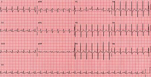
117
118 Differential Diagnosis of ST Segment Elevation 1-Normal Variant "Early Repolarization" (usually concave upwards, ending with symmetrical, large, upright T waves) 2- Ischemic Heart Disease (usually convex upwards, or straightened) Acute transmural injury - as in this acute anterior MI 3- Persistent ST elevation after acute MI suggests ventricular aneurysm 4-ST elevation may also be seen as a manifestation of Prinzmetal's (variant) angina (coronary artery spasm) 5-ST elevation during exercise testing suggests extremely tight coronary artery stenosis or spasm (transmural ischemia)
119 Differential Diagnosis of ST Segment Elevation 6-Acute Pericarditis #Concave upwards ST elevation in most leads except avr # No reciprocal ST segment depression (except in avr) #Unlike "early repolarization", T waves are usually low amplitude, and heart rate is usually increased. #May see PR segment depression, a manifestation of atrial injury

120

121 Pericarditis

122 Ventricle aneurysm
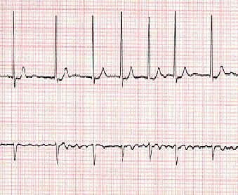
123 ST depression >2mm usually indicates ischemia Common in normal ECG, especially in pregnancy But: Non specific not more than 2mm below baseline It is convex downward or slopes upwards from the S wave
124 Differential Diagnosis of ST Segment Depression 1-Normal variants or artifacts: Pseudo-STdepression (wandering baseline due to poor skin-electrode contact) 2-Physiologic J-junctional depression with sinus tachycardia (most likely due to atrial repolarization) 3-Hyperventilation-induced ST segment depression
125 Differential Diagnosis of ST Segment Depression 4-Ischemic heart disease Subendocardial ischemia (exercise induced or during angina attack ) ST segment depression is often characterized as "horizontal", "upsloping", or "downsloping" 5-Non Q-wave MI 6- Reciprocal changes in acute Q-wave MI (e.g., ST depression in leads I & avl with acute inferior MI)
126 Differential Diagnosis of ST Segment Depression 7-Nonischemic causes of ST depression #RVH (right precordial leads) or LVH (left precordial leads, I, avl) # Digoxin effect on ECG # Hypokalemia #Mitral valve prolapse (some cases) #Secondary ST segment changes with IV conduction abnormalities (e.g., RBBB, LBBB, WPW, etc)

127 Acute inferoposterior MI (note tall R waves V1-3, marked ST depression V1-3, ST elevation in II, III, avf)

128
129 The ECG signs of Infarct! Abnormal Q waves ST segment elevation (Greater than 1mm in 2 or more adjacent leads) Inverted T waves
130 ST Elevation - Myocardial Infarction ST elevation in two or more leads Must be at least 1mm in limb leads Must be at least 2mm in chest leads
131

132 Antero -Lateral MI

133 Old inferoposterior MI (note tall R in V1-3, upright T waves and inferior Q waves)
134 T wave

135
136
137 T wave: tall T waves( more than 2 big squares) Hyperkalaemia Hyperacute myocardial infarction Left bundle branch block (LBBB)
138 T waves: small, flattened or inverted Ischemia age, race hyperventilation, anxiety, drinking iced water LVH drugs (e.g. digoxin) pericarditis, PE intraventricular conduction delay (e.g. RBBB) electrolyte disturbance
139

140

141
142 QT Interval
143
144 QT interval measured from beginning of QRS to end of T wave QT Interval (QT c < 0.40 sec) upper limit for QT c = 0.44 sec 1-Bazett's Formula: QT c = (QT)/SqRoot RR (in seconds) 2-Poor Man's Guide to upper limits of QT: For HR = 70 bpm, QT<0.40 sec; for every 10 bpm increase above 70 subtract 0.02 sec, and for every 10 bpm decrease below 70 add 0.02 sec. For example: QT < 80 bpm QT < 60 bpm
145
146 QT interval Prolonged QT : A. Familial long QT Syndrome (LQTS) B. Congestive Heart Failure C. Myocardial Infarction D. Hypocalcemia & Hypokalaemia E. Hypomagnesemia F. Type I Antiarrhythmic drugs & Cispride G. Rheumatic Fever H. Myocarditis I. Congenital Heart Disease Short QT : A. Digoxin (Digitalis) B. Hypercalcemia C. Hyperkalemia

147
148 The U wave is the only remaining enigma of the ECG, and probably not for long. The origin of the U wave is still in question, although most authorities correlate the U wave with electrophysiologic events called "afterdepolarizations" in the ventricles.. The normal U wave has the same polarity as the T wave and is usually less than one-third the amplitude of the T wave. U waves are usually best seen in the right precordial leads especially V2 and V3. The normal U wave is asymmetric with the ascending limb moving more rapidly than the descending limb (just the opposite of the normal T wave).
149 Prominent upright U waves 1-Sinus bradycardia accentuates the U wave 2-Hypokalemia (remember the triad of ST segment depression, low amplitude T waves, and prominent U waves) 3- Quinidine and other type 1A antiarrhythmics
150 Negative or "inverted" U waves 1- Ischemic heart disease (often indicating left main or LAD disease) Myocardial infarction (in leads with pathologic Q waves) 2-During episode of acute ischemia (angina or exercise-induced ischemia) 3- During coronary artery spasm (Prinzmetal's angina) 4- Nonischemic causes Some cases of LVH or RVH (usually in leads with prominent R waves)

151 Thanks
Understanding the Electrocardiogram. David C. Kasarda M.D. FAAEM St. Luke s Hospital, Bethlehem
 Understanding the Electrocardiogram David C. Kasarda M.D. FAAEM St. Luke s Hospital, Bethlehem Overview 1. History 2. Review of the conduction system 3. EKG: Electrodes and Leads 4. EKG: Waves and Intervals
12-Lead EKG Interpretation. Judith M. Haluka BS, RCIS, EMT-P
 12-Lead EKG Interpretation Judith M. Haluka BS, RCIS, EMT-P ECG Grid Left to Right = Time/duration Vertical measure of voltage (amplitude) Expressed in mm P-Wave Depolarization of atrial muscle Low voltage
BIPOLAR LIMB LEADS UNIPOLAR LIMB LEADS PRECORDIAL (UNIPOLAR) LEADS VIEW OF EACH LEAD INDICATIVE ECG CHANGES
 LEADS VIEW OF EACH LEAD INDICATIVE ECG CHANGES") BIPOLAR LIMB LEADS Have both a distinctive positive and negative pole. Lead I LA (positive) RA (negative) Lead II LL (positive) RA (negative) Lead III LL (positive) LA (negative) UNIPOLAR LIMB LEADS Have
BIPOLAR LIMB LEADS Have both a distinctive positive and negative pole. Lead I LA (positive) RA (negative) Lead II LL (positive) RA (negative) Lead III LL (positive) LA (negative) UNIPOLAR LIMB LEADS Have
ECG made extra easy. medics.cc
 ElectroCardioGraphyraphy ECG made extra easy Overview Objectives for this tutorial What is an ECG? Overview of performing electrocardiography on a patient Simple physiology Interpreting the ECG Objectives
EKG Abnormalities. I. Early repolarization abnormality:
 I. Early repolarization abnormality: EKG Abnormalities A. A normal variant. Early repolarization is most often seen in healthy young adults. Look for ST elevation, tall QRS voltage, "fishhook" deformity
ST Segment Elevation Nothing is ever as hard (or easy) as it looks
 as it looks") ST Segment Elevation Nothing is ever as hard (or easy) as it looks Cameron Guild, MD Division of Cardiology University of Mississippi Medical Center February 17, 2012 Objectives 1. Describe the electrical
ST Segment Elevation Nothing is ever as hard (or easy) as it looks Cameron Guild, MD Division of Cardiology University of Mississippi Medical Center February 17, 2012 Objectives 1. Describe the electrical
RAPID INTERPRETATION OF. EKG s
 Personal Quick Reference Sheets 333 (pages 333 to 346) There is no need to remove these reference pages from your book. To download and print them in full color, go to: www.themdsite.com Reference Sheets
Systematic Approach to 12 Lead EKG Interpretation
 Systematic Approach to 12 Lead EKG Interpretation Maureen Knechtel MPAS, PA-C Wellmont CVA Heart Institute Disclosure Statement of Financial Interest I, Maureen Knechtel, do not have a financial interest/arrangement
the basics Perfect Heart Institue, Piyavate Hospital
 ECG INTERPRETATION: the basics Damrong Sukitpunyaroj MD Damrong Sukitpunyaroj, MD Perfect Heart Institue, Piyavate Hospital Overview Conduction Pathways Systematic Interpretation Common abnormalities in
NEONATAL & PEDIATRIC ECG BASICS RHYTHM INTERPRETATION
 NEONATAL & PEDIATRIC ECG BASICS & RHYTHM INTERPRETATION VIKAS KOHLI MD FAAP FACC SENIOR CONSULATANT PEDIATRIC CARDIOLOGY APOLLO HOSPITAL MOB: 9891362233 ECG FAX LINE: 011-26941746 THE BASICS: GRAPH PAPER
Introduction to Electrocardiography. The Genesis and Conduction of Cardiac Rhythm
 Introduction to Electrocardiography Munther K. Homoud, M.D. Tufts-New England Medical Center Spring 2008 The Genesis and Conduction of Cardiac Rhythm Automaticity is the cardiac cell s ability to spontaneously
Electrophysiology Introduction, Basics. The Myocardial Cell. Chapter 1- Thaler
 Electrophysiology Introduction, Basics Chapter 1- Thaler The Myocardial Cell Syncytium Resting state Polarized negative Membrane pump Depolarization fundamental electrical event of the heart Repolarization
Electrocardiography Review and the Normal EKG Response to Exercise
 Electrocardiography Review and the Normal EKG Response to Exercise Cardiac Anatomy Electrical Pathways in the Heart Which valves are the a-v valves? Closure of the a-v valves is associated with which heart
ECG Measurments and Interpretation Programs
 ECG Measurments and Interpretation Programs Physician s Guide Distributed by Welch Allyn 4341 State Street Road, PO Box 220 Skaneateles Falls, NY 13153-0220 www.welchallyn.com Sales and Service information:
Interpreting a rhythm strip
 3 Interpreting a rhythm strip Just the facts In this chapter, you ll learn: the components of an ECG complex and their significance and variations techniques for calculating the rate and rhythm of an ECG
Normal Sinus Rhythm. Sinus Bradycardia. Sinus Tachycardia. Rhythm ECG Characteristics Example (NSR) & consistent. & consistent.
 & consistent. & consistent.") Normal Sinus Rhythm (NSR) Rate: 60-100 per minute Rhythm: R- R = P waves: Upright, similar P-R: 0.12-0.20 second & consistent P:qRs: 1P:1qRs Sinus Tachycardia Exercise Hypovolemia Medications Fever Hypoxia
Normal Sinus Rhythm (NSR) Rate: 60-100 per minute Rhythm: R- R = P waves: Upright, similar P-R: 0.12-0.20 second & consistent P:qRs: 1P:1qRs Sinus Tachycardia Exercise Hypovolemia Medications Fever Hypoxia
INTRODUCTORY GUIDE TO IDENTIFYING ECG IRREGULARITIES
 INTRODUCTORY GUIDE TO IDENTIFYING ECG IRREGULARITIES NOTICE: This is an introductory guide for a user to understand basic ECG tracings and parameters. The guide will allow user to identify some of the
ACLS Chapter 3 Rhythm Review Instructor Lesson Plan to Accompany ACLS Study Guide 3e
 ACLS Chapter 3 Rhythm Review Lesson Plan Required reading before this lesson: ACLS Study Guide 3e Textbook Chapter 3 Materials needed: Multimedia projector, computer, ACLS Chapter 3 Recommended minimum
Diagnosis Code Crosswalk : ICD-9-CM to ICD-10-CM Cardiac Rhythm and Heart Failure Diagnoses
 Diagnosis Code Crosswalk : to 402.01 Hypertensive heart disease, malignant, with heart failure 402.11 Hypertensive heart disease, benign, with heart failure 402.91 Hypertensive heart disease, unspecified,
MULTIPLE CHOICE. Choose the one alternative that best completes the statement or answers the question.
 Exam Name MULTIPLE CHOICE. Choose the one alternative that best completes the statement or answers the question. 1) What term is used to refer to the process of electrical discharge and the flow of electrical
ECG INTERPRETATION MANUAL
 Lancashire & South Cumbria Cardiac Network ECG INTERPRETATION MANUAL THE ABNORMAL ECG Lancashire And South Cumbria Cardiac Physiologist Training Manual AV NODAL BLOCKS (HEART BLOCKS) Disturbances of intra
Objectives. The ECG in Pulmonary and Congenital Heart Disease. Lead II P-Wave Amplitude during COPD Exacerbation and after Treatment (50 pts.
 The ECG in Pulmonary and Congenital Heart Disease Gabriel Gregoratos, MD Objectives Review the pathophysiology and ECG signs of pulmonary dysfunction Review the ECG findings in patients with: COPD (chronic
The P Wave: Indicator of Atrial Enlargement
 Marquette University e-publications@marquette Physician Assistant Studies Faculty Research and Publications Health Sciences, College of 8-12-2010 The P Wave: Indicator of Atrial Enlargement Patrick Loftis
QRS Complexes. Fast & Easy ECGs A Self-Paced Learning Program
 6 QRS Complexes Fast & Easy ECGs A Self-Paced Learning Program Q I A ECG Waveforms Normally the heart beats in a regular, rhythmic fashion producing a P wave, QRS complex and T wave I Step 4 of ECG Analysis
By the end of this continuing education module the clinician will be able to:
 EKG Interpretation WWW.RN.ORG Reviewed March, 2015, Expires April, 2017 Provider Information and Specifics available on our Website Unauthorized Distribution Prohibited 2015 RN.ORG, S.A., RN.ORG, LLC Developed
How to read the ECG in athletes: distinguishing normal form abnormal
 How to read the ECG in athletes: distinguishing normal form abnormal Antonio Pelliccia, MD Institute of Sport Medicine and Science www.antoniopelliccia.it Cardiac adaptations to Rowing Vagotonia Sinus
The abbreviation EKG, for electrocardiogram,
 CLIN PEDIATR OnlineFirst, published on January 28, 2010 as doi:10.1177/0009922809336206 Simplified Pediatric Electrocardiogram Interpretation Clinical Pediatrics Volume XX Number X Month XXXX xx-xx 2009
An ECG Primer. Quick Look. I saw it, but I did not realize it. Elizabeth Peabody
 4 An ECG Primer Quick Look Cardiac Monitoring System - p. 64 ECG Paper - p. 73 Lead Polarity and Vectors - p. 77 Basic ECG Components - p. 79 Heart Rate and Pulse Rate - p. 91 Summary - p. 94 Chapter Quiz
Copyright 2006 Blaufuss Multimedia. All rights reserved. Page 1
 Copyright 2006 Blaufuss Multimedia. All rights reserved. Page 1 002 Sinus Rhythm, atrial rate 90 Mobitz II AVB, Ventricular rate 50 Left Atrial Enlargement Left Ventricular Hypertrophy RBBB a) Long R-R
Basics of EKG Interpretation: A Programmed Study - Barbara Ritter Ed.D, FNP
 Basics of EKG Interpretation: A Programmed Study - Barbara Ritter Ed.D, FNP Acknowledgement is given to Leslie K. Muma, MS, RN, NP for assistance in preparation of this learning module. Description The
HTEC 91. Topic for Today: Atrial Rhythms. NSR with PAC. Nonconducted PAC. Nonconducted PAC. Premature Atrial Contractions (PACs)
") HTEC 91 Medical Office Diagnostic Tests Week 4 Topic for Today: Atrial Rhythms PACs: Premature Atrial Contractions PAT: Paroxysmal Atrial Tachycardia AF: Atrial Fibrillation Atrial Flutter Premature Atrial
HTEC 91 Medical Office Diagnostic Tests Week 4 Topic for Today: Atrial Rhythms PACs: Premature Atrial Contractions PAT: Paroxysmal Atrial Tachycardia AF: Atrial Fibrillation Atrial Flutter Premature Atrial
Atrial & Junctional Dysrhythmias
 Atrial & Junctional Dysrhythmias Atrial & Junctional Dysrhythmias Atrial Premature Atrial Complex Wandering Atrial Pacemaker Atrial Tachycardia (ectopic) Multifocal Atrial Tachycardia Atrial Flutter Atrial
Table of Contents Error! Bookmark not defined.
 Table of Contents EKG TRACING...1 Figure 1 - EKG Tracing... Error! Bookmark not defined. STEP 1...1 Rate... 1 Figure 2 - Determining the Rate... 1 Step 2...2 Rhythm... 2 Figure 3 - Determining the Rhythm
Tips and Tricks to Demystify 12 Lead ECG Interpretation
 Tips and Tricks to Demystify 12 Lead ECG Interpretation Mission: Lifeline North Dakota Regional EMS and Hospital Conference Samantha Kapphahn, DO Essentia Health- Interventional Cardiology June 5th, 2014
HOW TO READ AN ECG. Rate = 300 / big squares 1 line = 300 2 line = 150 3 line = 75 4 line = 60 5 line = 50 6 line = 42 7 line = 38
 HOW TO READ AN ECG Pathophysiology Pacemaker Rates: SAN 60-100 AVN 40-60 Ventricle 20-40 Areas of ECG Horizontal scale: 1mm = 0.04s 5mm = 0.2s Calculating Rate Rate = 300 / big squares 1 line = 300 2 line
Introduction to Electrophysiology. Wm. W. Barrington, MD, FACC University of Pittsburgh Medical Center
 Introduction to Electrophysiology Wm. W. Barrington, MD, FACC University of Pittsburgh Medical Center Objectives Indications for EP Study How do we do the study Normal recordings Abnormal Recordings Limitations
2 ECG basics. Leads and planes. Leads. Planes. from different perspectives, which are called leads and planes.
 558302.qxp 3/14/12 10:47 PM Page 12 2 ECG basics One of the most valuable diagnostic tools available, an electrocardiogram (ECG) records the heart s electrical activity as waveforms. By interpreting these
Biology 347 General Physiology Lab Advanced Cardiac Functions ECG Leads and Einthoven s Triangle
 Biology 347 General Physiology Lab Advanced Cardiac Functions ECG Leads and Einthoven s Triangle Objectives Students will record a six-lead ECG from a resting subject and determine the QRS axis of the
Equine Cardiovascular Disease
 Equine Cardiovascular Disease 3 rd most common cause of poor performance in athletic horses (after musculoskeletal and respiratory) Cardiac abnormalities are rare Clinical Signs: Poor performance/exercise
Lead avr: The Neglected Lead
 Chapter 22 Lead avr: The Neglected Lead M Chenniappan INTRODUCTION Lead avr, one of the 12 electrocardiographic leads, is frequently ignored in clinical medicine. In fact, many clinicians refer to the
ECG Measurement and Interpretation
 ECG Measurement and Interpretation Statement of accuracy for analysing ECG units *2.530036* Physicians Guide Sales and Service Information The SCHILLER sales and service centre network is world-wide. For
The new generation in ECG interpretation
 The new generation in ECG interpretation Philips DXL ECG Algorithm, Release PH100B The Philips DXL ECG Algorithm, developed by the Advanced Algorithm Research Center, uses sophisticated analytical methods
Adult Cardiac Surgery ICD9 to ICD10 Crosswalks
 164.1 Malignant neoplasm of heart C38.0 Malignant neoplasm of heart 164.1 Malignant neoplasm of heart C45.2 Mesothelioma of pericardium 198.89 Secondary malignant neoplasm of other specified sites C79.89
Sleep Heart Health Study (SHHS) ECG Protocol
 ECG Protocol") Sleep Heart Health Study (SHHS) ECG Protocol SHHS 1 Electrocardiography (ECG) Baseline ECG is performed in all parent study clinic visits preceding the PSG. All sites perform a standard resting 12-lead
Sleep Heart Health Study (SHHS) ECG Protocol SHHS 1 Electrocardiography (ECG) Baseline ECG is performed in all parent study clinic visits preceding the PSG. All sites perform a standard resting 12-lead
Tachyarrhythmias (fast heart rhythms)
") Patient information factsheet Tachyarrhythmias (fast heart rhythms) The normal electrical system of the heart The heart has its own electrical conduction system. The conduction system sends signals throughout
Patient information factsheet Tachyarrhythmias (fast heart rhythms) The normal electrical system of the heart The heart has its own electrical conduction system. The conduction system sends signals throughout
Scott Hubbell, MHSc, RRT-NPS, C-NPT, CCT Clinical Education Coordinator/Flight RRT EagleMed
 Scott Hubbell, MHSc, RRT-NPS, C-NPT, CCT Clinical Education Coordinator/Flight RRT EagleMed Identify the 12-Lead Views Explain the vessels of occlusion Describe the three I s Basic Interpretation of 12-Lead
ACLS RHYTHM TEST. 2. A 74-year-old woman with chest pain. Blood pressure 192/90 and rates her pain 9/10.
 ACLS RHYTHM TEST Name Date Choose the best answer for each of the following questions. Each of the following strips is 6 seconds in length. 1. Identify the following rhythm a. Sinus bradycardia with 2
An Introduction to Tachyarrhythmias R. A. Seyon MN, NP, CCN(C) & Dr. R. G. Williams
 & Dr. R. G. Williams") Arrhythmias 1 An Introduction to Tachyarrhythmias R. A. Seyon MN, NP, CCN(C) & Dr. R. G. Williams Things to keep in mind when analyzing arrhythmias: Electrical activity recorded in 12 and 15 leads Examine
Arrhythmias 1 An Introduction to Tachyarrhythmias R. A. Seyon MN, NP, CCN(C) & Dr. R. G. Williams Things to keep in mind when analyzing arrhythmias: Electrical activity recorded in 12 and 15 leads Examine
Electrophysiology Daymar College. Lisa H. Young, RN, BSN, MAE 2011
 Electrophysiology Daymar College Lisa H. Young, RN, BSN, MAE 2011 Electrical Conduction Pathway Chemical Basis for Impulse Formation Cardiac Action Potential Phases http://www.youtube.com/watch?v=oqpffilde0e
Review of Important ECG Findings in Patients with Syncope Joseph Toscano, MD
 92 Review of Important ECG Findings in Patients with Syncope Joseph Toscano, MD Abstract Guidelines recommend 12-lead ECG as an important test to perform in patients with syncope. Though the incidence
The Electrocardiogram (ECG)
") The Electrocardiogram (ECG) Preparation for RWM Lab Experiment The first ECG was measured by Augustus Désiré Waller in 1887 using Lippmann's capillary electrometer. Recorded ECG: http://www.youtube.com/watch_popup?v=q0jmfivadue&vq=large
The Electrocardiogram (ECG) Preparation for RWM Lab Experiment The first ECG was measured by Augustus Désiré Waller in 1887 using Lippmann's capillary electrometer. Recorded ECG: http://www.youtube.com/watch_popup?v=q0jmfivadue&vq=large
Morphology of the Electrocardiogram
 TETC01 12/2/05 18:09 Page 1 1 The Morphology of the Electrocardiogram ntoni ayés de Luna, Velislav N. atchvarov and Marek Malik Summary The 12-lead electrocardiogram (ECG) is the single most commonly performed
ECG Findings. IV Access. 12 Lead Interpretation: STEMI and NSTEMI. ACLS Acute Coronary Syndrome Chest Pain Suggestive of Ischemia.
 12 Lead Interpretation: STEMI and NSTEMI Presented by Annmarie Keck, RN, BSN, CEN Northwest MedStar Clinical Outreach Educator ACLS Acute Coronary Syndrome Chest Pain Suggestive of Ischemia Immediate Assessment
Electrodes placed on the body s surface can detect electrical activity, APPLIED ANATOMY AND PHYSIOLOGY. Circulatory system
 4 READING AND INTERPRETING THE ELECTROCARDIOGRAM Electrodes placed on the body s surface can detect electrical activity, which occurs in the heart. The recording of these electrical events comprises an
BASIC CARDIAC ARRHYTHMIAS Revised 10/2001
 BASIC CARDIAC ARRHYTHMIAS Revised 10/2001 A Basic Arrhythmia course is a recommended prerequisite for ACLS. A test will be given that will require you to recognize cardiac arrest rhythms and the most common
ECG Signal Analysis Using Wavelet Transforms
 Bulg. J. Phys. 35 (2008) 68 77 ECG Signal Analysis Using Wavelet Transforms C. Saritha, V. Sukanya, Y. Narasimha Murthy Department of Physics and Electronics, S.S.B.N. COLLEGE (Autonomous) Anantapur 515
Electrocardiography I Laboratory
 Introduction The body relies on the heart to circulate blood throughout the body. The heart is responsible for pumping oxygenated blood from the lungs out to the body through the arteries and also circulating
Provider Checklist-Outpatient Imaging. Checklist: Nuclear Stress Test, Thallium/Technetium/Sestamibi (CPT Code 78451-78454 78469)
") Provider Checklist-Outpatient Imaging Checklist: Nuclear Stress Test, Thallium/Technetium/Sestamibi (CPT Code 78451-78454 78469) Medical Review Note: Per InterQual, if any of the following are present,
Provider Checklist-Outpatient Imaging Checklist: Nuclear Stress Test, Thallium/Technetium/Sestamibi (CPT Code 78451-78454 78469) Medical Review Note: Per InterQual, if any of the following are present,
22 Arrhythmias. C. Scharf and F. Duru. Siegenthaler, Differential Diagnosis in Internal Medicine (ISBN9783131421418), 2007 Georg Thieme Verlag
, 2007 Georg Thieme Verlag") 22 22 Arrhythmias C. Scharf and F. Duru 22 712 Arrhythmias 22.1 Differential Diagnosis of Arrhythmias 714 Medical History 714 Clinical Examination 714 Electrocardiogram (ECG) 715 Additional Tools for the
22 22 Arrhythmias C. Scharf and F. Duru 22 712 Arrhythmias 22.1 Differential Diagnosis of Arrhythmias 714 Medical History 714 Clinical Examination 714 Electrocardiogram (ECG) 715 Additional Tools for the
Evaluation copy. Analyzing the Heart with EKG. Computer
 Analyzing the Heart with EKG Computer An electrocardiogram (ECG or EKG) is a graphical recording of the electrical events occurring within the heart. In a healthy heart there is a natural pacemaker in
Interpreting AV (Heart) Blocks: Breaking Down the Mystery
 Blocks: Breaking Down the Mystery") Interpreting AV (Heart) Blocks: Breaking Down the Mystery 2 Contact Hours Copyright 2012 by RN.com. All Rights Reserved. Reproduction and distribution of these materials is prohibited without the express
Interpreting AV (Heart) Blocks: Breaking Down the Mystery 2 Contact Hours Copyright 2012 by RN.com. All Rights Reserved. Reproduction and distribution of these materials is prohibited without the express
Medtronic Cardiac Rhythm and Heart Failure ICD-10 Coding for Physicians
 Medtronic Cardiac Rhythm and Heart Failure ICD-10 Coding for Physicians May 19, 2015 Disclaimer This presentation is intended for educational use. Any duplication is prohibited without written consent
MEANS ECG Physicians Manual for Welch Allyn CP Series Electrocardiographs
 MEANS ECG Physicians Manual f Welch Allyn CP Series Electrocardiographs Welch Allyn 4341 State Street Road PO Box 220 Skaneateles Falls, NY 13153-0220 USA 2013, DIR: 80011564, Ver: E www.welchallyn.com
Cardiac Arrhythmias. Introduction. Sinus Rhythms. Premature Beats. Secondary article. John A Kastor, University of Maryland, Baltimore, Maryland, USA
 John A Kastor, University of Maryland, Baltimore, Maryland, USA Cardiac arrhythmias are disturbances in the rhythm of the heart manifested by irregularity or by abnormally fast rates ( tachycardias ) or
The Basics of 12 Lead EKG s
 EMS Solutions Presents The Basics of 12 Lead EKG s NOTICE: You DO NOT Have the Right to Reprint or Resell this Publication. However, you MAY give this report away, provided you do not change or alter the
ELECTROCARDIOGRAPHY (I) THE GENESIS OF THE ELECTROCARDIOGRAM
 THE GENESIS OF THE ELECTROCARDIOGRAM") ELECTROCARDIOGRAPHY (I) THE GENESIS OF THE ELECTROCARDIOGRAM Scridon Alina, Șerban Răzvan Constantin 1. Definition The electrocardiogram (abbreviated ECG or EKG) represents the graphic recording of electrical
ELECTROCARDIOGRAPHY (I) THE GENESIS OF THE ELECTROCARDIOGRAM Scridon Alina, Șerban Răzvan Constantin 1. Definition The electrocardiogram (abbreviated ECG or EKG) represents the graphic recording of electrical
For more information about the use of the Propaq monitor, refer to the Propaq Directions For Use.
 Clinical Support 8500 S.W. Creekside Pl. Beaverton, OR 97008-7107 U.S.A. Telephone: 503-526-4200 Toll Free: 800-289-2500 clinicalsupport@protocol.com ELECTROCARDIOGRAPHY Introduction This article provides
Clinical Observations with the Lead System
 Clinical Observations with the Lead System Frank Precordial By J. A. ABILDSKOv, M.D., W. W. STREET, M.D., N. SOLOMON, B.A., AND A. H. TOOMAJIAN, B.A. Several new lead systems for electrocardiography and
ST Segment Monitoring. IntelliVue Patient Monitor and Information Center, Application Note
 ST Segment Monitoring ST/AR Algorithm IntelliVue Patient Monitor and Information Center, Application Note This application note describes principles and uses for continuous ST segment monitoring. It also
HEART HEALTH WEEK 3 SUPPLEMENT. A Beginner s Guide to Cardiovascular Disease HEART FAILURE. Relatively mild, symptoms with intense exercise
 WEEK 3 SUPPLEMENT HEART HEALTH A Beginner s Guide to Cardiovascular Disease HEART FAILURE Heart failure can be defined as the failing (insufficiency) of the heart as a mechanical pump due to either acute
Section Four: Pulmonary Artery Waveform Interpretation
 Section Four: Pulmonary Artery Waveform Interpretation All hemodynamic pressures and waveforms are generated by pressure changes in the heart caused by myocardial contraction (systole) and relaxation/filling
Guidelines for the interpretation of the neonatal electrocardiogram
 European Heart Journal (2002) 23, 1329 1344 doi:10.1053/euhj.2002.3274, available online at http://www.idealibrary.com on Task Force Report Guidelines for the interpretation of the neonatal electrocardiogram
Potential Causes of Sudden Cardiac Arrest in Children
 Potential Causes of Sudden Cardiac Arrest in Children Project S.A.V.E. When sudden death occurs in children, adolescents and younger adults, heart abnormalities are likely causes. These conditions are
Premature Ventricular Contractions. Ralph Augostini, MD FACC FHRS
 Premature Ventricular Contractions Ralph Augostini, MD FACC FHRS Orlando, Florida October 7-9, 2011 Premature Ventricular Contractions: ACC/AHA/ESC 2006 Guidelines for Management of Patients With Ventricular
School of Health Sciences
 School of Health Sciences Cardiology Teaching Package A Beginners Guide to Normal Heart Function, Sinus Rhythm & Common Cardiac Arrhythmias Welcome This document extends subjects covered in the Cardiology
How To Understand What You Know
 Heart Disorders Glossary ABG (Arterial Blood Gas) Test: A test that measures how much oxygen and carbon dioxide are in the blood. Anemia: A condition in which there are low levels of red blood cells in
Feature Vector Selection for Automatic Classification of ECG Arrhythmias
 Feature Vector Selection for Automatic Classification of ECG Arrhythmias Ch.Venkanna 1, B. Raja Ganapathi 2 Assistant Professor, Dept. of ECE, G.V.P. College of Engineering (A), Madhurawada, A.P., India
Advanced EKG Interpretation
 Advanced EKG Interpretation JUNCTIONAL RHYTHMS AND NURSING INTERVENTIONS Objectives Identify specific cardiac dysrhythmias Describe appropriate nursing interventions for specific dysrhythmias Junctional
NAME OF THE HOSPITAL: 1. Coronary Balloon Angioplasty: M7F1.1/ Angioplasty with Stent(PTCA with Stent): M7F1.3
: M7F1.3") 1. Coronary Balloon Angioplasty: M7F1.1/ Angioplasty with Stent(PTCA with Stent): M7F1.3 1. Name of the Procedure: Coronary Balloon Angioplasty 2. Select the Indication from the drop down of various indications
1. Coronary Balloon Angioplasty: M7F1.1/ Angioplasty with Stent(PTCA with Stent): M7F1.3 1. Name of the Procedure: Coronary Balloon Angioplasty 2. Select the Indication from the drop down of various indications
DEPARTMENT OF HEALTH AND HUMAN SERVICES Centers for Medicare & Medicaid Services
 DEPARTMENT OF HEALTH AND HUMAN SERVICES Centers for Medicare & Medicaid Services NEW product from the Medicare Learning Network (MLN) Provider Compliance Tips for Computed Tomography (CT) Scans Podcast,
QT analysis: A guide for statistical programmers. Prabhakar Munkampalli Statistical Analyst II Hyderabad, 7 th September 2012
 QT analysis: A guide for statistical programmers Prabhakar Munkampalli Statistical Analyst II Hyderabad, 7 th September 2012 Agenda ECG ICH E14 Thorough QT/QTc study Role of Statistical Programmer References
Diagnostic and Therapeutic Procedures
 Diagnostic and Therapeutic Procedures Diagnostic and therapeutic cardiovascular s are central to the evaluation and management of patients with cardiovascular disease. Consistent with the other sections,
VCA Veterinary Specialty Center of Seattle
 An electrocardiogram (ECG) is a graph of the heart`s electrical current, which allows evaluation of heart rate, rhythm and conduction. Identification of conduction problems within the heart begins with
Rigel 333 Multi Parameter Patient Simulator Version 2.0
 INSTRUCTION MANUAL Rigel 333 Multi Parameter Patient Simulator Version 2.0 Rigel Medical Seaward Group 18 Bracken Hill Peterlee, County Durham SR8 2SW England www.rigelmedical.com Dear User: We appreciate
Electrolyte Physiology. Something in the way she moves
 Electrolyte Physiology Something in the way she moves me Electrolyte Movement CONCENTRATION GRADIENT ELECTRICAL GRADIENT DRIVING FORCE NERNST NUMBER (E-ion) CONDUCTANCE (G-ion) PERMEABILITY CHANNELS: small
CARDIAC ELECTROPHYSIOLOGY, ARRHYTHMIAS AND PACING. Medical Knowledge. Goals and Objectives PF EF MF LF Aspirational
 Know the histology and gross anatomy of the normal sinoatrial node, atrial conduction pathways, atrioventricular (AV) junction and nod, His bundle, conduction fascicles and terminal intra-ventricular conduction
Banner Staff Service ECG Study Guide
 Banner Staff Service ECG Study Guide Edited by Larry H. Lybbert, MS, RN Table of Contents ECG STUDY GUIDE... 3 ECG INTERPRETATION BASICS... 4 EKG GRAPH PAPER...4 RATE MEASUREMENT...9 The Six Second Method...9
Activity 4.2.3: EKG. Introduction. Equipment. Procedure
 Activity 4.2.3: EKG The following is used with permission of Vernier Software and Technology. This activity is based on the experiment Analyzing the Heart with EKG from the book Human Physiology with Vernier,
DEFIBRILLATION AND THE AUTOMATIC EXTERNAL DEFIBRILLATOR A GUIDE
 DEFIBRILLATION AND THE AUTOMATIC EXTERNAL DEFIBRILLATOR A GUIDE Defibrillation and the AED, A guide Revised February 2012 Page 1 of 22 Original document complied by Pat Standen, Grampians Trauma, Emergency
The heart then repolarises (or refills) in time for the next stimulus and contraction.
 in time for the next stimulus and contraction.") Atrial Fibrillation BRIEFLY, HOW DOES THE HEART PUMP? The heart has four chambers. The upper chambers are called atria. One chamber is called an atrium, and the lower chambers are called ventricles. In
Atrial Fibrillation BRIEFLY, HOW DOES THE HEART PUMP? The heart has four chambers. The upper chambers are called atria. One chamber is called an atrium, and the lower chambers are called ventricles. In
38 year old female with mild obesity. She is planning an exercise program to loose weight. She has no other known risk factors for CAD.
 Stress Testing: Wael A. Jaber, MD,FACC 38 year old female with mild obesity She is planning an exercise program to loose weight. She has no other known risk factors for CAD. You recommend: A. Exercise
Anatomi & Fysiologi 060301. The cardiovascular system (chapter 20) The circulation system transports; What the heart can do;
 The circulation system transports; What the heart can do;") The cardiovascular system consists of; The cardiovascular system (chapter 20) Principles of Anatomy & Physiology 2009 Blood 2 separate pumps (heart) Many blood vessels with varying diameter and elasticity
The cardiovascular system consists of; The cardiovascular system (chapter 20) Principles of Anatomy & Physiology 2009 Blood 2 separate pumps (heart) Many blood vessels with varying diameter and elasticity
12 Lead ECGs: Ischemia, Injury & Infarction Part 2
 12 Lead ECGs: Ischemia, Injury & Infarction Part 2 McHenry Western Lake County EMS Localization: Left Coronary Artery Right Coronary Artery Right Ventricle Septal Wall Anterior Descending Artery Left Main
Management of Pacing Wires After Cardiac Surgery
 Management of Pacing Wires After Cardiac Surgery David E. Lizotte, Jr. PA C, MPAS, FAPACVS President, Association of Physician Assistants in Cardiovascular Surgery Conflicts: None Indications 2008 Journal
Guideline for the management of arrhythmias
 Guideline for the management of arrhythmias The following guideline is approved only for use at University College London Hospitals NHS Foundation Trust. It is provided as supporting information for the
Relax and Learn at the FARM 2012. History of Cardiac Monitoring. History of Cardiac Monitoring 10/17/2012
 Relax and Learn at the FARM 2012 Session 3: Bedside Cardiac Monitoring: Arrhythmias, ST Segments, and QT Interval Karen MarzlinDNP, RN, CCNS, CCRN-CMC, CHFN Cardiovascular Nursing Education Associates
Atrial Fibrillation Peter Santucci, MD Revised May, 2008
 Atrial Fibrillation Peter Santucci, MD Revised May, 2008 Atrial fibrillation (AF) is an irregular, disorganized rhythm characterized by a lack of organized mechanical atrial activity. The atrial rate is
Automatic External Defibrillators
 Last Review Date: May 27, 2016 Number: MG.MM.DM.10dC2 Medical Guideline Disclaimer Property of EmblemHealth. All rights reserved. The treating physician or primary care provider must submit to EmblemHealth
Bradycardia CHAPTER 12 CODE SCENARIO
 Senecal-12.qxd 14/04/2005 09:44 AM Page 69 CHAPTER 12 Bradycardia CODE SCENARIO A code is called for a 78-year-old man who was admitted to the hospital for syncope of unknown etiology. He was resting comfortably
Electrophysiology Heart Study - EPS -
 Electrophysiology Heart Study - EPS - What is an EPS? EPS is short for ElectroPhysiology heart Study. This procedure looks at the electrical system of your heart. An EPS will show if you have a heart rhythm